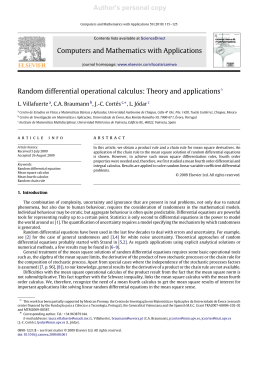
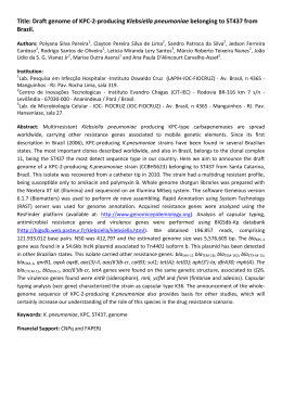
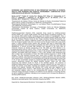
Revista Brasileira de FisiologiaVegetal, 10(2):161-164, 1998.
MINI-REVIEW
DIFFERENTIAL DISPLAY: A NOVEL PCR-BASED
METHOD FOR GENE ISOLATION AND CLONING1
José Donizeti Alves2, Tara T. VanToai3 and Namik Kaya4
Departamento de Biologia, Universidade Federal de Lavras, C.P. 37, Lavras, MG, 37200000, Brazil.
ABSTRACT - Differential Display (DDRT-PCR) is a powerful technique for analyzing differences in
gene expression. DDRT-PCR has the following essential steps: total RNA isolation and purification,
cDNA synthesis from mRNAs, PCR amplification of cDNAs, visualization of PCR products, reamplification
and cloning of the differentially expressed PCR products, confirmation by northern blot, sequencing the
confirmed clones, and finally cDNA library screening to isolate the genes of interest. After its invention
in 1992, a number of modifications have been introduced to optimize the technique and specifically to
reduce the major problem of “false positives”. Since understanding of specific gene expression patterns
that regulate developmental and stress responses is a major concern of molecular biology, DDRT-PCR
has become a very popular molecular technique during the past decade.
Additional index terms: flooding, gene expression, mRNA differential display.
DIFFERENTIAL DISPLAY: UM NOVO MÉTODO DE PCR PARA ISOLAMENTO E
CLONAGEM DE GENE.
RESUMO - Differential Display (DDRT-PCR) é uma poderosa técnica utilizada para análise de diferenças na expressão de genes. DDRT-PCR engloba as seguintes etapas: isolamento e purificação de
RNA total, síntese de cDNA, PCR, visualização dos produtos de PCR, reamplificação e clonagem dos
fragmentos de interesse (diferencialmente expressos), confirmação por análise de “northern blot”,
sequenciamento dos clones confirmados e, finalmente, seleção em bibliotecas de cDNA para isolamento do gene de interesse. Após seu desenvolvimento em 1992, DDRT-PCR tem sofrido uma série de
modificações com o propósito de melhorar e otimizar a técnica principalmente no que se refere a eliminação ou diminuição de “falsos positivos”. Visto que o entendimento do padrão de expressão gênica e à
comparação de genes diferencialmente expressos em vários tecidos são de fundamental importância
nas diferentes áreas da biologia molecular, DDRT-PCR tem se tornado, dentro de seus propósitos, uma
das ferramentas mais eficientes e práticas da última década.
Termos adicionais para indexação: alagamento, expressão gênica, mRNA differential display.
1 Received 07/01/1988 and accepted 07/08/1998.
2
Corresponding author. Professor Adjunto, Departamento de
Biologia, Universidade Federal de Lavras, C.P. 37, Lavras, MG,
37200-000, Brasil. - Bolsista do CNPq - [email protected].
3 USDA-ARS, Soil Drainage Research, Columbus, OH, USA.
4 Department of Horticulture, Agricultural Engineering Faculty,
Van, Turkey, Yuzuncu Yil University.
161
Alves et al
162
INTRODUCTION
Identification of differentially expressed genes in
various cells or under different conditions is one of the
main areas of molecular biology. Until 1992, subtractive
hybridization was the only method that could isolate
differentially expressed genes. Although subtractive
hybridization is a reliable method, it is tedious, time
consuming and difficult to perform. It also requires large
amounts of mRNA that can be limited in many situations.
In 1992, Liang and Pardee developed a new PCR-based
technique called Differential Display (DDRT-PCR) (Liang
& Pardee, 1992). This technique focused on detecting
differentially expressed genes among nearly 15000 individual mRNA sequences in mammalian cells. It was
first described to compare messages that differ between
normal and tumorigenic cells.
The method is based on the detection of the
differentially expressed cDNAs from two or more samples
that are separated on adjacent lanes of sequencing gels.
The differentially expressed bands can readily be cloned
and used as probes in Northern or Southern (DNA) blots
and to isolate genes from cDNA or genomic libraries.
Compared to the subtractive hybridization method,
DDRT-PCR is simpler, quicker and more sensitive.
However, false-positive results can generate a large
number of spurious sequences that do not represent
differential expressed genes. A number of technical
modifications have been introduced to reduce the
problem of false positives and to increase the
reproducibility of the technique (Liang & Pardee, 1995a;
Huang et al., 1996; Jones et al. 1997; Zhao et al. 1995;
Doss, 1996; Pfeffer et al. 1995; Averboukh et al., 1996;
Chen & Peck, 1996; Callard et al. 1994)). Modifications
that allowed the display of longer cDNAs have also been
reported (Averboukh et al., 1996). Therefore, with properly
designed primers and controls, DDRT-PCR could
produce results that truly reflect gene expression patterns
of different tissues. Recently a comprehensive text
containing all the procedure involving this technique, as
well as additional information on materials, equipment,
reagents and suppliers was published by Collona-Romano et al. (1998).
Total RNA 5’
N’M’AAAAAAAn
Reverse transcription
5’T11MN
dNTP
cDNA
N’M’AAAAAAAn
N M TTTTTTTT
PCR
5’T11MN
Decamer (
dNTP
)
N M TTTTTTTT
PCR
N M TTTTTTTT
PAGE
FIGURE 1 - Schematic representation of DDRT-PCR. T11MN,
degenerate oligo (dT) primer; M indicates A, C or G (degenerate);
N can be A, C, G or T. (adapted from Liang & Pardee, 1995b).
Arrowhead indicates a differentially expressed fragment.
METHOD
In this technique (Fig. 1) mRNAs are first reverse
transcribed into cDNA using each of the four sets of
generate 3’two-base anchored poly-T primers (T11MN),
where M can be G, A or C and N is G, A, T, or C. The use
of these primers allows the sampling of mRNA into 12
fractions, which should represent, in theory, 1/12 of the
total mRNA population expressed in a particular tissue.
The cDNAs are then amplified by the Polymerase
Chain Reaction (PCR) using the same 3’ anchored primer
and an arbitrary decamer. The DDRT-PCR products are
separated on sequencing gels and visualized either by
autoradiography or silver staining (Fig. 2). The pieces of
FIGURE 2 - Non-radioisotopic differential display by silver
staining. Total RNA (1 µg) was reverse transcribed using the
protocol supplied by Promega. The derived cDNA was used as
template in RT-PCR as described by Liang & Pardee (1994).
The reaction contained one of each arbitrary decamer
(AGTCCGCTGC, CGTCGGGCCT) in combination with one of
the degenerated anchored oligo dT primer set (T11NM, where
M can be G, A, or C and N can be G, A, T, or C). RT-PCR of the
cDNA from Huai 849 and Nizhen roots before (b) and after (a)
72 h of submergence using two random decamers and four sets
of degenerated anchored oligo (dT) primers produced over 960
bands on the 5% polyacrylamide 8 M urea sequencing gels,
and forty three of the bands (see arrowheads) showed
differentially expressed patterns (Alves & Vantoai, 1997).
R. Bras. Fisiol. Veg., 10(2):161-164, 1998.
Differential display . . .
163
FIGURE 3 - Northern analysis of differential display clones. b and a: before and after 3 days of flooding, respectively; 1, 2 ,3,
4: class of gene expression (Alves & Vantoai, 1997).
acrylamide bands corresponding to the differentially
expressed PCR products are cut from the gel, reamplified
with the same primers and used as probes in northern
analysis (Fig. 3). The sequences that reproducibly
express the differential pattern are cloned, characterized
by restriction mapping and sequenced.
DISCUSSION
In the five years since its introduction, DDRT-PCR
has been widely used to identify and analyse the
expression patterns of previously uncharacterized genes
in many different species. In 1996 the Medline database
listed several hundred publications describing genes
identified using DDRT-PCR. A great deal of research have
focused on understanding the different aspects of
developmentally regulated genes that are differentially
expressed under stressful conditions in plants (Sharma
& Davis, 1995; Knaap & Kende, 1995; Tsengh et al.;
1995; Momiyama et al., 1995; Wilkinson et al., 1995;
Tieman & Handa, 1996; Alves & Vantoai, 1997; Paiva,
1997). Since this technique is very efficient, a large
number of differentially expressed genes can usually be
identified in a given condition. For example, using this
technique, Mathews and Heinz (1998) reported that over
50 genes were identified in soybean roots in response
to cyst nematode infection. However, the role of these
genes in the resistance or susceptibility to cyst nematode
remains unknown. Alves and Vantoai (1997) cloned 15
bands which reproducibly expressed the differential
pattern in Northern analysis (Fig. 3). Of these, eight clones
with unique restriction maps were sequenced. A
Genbank search showed no significant homology
between six of the eight sequences to known genes.
The others two clones showed over 90% homology with
a shaggy kinase gene. In Arabidopsis, one of the
important functions of this gene is the regulation of the
DNA binding activity of a transcription factor (Bianchi et
al., 1994). Its role in the expression of flooding tolerance
in plants remains to be worked out.
While gene identification has received a great deal
of attention, the application of molecular biology to
agriculture or medicine requires an understanding of
gene function which will remain the focus of molecular
biology during the next decade.
REFERENCES
ALVES, J.D. & VANTOAI, T.T. Characterization of genes expressed in the
flooding tolerant response of soybean by mRNA differential display
technique. Annals of Botany (in press), 1997.
AVERBOUKH, L.; DOUGLAS, S.A.; LOWE, K.; MAHER, J. & PARDEE,
A.B. Better gel resolution and longer cDNAs increase the precision
of differential display. BioTechniques, 20: 918-921, 1996.
R. Bras. Fisiol. Veg., 10(2):161-164, 1998.
164
Alves et al.
BIANCHI, M. W.; GUIVARC’H, D.; THOMAS, M.; WOODGETT, J.R. &
KREIS M. Arabidopsis homologs of the shaggy and GSK-3 protein
kinases: Molecular cloning and functional expression in
Escherichia coli. Molecular & General Genetics, 242:337-345, 1994.
CALLARD, D.; LESCURE, B. & MAZZOLINI, L. A method for the
elimination of false positives generated by the mRNA differential
display technique. BioTechniques, 16:1096-1103, 1994.
CHEN, J.J.W. & PECK, K. Non-radioisotopic differential display method
to directly visualize and amplify differential bands on nylon membrane.
Nucleic Acids Research, 24: 793-794, 1996.
COLONNA-ROMANO, S.; LEONE, A. & MARESCA, B. Differentialdisplay reverse transcription-PCR (DDRT-PCR). Berlin Heidelberg,
Springer-Verlag, 1998. 124p.
DOSS, R.P. Differential display without radioactivity- A modified
procedure. BioTechniques, 21: 408-412, 1996.
HUANG, Z.; FASCO, M.J. & KAMINSKY, L.S. Optimization of Dnase I
removal of contaminating DNA from RNA for use in quantitative RNAPCR. BioTechniques, 20:1012-1020, 1996.
JONES, S.W.; CAI, D.; WEISLOW, O.S. & BABAK, E.A. Generation of
Multiple mRNA fingerprints using fluorescence –based differential
display and automated DNA sequencer. BioTechniques, 22:536543, 1997.
KNAAP, E. & KENDE H. Identification of a gibberellin-induced gene in
deepwater rice using differential display of mRNA. Plant Molecular
Biology, 28:589-592, 1995.
LIANG, P. & PARDEE, A.B. Differential display of eukaryotic messenger
RNA by means of the polymerase chain reaction. Science,
257:967-971, 1992.
LIANG, P & PARDEE, A.B. Recent advances in differential display.
Currents Protocols in Molecular Biology 7:274-280, 1995a.
LIANG, P & PARDEE, A.B. Differential display of mRNA by PCR.
Currents Protocols in Molecular Biology. Supplement 26: 15.8.115.8.8, 1995b.
MATHEWS, B.F. & R. HEINZ. Differentially expressed genes of soybean
in response to invasion by soybean cyst nematode. Plant & Animal
Genome VI Abstract: 48 . 1998.
MOMIYAMA, T.; AFELE, J.C.; SAITO, T.; KAYANO, T.; TABEI, Y.;
TAKAIWA, K. & NISHIMURA, S. Differential display identifies
developmentally regulated genes during somatic embryogenesis
in eggplant ( Solanum melongena L.) . Biochemical and
Biophysical Communications, 213:376-382, 1995.
PAIVA, L.V. Marcadores bioquímicos para o declínio dos citros.
Universidade Federal de Lavras, 1997. 69 p. Tese de Doutorado
PFEFFER, U.; FECAROTTA, E. & VIDALI, G. Efficient one-tube RTPCR amplification of rare transcripts using short sequence-specific
reverse transcription primers. BioTechniques, 18: 204-205, 1995.
SHARMA, Y.K. & DAVIS, K.R. Isolation of a novel Arabdopsis ozoneinduced cDNA by differential display. Plant Molecular Biology,
29:91-98, 1995.
TIEMAN, D. & HANDA, A.K. Molecular cloning and characterization of
genes expressed during early tomato (Lycopersicum esculentum
Mill.) fruit development by mRNA differential display. Journal of
American Society of Horticulture Science, 121:52-56, 1996.
TSENG, T.C.; TSAI, T.H.; LUE, M.Y & LEE, H.T. Identification of sucroseregulated genes in cultured rice using mRNA differential display.
Gene, 161:179-182, 1995.
WILKINSON, J.Q.; LANAHAN, M.B.; CONNER, T.W. & KLEE, H.J.
Identification of mRNAs with enhanced expression in ripening
strawberry fruit using polymerase chain reaction differential display.
Plant Molecular Biology, 27:1097-1108, 1995.
ZHAO S.; OOI, S.L. & PARDEE, A.B. New primer strategy improves
precision of differential display. BioTechniques, 18: 842-850, 1995.
R. Bras. Fisiol. Veg., 10(2):161-164, 1998.
Baixar