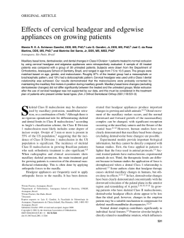
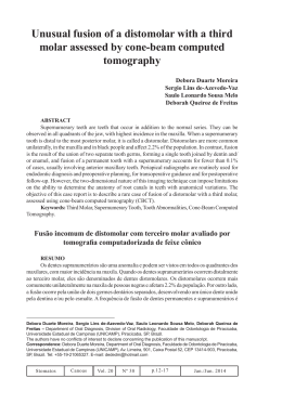
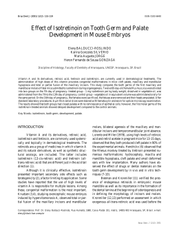
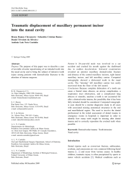
Revista de Odontologia da Universidade Cidade de São Paulo
2009 set-dez; 21(3): 233-8
epideMiologicAl investigAtion oF second preMolAr Agenesis And its relAtionsHip with
Agenesis oF otHer perMAnent teetH
ESTUDO EPIDEMIOLÓGICO DA AGENESIA DE SEGUNDOS PRÉ-MOLARES E SUA RELAÇãO COM A AGENESIA DE
OUTROS DENTES PERMANENTES
Simone Carinhena Gomes *
Daniela Gamba Garib **
Paulo Eduardo Guedes Carvalho **
Flávio Augusto Cotrim-Ferreira **
Bárbara Maria de Alencar ***
ABstrAct
Objective – This study aims to evaluate the epidemiological characteristics of a sample of patients with agenesis of second premolars, as well as to investigate its association with agenesis of other permanent teeth.
Material and methods – A Brazilian sample of 203 patients aged from 8 to 22 years was selected, all patients
presenting agenesis of at least 1 second premolar. Patient age, gender and ethnicity were recorded, as well
as the history of extraction of permanent teeth and associated agenesis of other permanent teeth. Results:
– Most individuals presenting second premolar agenesis were female (66%), a sex ratio of 2F:1M. The frequency of second premolar agenesis was higher in the mandible (61.5%) than in the maxilla (16.7%), while
21.6% of the patients showed second premolar agenesis affecting both arches. The prevalence of unilateral
and bilateral agenesis was similar (50%) in the maxilla, while in the mandible 60% of the sample exhibited
unilateral agenesis. The mandibular left hemi arch was the most frequently affected. Most of the patients
(45.3%) had agenesis of just 1 second premolar. There was a high prevalence of agenesis of other permanent
teeth associated with second premolar agenesis, especially of maxillary lateral incisors (16%) and third
molars (48%). Conclusion: Agenesis of second premolars was more prevalent in females and at the left side
of the mandibular arch and was often associated with agenesis of other permanent teeth.
descriptors: Anodontia • Tooth abnormalities • Genetics.
resUMo
Objetivo – O presente trabalho visa avaliar as características epidemiológicas de uma amostra de pacientes
com agenesia de segundos pré-molares, assim como verificar a sua associação com a agenesia de outros
dentes permanentes. Material e métodos – Foi avaliada a documentação ortodôntica de 203 pacientes brasileiros com idade entre 8 e 22 anos, apresentando agenesia de pelo menos um segundo pré-molar. Foi feito
o registrado de: idade do paciente, sexo e etnia, histórico de extrações de dentes permanentes e presença de
agenesias de outros dentes permanentes associadas. Resultados – A maior parte da amostra era composta
pelo sexo feminino (66%), apresentando oddes ratio de 2F:1M. A agenesia de segundo pré-molar mostrouse mais prevalente no arco inferior (61,5%) do que no superior (16,7%), enquanto que 21,6% mostraram a
referida agenesia em ambos os arcos. Os valores percentuais obtidos para agenesia tanto unilateral quanto
bilateral foram semelhantes (50%) no arco dentário superior, enquanto que, no arco dentário inferior,
60% da amostra apresentaram agenesia unilateral. A maioria dos pacientes (45,3%) apresentou agenesia de
apenas 1 segundo pré-molar. Observou-se uma alta prevalência de agenesia de outros dentes permanentes
associada à agenesia de segundos pré-molares, principalmente dos incisivos laterais superiores (16%) e
dos terceiros molares (48%). Conclusão: A agenesia dos segundos pré-molares foi mais prevalente no sexo
feminino, no arco inferior do lado esquerdo e mostrou-se frequentemente associada a outras agenesias de
dentes permanentes.
descritores: Anodontia • Anormalidades dentárias • Genética.
*** Master in Orthodontics, University of São Paulo City- UNICID, São-Paulo, Brazil.
*** Associate professor, Department of Orthodontics, University of São Paulo City – UNICID, São-Paulo, Brazil.
*** Graduate Student, Department of Orthodontics, University of São Paulo City- UNICID, São-Paulo, Brazil.
233
Gomes SC, Garib DG, Carvalho PEG, Cotrim-Ferreira FA, Alencar BM. Epidemiological investigation of second premolar agenesis and
its relationship with agenesis of other permanent teeth. Revista de Odontologia da Universidade Cidade de São Paulo 2009 set-dez;
21(3): 233-8
introdUction
Tooth agenesis is the most common developmental
disorder of human dentition, affecting 25% of the population1. The third molar is the most affected tooth by
this disorder, with prevalence of 20.7%2-3.
Except for the third molars, the prevalence of tooth
agenesis is nearly 4.3 to 7.8%; the mandibular second
molars are the most affected, with prevalence of 2.2 to
4.1%4-8. In fact, the second premolars present a remarkable instability of development. In addition to the high
prevalence of agenesis, these teeth commonly exhibit
delayed development, especially when associated with
agenesis of other permanent teeth9. Initial mineralization of the mandibular second premolars occurs at three
years of age in the average (ranging from two years and
three months to three years and seven months); however,
this tooth may appear up to 6 years, after 9 years, or even
at 13 years of age10-13.
Previous studies revealed that females are more affected by tooth agenesis than males 4, 7, 14-19. Polder et al.7
conducted a meta-analysis on the prevalence of agenesis
of permanent teeth in Caucasians and concluded that
unilateral occurrence of tooth agenesis is more common
than bilateral, and that the overall prevalence of agenesis
in the maxilla is similar to that in the mandible. Additionally, they stated that one or two missing permanent teeth were observed in 83% of subjects with tooth
agenesis. Endo et al.20 evaluated a sample of Japanese patients and observed that agenesis was more frequent in
the mandible than in the maxilla, and 76.3% of patients
exhibited agenesis of one or more teeth.
Only one epidemiological study has specifically addressed the agenesis of second premolars. Stritzel, Symons and Gage21 evaluated 176 white European patients
with agenesis of second premolars and observed that the
mandible was more affected than the maxilla; agenesis
tended to be more symmetrical in the maxilla; and agenesis of one or two second premolars occurred in 75% of
cases.
This study evaluated the detailed epidemiological
characteristics of a sample of Brazilian patients with agenesis of second premolars and investigated the prevalence
of agenesis of other permanent teeth in this sample.
MAteriAl And MetHods
A sample of 203 Brazilians with agenesis of 1 or
more second premolars was selected from the orthodon234
tic patient files of a university dental school and 8 private dental offices. The subjects ranged in age from 8 to
22 years at the time of construction of the diagnostic
records used in this study. Given the widely heterogeneous backgrounds within the Brazilian population, a
rough estimate of the ethnic make-up of the sample was
derived subjectively from facial photographic records:
white (84%), black (13%) and Japanese (3%).
Two examiners simultaneously analyzed the clinical
records and panoramic radiographs in a detailed manner. The following data were recorded for each patient:
• Age, gender and ethnicity;
• History of extraction of permanent teeth to assure
that previous tooth extractions would not be diagnosed as agenesis;
• Identification of the number and location of missing second premolars;
• Agenesis of other permanent teeth.
The third molar was considered to be missing only after 14 years of age. According to Garn & Lewis (1962),
this is the age limit for late appearance of third molars.
Descriptive statistics included calculation of the
prevalence of agenesis of second premolars, in percentage, according to gender, affected arches, affected sides,
and number of missing teeth. The frequency of agenesis
of other permanent teeth in the sample was also calculated.
resUlts
Most of the sample was composed of Caucasian patients (84.2%, n=171), followed by African (13.3%,
n=27) and Asian patients (2.5%, n=5). There was higher
prevalence of females in the sample, with a male to female ratio of 1:2 (Figure 1).
Figure 1 – Sample distribution according to gender.
Figure 2 reveals that most patients exhibited agenesis of only one second premolar, followed by absence
Gomes SC, Garib DG, Carvalho PEG, Cotrim-Ferreira FA, Alencar BM. Epidemiological investigation of second premolar agenesis and
its relationship with agenesis of other permanent teeth. Revista de Odontologia da Universidade Cidade de São Paulo 2009 set-dez;
21(3): 233-8
of two second premolars and three second premolars.
Few patients presented agenesis of all second premolars.
Overall, 81.8% of the sample exhibited agenesis of one
or two second premolars.
Figure 4 – Sample distribution according to hemiarch affected
by agenesis of second premolars.
Figure 2 – S ample distribution according to the number of
missing second premolars.
In 21.7% of the sample, agenesis of second premolars was observed in both dental arches. Agenesis affected
only one dental arch in 78.3% of cases, being more frequent in the mandibular arch compared to the maxillary
arch, at an approximate ratio of 4:1 (Figure 3).
Figure 5 – Distribution of unilateral/bilateral agenesis in the
maxillary arch.
Figure 3 – S ample distribution according to dental arches affected by agenesis of second premolars.
With regard to distribution of agenesis according to
hemiarch, the mandibular left hemiarch was the most affected, followed by the mandibular right, maxillary right
and maxillary left hemiarches, as presented in Figure 4.
In the maxillary dental arch, the percent values of
prevalence of unilateral and bilateral agenesis was similar (50%), different from the mandibular arch, in which
60% of the sample exhibited unilateral agenesis (Figures
5 and 6, respectively).
Figures 7, 8 and 9 display the distribution of prevalence of missing teeth in patients with agenesis of one,
Figure 6 – Distribution of unilateral/bilateral agenesis in the
mandibular arch.
two or three second premolars, respectively.
- Evaluation of prevalence of other tooth abnormalities
The prevalence of agenesis of other permanent teeth
in individuals with agenesis of second premolars, except
for third molars, corresponded to 21%. In decreasing
order of prevalence, the sample exhibited agenesis of
maxillary lateral incisors, mandibular central incisors,
mandibular first premolars, maxillary first premolars,
235
Gomes SC, Garib DG, Carvalho PEG, Cotrim-Ferreira FA, Alencar BM. Epidemiological investigation of second premolar agenesis and
its relationship with agenesis of other permanent teeth. Revista de Odontologia da Universidade Cidade de São Paulo 2009 set-dez;
21(3): 233-8
Figure 7 – D
istribution of patients with agenesis of one second
premolar, according to the missing teeth.
Figure 10 – Prevalence of agenesis of other permanent teeth, excluding the third molars, in the sample with second
premolar agenesis.
Figure 8 – D
istribution of patients with agenesis of two second
premolars, according to the missing teeth.
Figure 11 – Prevalence of agenesis of third molars in the
sample.
Figure 9 – Distribution of patients with agenesis of three second
premolars, according to the missing teeth.
mandibular second molars, maxillary second molars,
mandibular lateral incisors, maxillary first molars, maxillary canines and mandibular first molars. No patients
with agenesis of second premolars presented agenesis of
maxillary central incisors or mandibular canines (Figure
10).
Interestingly, the higher number of missing second
premolars, the higher was the prevalence of agenesis of
236
other permanent teeth. The prevalence of agenesis of
other permanent teeth in patients with one or two missing second premolars was approximately 15%.
Conversely, nearly 50% of patients with three or four
missing second premolars exhibited agenesis of other
permanent teeth.
Considering only the subgroup in the age range from
14 to 22 years (n=77), the prevalence of agenesis of third
molars in patients with agenesis of second premolars was
48% (Figure 11).
discUssion
In the present sample of patients with agenesis of
second premolars, 1/3 of subjects were males and 2/3
were females (Figure 1). Therefore, the sex ratio corresponded to 1:2. These data agree with several studies in
the literature, which report that agenesis, in general, is
more prevalent in females 4, 7, 14-19. Stritzel, Symons and
Gomes SC, Garib DG, Carvalho PEG, Cotrim-Ferreira FA, Alencar BM. Epidemiological investigation of second premolar agenesis and
its relationship with agenesis of other permanent teeth. Revista de Odontologia da Universidade Cidade de São Paulo 2009 set-dez;
21(3): 233-8
Gage21 also observed higher prevalence among females
compared to males in a sample of patients selected according to agenesis of second premolars, even though
no statistically significant difference was found. Conversely, Rolling18 1980, Endo et al.6 2006 and Grieco9
2007 did not find statistically significant differences
between genders as to agenesis of permanent teeth in
samples of Danish, Japanese and Brazilian patients, respectively. Other tooth abnormalities, such as transposition between maxillary canines and first premolars, were
also more prevalent in females22. Analysis of the sample
according to the number of missing second premolars
(Figure 2) revealed that most patients exhibited agenesis of one or two teeth, while few presented agenesis
of all second premolars. General epidemiological studies
reported that the absence of one or two teeth is the most
common condition in patients with agenesis, either
Caucasians 5, 7 or Asians20. Similar results were reported
in a sample selected exclusively according to agenesis of
second premolars21.
The mandibular arch was the most affected by agenesis of second premolars (Figure 3). The current results
agree with previous epidemiological studies that reported higher occurrence of agenesis of second premolars23,
21, 15
and permanent teeth5 in the mandibular arch.
Evaluation of the hemiarch most affected by agenesis of second premolars revealed a decreasing order of
occurrence in the mandibular left hemiarch, followed
by the mandibular right, maxillary right and maxillary
left hemiarches, as presented in Figure 4. Therefore, the
maxillary left second premolar was the most commonly
affected by agenesis.
The unilateral or bilateral occurrence of agenesis
was related to the dental arch analyzed. In the maxillary arch, half of the cases were unilateral and half were
bilateral. Conversely, in the mandibular arch, unilateral
occurrence was more common (60%). Stritzel, Symons
and Gage21 1990 observed similar results, reporting that
agenesis was more symmetrical in the maxilla compared
to the mandible. Based on a meta-analysis on agenesis
of permanent teeth, Polder et al.16 2004 observed that
unilateral occurrence is more common than bilateral for
agenesis of all permanent teeth, except for the maxillary
lateral incisors. However, Endo et al.6 2006, reported
that “symmetrical” agenesis was more prevalent in Japanese individuals.
The prevalence of agenesis of other permanent teeth
was remarkably increased in patients with agenesis of
one or more second premolars. According to the results,
the probability of agenesis of other permanent teeth in
patients with agenesis of second premolars, except for
the third molars, was quite increased (Figure 10). Analysis of the sample revealed that agenesis may affect all
types of teeth, except for the maxillary central incisors
and mandibular canines, with higher prevalence of agenesis of maxillary lateral incisors (Figure 7). Interestingly,
the higher the number of missing second premolars, the
higher was the prevalence of agenesis of other permanent teeth. The probability of agenesis of third molars
in patients with agenesis of second premolars was also
higher (Figure 11).
Only one study in the literature has previously addressed the association of occurrence of tooth agenesis.
In the 1960s, Garn and Lewis7 1963 observed that patients with agenesis of third molars presented increased
prevalence of agenesis of other permanent teeth. The
prevalence of agenesis of permanent teeth in the group
with agenesis of third molars was 13 times higher than
the prevalence of agenesis in the control group. Even stable teeth as central incisors, canines and first premolars
were missing in the sample with agenesis of third molars. Specifically concerning the second premolars, the
prevalence of agenesis in the study group corresponded
to 11%, compared to 1.5% in the control group. This
is explained because a single genetic defect may cause
several anomalies; that is to say, agenesis of two or more
teeth in a single patient may have a common genetic origin. The present results revealed increased prevalence of
agenesis of permanent teeth associated with agenesis of
second premolars, thereby corroborating these findings.
conclUsion
Agenesis of second premolars was more prevalent in
females, in the mandibular arch and at the left side, and
was often associated with agenesis of other permanent
teeth.
237
Gomes SC, Garib DG, Carvalho PEG, Cotrim-Ferreira FA, Alencar BM. Epidemiological investigation of second premolar agenesis and
its relationship with agenesis of other permanent teeth. Revista de Odontologia da Universidade Cidade de São Paulo 2009 set-dez;
21(3): 233-8
REFERences
1. Alexander-Abt J. Apparent hypodontia: A case of
13. Meza RS. Radiographic assessment of congenitally
2. Bredy E, Erbring C, Hubenthal B. The incidence of
13. Moorrees CF, Fanning EA, Hunt EE, Jr. Age Varia-
misdiagnosis. Am J Orthod Dentofacial Orthop.
1999;116:321-3.
hypodontia with the presence and absence of wisdom teeth. Dtsch Zahn Mund Kieferheilkd Zentralbl. 1991;79:357-63.
3. Brook AH. A unifying aetiological explanation for
anomalies of human tooth number and size. Arch
Oral Biol. 1984;29:373-8.
4. Castilho JCM, Nicodemo RA, Bazzarella CB, Mora-
es LC. Prevalência de anodontia entre estudantes do
2º grau da cidade de São José dos Campos – correlação dessa anomalia entre terceiros molares e outros
órgãos dentários. Rev Odont UNESP. 1990;19:269276.
5. Coupland MA. Apparent hypodontia. Br Dent J.
1982;152:388.
6. Endo T, Ozoe R, Kubota M, Akiyama M, Shimooka
S. A survey of hypodontia in japonese orthodontics patients. Am J Orthod Dentofacial Orthop.
2006;129:29-35.
7. Garn SM, Lewis AB, Vicinus JH. Third molar poly-
morphism and its significance to dental genetics. J
Dent Res. 1963;42:1344-63.
8. Garn SM, Lewis AB. The relationship between third
missing teeth in orthodontic patients. Int J Paediatr
Dent. 2003;13:112-6.
tion of Formation Stages for Ten Permanent Teeth.
J Dent Res. 1963;42:1490-502.
15. Peck L, Peck S, Attia Y. Maxillary canine-first pre-
molar transposition, associated dental anomalies
and genetic basis. Angle Orthod. 1993;63:99-109.
16. Polder BJ, Van’t Hof MA, Van der Linden FPGM,
Kuijpers-Jagtman AM. A meta-analysis of the prevalence of dental agenesis of permanent teeth.
Community Dent Oral Epidemiol. 2004;32:217-26.
17. Ravn JJ, Nielsen HG. A longitudinal radiographic
study of the mineralization of 2nd premolars. Scand
J Dent Res. 1977;85:232-6.
18. Rolling S. Hypodontia of permanent teeth in Danish
schoolchildren. Scand J Dent Res. 1980;88:365-9.
19. Rose JS. A survey of congenitally missing teeth, ex-
cluding third molars, in 6000 orthodontic patients.
Dent Pract Dent Rec. 1966;17:107-13.
20. Silva AC, Luca DN, Lacerda M. Anodontia parcial
congênita: estudo da prevalência em dentes permanentes. Rev Odontol UNICID. 2004;16:41-5.
21. Stritzel F, Symons AL, Gage JP. Agenesis of second
premolar in males and females: distribution, number
and sites affected. J Clin Pediatr Dent. 1990;15:3041.
molar agenesis and reduction in tooth number. Angle Orthod. 1962;32:14-18.
9. Grieco FAD, Carvalho PEG, Guedes – Pinto E, Ga-
rib DG, Valle – Corotti KM. Prevalência de agenesia
dentária em pacientes ortodônticos da cidade de São
Paulo. RPG Rev Pós Grad. 2007;13:312 -7.
10. Kirzioglu Z, Sentut TK, Erturk MSO, Karayilmaz
H. Clinical features of hypodontia and associated
dental anomalies: a retrospective study. Oral Dis.
2005;11:399-409.
11. Kotsomitis N, Freer TJ. Inherited dental anomalies
and abnormalities. J Dent Child. 1997;64:405-8.
12. Lynham A. Panoramic radiographic survey of hy-
podontia in Australian Defense Force recruits. Aust
Dent J. 1990;35:19-22.
238
22. Svedmyr B. Genealogy and consequences of con-
genitally missing second premolars. J Int Ass Dent
Child.1983;14:77-82.
23. Tristão MC, Gomes AMM, Valle MAS, Gomes AA.
Avaliação radiográfica da ocorrência de agenesia
de dentes permanentes. Rev Assoc Paul Cir Dent.
2003;57:337-410.
Recebido em: 13/7/2009
Aceito em: 1/10/2009
Baixar