PO Box 2345, Beijing 100023, China
www.wjgnet.com
[email protected]
World J Gastroenterol 2006 October 14; 12(38): 6207-6211
World Journal of Gastroenterology ISSN 1007-9327
© 2006 The WJG Press. All rights reserved.
RAPID COMMUNICATION
Interrelationship between chromosome 8 aneuploidy, C-MYC
amplification and increased expression in individuals from
northern Brazil with gastric adenocarcinoma
Danielle Queiroz Calcagno, Mariana Ferreira Leal, Aline Damaceno Seabra, André Salim Khayat, Elizabeth Suchi Chen,
Samia Demachki, Paulo Pimentel Assumpção, Mario Henrique Girão Faria, Silvia Helena Barem Rabenhorst,
Márcia Valéria Pitombeira Ferreira, Marília de Arruda Cardoso Smith, Rommel Rodríguez Burbano
Danielle Queiroz Calcagno, Aline Damaceno Seabra,
André Salim Khayat, Rommel Rodríguez Burbano, Human
Cytogenetics and Toxicological Genetics Laboratory, Department
of Biology, Center of Biological Sciences, Federal University of
Pará, Belém, PA, Brazil
Mariana Ferreira Leal, Elizabeth Suchi Chen, Marília de
Arruda Cardoso Smith, Rommel Rodríguez Burbano, Genetics
Division, Department of Morphology, Federal University of São
Paulo, São Paulo, SP, Brazil
Samia Demachki, Department of Pathology and Surgery Service,
Federal University of Pará, Belém, PA, Brazil
Paulo Pimentel Assumpção, João de Barros Barreto University
Hospital, Federal University of Pará, Belém, PA, Brazil
Mario Henrique Girão Faria, Silvia Helena Barem Rabenhorst,
Márcia Valéria Pitombeira Ferreira, Molecular Genetics
Laboratory Department of Pathology, Medical School, Federal
University of Ceará, Fortaleza, CE, Brazil
Supported by Financiadora de Estudos e Projetos (FINEP CTINFRA/FADESP), No. 0927-03 and Fundação de Amparo à
Pesquisa do Estado de São Paulo (FAPESP) No. 2003/06540-5;
DQC had a master fellowship, No. 151127/2002-6, granted by
Coordenação de Aperfeiçoamento de Pessoal de Nível Superior
Correspondence to: Rommel Rodríguez Burbano, Laboratório
de Citogenética Humana e Genética Toxicológica, Departamento
de Biologia, Centro de Ciências Biológicas, Universidade Federal
do Pará, Campus Universitário do Guamá, Av. Augusto Correa,
01, CEP 66075-900, Belém, PA, Brazil. [email protected]
Telephone: +55-91-2111727 Fax: +55-91-2111601
Received: 2006-05-12
Accepted: 2006-06-16
Abstract
AIM: To investigate chromosome 8 numerical aberrations, C-MYC oncogene alterations and its expression in
gastric cancer and to correlate these findings with histopathological characteristics of gastric tumors.
METHODS: Specimens were collected surgically from
seven patients with gastric adenocarcinomas. Immunostaining for C-MYC and dual-color fluorescence in situ
hybridization (FISH) for C-MYC gene and chromosome 8
centromere were performed.
RESULTS: All the cases showed chromosome 8 aneuploidy and C-MYC amplification, in both the diffuse and
intestinal histopathological types of Lauren. No significant
difference (P < 0.05) was observed between the level of
chromosome 8 ploidy and the site, stage or histological
type of the adenocarcinomas. C-MYC high amplification,
like homogeneously stained regions (HSRs) and double
minutes (DMs), was observed only in the intestinal-type.
Structural rearrangement of C-MYC , like translocation,
was observed only in the diffuse type. Regarding C-MYC
gene, a significant difference (P < 0.05) was observed
between the two histological types. The C-MYC protein
was expressed in all the studied cases. In the intestinaltype the C-MYC immunoreactivity was localized only in
the nucleus and in the diffuse type in the nucleus and
cytoplasm.
CONCLUSION: Distinct patterns of alterations between
intestinal and diffuse types of gastric tumors support the
hypothesis that these types follow different genetic pathways.
© 2006 The WJG Press. All rights reserved.
Key words: Chromosome 8 aneuploidy; C-MYC amplification; Immunostaining; Gastric adenocarcinoma
Calcagno DQ, Leal MF, Seabra AD, Khayat AS, Chen ES,
Demachki S, Assumpção PP, Faria MHG, Rabenhorst SHB,
Ferreira MVP, de Arruda Cardoso Smith M, Burbano RR.
Interrelationship between chromosome 8 aneuploidy,
C-MYC amplification and increased expression in individuals
from northern Brazil with gastric adenocarcinoma. World J
Gastroenterol 2006; 12(38): 6207-6211
http://www.wjgnet.com/1007-9327/12/6207.asp
INTRODUCTION
Gastric cancer is the third most frequent type of cancer
in the world [1]. In Northern Brazil, the State of Pará
presents a high incidence of this neoplasia type and its
capital, Belém, is ranked eleventh in number of gastric
cancers per inhabitant among all cities in the world with
cancer records [2] . Food factors may be related to the
high incidence of this neoplasia in Pará, specially high
consumption of salt-conserved food, reduced use of
refrigerators and little consumption of fresh fruit and
www.wjgnet.com
6208
ISSN 1007-9327
CN 14-1219/R
World J Gastroenterol
October 14, 2006
Volume 12
Number 38
vegetables[3].
Gastrointestinal tract tumors are notorious for being
difficult to analyze by standard cytogenetic techniques[4-8].
Fluorescence in situ hybridization (FISH) technique
with specific DNA probes allows rapid detection of
chromosome aberrations in tumor interphase nuclei. In
FISH studies, numerical aberrations in chromosomes 1,
7, 8, 9, 17, 20, X and Y are common[9-12]. Chromosome 8
abnormalities are frequent, not only in gastric neoplasias,
but also in several types of hematopoietic proliferations
and solid tumors[13-15].
Among the genes found on chromosome 8, C-MYC,
located at 8q24, has been the most studied. C-MYC
gene is a regulator of cell cycle and plays a major role
in control of cell growth, differentiation, apoptosis and
neoplastic transformation[16]. C-MYC gene overexpression
is a frequent alteration and has been described in several
types of human cancer[17-19]. An increased C-MYC gene
expression has been found in gastric neoplasias[20-23].
The aim of this study was to investigate chromosome
8 numerical aberrations, C-MYC oncogene alterations
and its expression in g astric cancer samples from
the State of Pará, using the FISH technique and
immunohistochemistry. Possible correlations between
these findings and histopathological characteristics were
also evaluated.
was performed on slides with cells fixed in methanol/
acetic acid. A directly labeled dual-color probe was used
for chromosome 8 alpha-satellite region (8q11) and for
C-MYC gene region (8q24). Slides were washed in 2 × saline sodium citrate solution (SSC) and dehydrated in 70%,
80% and 95% ethanol, respectively. Samples were then
denatured with 70% formamide/2 × SSC (pH 7.0) at 70℃
for 2 min and transferred to an iced ethanol (-20℃) series
at 70%, 80% and 95%. Probes were denatured at 96℃
for 5 min. Then, 10 μL was applied onto the slide under
a glass coverslip. In situ hybridization occurred at 37℃ in
a moist chamber overnight. Post-hybridization washings
were done, and nuclei were counterstained with DAPI/antifade. Molecular cytogenetic analysis was carried out under an Olympus BX41 fluorescence microscope with triple
DAPI/FITC/TRICT filter and an Applied Spectral Imaging image analysis. For each sample, 200 interphase nuclei
were analyzed. To avoid misinterpretation due to technical
error, normal lymphocyte nuclei and normal gastric tissue
were used as a control.
MATERIALS AND METHODS
All seven samples studied were histologically classified as
gastric adenocarcinomas, 2 of them were diffuse type and
5 were intestinal type, according to Laurén’s classification
(Table 1).
C-MYC was expressed in all cases. C-MYC immunoreactivity
was localized in nucleus and cytoplasm of diffuse type
samples, but only in nucleus of intestinal type (Figure 1A
and B).
In peripheral blood lymphocytes, two signals were
observed in 98.5% of analyzed nuclei for chromosome 8
probe and 99.5% for C-MYC gene probe. Normal stomach
tissue showed two signals in 96% of analyzed nuclei for
chromosome 8 probe and 98% for C-MYC gene probe. All
gastric adenocarcinoma cases showed numerical increase
of chromosome 8 and C-MYC gene (Table 1).
Chromosome 8 trisomy was detected in all cases, varying
from 19% (case 3) to 33% (case 1), and chromosome 8
tetrasomy (observed in all cases) varied from 1% (case
4) to 18% (case 3). Five signals for chromosome 8 were
observed in 6 cases (85.7%) and the highest frequency was
found in case 7 (4%). Six or more signals were observed in
1 case (case 7).
Presence of 6 or more signals for C-MYC gene was
considered as an intermediary degree of amplification,
whereas the cases which presented double minutes (DMs)
and/or homogeneously stained regions (HSRs) were
classified as presenting a high degree of gene amplification.
Cells with more than six signals were found in 5 cases
(71.42%). The frequency of cells with high amplification
(Figure 1C) of this gene varied from 1% (case 4 and 5)
to 6% (case 7) (Table 1). Thus, all intestinal type cases
and none diffuse type presented intermediary and high
amplification of C-MYC.
Cases studied
Seven samples of primary tumors submitted to surgical
resection were obtained from male patients in Pará State
João de Barros Barreto University Hospital (HUJBB).
Patients’ ages and tumors’ anatomical sites were obtained
from tumor registries (Table 1). The patients had never
been submitted to chemotherapy or radiotherapy prior to
surgery, nor had they any other diagnosed cancer. Genetic
study of samples was approved by the Ethics Committee
of HUJBB. A fraction of each sample was used for
routine histopathological diagnosis according to Laurén’s
classification[24].
Immunostaining
For antigen retrieval, deparaffinized sections (5 μ m)
were pretreated by heating in a microwave oven in citrate
buffer 10 mmol/L, pH 6.0 for 20 min. After cooling,
sections were immersed in PBS containing 3% hydrogen
peroxide for 10 min to block endogenous peroxidase
activity. Sections were then incubated in a humid chamber
overnight at 4℃ with primary antibody C-MYC (clone
9E10.3; dilution 1:100). After rinsing with PBS, slides
were incubated with secondary antibody followed by
streptavidin-biotin-peroxidase complex, both for 30 min
at room temperature with a PBS wash between each step.
Slides were visualised with diaminobenzidine-H2O2 and
counterstained with Harry’s hematoxylin.
Fluorescence in situ hybridization
Tumor samples from all patients were processed for cytogenetic study as described previously[25]. FISH assay
www.wjgnet.com
Statistical analysis
For statistical evaluation, chi-square test was used. P < 0.05
was taken as significant.
RESULTS
Calcagno DQ et al . Chromosome 8 and C-MYC in gastric cancer
6209
Table 1 Number of signals by percentage of analyzed nuclei
Percentage of nuclei
Number of signals
Case
Age (yr) Origin
HT
UICC
Immunostaining
Probe
1
2
3
4
5
≥6
HA
1
77
Antrum
Intestinal
T2N1M0
Nuclear
2
48
Antrum
Intestinal
T4N0M0
Nuclear
3
74
Antrum
Diffuse
T1N0M0
Nuclear/Cytoplasmatic
4
74
Antrum/body
Intestinal
T4N0M0
Nuclear
5
41
Antrum
Intestinal
T2N0M0
Nuclear
6
56
Antrum/body
Diffuse
T2N1M0
Nuclear/Cytoplasmatic
7
55
Antrum
Intestinal
T4N2M1
Nuclear
77
Stomach tissue
-
-
Without staining
Control
37
Lymphocytes
-
-
Without staining
0
0
0
0
1
2
0
0
0
4
0
3
2
2
2
1
0.5
1
34
58
35
70
38
59
40
63
36
61
31
60
48
54
98
96
99.5
98.5
23
33
10
21
29
19
12
25
18
30
38
32
27
27
0
3
0
0.5
25
9
20
6
30
18
28
1
30
4
17
5
7
5
0
0
0
0
14
0
29
3
2
2
17
1
13
1
14
1
7
4
0
0
0
0
2
0
2
0
0
0
2
0
2
0
0
0
3
8
0
0
0
0
2
4
0
1
1
0
6
Control
C-MYC
Chrom.8
C-MYC
Chrom.8
C-MYC
Chrom.8
C-MYC
Chrom.8
C-MYC
Chrom.8
C-MYC
Chrom.8
C-MYC
Chrom.8
C-MYC
Chrom.8
C-MYC
Chrom.8
0
0
-
HT: Histological type; UICC: Union Internationale Contre le Cancer; HA: High amplification.
Rearrangements between C-MYC gene and chromosome
8 centromere were discriminated by evident separation
of the two signals. In tumor samples, translocation was
observed between C-MYC gene and another chromosome
in case 3 (2%) and case 6 (4%), both of them of diffuse
type (Figure 1D).
Analyzing the total number of signals for C-MYC gene
and for chromosome 8 centromere, it was found that
the number of signals for the gene was greater than the
number of signals for chromosome 8 ploidy in all cases
studied.
No significant difference (P < 0.05) was observed
between chromosome 8 ploidy level and adenocarcinoma
site, stage or histological type. Regarding C-MYC gene,
a significant difference was observed between the two
histological types. This difference was due to the presence
of HSRs and/or DMs in Laurén’s intestinal type, where
multiple (uncountable) signals were found per cell.
DISCUSSION
Gastric cancer is one of the most common neoplasias and
both environmental and genetic factors contribute to its
occurrence[26]. The present study used interphase dual-color
FISH with direct fluorescent labeling for the chromosome
8 centromere/C-MYC gene and compared the copy
number observed in 7 gastric adenocarcinoma samples
with C-MYC expression by immunohistochemistry.
C-MYC amplification has been reported in a small
percent of gastric carcinomas[21,27,28]. C-MYC expression
was shown to be more frequent in diffuse than in
intestinal type gastric cancer cells, and more frequent
in gastric adenocarcinoma than in adenoma[29,30]. In the
present study, C-MYC was expressed in all cases. C-MYC
immunoreactivity was localized in the nucleus in intestinaltype and in cytoplasm and nucleus in diffuse type. More
samples need to be investigated, in order to clarify if this
A
B
C
D
Figure 1 Cells submitted to immunohistochemistry and FISH techniques. A:
Infiltrating gastric adenocarcinoma of intestinal type shows intense nuclear
marcation for C-MYC, × 400; B: Gastric adenocarcinoma of diffuse type nuclear
marcation and cytoplasmatic light marcation for C-MYC, × 400; C: Interphase
nuclei presenting C-MYC high amplification (red) and chromosome 8 (green); D:
Interphase nuclei presenting chromosome 8/C-MYC rearrangement.
immunostaining differences exist.
Our laboratory has previously observed the presence
of chromosome 8 trisomy in all 16 cases studied by direct
chromosome analysis and centromeric FISH[31] and in 60%
of analyzed cells of ACP01 gastric adenocarcinoma cell
line[32]. The gain of signals for the two probes analyzed in
all samples in the present study corroborates with these
data.
Panani et al[9] analyzed 33 gastric tumor samples by
FISH, using a chromosome 8 alpha-satellite probe.
Numerical aberrations in this chromosome were observed
in 62.16% of studied samples, in which trisomy was
detected in 43.24%, tetrasomy in 10.81% and monosomy
www.wjgnet.com
6210
ISSN 1007-9327
CN 14-1219/R
World J Gastroenterol
in 8.10%. Our results confir med that chromosome
8 trisomy is a common biological phenomenon in
adenocarcinoma of stomach and can be used as a gastric
mucosa malignancy marker. In our study, 100% of samples
presented a gain of chromosome 8 as a clonal alteration.
Chromosome 8 numerical abnormalities, in which
C-MYC is located, are suggested to be an important
mechanism in C-MYC copy number increase. Xia et al[5]
suggested that chromosome 8 trisomy, associated or not
with other chromosomal aberrations, could occur even in
less advanced stages of the disease, possibly prior to the
occurrence of metastases.
Kitayama et al[12] analyzed interphase nuclei of 51 gastric
cancer cases from pathology archives using 18 centromeric
probes, including chromosome 8 probe, and a probe for
C-MYC gene. They observed chromosome 8 numerical
abnormalities in 56.9% of samples, which placed them
among the most frequent alterations; in 12 cases, a C-MYC
gain was observed and all of them presented chromosome
8 gain. Amplification of this oncogene was also reported
in other FISH studies in gastric neoplasias[33-35].
C-MYC oncogene seems to be fundamental in the
oncogenesis process. Thus, increased C-MYC allele number
is directly related to the degree of tumor aggressiveness,
considering that the greater the gene copy number, the
higher its level of expression. This gene was amplified in
all samples studied, but without numerically accompanying
chromosome 8 ploidy; that is, in all cases there were more
C-MYC gene alleles than this chromosome copies.
In another study conducted in our laboratory [36] ,
90.9% of cases presented intermediary amplification,
60% of which presented DMs and HSRs. It seems that,
as our sample presented chromosome 8 gain as a clonal
characteristic, C-MYC amplification is a later step, a
consequence of carcinogenesis clonal expansion.
We have previously demonstrated, by comparative
genomic hybridization, gains at region 8q24.1 (38.1%),
which were found exclusively, in all intestinal type
adenocarcinomas with systemic metastasis (M1). Results
from this work also showed the highest amplification
level in gastric cancer of intestinal type. C-MYC locus
amplification may be an aggressiveness predictor in
intestinal type gastric cancer, playing an important role
in its development and progression. These results should
provide useful information for developing more effective
strategies in management of gastric cancer[37].
Stamouli et al [25] studied, by multicolor FISH, two
primary gastric adenocarcinoma cases: a well-differentiated
intestinal-type and a poorly differentiated diffuse type.
The intestinal-type exhibited few structural abnormalities,
in contrast to the diffuse type. In our analysis, all diffuse
type cases presented at least four cells with translocation.
It seems that this histological type is more susceptible to
chromosomal rearrangements than intestinal type.
Correa [38] suggested that the intestinal type fits the
multiple-step process. Thus, it is plausible that the
intestinal type presents a greater number of DMs and/
or HSRs than the diffuse type. Our findings support
that these two histological types follow different genetic
tumorigenesis mechanisms [39] . Moreover, it could be
observed that translocations were restricted to diffuse
www.wjgnet.com
October 14, 2006
Volume 12
Number 38
type. This result can be explained by the fact that gene
amplification is not necessarily associated or required for
its overexpression. Leukemias, lymphomas and sarcomas
commonly elicit specific balanced translocations which
mediate proto-oncogenes activation, by their juxtaposition
with promoter sequences or generating gene fusion[40].
Enhanced C-MYC protein expression contributes to
almost every aspect of tumor cell biology. Although the
ability of C-MYC to drive unrestricted cell proliferation
and to inhibit cell differentiation had been well recognized,
a recent work showed that deregulated C-MYC expression
can drive cell growth and vasculogenesis, reduce cell
adhesion, and promote metastasis and genomic instability.
On the other hand, C-MYC loss not only inhibits cell
proliferation and cell growth, but can also accelerate
differentiation, increase cell adhesion and lead to an
excessive response to DNA damage. Studies in animal
models suggest that C-MYC may be a target for human
cancer treatment, but it is still unknown whether such
drugs will be useful[41].
The alterations found in this study have been described
in the literature, even though their frequency was higher
in our sample. Considering that external factors, such as
eating habits and other environmental agents, have a direct
influence on the development of this neoplasia, many
genetic alterations may be regional characteristics of a
given population.
Based on our findings we could also affirm that gastric
adenocarcinomas of differing histopathological features
are associated with distinct patterns of genetic alterations,
suggesting that they evolve through different genetic
pathways.
REFERENCES
1
2
3
4
5
6
7
8
9
10
Instituto Nacional do Câncer (INCA). Estimativa 2005:
incidência de câncer no Brasil. Rio de Janeiro: INCA, 2004: 33
Cancer Databases and Other Resourcess. International
Agency for Research on Cancer (IARC) page. Available from
URL: http://www.iarc.fr. Accessed January 28, 2005
Pereira LPLB, Waisberg J, André EA, Zanoto A, Mendes Jr JP,
Soares HP. Detection of Helicobacter pylori in gastric cancer.
Arq Gastroenterol 2001; 38: 240-246
Ferti-Passantonopoulou AD, Panani AD, Vlachos JD, Raptis
SA. Common cytogenetic findings in gastric cancer. Cancer
Genet Cytogenet 1987; 24: 63-73
Xia JC, Lu S, Geng JS, Fu SB, Li P, Liu QZ. Direct chromosome
analysis of ten primary gastric cancers. Cancer Genet Cytogenet
1998; 102: 88-90
Ochi H, Douglass HO Jr, Sandberg AA. Cytogenetic studies
in primary gastric cancer. Cancer Genet Cytogenet 1986; 22:
295-307
Gomyo Y, Osaki M, Kaibara N, Ito H. Numerical aberration
and point mutation of p53 gene in human gastric intestinal
metaplasia and well-differentiated adenocarcinoma: analysis
by fluorescence in situ hybridization (FISH) and PCR-SSCP.
Int J Cancer 1996; 66: 594-599
Kitayama Y, Igarashi H, Sugimura H. Different vulnerability
among chromosomes to numerical instability in gastric
carcinogenesis: stage-dependent analysis by FISH with the use
of microwave irradiation. Clin Cancer Res 2000; 6: 3139-3146
Panani AD, Ferti AD, Avgerinos A, Raptis SA. Numerical
aberrations of chromosome 8 in gastric cancer detected by
fluorescence in situ hybridization. Anticancer Res 2004; 24:
155-159
Han K, Oh EJ, Kim YS, Kim YG, Lee KY, Kang CS, Kim
Calcagno DQ et al . Chromosome 8 and C-MYC in gastric cancer
11
12
13
14
15
16
17
18
19
20
21
22
23
24
25
BK, Kim WI, Shim SI, Kim SM. Chromosomal numerical
aberrations in gastric carcinoma: analysis of eighteen cases
using in situ hybridization. Cancer Genet Cytogenet 1996; 92:
122-129
Beuzen F, Dubois S, Flejou JF. Chromosomal numerical
aberrations are frequent in oesophageal and gastric
adenocarcinomas: a study using in-situ hybridization.
Histopathology 2000; 37: 241-249
Kitayama Y, Igarashi H, Watanabe F, Maruyama Y, Kanamori
M, Sugimura H. Nonrandom chromosomal numerical
abnormality predicting prognosis of gastric cancer: a
retrospective study of 51 cases using pathology archives. Lab
Invest 2003; 83: 1311-1320
Bofin AM, Ytterhus B, Fjosne HE, Hagmar BM. Abnormal
chromosome 8 copy number in cytological smears from
breast carcinomas detected by means of fluorescence in situ
hybridization (FISH). Cytopathology 2003; 14: 5-11
Paulsson K, Fioretos T, Strombeck B, Mauritzson N, Tanke HJ,
Johansson B. Trisomy 8 as the sole chromosomal aberration
in myelocytic malignancies: a multicolor and locus-specific
fluorescence in situ hybridization study. Cancer Genet
Cytogenet 2003; 140: 66-69
Mark HF, Feldman D, Samy M, Sun C, Das S, Mark S, Lathrop J.
Assessment of chromosome 8 copy number in cervical cancer
by fluorescent in situ hybridization. Exp Mol Pathol 1999; 66:
157-162
Dang CV. c-Myc target genes involved in cell growth,
apoptosis, and metabolism. Mol Cell Biol 1999; 19: 1-11
Little CD, Nau MM, Carney DN, Gazdar AF, Minna JD.
Amplification and expression of the c-myc oncogene in human
lung cancer cell lines. Nature 1983; 306: 194-196
Escot C, Theillet C, Lidereau R, Spyratos F, Champeme
MH, Gest J, Callahan R. Genetic alteration of the c-myc
protooncogene (MYC) in human primary breast carcinomas.
Proc Natl Acad Sci USA 1986; 83: 4834-4838
Wang ZR, Liu W, Smith ST, Parrish RS, Young SR. c-myc
and chromosome 8 centromere studies of ovarian cancer by
interphase FISH. Exp Mol Pathol 1999; 66: 140-148
Han S, Kim HY, Park K, Cho HJ, Lee MS, Kim HJ, Kim
YD. c-Myc expression is related with cell proliferation and
associated with poor clinical outcome in human gastric cancer.
J Korean Med Sci 1999; 14: 526-530
Ishii H, Gobe G, Kawakubo Y, Sato Y, Ebihara Y.
Interrelationship between Epstein-Barr virus infection in
gastric carcinomas and the expression of apoptosis-associated
proteins. Histopathology 2001; 38: 111-119
Xu AG, Li SG, Liu JH, Gan AH. Function of apoptosis and
expression of the proteins Bcl-2, p53 and C-myc in the
development of gastric cancer. World J Gastroenterol 2001; 7:
403-406
Zhang GX, Gu YH, Zhao ZQ, Xu SF, Zhang HJ, Wang HD,
Hao B. Coordinate increase of telomerase activity and c-Myc
expression in Helicobacter pylori-associated gastric diseases.
World J Gastroenterol 2004; 10: 1759-1762
Lauren P. The two histological main types of gastric
carcinoma: diffuse and so-called intestinal-type carcinoma. An
attempt at a histo-clinical classification. Acta Pathol Microbiol
Scand 1965; 64: 31-49
Stamouli MI, Ferti AD, Panani AD, Raftakis J, Consoli C,
Raptis SA, Young BD. Application of multiplex fluorescence
in situ hybridization in the cytogenetic analysis of primary
gastric carcinoma. Cancer Genet Cytogenet 2002; 135: 23-27
26
27
28
29
30
31
32
33
34
35
36
37
38
39
40
41
6211
Ponz de Leon M. Genetic predisposition and environmental
factors in gastric carcinoma. Recent Results Cancer Res 1994;
136: 179-202
Peng H, Diss T, Isaacson PG, Pan L. c-myc gene abnormalities
in mucosa-associated lymphoid tissue (MALT) lymphomas. J
Pathol 1997; 181: 381-386
O'Leary JJ, Landers RJ, Crowley M, Healy I, Kealy WF, Hogan
J, Doyle CT. Alterations in exon 1 of c-myc and expression of
p62c-myc in cervical squamous cell carcinoma. J Clin Pathol
1997; 50: 896-903
Hajdu J, Kozma L, Kiss I, Szentkereszty Z, Szakall S, Ember
I. Is the presence of distant metastasis associated with c-myc
amplification in gastric cancer? Acta Chir Hung 1997; 36: 119-121
Amadori D, Maltoni M, Volpi A, Nanni O, Scarpi E, Renault
B, Pellegata NS, Gaudio M, Magni E, Ranzani GN. Gene
amplification and proliferative kinetics in relation to prognosis
of patients with gastric carcinoma. Cancer 1997; 79: 226-232
Assumpção PP, Chen ES, Takeno SS, Leal MF, Calcagno DQ,
Guimarães AC, Khayat AS, Demachki S, Smith MAC, Burbano
RR. Numerical aberrations of chromosome 8 detected by
classic cytogenetic and Fluorescence In Situ Hybridization in
individuals from Northern Brazil with gastric adenocarcinoma.
Cancer Genet Cytogenet 2006: 167
Lima EM, Rissino JD, Harada ML, Assumpcao PP, Demachki
S, Guimaraes AC, Casartelli C, Smith MA, Burbano RR.
Conventional cytogenetic characterization of a new cell line,
ACP01, established from a primary human gastric tumor. Braz
J Med Biol Res 2004; 37: 1831-1838
Takahashi Y, Shintaku K, Ishii Y, Asai S, Ishikawa K, Fujii M.
Analysis of MYC and chromosome 8 copy number changes
in gastrointestinal cancers by dual-color fluorescence in situ
hybridization. Cancer Genet Cytogenet 1998; 107: 61-64
Hara T, Ooi A, Kobayashi M, Mai M, Yanagihara K, Nakanishi
I. Amplification of c-myc, K-sam, and c-met in gastric cancers:
detection by fluorescence in situ hybridization. Lab Invest 1998;
78: 1143-1153
Koo SH, Kwon KC, Shin SY, Jeon YM, Park JW, Kim SH, Noh
SM. Genetic alterations of gastric cancer: comparative genomic
hybridization and fluorescence In situ hybridization studies.
Cancer Genet Cytogenet 2000; 117: 97-103
Calcagno DQ, Leal MF, Taken SS, Assumpcao PP, Demachki S,
Smith Mde A, Burbano RR. Aneuploidy of chromosome 8 and
C-MYC amplification in individuals from northern Brazil with
gastric adenocarcinoma. Anticancer Res 2005; 25: 4069-4074
Burbano RR, Assumpção PP, Leal MF, Calcagno DQ, Guimarães
AC, Khayat AS, Takeno SS, Chen ES, Smith MAC. C-MYC locus
amplification as metastasis predictor in intestinal- type gastric
adenocarcinomas: CGH study in Brazil. Anticancer Res 2006: 26
Correa P. Bacterial infections as a cause of cancer. J Natl Cancer
Inst 2003; 95: E3
Tahara E. Genetic pathways of two types of gastric cancer.
IARC Sci Publ., 157: 327-349, 20044 Mitelman Database of
Chromosome Aberrations in Cancer. National Center for
Biotechnology Information (NCI) page. Available from URL:
http://cgap.nci.nih.gov/Chromosomes/Mitelman. Accessed
January 28, 2005
Popescu NC, Zimonjic DB. Chromosome-mediated alterations
of the MYC gene in human cancer. J Cell Mol Med 2002; 6:
151-159
Adhikary S, Eilers M. Transcriptional regulation and
transformation by Myc proteins. Nat Rev Mol Cell Biol 2005; 6:
635-645
S- Editor Liu Y L- Editor Zhu LH E- Editor Bi L
www.wjgnet.com
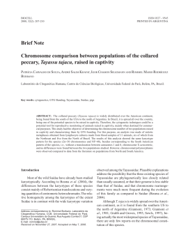
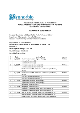
Baixar