Immunology and Cell Biology (2001) 79, 213–221
Research Article
Real-time reverse transcriptase–polymerase chain reaction
(RT–PCR) for measurement of cytokine and growth factor mRNA
expression with fluorogenic probes or SYBR Green I
J I A N L I N Y I N, 1 N I C H O L A S A S H AC K E L , 2 A M A N Y Z E K RY, 2 P E T E R H M c G U I N N E S S , 2
C R A I G R I C H A R D S , 2 K A R I E N VA N D E R P U T T E N, 2 G E O F F R E Y W M c C AU G H A N, 2
JOSETTE M ERIS1 and G ALEX BISHOP2
1
Department of Renal Medicine and 2Centenary Institute, Royal Prince Alfred Hospital and University of Sydney,
New South Wales, Australia
Summary Real-time quantitative reverse transcriptase–polymerase chain reaction (RT–PCR) is the method of
choice for rapid and reproducible measurements of cytokine or growth factor expression in small samples. Fluorescence detection methods for monitoring real-time PCR include fluorogenic probes labelled with reporter and
quencher dyes, such as Taqman probes or Molecular Beacons and the dsDNA-binding dye SYBR Green I. Fluorogenic (Taqman) probes for a range of human and rat cytokines and growth factors were tested for sensitivity and
compared with an assay for SYBR Green I quantification using real-time fluorescence monitoring (PE Applied
Biosystems Model 7700 sequence detector). SYBR Green I detection involved analysis of the melting temperature
of the PCR product and measurement of fluorescence at the optimum temperature. Fluorogenic probes provided
sensitive and reproducible detection of targets that ranged from low (<10 copies/reaction) to high (>107 copies/
reaction) expression. SYBR Green I gave reproducible quantification when the target gene was expressed at moderate to high levels (≥1000 copies/reaction), but did not give consistently reproducible quantification when the
target gene was expressed at low levels. Although optimization of melting temperature improved the specificity of
SYBR Green I detection, in our hands it did not equal the reproducible sensitivity and specificity of fluorogenic
probes. The latter method is the first choice for measurement of low-level gene expression, although SYBR Green
I is a simple and reproducible means to quantify genes that are expressed at moderate to high levels.
Key words: fluorescent resonance energy transfer (FRET) probe, quantitative real-time PCR, SYBR Green I,
Taqman.
Introduction
A number of methods are available for measurement of
cytokine and growth factor mRNA expression including
Northern Blot, RNase protection assays, in situ hybridization
and quantitative reverse transcription–polymerase chain reaction (RT–PCR). Of these, quantitative RT–PCR is the only
method that provides sufficient sensitivity and specificity
to measure low level mRNA expression in small amounts of
tissue, such as biopsy samples.1 This is especially the case for
some cytokines, such as IL-2 and IL-4, that are expressed at
very low levels.1 In spite of its advantages, previous methods
for quantitative RT–PCR have been laborious and difficult to
make consistently reproducible.2 Conventional methods for
quantifying PCR include measurement of end-stage product
by Southern blotting or incorporation of radioactive label;3
Correspondence: GA Bishop, Centenary Institute of Cancer
Medicine and Cell Biology, Royal Prince Alfred Hospital, Missenden
Road, Camperdown, NSW 2050, Australia.
Email: [email protected]
Received 20 September 2000; accepted 2 January 2001.
use of known numbers of competitor DNA fragments during
PCR,2 end-point titration of cDNA and removal of aliquots of
PCR reaction at successive cycle numbers to determine qualitative4 or quantitative5 differences in cytokine mRNA
expression.
Recent parallel developments of equipment for real-time
monitoring of fluorescence within PCR tubes and in the
chemistry of fluorescent detection of PCR product have led
to a major improvement in the ease and reproducibility of
quantitative RT–PCR. Equipment for monitoring of PCR
includes the Model 7700 and Model 5700 sequence detectors
(PE Applied Biosystems, Foster City, CA, USA), and the
LightCycler (Roche, Basel, Switzerland), Sentinel (Stratagene, La Jolla, CA, USA) and Rotorgene (Corbett Research,
Mortlake, Australia) real-time PCR machines. Methods for
fluorescence monitoring of PCR product employ fluorogenic
probes based on fluorescent resonance energy transfer
(FRET) systems such as Taqman and Molecular Beacons or
the dsDNA-binding dye SYBR Green I.6–8 Real-time monitoring of the PCR reaction using these technologies allows
faster and more accurate measurement of gene expression
than conventional PCR quantification. In-tube fluorescent
monitoring of the appearance of PCR product eliminates
214
JL Yin et al.
post-PCR analysis of product, thus removing the errors and
time associated with these steps. It also enables quantification to be based on the early, exponential amplification phase
of the reaction where substrates are not limiting and differences in efficiency of amplification have less effect on the
results. There have been few reports to date of the use of
real-time quantitative PCR for analysis of human or rat
cytokines.7,9,10 Also the conditions used for quantitative PCR
and the monitoring methods for fluorescent detection of PCR
product have not been well characterized. Here we examine
the optimum conditions for use of fluorogenic Taqman probes
and SYBR Green I and compare the sensitivity and specificity of the two systems for measurement of cytokine and
growth factor genes in human and rat cDNA.
Materials and Methods
RNA extraction and cDNA synthesis
Human liver core biopsies (1 mm diameter and 10 mm length) and
rat kidney tissue samples (3 mm3) were immediately frozen in liquid
nitrogen. Total RNA was prepared by the acid phenol-guanidine
method11 and reverse transcribed by methods we have previously
described.12 A yield of 5–15 µg of RNA was obtained from approximately 10 mg of tissue. All samples were reverse transcribed at the
same time, diluted 1:10 in diethylpyrocarbonate-treated autoclaved
H2O (Sigma, St Louis, Mo, USA) and stored in aliquots at –70°C.
PCR standards
The PCR standards for each cytokine or growth factor consisted of
known numbers of molecules of purified PCR product, prepared as
previously described.5 PCR product was purified by gel electrophoresis followed by excision of the band of the correct molecular
weight and separation of DNA (Wizard Minipreps, Promega,
Madison, WI, USA). The concentration of the purified PCR product
DNA was estimated by OD260 and the number of copies/mL of
standard were calculated according to the following formula:
copies/mL =
6.023 × 10²³ × C × OD260
MWt
where C = 5 × 10⁻⁵ g/mL for DNA and MWt = molecular weight of
cytokine PCR product (base pairs × 6.58 × 10² g).
Standards were made to a concentration of 108 copies/µL.
Sodium azide preservative was added to a final concentration of
0.025% w/v and the solution sterile filtered through a 0.22-mm filter
(Millipore, Bedford, MA, USA) and stored in aliquots at 4°C.
Primers and probes
Primers for amplification using SYBR Green I were based on
published sequences for IFN-γ13 and glyceraldehyde-3-phosphate
dehydrogenase (G3PDH).14 For growth arrest-specific gene
6 (Gas6) the sequences used were: forward primer, 5′-GAGGTGTTCGAGAACGACCC-3′; reverse primer, 5′-CTGCATTCGTTGACATCTTTGTC-3′. Sizes of PCR products amplified with
primers for SYBR Green I analysis were: IFN-γ, 285 base pairs
(bp); G3PDH, 456 bp; Gas6, 254 bp. Primers and fluorogenic
probes were designed using Primer Express version 1.0 (PE Applied
Biosystems) and are listed in Table 1. All probes were synthesized
by PE Applied Biosystems. All primers and probes were designed
either to cross large expanses of intronic sequence or to span an
intron–exon boundary except for Gas6, for which the genomic
sequence has not been described. Fluorogenic primers and probes
were limited to amplify products of 150 bases or less and could not
be designed to cross large introns. In this case, one or more of the
primers and probe were designed to cross an exon–intron boundary
(Table 1). Non-reactivity of the primers and probe with contaminating genomic DNA was tested by the inclusion of controls that
omitted the reverse transcriptase enzyme from the cDNA synthesis
reaction (no RT controls).
Real-time PCR quantification using fluorogenic probes
Reaction conditions for fluorogenic PCR mixtures are shown in
Table 2 (columns 2 and 3). The optimum concentration of probe,
primers and MgCl2 was determined in preliminary experiments. The
thermal cycling profile consisted of: stage 1, 50°C for 2 min; stage
2, 95°C for 10 min; stage 3, 95°C for 15 s followed by 60°C for
1 min. Stage 3 was repeated for 40 cycles. In experiments using
fluorogenic probe and primer sets either Buffer A or Universal
Master Mix was used (Table 2).
Real-time PCR quantification using SYBR Green I
SYBR Green I dye (Sigma S9430) was supplied at 10 000× concentration in DMSO. It was stored in 10 µL aliquots at –80°C, and a
working dilution of 50× solution in water was stored as multiple
aliquots at –20°C. Each aliquot of working dilution was discarded
after a single use. The reaction conditions and buffers used are shown
in Table 2. For some experiments 6-carboxy-X-rhodamine (ROX)
(Molecular Probes, Eugene, OR, USA) was used as a positive internal reference. Fifteen milligrams of ROX was dissolved in 0.5 mL
DMSO (Sigma) and diluted to 600 nmol/L concentration with H2O
and stored at –20°C. All SYBR Green I or ROX solutions were protected from light. In experiments using SYBR Green I dye either
Buffer A or Buffer II + ROX was used (Table 2).
As SYBR Green I is not specific for the PCR product and binds
to primer dimers formed non-specifically during all PCR reactions,
it was necessary to obtain an optimum temperature for analysis of
specific product as previously described.7 This was performed on the
PCR product obtained after amplification by raising the temperature
successively through 1°C steps and comparing the melting temperature of specific cytokine or growth factor product with non-specific
product obtained from a no-template control (NTC). The melting
temperature profile on the Model 7700 Sequence Detector consisted
of: stage 1, 95°C for 1 min then 55°C for 2 min; stage 2, 77°C for
1 min then a 1°C increase in temperature every 1 min up to a final
temperature of 92°C at stage 17. Each cytokine or growth factor
analysed by SYBR Green I was examined by this melting temperature profile. The optimum temperature was that which gave the
maximum reading for specific product when the non-specific
product could no longer be detected.
The optimum temperature determined from melting point analysis was then used for quantitative PCR using the following thermal
cycling programme: stage 1, 95°C for 10 min. Stage 2 consisted of
three steps: 95°C for 30 s with a 40-s ramp time to 55°C for 30 s.
After a 20-s ramp time to 72°C for 1 min there was a 17-s ramp time
before data acquisition at the optimum melting temperature for 30 s.
Stage 2 was repeated for 40 cycles.
PCR buffers
The buffers used for these experiments were obtained commercially.
They were PCR Buffer II, PCR Buffer A and Universal Master
Mix (PE Applied Biosystems). PCR Buffer II when diluted to the
final concentration in the PCR reaction mixture (1×) contained
Real-time quantitative RT–PCR method
Table 1
215
Fluorogenic probe and primer sets
Species
Target gene
Primer/probe
Human
IL-2
Forward
Reverse
Probe
Forward
Reverse
Probe
Forward
Reverse
Probe
Forward
Reverse
Probe
Forward
Reverse
Probe
CCAGGATGCTCACATTTAAGTTTTAC
GAGGTTTGAGTTCTTCTTCTAGACACTGA
6FAM-TGCCCAAGAAGGCCACAGAACTGAA-TAMRA
AACAGCCTCACAGAGCAGAAGAC
GCCCTGCAGAAGGTTTCCTT
6FAM-TGCTGCCTCCAAGAACACAACTGA-TAMRA
ACGGCGCTGTCATCGATT
TTGGAGCTTATTAAAGGCATTCTTC
6FAM-CTTCCCTGTGAAAACAAGAGCAAGGCC-TAMRA
TCTCGAACCCCGAGTGACA
GGCCCGGCGGTTCA
6FAM-TAGCCCATGTTGTAGCAAACCCTCAAGC-TAMRA
AGCGGATAATGGAACTCTTTTCTTAG
AAGTTTGAAGTAAAAGGAGACAATTTGG
6FAM-TCTGTCACTCTCCTCTTTCCAATTCTTCAAAATG-TAMRA
Forward
Reverse
Probe
Forward
Reverse
Probe
TGCACCACCAACTGCTTAGC
GGCATGGACTGTGGTCATGAG
VIC-CCTGGCCAAGGTCATCCATGACAACTT-TAMRA
GACCTCGTCCAGCCGATTAA
TGCATTTTTGTATTGGCCTTGA
6FAM-TCAGCCAGTTCCAGCTCCTCATGCA-TAMRA
Forward
Reverse
Probe
Forward
Reverse
Probe
Forward
Reverse
Probe
CCCCATGATGCTCACGTTTA
ATTTTCCAGGCACTGAAGATGTTT
6FAM-TTGCCCAAGCAGGCCACAGAATTG-TAMRA
AGTCTGAAGAACTATTTTAACTCAAGTAGCAT
CTGGCTCTCAAGTATTTTCGTGTTAC
6FAM-CCTTTTGCCAGTTCCTCCAGATATCCAAGA
CGTGGAAATCAATGGGATCAG
GGAAGGGTCGGTTCATGTCA
6FAM-CGAGGTGACCTGGGCACCATCC-TAMRA
IL-4
IL-10
TNF-α
IFN-γ
Human/Rat
G3PDH
Gas6
Rat
IL-2
IFN-γ
TGF-β
Sequence
Length (bp)
87
101
82
87
103
87
113
76
117
74
Exon/intron boundaries are shown as underlined bases. All primer/probe sets were designed to span intronic sequence, except Gas6 for which
the genomic structure is unknown. bp, base pairs; G3PDH, glyceraldehyde-3-phosphate dehydrogenase; Gas6, growth arrest-specific gene 6.
Table 2
Polymerase chain reaction mixtures for fluorogenic probes or SYBR Green I
Buffer II + ROX
(SYBR Green I)
Buffer II (10×)
Buffer A (10×)
Universal Master Mix (2×)
dNTP (10 mmol/L)
MgCl2 (25 mmol/L)
Taq polymerase (5 U/µL)
Gold Taq polymerase (5 U/µL)
SYBR Green I (50×)‡
Probe (20 µmol/L)
ROX (600 nmol/L)
Forward primer (30 µmol/L)
Reverse primer (30 µmol/L)
cDNA (1:10 dilution)
H2O
Total volume
2.5*
–
–
0.5
3.0
0.125
–
0.25
–
2.5
0.25
0.25
5
10.625
25
Buffer A
(SYBR Green I
or Probe)
[1×]†
–
2.5
–
[200 µmol/L]
0.5
[3 mmol/L] 3.0
[0.625 U]
–
0.125
[0.5×]
0.25§
0.25§
[60 nmol/L]
–
[300 nmol/L]
0.25
[300 nmol/L]
0.25
5
12.875
25
[1×]
[200 µmol]
[3 mmolL]
[0.625 U]
[0.5×]
[200 nmol/L]
[300 nmol/L]
[300 nmol/L]
Universal Master
Mix
(Probe)
–
–
12.5
–
–
–
–
–
0.25
–
0.25
0.25
5
6.75
25
[1×]
[200 µmol/L]
[200 nmol/L]
[300 nmol/L]
[300 nmol/L]
*Figures denote µl/reaction. †Figures in [ ] denote the final concentration in the reaction. ‡SYBR Green I was diluted from the supplied
10 000× concentrate as described in the methods section. §Either SYBR Green I or fluorogenic probe was used. dNTP, deoxyribonucleoside
triphosphate; ROX, 6-carboxy-X-rhodamine.
216
JL Yin et al.
Figure 1 Efficiency of fluorogenic probe amplification in two
different buffers. Amplification
plots of IL-4 standards in (a)
Buffer A and (b) Universal Master
Mix. Amplification plots of
standards ranged from 107 copies/
reaction to 10 copies/reaction.
Duplicate reactions are shown
and often appear superimposed.
No-template controls (NTC) were
also included and did not amplify
in either buffer system. The horizontal line shows the setting of
the baseline. The point at which
the amplification plot crosses the
baseline is the CT value.
50 mmol/L KCl and 10 mmol/L Tris HCl (pH 8.3). PCR Buffer
A (1×) contained 50 mmol/L KCl, 10 mmol/L Tris HCl (pH 8.3),
10 mmol/L EDTA and 60 nmol/L ROX. Universal Master Mix (1×)
contained 6% Glycerol, 50 mmol/L KCl, 10 mmol/L Tris HCl (pH
8.3), 7.5 mmol/L MgCl2, 200 µmol/L dATP, 200 µmol/L dCTP,
200 µmol/L dGTP, 400 µmol/L dUTP, 0.05 U/µL AmpliTaq Gold,
0.01 U/µL Amperase UNG and 60 nmol/L ROX.
Statistical analysis
Statview (Abacus Concepts Inc, Berkeley, CA, USA) was used for
statistical analysis. The results are shown as mean ± SD for three
different PCR reactions. Reproducibility between assays was determined by linear regression analysis.
Results
Data expression and analysis
For each assay a standard curve was prepared by serial dilution of a
known number of cytokine or growth factor PCR product molecules.
All samples to be compared were run in the same assay. After
completion of the PCR amplification, data was analysed with the
Sequence Detector version 1.7 software (PE Applied Biosystems).
To maintain consistency, the baseline was set automatically by the
software using data collected from cycle 3 to cycle 15 in most experiments. In some experiments it was necessary to manually override
this setting to optimize analysis. The increase in intensity of fluorescence of the reporter dye (∆Rn)6 was plotted against the cycle
number. The threshold cycle (CT) was calculated by the sequence
detection software as the cycle number at which the ∆Rn crossed the
baseline. Quantification of the samples by the software was calculated from the CT by interpolation from the standard curve to yield a
copy number of cytokine or growth factor cDNA.
Fluorogenic probe optimization and buffer reaction
efficiency
Initial experiments examined the effect of concentration of
primers, probe, Mg2+ and enzyme on efficiency of amplification in PCR. The optimum concentration of these is shown
in Table 2. In addition, the effect of different PCR buffers was
examined. This showed that Universal Master Mix was superior to other buffers tested for amplification of most fluorogenic probe and primer sets. Figure 1 shows an example of
an amplification plot of human IL-4 standards, derived from
IL-4 PCR product, in Universal Master Mix compared to
Buffer A. Buffer A gave poor amplification of this target as
shown by the high CT values for the standards and the shallow
slope of the amplification plot (Fig. 1a). Universal Master
Mix gave much more efficient amplification as shown by the
Real-time quantitative RT–PCR method
Table 3
Sensitivity of fluorogenic probe and primer sets for cytokine detection
Fluorogenic
probe
G3PDH
IL-2
IL-4
IL-10
IFN-γ
TNF-α
Gas6
TGF-β
217
Sensitivity
(copies/reaction)
NTC
(CT)
10 copies
(CT)
103 copies
(CT)
105 copies
(CT)
≤10
≤10
≤10
≤10
≤10
≤10
≤10
≤10
> 40*
> 40
> 40
> 40
> 40
> 40
39.0 ± 1.6
> 40
33.9 ± 0.6
37.2 ± 0.6
36.2 ± 0.7
36.3 ± 0.3
36.1 ± 1.0
34.9 ± 1.0
31.7 ± 1.1
29.1 ± 0.2
27.8 ± 0.7
30.5 ± 0.9
29.1 ± 0.1
29.0 ± 0.8
28.4 ± 1.6
28.8 ± 1.2
26.8 ± 0.5
22.9 ± 0.2
18.9 ± 2.8
23.5 ± 1.1
22.4 ± 0.2
22.1 ± 0.4
23.3 ± 0.5
22.4 ± 1.2
20.2 ± 0.1
17.4 ± 0.1
*The no-template control (NTC) was not amplified in the 40 cycle PCR reaction and a value of > 40 was allotted. All primers and probes
were specific for human sequence and the results show amplification of human standards, except for the growth factors Gas6 and TGF-β, which
were specific for rat sequence and used rat standards. CT, threshold cycle; G3PDH, glyceraldehyde-3-phosphate dehydrogenase; Gas6, growth
arrest-specific gene 6.
lower CT values for all standards and a steep amplification plot
(Fig. 1b). Similar results were obtained when Universal Master
Mix was compared to Buffer II and amplification in Universal
Master Mix equalled or exceeded the efficiency compared to
the other buffers for all primers and probes tested.
No amplification for cytokine or growth factor cDNA was
observed in the no RT controls, showing that the primer and
probe combinations did not amplify potential contaminating
genomic DNA in samples. It also showed the absence of PCR
contamination, a situation that was brought about by a combination of factors. The real-time system allowed analysis
without requirement to open the PCR tube and the PCR setup area was in a separate laboratory to the sequence detector.
Also there were contamination control reagents in the Universal Master Mix that incorporated uracil-n-glycosylase and
dUTP to prevent PCR product carry-over.
There was consistent amplification of G3PDH in the no
RT control, although at a level of expression of <1% of that
observed when reverse transcriptase enzyme was included.
These results for G3PDH were not due to contamination of
the reactions with PCR product as the NTC were consistently
negative. G3PDH appeared to be present in the samples at
low levels, possibly due to small amounts of processed
pseudogenes in genomic DNA contamination in the purified
RNA. Although the G3PDH probe was labelled with VIC
fluorophore, which allows multiplexing of the PCR reaction,
multiplex reactions were not used for these experiments.
was allotted the arbitrary value of >40 in these 40 cycle
reactions. The sensitivity of this technique is shown by the
difference between the CT for the NTC and for the 10 copy
standard, which ranged from >2.8 for IL-2 to >10.9 for
TGF-β (Table 3). This sensitivity reflects the high efficiency
of amplification in these reactions, shown by the slopes of the
standard curves, which were close to the theoretical optimum
of –3.32 and correspond to 100% efficiency of amplification.
This is shown in Table 3 where the difference in CT between
100-fold differences in copy numbers is approximately 6.6.
The primers and probes used for analysis of the human
cytokines were thus able to reproducibly detect as few as 10
copies of standard per reaction. The results for rat G3PDH,
IL-2, IL-4, IL-10 and IFN-γ were similar and each was able
to amplify ≥10 copies of standards (data not shown).
This absolute quantification was obtained using a DNAbased standard curve derived from PCR product. It is also
possible to obtain relative gene expression by serial dilution
of a cDNA sample with high-level expression. We prepared
standard curves of diluted cDNA and found that cDNA
standard curves were linear with high correlations
(R2 = 0.986 ± 0.013) similar to the linear standard curves of
DNA standards. The mean slope of the cDNA standard
curves (–4.0 ± 0.7) was different from that of the DNA standards (–3.25 ± 0.42) (P = 0.01) showing that cDNA standards amplify less efficiently than DNA standards.
Reproducibility of fluorogenic probe detection
Sensitivity of fluorogenic probe detection
Sensitivity of detection of cytokine and growth factor standards with each of the primer/probe combinations is shown in
Table 3. These results were obtained using the optimum
concentration of probe and primer in the Universal Master
Mix. The sensitivity of the method was indicated by the
minimum number of copies of standard that could be consistently amplified and was <10 copies for each cytokine or
growth factor. A further indication of the sensitivity of the
method was the CT at low copy numbers. This is shown in
Table 3 and for amplification of 10 copies/reaction the CT
ranged from 29.1 ± 0.2 to 37.2 ± 0.6. The NTC was not
amplified for all but one template, which meant that the CT
The reproducibility of the fluorogenic probe detection system
was tested in 25 kidney biopsy samples for a highly
expressed target (G3PDH), for intermediate levels of expression (Gas6) and for low-level expression (IFN-γ). Two separate PCR reactions run on different days from stored aliquots
of the same cDNA were compared by linear regression
analysis (Fig. 2). There was highly reproducible measurement at all levels of gene expression when the CT from each
run were compared. The correlation coefficients (R2) for CT
ranged from 0.74 (P < 0.0001) for G3PDH to 0.98
(P < 0.0001) for IFN-γ, showing that even at low levels of
expression there was good reproducibility of amplification.
The lower correlation coefficient for G3PDH compared to
218
JL Yin et al.
Figure 2 Reproducibility of amplification of samples using fluorogenic probes compared to SYBR Green I. Results are shown for
glyceraldehyde-3-phosphate dehydrogenase (G3PDH), growth arrest-specific gene 6 (Gas6), and IFN-γ. Linear regression analysis of two
separate PCR reactions amplified from aliquots of the same cDNA synthesis is shown. The horizontal axis shows the results of PCR1 and
the vertical axis of PCR2. Comparison of reproducibility of threshold cycle (CT) values for fluorogenic probe (column 1) and for SYBR
Green I (column 2) and of the corresponding copy numbers calculated using external standards for fluorogenic probe (column 3) and
SYBR Green I (column 4) are shown. The regression coefficient (R2) is shown for each analysis. All results showed high reproducibility
(P < 0.0001), except for SYBR Green I analysis of IFN-γ (P value = 0.076 for CT and 0.004 for copy numbers).
IFN-γ was due in part to the smaller range of expression of
G3PDH target in the samples, with most samples having CT
between 17 and 19, while for IFN-γ the high correlation
reflects the wider range of CT (from 29 to 40).
There was also very good reproducibility based on the
number of copies of target, calculated from the external standards and shown in Fig. 2. There was a good correlation
between PCR runs with R2 ranging from 0.68 for Gas6 to
0.94 for IFN-γ. The correlation for estimated copy numbers
for each of these three targets was in all cases lower than the
correlation for the corresponding CΤ, which was a pattern that
was observed for all the cytokines and growth factors tested.
This probably reflects the combined variations in amplification of both the sample and the standards required for the
derivation of the number of copies.
Optimization of SYBR Green I quantification
Initial experiments examined the optimum concentration of
primers, Taq polymerase enzyme and SYBR Green I dye.
These reaction conditions were found to be relatively constant for different primer/template combinations and are
shown in Table 2. To obtain optimum specificity of detection
of dsDNA with SYBR Green I, the temperature at which
non-specific product had disappeared was determined by
melting temperature analysis. Results of a typical analysis for
no-template controls compared to specific targets are shown
in Fig. 3. The multicomponent plot of the NTC (Fig. 3a)
showed that at a temperature of 86°C the fluorescence signal
from SYBR Green I was reduced to background levels. In
contrast, there was still a high level of SYBR Green I fluorescence at a temperature of 86°C in the sample that contained
cDNA template and the fluorescent signal did not decrease to
background until a temperature of greater than 89°C (Fig.
3b). A melting temperature profile was performed for all
cytokines and growth factors analysed by SYBR Green I and
showed an almost identical pattern of melting. Consequently,
a temperature of 86°C was selected as the optimum for IFNγ, Gas6 and G3PDH, which had sizes ranging from 254 to
456 base pairs.
Real-time quantitative RT–PCR method
Sensitivity of SYBR Green I detection
The sensitivity of detection was tested in Buffer A and Buffer
II + ROX and gave high efficiency in both buffer systems.
The CT for human/rat G3PDH and Gas6 in the two different
buffer systems are shown in Table 4. There was little or no
difference between the two buffers for efficiency of amplification as shown by the similar CT values over the range of
copy numbers tested, or of reproducibility, as shown by the
low variability of the analysis. The main difference was that
Figure 3 Optimization of fluorescence measurement for SYBR
Green I by melting temperature analysis. The fluorescence signal
generated by SYBR Green I binding to dsDNA amplified with
primers for glyceraldehyde-3-phosphate dehydrogenase (G3PDH)
is shown in the no-template control (NTC) (a), compared to the
positive control containing cytokine template (b). The melting
temperature profile of the reaction consisted of an initial denaturation followed by re-annealing then a stepwise increase in
temperature from 77°C to 92°C as described in the methods. At
86 °C there was no signal from the PCR artifact including primerdimers present in the NTC while there was still significant signal
in the positive control reaction.
Table 4
G3PDH
Gas6
219
Buffer A gave a lower background level for the NTC, shown
by the CT value of >40 cycles for both primer combinations
tested.
Reproducibility of SYBR Green I detection
The reproducibility of the SYBR Green I detection system,
tested in 25 kidney biopsy samples for a highly expressed
target (G3PDH) for intermediate expression (Gas6) and for
low-level expression (IFN-γ) is shown in Fig. 2. The aliquots
of cDNA used for these experiments were from the same
cDNA as used for fluorogenic probe quantification. Two
separate PCR reactions run on different days from stored
aliquots of the same cDNA were compared by linear regression analysis. There was highly reproducible amplification,
based on comparison of CT, for the highly expressed target
G3PDH (R2 = 0.94), where most samples expressed between
1 × 10⁵ and 1.8 × 10⁶ copies/reaction. There was also acceptable reproducibility for Gas6 (R2 = 0.68; P < 0.0001), where
most samples were expressed at intermediate levels of
approximately 1 × 10⁴–3 × 10⁵ copies/reaction. Reproducibility of IFN-γ measurement using SYBR Green I was poor
compared to the reproducibility of fluorogenic probes. SYBR
Green I gave a poor correlation between two different PCR
reactions (R2 = 0.10) for CT analysis compared to an R2 of
0.98 for fluorogenic probe analysis of the same samples
(Fig. 2). Similarly there was poor reproducibility of the
SYBR Green I analysis for IL-2, which was also expressed
at low levels (data not shown).
A further difference between SYBR Green I and fluorogenic probes was the different estimates of copy numbers
obtained, particularly at low-level gene expression. This was
most obvious for the number of copies/reaction of IFN-γ
obtained with SYBR Green I (range 0–9.3 × 10³, median
675) compared to fluorogenic probe (0–117, median 0.5). It
was less apparent at higher level expression as shown by
Gas6 (SYBR Green I range 1.2 × 10⁴–3.2 × 10⁵, median
8.4 × 10⁴ compared to fluorogenic probe range 2.2 × 10³–
7.1 × 10⁴, median 2.4 × 10⁴). It was much less marked at the
high level of expression of G3PDH (SYBR Green I range
2.1 × 10⁴–2.0 × 10⁶, median 6.4 × 10⁵ compared to fluorogenic probe range 1.1 × 10⁴–1.6 × 10⁶, median 2.8 × 10⁵).
Discussion
Two methods for fluorescence detection in real-time quantitative RT–PCR analysis of cytokine and growth factor expression were optimized and compared. Fluorogenic probes were
Sensitivity of SYBR Green I detection in Buffer A and Buffer II + 6-carboxy-X-rhodamine (ROX)
Buffer
Sensitivity
(copies/reaction)
NTC
10 copies
(CT)
103 copies
(CT)
105 copies
(CT)
II + ROX
A
II + ROX
A
≤1000
≤1000
≤1000
≤1000
33.7 ± 7.4
> 40.0*
35.2 ± 5.6
> 40.0
31.3 ± 3.5
29.2 ± 1.6
25.8 ± 0.5
27.7 ± 0.7
22.4 ± 2.4
22.6 ± 1.9
20.8 ± 1.5
22.3 ± 1.6
14.5 ± 0.9
14.6 ± 1.1
14.0 ± 0.1
14.9 ± 0.9
*The no-template control (NTC) was not amplified in the 40 cycle PCR reaction and a value of > 40 was allotted. G3PDH, glyceraldehyde3-phospate dehydrogenase; Gas6, growth arrest-specific gene 6; ROX, 6-carboxy-X-rhodamine.
220
JL Yin et al.
extremely sensitive, detecting fewer than 10 copies of target
per reaction and providing excellent reproducibility of analysis at this level of detection. These probes were specific for
the processed form of the genes, present in mRNA and not
the genomic form, as no RT controls were consistently negative for all cytokines and growth factors. No RT controls for
G3PDH were always positive, due to the presence of
processed pseudogenes in contaminating DNA in the RNA
preparation.12 In spite of this, the level of false positive
expression of G3PDH in the no RT controls was always
markedly less than the level of mRNA expression in the
samples and did not affect accurate quantification. SYBR
Green I analysis of genes expressed at moderate to high
levels gave excellent reproducibility; however, the reproducibility was poor for IFN-γ, which was expressed at lower
levels.
The fluorogenic probe system proved to be more difficult
to optimize than SYBR Green I and was more sensitive to the
buffer conditions used. The sensitivity of this system to the
reaction conditions might have been due to the fluorogenic
probe interfering with amplification by the forward and
reverse primers or to dependence of probe cleavage on the
reaction conditions. The original description, which used
exonuclease cleavage of labelled probe for quantitative PCR,
found that the presence of probe did not affect the efficiency
of amplification.15 Subsequent studies have found that the
efficiency of probe cleavage was dependent on a range of
conditions, including the type and concentration of divalent
cation16 or the type of DNA polymerase used.17 In spite of the
dependence of the fluorogenic probe system on the reaction
conditions, under optimum conditions, the reaction progressed with high efficiency (approximately 100%) leading
to high sensitivity of detection. SYBR Green I amplified at
optimal efficiency in a wider range of buffers than the fluorogenic probe system, possibly because it was not dependent
on a labelled oligonucleotide that could potentially reduce the
efficiency of amplification or that had a requirement for efficient nucleolytic cleavage.
The experiments shown used external standards for the
quantification of cytokines or growth factors. These consisted of purified PCR products that spanned the target
regions for forward and reverse primers used for fluorogenic
probe or SYBR Green I detection. These standards were
useful to estimate the sensitivity of the reaction and could
also be used to derive an estimate of the number of copies of
cytokine mRNA present in the unknown samples as we have
previously described.12 It is unlikely that this estimate is an
accurate reflection of the actual number of cytokine mRNA
molecules present in the sample for several reasons. The
estimate does not take account of the efficiency of purification of RNA from the original sample or of cDNA synthesis
during reverse transcription. Neither does it account for differences in efficiency of amplification between cDNA of
samples and PCR product of standards. The results here show
that DNA standards made from PCR product amplified more
efficiently than cDNA standards in the fluorogenic probe
assay. This latter difference might have been the basis of
discrepancies between fluorogenic probes and SYBR Green
I in the estimation of gene copy numbers. Measurement of
IFN-γ expression by the fluorogenic probe method gave an
estimate of copy numbers approximately 100-fold less than
the estimate using SYBR Green I. This difference was less
marked for Gas6, which was expressed at higher levels and
was barely detected for G3PDH, the gene expressed at the
highest level. Less efficient amplification of samples than
standards in the fluorogenic probe system would provide an
explanation for this difference.
In spite of this, the use of the same external standards
allows for the comparison of results obtained in separate PCR
runs on different machines or in separate laboratories. They
also provide a numerical estimate of the differences in cytokine expression levels between samples. Ribonucleic acid
standards are likely to overcome some of the limitations of
standards prepared from PCR product described, but are
much more time consuming to prepare and are less stable
than DNA standards.
There have been few reports examining the reproducibility and sensitivity of fluorogenic probes to measure lowlevel human cytokine mRNA expression especially with
primers and probes for short amplicons.9,10 The efficiency of
PCR amplification is higher for short amplicons, which
should lead to improved sensitivity of detection.18 Quantification of murine,19 equine20 and feline21 mRNA expression
has also been reported using fluorogenic probes and the
sensitivity of this system for murine IL-12 and β-actin has
been examined.19 In this latter case the method was able to
detect ≤1000 copies of IL-12 plasmid DNA and ≤100 copies
of β-actin plasmid. Sensitivity of feline G3PDH primers and
probe was reported to be ≤10 copies/reaction.21 The efficiency of all our fluorogenic primer/probe combinations,
which were designed to amplify short amplicons, was tested
and found to be highly sensitive, with detection of ≤10
copies/reaction.
The sensitivity of detection of cytokines or growth factors
using SYBR Green I dye for the target genes that we examined was ≤1000 copies/reaction. SYBR Green I has previously been used for quantification of rat cytokines IL-1β,
IL-6, TNF-α, GM-CSF, granulocyte colony-stimulating
factor (G-CSF) and MCP-1.7 Although this method was
initially unable to measure <1000 copies of template per
reaction, its sensitivity was improved to ≤10 copies per reaction by increasing the temperature of fluorescence reading to
86°C and by using a heat-denaturable antibody to Taq polymerase enzyme to prevent PCR artifact formation.22 Our
experiments using SYBR Green I dye also used hightemperature measurement of fluorescence at 86°C and a hotstart enzyme that was inactive until commencement of
thermal cycling. This method reproducibly amplified G3PDH
and Gas6, which were expressed at ≥10 000 copies per reaction. We were, however, unable to obtain reproducible
measurement of IFN-γ, which was expressed at lower levels,
ranging from 0 to 9300 (median 900) copies per reaction.
Our results demonstrate that the ability of SYBR Green I
dye to reproducibly measure gene expression was thus, in our
hands, limited to genes that were expressed at ≥1000
copies/reaction. The ability of SYBR Green I to reproducibly
measure cytokines or growth factors that are expressed at
lower levels might be improved by the use of alternative
primers that are better able to specifically amplify the target.
Further optimization of the reaction to favour amplification
of the specific PCR product and minimization of PCR artifact could also improve sensitivity. Also, dedicated software
Real-time quantitative RT–PCR method
that can more accurately measure and control the temperature
of PCR artifact melting, such as that available with some
recently developed real-time PCR machines, might improve
the sensitivity of detection.
In conclusion, we report the rapid and reproducible quantification of cytokine and growth factor mRNA in small
samples using real-time RT-PCR. Two methods were used for
fluorescent detection of PCR product: fluorogenic probes and
SYBR Green I dye. Both methods gave rapid and reproducible amplification of gene expression at expression levels
of >1000 copies/reaction. SYBR Green I was simpler and
cheaper than fluorogenic probes as it could be used with any
set of amplification primers. In contrast, fluorogenic probes
were more difficult to design; more demanding in terms of
the reaction conditions required for optimum amplification
and were expensive to synthesize. The advantage of fluorogenic probes was that they were exquisitely sensitive and
detected ≤10 copies of cytokine or growth factor per reaction
for all primer and probe sets tested. This resulted in reproducible measurement of cytokines at low-level expression.
Additionally, the internal probe increases the specificity of
detection. In our experience, fluorogenic probes were thus
superior to SYBR Green I for measurement of genes
expressed at low levels of <1000 copies per reaction.
Acknowledgements
This work was supported by grants from the Wellcome Trust
and from the Clive and Vera Ramaciotti Foundation. The
support of the New South Wales Health Department through
its research and development infrastructure grants programme is gratefully acknowledged. N. Shackel is the
recipient of a National Health and Medical Research Council
of Australia Sir Gustav Nossal Scholarship.
References
1 Dallman MJ. Cytokine gene expression: analysis using northern
blotting, polymerase chain reaction and in situ hybridization.
Immunol. Rev. 1991; 119: 163–80.
2 Gilliland G, Perrin S, Blanchard K, Bunn HF. Analysis of
cytokine mRNA and DNA: detection and quantitation by
competitive polymerase chain reaction. Proc. Natl Acad. Sci.
USA 1990; 87: 2725–9.
3 Takeuchi T, Lowry RP, Konieczny B. Heart allografts in murine
systems: The differential activation of TH2 like effector cells in
peripheral tolerance. Transplantation 1992; 53: 1281–94.
4 Dallman MJ, Larsen CP, Morris PJ. Cytokine gene expression
in vascularized organ grafts: analysis using semiquantitative
polymerase chain reaction. J. Exp. Med. 1991; 174: 493–6.
5 Bishop GA, Rokahr KL, McGuinness PH et al. Quantitative
reverse transcriptase-PCR amplification of cytokine mRNA in
liver biopsy specimens using a non-competitive method.
Immunol. Cell. Biol. 1997; 75: 142–7.
6 Gibson UE, Heid CA, Williams PM. A novel method for real
time quantitative RT–PCR. Genome Res. 1996; 6: 995–1001.
221
7 Morrison TB, Weis JJ, Wittwer CT. Quantification of low-copy
transcripts by continuous SYBR Green I monitoring during
amplification. Biotechniques 1998; 24: 954–9.
8 Tyagi S, Kramer FR. Molecular beacons: probes that fluoresce
upon hybridization. Nat. Biotechnol. 1996; 14: 303–8.
9 Hartel C, Bein G, Kirchner H, Kluter H. A human whole-blood
assay for analysis of T-cell function by quantification of
cytokine mRNA. Scand. J. Immunol. 1999; 49: 649–54.
10 Kruse N, Pette M, Toyka K, Rieckmann P. Quantification of
cytokine mRNA expression by RT–PCR in samples of previously frozen blood. J. Immunol. Methods 1997; 210: 195–203.
11 Chomczynski P, Sacchi N. Single-step method of RNA isolation
by acid guanidinium thiocyanate-phenol-chloroform extraction.
Anal. Biochem. 1987; 162: 156–9.
12 Bishop GA, Rokahr KL, Napoli J, McCaughan GW. Intragraft
cytokine mRNA levels in human liver allograft rejection
analysed by reverse transcription and semiquantitative polymerase chain reaction amplification. Transpl. Immunol. 1993;
1: 253–61.
13 Sun J, McCaughan GW, Matsumoto Y, Sheil AGR, Gallagher
ND, Bishop GA. Tolerance to rat liver allografts 1. Differences
between tolerance and rejection are more marked in the B cell
compared with the T cell or cytokine response. Transplantation
1994; 57: 1349–57.
14 Lowes MA, Bishop GA, Cooke BE, Barnetson RS, Halliday
GM. Keratoacanthomas have an immunosuppressive cytokine
environment of increased IL-10 and decreased GM-CSF compared to squamous cell carcinomas. Br. J. Cancer 1999; 80:
1501–5.
15 Holland PM, Abramson RD, Watson R, Gelfand DH. Detection
of specific polymerase chain reaction product by utilizing the
5′–3′ exonuclease activity of Thermus aquaticus DNA polymerase. Proc. Natl Acad. Sci. USA 1991; 88: 7276–80.
16 Lyamichev V, Brow MA, Dahlberg JE. Structure-specific
endonucleolytic cleavage of nucleic acids by eubacterial DNA
polymerases. Science 1993; 260: 778–83.
17 Kreuzer KA, Bohn A, Lass U, Peters UR, Schmidt CA. Influence of DNA polymerases on quantitative PCR results using
TaqMan probe format in the LightCycler instrument. Mol. Cell.
Probes 2000; 14: 57–60.
18 McCulloch RK, Choong CS, Hurley DM. An evaluation of
competitor type and size for use in the determination of mRNA
by competitive PCR. PCR Methods Applic. 1995; 4: 219–26.
19 Overbergh L, Valckx D, Waer M, Mathieu C. Quantification of
murine cytokine mRNAs using real time quantitative reverse
transcriptase PCR. Cytokines 1999; 11: 305–12.
20 Leutenegger CM, von Rechenberg B, Huder JB et al. Quantitative real-time PCR for equine cytokine mRNA in nondecalcified
bone tissue embedded in methyl methacrylate. Calcif. Tissue Int.
1999; 65: 378–83.
21 Leutenegger CM, Mislin CN, Sigrist B, Ehrengruber MU,
Hofmann-Lehmann R, Lutz H. Quantitative real-time PCR for
the measurement of feline cytokine mRNA. Vet. Immunol.
Immunopathol. 1999; 71: 291–305.
22 Menon RS, Chang YF, St. Clair J, Ham RG. RT–PCR artifacts
from processed pseudogenes. PCR Methods Applic. 1991; 1:
70–1.
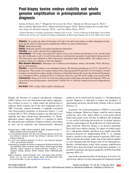
Download