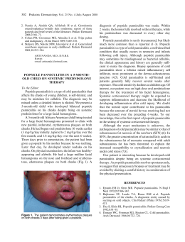
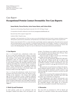
http://dx.doi.org/10.1590/0103-8478cr20150254
Ciência Rural, Santa Maria, Online
Pyogranulomatous dermatitis and panniculitis due to Nocardia
nova in a cat.
ISSN 0103-8478
Pyogranulomatous dermatitis and panniculitis due to Nocardia nova in a cat
Dermatite e paniculite piogranulomatosas causadas por Nocardia nova em um gato
Guilherme Reis BlumeI Christine Souza MartinsI Letícia Beatriz MatterII
Agueda Palmira Castagna de VargasII Letícia Batelli de OliveiraI
Janildo Ludolf Reis JuniorI Fabiano José Ferreira de Sant’AnaI*
– NOTE –
ABSTRACT
This report describes the clinical, pathological and
microbiological findings of an uncommon infection in a cat by
Nocardia nova. A 3-year-old male domestic short hair cat with
an ulcerated and exudative cutaneous wound was presented for
clinical examination. Samples were collected for histopathology
and bacteriology diagnosis. Microscopically, the lesion was
diagnosed as pyogranulomatous dermatitis and panniculitis with
large and irregular colonies of branching filamentous bacterium.
Skin bacteriological culture showed gram-positive rods and
partially acid-fast branching filaments by gram and kinyoun
staining, respectively. The identity of Nocardia nova was confirmed
by 16S rDNA sequencing and phylogenetic analysis. This is the
first case of pyogranulomatous dermatitis and panniculitis in a cat
caused by Nocardia nova reported in Brazil.
Key words: cat, dermatopathology, nocardiosis.
RESUMO
Este relato descreve os achados clínicos, patológicos
e microbiológicos de uma infecção incomum por Nocardia nova
em um gato. Um gato macho, sem raça definida, de 3 anos de
idade, apresentou ferida cutânea exsudativa e ulcerada. Amostras
da lesão foram coletadas para histopatologia e bacteriologia.
Histologicamente, a lesão consistiu de dermatite e paniculite
piogranulomatosas associadas a colônias grandes e irregulares
de bactérias filamentosas e ramificadas. O cultivo bacteriológico
revelou bacilos filamentosos, gram-positivos, parcialmente ácido
resistentes, visualizados pelas colorações de Gram e Kinyoun,
respectivamente. A identificação da Nocardia nova foi confirmada
pelo sequenciamento 16S rDNA e análise filogenética. Este é o
primeiro caso de paniculite e dermatite piogranulomatosas em um
gato causado por Nocardia nova registrado no Brasil.
Palavras-chave: gato, dermatopatologia, nocardiose.
Nocardia spp. are aerobic, grampositive, partial acid-fast, filamentous bacteria and
ubiquitous environmental saprophytes responsible
for granulomatous or suppurative infections
(RIBEIRO et al., 2008). The bacteria belong to the
order Actinomycetales, family Nocardiacea and
the main species that affect domestic animals are N.
asteroides, N. brasiliensis, N. otitidiscaviarum, and
N. nova (HARADA et al., 2009). N. brasiliensis is
the most common species affecting humans in tropical
countries, such as central and south America (SYKES,
2012), where as N. otitidiscaviarum was considered
the specie more isolated in dogs from Brazil (RIBEIRO
et al., 2008) and has been occasionally reported in a cat
from Spain (LUGUE et al., 2002).
Nocardiosis can affect many domestic
animals, but is uncommon in cats (RIBEIRO et al.,
2008). Immunosuppression associated to multidrug
therapy or concurrent infections is an important
predisposing factor to the development of the
disease in animals and humans (RIBEIRO et al.,
2002). Clinically, nocardios can present as systemic,
pulmonary, nervous, and cutaneous form (BEAMAN
& BEAMAN, 1994). Cutaneous nocardiosis is the
most common form in cats and causes localized
nodules, cellulitis, and abscesses (SYKES, 2012).
The route of infection and transmission occurs
by inhalation of aerosols containing the agent, ingestion
or direct contact with contaminated environment
I
Laboratório de Patologia Veterinária (LPV), Universidade de Brasília (UnB), Campus Universitário Darcy Ribeiro, 70910-900, Brasília,
DF, Brasil. E-mail: [email protected]. *Corresponding author.
II
Laboratório de Bacteriologia (LABAC), Centro de Ciências Rurais (CCR), Universidade Federal de Santa Maria (UFSM), Santa Maria,
RS, Brasil.
Received 02.23.15
Approved 03.28.15
Returned by the author 07.03.15
CR-2015-0254
Blume et al.
(SYKES, 2012). Diagnosis is made by history,
clinical signs and hematological, microbiological,
anatomopathological, and molecular tests (RIBEIRO
et al., 2002). This study aims to report the clinical,
pathological and microbiological findings of a case of
feline pyogranulomatous dermatitis and panniculitis
associated to Nocardia nova. This specie has not been
yet associated to nocardiosis in cats in Brazil.
A 3-year-old, spayed, male, short hair
cat presented history of a progressive skin lesion in
the thoracic region for about six months. Clinical
examination showed multiple channels connecting
the ulcerated skin with the subcutis and extended
from the right scapula to the right thorax that drained
serosanguineous secretion (Figure 1A). Hematologic
results revealed neutrophilic leukocytosis with leftshift and toxic neutrophils. Serological tests for feline
immunodeficiency (FIV) and feline leukemia virus
(FeLV) were negative.
The animal was treated with antibiotics
including enrofloxacin (5mg kg-1, Baytril® Flavour,
Bayer, São Paulo/SP, Brazil), amoxicillin associated
with potassium clavulanate (20mg kg-1, Synulox®,
Zoetis, Campinas/SP, Brazil) and ampicillin
(25mg kg-1, Ampicilina Veterinária Oral®, Vetnil,
Louveira/SP, Brazil) but there was no clinical
improvement. The animal was also treated with
methylprednisolone acetate (20mg kg-1, DepoMedrol®, Pfizer, Guarulhos/SP, Brazil) by another
veterinarian during a trip. After this last treatment,
the cutaneous lesion increased considerably.
Due to worsening of the skin lesions,
surgical excision was performed. Samples were
collected for bacteriological and molecular diagnosis.
Additional samples was fixed in buffered 10%
formalin, routinely processed for histopathology, and
stained by hematoxylin and eosin, Brown Y Brenn
(Gram), and modified Fite-Faraco.
Microscopically, expending, infiltrating
and distorting the dermis, panniculum and adjacent
skeletal muscle, there were focally extensive areas with
multiple pyogranulomas containing large, central, and
irregular colonies of branching filamentous bacteria
(Figure 1C). The organism was Brown Y Brenn- and
Fite-Faraco positive (Figure 1D). In addition, adjacent
to the bacteria there were numerous degenerate and
intact neutrophils with an outer layer of epithelioid
macrophages and fewer lymphocytes, plasma
cells, and rare multinucleated giant cells. Moderate
proliferation of loose fibrovascular stroma and edema
Figure 1 - Pyogranulomatous dermatitis and panniculitis due to Nocardia nova. A: Gross lesion shows the ulcerated skin with multiple channels
connecting with the subcutis. B: Skin smears showing gram-positive branching filamentous bacterium (arrows). 1000 X. C: Multiple
pyogranulomas containing large, central, and irregular colonies of branching filamentous bacteria (arrows) infiltrating and distorting
the dermis and the panniculum. D: Organisms were Fite-Faraco positive (arrows). E: Phylogenetic inference based on 16S rDNA
sequence showing the homology between N. nova isolate SBP 20/14 (GenBank accession number KP025810) and others 16S
rDNA sequences of Nocardia species. The phylogenetic tree was built using neighbor-joining algorithm implemented in MEGA
5.2 software program. The percentage of replicate trees in which the associated tax clustered together in the bootstrap test (1.000
replicates) is shown next to each branch. Rhodococcus opacus B4 (AP011115.1) was used as outgroup.
Pyogranulomatous dermatitis and panniculitis due to Nocardia nova in a cat.
around pyogranulomas were observed. Multifocally,
there are multiple anastomosing channels filled with
necrotic debris and inflammatory cells. Often, these
channels were interconnected with skin ulcers.
Gram staining of the skin smear showed
gram-positive branching filamentous bacterium
(Figure 1B). Colonies were isolated from the skin on
blood agar and brain heart infusion agar after 3 days
at 37°C of incubation. Bacteria isolation was possible
only from the removed tissue. All colonies had the
same aspect and resembled Nocardia colonies. Due
to the rapid and reliable result, molecular diagnosis
by 16S rDNA was performed instead the biochemical
tests. DNA from colonies (SBP 20/14) was extracted
by lysis with cetyltrimethylammonium bromide
(CTAB) (SAMBROOK & RUSSELL, 2001).
Molecular diagnosis was obtained by analyzing the
partial sequence (~1400 base pairs) of the16S rRNA
gene amplified with universal primers (FREDRICKS
& RELMAN, 1998). PCR product was sequenced
by ACT Gene Análises Moleculares (Centro de
Biotecnologia, UFRGS, Brazil) using the automatic
sequencer ABI-PRISM 3100 genetic analyzer
(Applied Biosystems). Chromatograms obtained by
sequencing were analyzed, and consensus sequences
were assembled using 4 sStaden package programs
(STADEN et al., 2000).
In order to compare the consensus
sequence (~1400bp) with other sequences available
on GenBank database (http://www.ncbi.nlm.nih.gov)
BLASTN tool was performed. The closest relative
16S rDNA sequence was used for the bacterial
identification. The sample showed 100% homology
to N. nova (Evalue 0.0).
In addition, phylogenetic inference of
the consensus with other GenBank sequences was
preceded. The phylogenetic inference showed
the strong homology of the isolate with others
N. nova strains and the relationship with others
species available in Genbank dataset (Figure 1E).
Furthermore, the cat presented clinical improvement
and there was no indication of recurrence of skin
lesions at one year after the surgical removal.
Nocardia spp. has a worldwide distribution,
and is considered an opportunistic pathogen. Bacteria
are facultative intracellular organisms which contain
important virulence factors, such as resistance
to phagocytosis and complex cell wall lipids
(BEAMAN & BEAMAN, 1994). Nocardiosis affects
especially dogs and cats, but can infect other species,
such as cattle, horses, pigs, poultry, and humans
(RIBEIRO et al., 2008). The main lesions observed
in companion animals include skin lesions, pyothorax
and pneumonia (MALIK et al., 2006). In the present
case, the lesions were restricted to skin and adjacent
tissues and there were no clinical signs compatible
with pulmonary and disseminated involvement.
In a study of 53 cases of canine nocardiosis
from California (USA), the disease was diagnosed
more frequently in male and young animals
(BEAMAN & SUGAR, 1983), as noted in the
current report. Some authors cite higher prevalence
of nocardiosis in male cats because they are often
roaming or fighting and the lesions are associated
to scratches or bite wounds (SYKES, 2012).
Usually, animals with cutaneous nocardiosis can
present transcutaneous transmission and contagion
by penetration of the agent into the skin, causing
cellulitis, pustules, pyoderma, or abscesses (MALIK
et al., 2006). In the current report, it was not possible
to determine the precise route and source of infection.
Renal transplantation, retroviral infection,
or glucocorticoid treatment are considered important
predisposing factors for the disease in cats (SYKES,
2012). However, these were not the cases. Serological
tests for feline immunodeficiency (FIV) and feline
leukemia virus (FeLV) were negative. Glucocorticoid
was used as treatment but it was before the conclusion
of the nocardiosis diagnosis, what increased
significantly the cutaneous lesion.
Hematologic findings were very similar
to a previous study that described also monocytosis
and hyperglobulinemia in a cutaneous nocardiosis
in a dog (GOLYŃSKY et al., 2006). In the current
case, the histopathology and mainly the molecular
analysis were paramount for the diagnosis of
pyogranulomatous dermatitis and panniculitis due to N.
nova. Histopathological lesions were similar to those
visualized in other previously reported cases (LUGUE
et al., 2002; MALIK et al., 2006). Actinomyces spp.,
Streptomyces spp., Mycobacterium spp., Rhodococcus
spp.,
Corynebacterium
spp.,
Dermatophilus
congolensis and saprophytic fungi should be considered
as differentials for cutaneous nocardiosis (RIBEIRO et
al., 2002; MALIK et al., 2006).
By indication of the owner and due
to prolonged time of skin infection, the surgical
treatment with removal of the affected tissues was
performed. This treatment was considered effective
and curative, because there was no recurrence of the
lesion after 1 year of procedure.
Further
epidemiologic,
pathological
and molecular studies are necessary to determine
the real prevalence, pathogenesis and specific
route of the infection by N. nova in cats. To the
author’s knowledge, this is the first feline case of
Blume et al.
pyogranulomatous dermatitis and panniculitis due to
N. nova reported in Brazil.
ETHICS COMITTE AND BIOSECURITY
This report met with an animal runs in the routine of
the University Veterinary Hospital – UnB – and was not part of any
project, with only one case report.
REFERENCES
BEAMAN, B.L.; SUGAR, A.M. Nocardia is naturally acquired
and experimental infections in animals. Journal of Hygiene, v.91,
p.393-419, 1983.
BEAMAN, B.L.; BEAMAN, L. Nocardia species: host-parasite
relationships. Clinical Microbiology Reviews, v.7, p.213-264,
1994. Available from: <http://cmr.asm.org/content/7/2/213.full.
pdf+html>. Accessed: May 12, 2015. doi: 10.1128/CMR.7.2.213.
FREDRICKS, D.N.; RELMAN D.A. Improved amplification of
microbial DNA from blood cultures by removal of the PCR inhibitor
sodium polyanetholesulfonate. Journal of Clinical Microbiology,
v.36, p.2810-2816, 1998. Available from: <http://jcm.asm.org/
content/36/10/2810.full.pdf+html>. Accessed: May 12, 2015.
GOLYŃSKY, M. et al. Cutaneous Nocardiosis in a dog – Clinical
case presentation. Bulletin of the Veterinary Institute in Pulawy,
v.50, p.47-50, 2006.
HARADA, H. et al. Cutaneous nocardiosis in a cat. Journal of
Veterinary Medical Science, v.71, p.785-787, 2009. Available
from: <https://www.jstage.jst.go.jp/article/jvms/71/6/71_6_785/_
pdf>. Accessed: May 12, 2015. doi: 10.1292/jvms.71.785.
LUGUE, I. et al. Nocardia otitidiscaviarum infection in a cat.
Veterinary Record, v.151, p.488, 2002.
MALIK, R. et al. Nocardia infections in cats: a retrospective multiinstitutional study of 17 cases. Australian Veterinary Journal,
v.84, p.235-245, 2006. Available from: <http://onlinelibrary.wiley.
com/doi/10.1111/j.1751-0813.2006.00004.x/epdf>. Accessed: May
12, 2015. doi: 10.1111/j.1751-0813.2006.00004.x.
RIBEIRO, M.G. et al. Nocardiose cutânea associada à cinomose em
cães. Relato de dez casos. Clínica Veterinária, v.39, p.34-42, 2002.
RIBEIRO, M.G. et al. Nocardiosis: an overview and additional
report of 28 cases in cattle and dogs. Revista do Instituto de
Medicina Tropical, v.50, p.177-185, 2008. Available from:
<http://www.scielo.br/pdf/rimtsp/v50n3/7407.pdf>.
Accessed:
May 12, 2015. doi: 10.1590/S0036-46652008005000004.
SAMBROOK, J.; RUSSELL, D.W. Molecular cloning. New
York: CSHL, 2001. 2344p.
STADEN, R. et al. The staden package, 1998. Methods in
Molecular Biology, v.132, p.115-130, 2000.
SYKES, J.E. Actinomycosis and nocardiosis. In: GREENE,
C.E. In: Infectious diseases of the dog and cat. 4.ed. St. Louis:
Elsevier, 2012. p.451-461.
Download