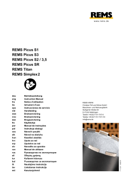
LOBO'S DI S EA S E No. 1 • Azulay 41 Fig. 3. Top, mi croscopic picture of the gland show ing a heavy infil trate of Glenospo loboi (low power). Fig. 4. Bot tom, microscopic picture of the gland showing a rella heavy infiltrate of Glenospore//a lo boi (high power). Case Report merous round bodies of nodule mem A 34-year-old man who was born in brane, presenting as single or multiple the Amazon area had a tumor on the left buds ear which had been present for the past (catenular). 3 years. At first, the area was pruritic, but grown. gradually a small nodule appeared at the site. The lesion became tender and eventually developed into a lobulated, springs, with linking disposition The organism could not be Histological study revealed epidermal atrophy. In the dermis, there was lym phocytic and histiocytic infiltration with glossy tumor which resembled a keloid. several Langhans and foreign body giant At the left postauricular area, there was cells. Numerous rounded parasites with a 2 cm lymph node. Mycological examination revealed nu- birefringent membranes and central nu clei were seen. Some showed a budding INTERNATIONAL JOURNAL OF DERMATOLOGY 42 picture, and the catenular aspect both inside and outside the giant cells was observed. Microscopic examination of the lymph node demonstrated a lymphocytic exu date with giant cells. Numerous parasites, both grouped and isolated, were seen. In addition, birefringent membranes similar to Glenosporella loboi, inside and 3-(p-Chloroanilino) 1 O-(p-chlorophenyl)-2, ·1 O-dihydro-2-(isopropylimino) phenazine Clofazimine References 1. conhecimento das granulomatose blasto micoiles; o agente etiol6gico da micose de Jorge Lobo. Med. Cirurg. Brasil. 48:143, 1940. 3. Azulay, R. D., Andrade, L. C., Silva, D., e Carneiro, J. A., Reprodu<;ao experimental da blastomicose de Jorge Lobo. An. Bras. 4. Azulay, R. improvement. Dermatol. 434:161, 1968. Discussion The disease is characterized by iso seu like aspect is rather common. In addi tion, infiltrative, gummatous, ulcerated and verrucous lesions may be observed. Isolation of the parasite is difficult. In 5. obtained an isolate in an artificial me 7. round forms identical to those found in tissues, using 199 medium. This medium 8. cultures. the etiological agent of Lobo's disease has behaved as an anthro pophile and has not been isolated from animals or vegetables. Good climatic conditions for its growth are found in the Brazilian Amazon area and in those neighboring countries where the average annual temperature is above 24°C and the rainfall at least 2.000 mm. All of the reported patients have been from such areas except for one from Costa Rica and another from Panama.9 Inflammatory reactions are not neces sari'ly found; however, the lymph nodes have been involved in 7 of the 98 re ported cases.11-1s We can offer no satis factory explanation for these variations. no 1968. Azulay, R. D., Andrade, L. C., e Carneiro, J. A., Lobo's blastomycoses: new experiments L. C., Blastomicose de Jorge Lobo. Con tribui<;ao ao estudo da etiologia, inocula <;ao experimental, imunologia e patologia sibility of this, but Carneiro et al.7 have is one of the few appropriate to tissue experimental-inocula<;ao on culture, immunology and inoculation. Cong. Int. Dermatol. Munich, 1967, pg. 858859. 6. Azulay, R. D., Carneiro, J. A., e Andrade, fact, some observers question the pos Last year, M. Sampaios isolated estudo homen e animais de laborat6rio e investi ga<;ao imunol6gica. 0 Hospital 73:1167, glossy, variously sized nodules, of cafe au lait or burnt ivory color. The keloid D., Carneiro, J. A., e Andrade, L. C., Micose de Jorge Lobo. Contribui<;ao ao lated or grouped painless, hard, smooth, Until now, Lobo, J., Um caso de blastomicose produ zido por uma especie nova, encontrada no recife Rev. Med. Pernambuco 1:763, 1931. 2. Fonseca, F. 0., Leao, A., Contribui<;ao para o The patient was treated with clofazi dium. Vol. 15 Drug Names outside the giant cells, were found. mine 100 mg daily and has shown some Jan.-Feb. 1976 9. 10. 11. da doen<;a. An. Bras. Dermato I. 45:47, 1970. Carneiro, J. A., Azulay, R. D., e Andrade, L. C., Micose de Jorge Lobo. lsolamento do parasito em cultura artificial. 0 Hospital 73: 1151, 1968. Sampaio, M., A note on the cultivation of the etiological agent of Jorge Lobo's disease in 199 Tc. Inst. Med. Trap. S.P. 16:121, 1974. Lobo, Jorge, Blastomicose queloidiforme. J. Bras. Med. 1 1:120, 1966. Fialho, A., Blastomicose do tipo Jorge Lobo. 0 Hospital 14:903, 1938. Trejos, Romero (V Cong. Int. Microbiologia, 1950) citado porr Jorge Lobo. J. Bus. Med. 11: 137, 1966. 12. Guimarraes, F. N., e Macedo, D. G., Con tribui<;ao ao estudo das blastomicose na Amazonia (blastomicose queloidiana e blas tomicose Sul-americana) 0 Hospital 38:223, 1950. 13. Correia, P., Colombia, 1958. Citado por Jorge Lobo (9). 14. Cerruti, H., Zamith, V.) Um caso de blasto micose de Jorge Lobo. Rev. Paulista Med. 34:210, 1949. 15. Silverie, C. R., Ravisse, P., Villar, J. P., e Moulins, C., La blastomycose cheloidienne on maladie de Jorge Lobo en Guyane Fran <;aise. Bull. Societe Pathol. Exotique. 56:29, 1963.
Download