Volume 57, Number 2
INTERNATIONAL JOURNAL OF LEPROSY ^
1',,nted in the U.S.A.
Studies of Human Leprosy Lesions
in Situ Using Suction-induced Blisters
1. Cellular Components of New, Uncomplicated Lesions'
Samreung Rangdaeng, David M. Scollard, Vinai Suriyanon,
Trevor Smith, Kamthorn Thamprasert, and Choti Theetranont 2
Human immunity to MPcobactcriuin leprae is manifested by a wide spectrum of
local intlammatory responses to M. leprae
in the skin, producing the wide range of
clinicai and histologic changes observed in
this disease (" 15 ). Most studies of the immunology of leprosy, however, have evaluated in nitro the functional status of cells
obtained from the peripheral blood of patients, rather than examining cells from
tissue lesions ('). The development of
monoclonal antibodies against different lymphocyte membrane antigens has made it
possible to examine the local infiltrate in
siar in more detail (reviewed in 12 ). Such
studies have shown that the percentages of
phenotypically distinct lymphocyte subpopulations are different in the peripheral
blood and the cutaneous lesions, and between lesions of different types of leprosy
(t. 16)
Although biopsies have the advantage of
examining the site of immunologic activity
and injury in sito, the trauma of the procedure does not readily permit the sequential, functional studies possible with peripheral blood celis in nino. Moreover, if
viable cells from biopsies are desired for
functional studies, harsh physical or enzymatic methods are required to cxtract them
from the connective tissue of the dermis.
We have attempted to combine the advantages of these two approaches by aspirating cells from blisters which have been
induced by suction directly over skin lesions—a painless, nonscarring procedure
( 6 . 7 ). We Itere report the results of immunocytologic studies of the cells obtained from
such blisters over representative skin lesions in 27 untreated patients across the immunopathologic spectrum of leprosy.
' Reccivcd for publication on 17 October 1988; acccpted for publication in revised forro on 11 January
1989.
S. Rangdaeng, M.D.; V. Suriyanon, M.D.; K.
Thamprasert, M.D., and C. Theetranont, M.D., Faculty of Medicine, Chiang Mai University, Chiang Mai,
Thailand. D. M. Scollard, M.D., Ph.D., Department
of Pathology, John A. Burns School of Medicine, University of Hawaii, Honolulu, Hawaii 96816, U.S.A. T.
Smith, M.13.13.S., McKcan Rehabilitation Institute,
Chiang Mai, Thailand.
Reprint requests to Dr. Scollard.
Four blisters were induced painlessly by
gentle continuous suction for 1-3 hr through
a plastic template taped to the skin, as previously described ( 6 ). The template was
placed directly over a representative skin
lesion on the trunk or extremities in patients
with active leprosy, or on the volar surface
of the forearm in controls. Blisters were protected from injury by taping a small inverted cup over them.
.
MATERIALS AND METHODS
Patients. Volunteers were recruited from
newly diagnoscd patients at McKean Rehabilitation Institute, Chiang Mai, Thailand. Patients with active leprosy were elassified according to the clinicai features of
the lesions by two experienced physicians
(TCS and VS) and histologic criteria in representative biopsies (DMS) according to a
five-part scale ("). These included 11 lepromatous (LL), 9 borderline lepromatous
(BL), 2 midborderline (BB), and 5 borderline-tuberculoid (BT) leprosy patients. Controls were recruited from cured patients with
no evidence of reactivation, whose leprosy
type was known from initial diagnostic examination by the same physicians in previous studies, confirmed in most cases by
biopsy results ( 2 - 3 ). These included 9 former
LL, 2 former BL, 1 former BB, 12 former
BT, and 5 former tuberculoid (TT) patients.
Blister formation and cell preparation.
492
57, 2^Rangdaeng, et al.: Suction-induced Blisters^493
Fluid was aspirated from blisters in a heparinized 1.0 cc "tubcrculin" syringc in which
the volume was measured. One blister was
aspirated cach day for 4 days; each blister
was used only once.
The blister fluid was diluted to 1.0 cc with
RPM I 1640 tissue culture mediam, and the
cells were collccted on a 13 mm, 0.22 µm
cellulose acetato filter (Millipore Corp.,
Bedford, Massachusetts, U.S.A.) ( 6 ). The filter preparations were fixed immediately in
cold formol-acetone ( 14 ) for 30 sec, and
washed in modified potassium phosphatebufTered saline (PBS) ( 17 ). Whilc immersed
in PBS, thc filters were divided roto several
pieces, each of which was then stained with
a difíerent antibody or substrate.
Cell staining and counting. An indirect,
two stagc avidin-biotin method was used to
stain individual pieces of each filter, using
monoclonal antibodies Leu4, Leu3 or
OKT4, and Leu2 or OKT8, to identify
all T cells, T-helper and T-suppressor lymphocyte phenotypes, respectively. The secondary antibody was biotinylatcd shecp
anti-mouse F(ab'), followed by an avidinbiotin-horseradish peroxidase complex
(ABC). This substrate was deveioped using
diaminobenzidine or aminoethyl carbazole
( 6 ). Monooytcs/maorophages (MAC) were
stained for nonspecific esterase (NSE) ('x),
and preparations were countcrstained with
hematoxylin or methyl green. (Attempts to
stain monocytes in this system with a variety of commercially available monoclonal
antibodies did not produce sufficiently reproducible results, for reasons which are not
yet olear.)
Cytologic detail was well preserved, easily
permitting discrimination between mononuclear cens and polymorphs. Similarly, the
dif erent patterns of NSE staining between
lymphocytes and monocytes were easily
discerned.
Thc percentage of positively stained cells
(i.e., cach subset) was determined by a differential count of at least 200 cells in each
scgment of the filter; the mean number of
cells in 20 oil immersion fields was used to
calculate thc total number of cells on thc
filter, finally expressed as the number of cells
per mm 3 of blister fluid.
From each filter one control segment was
incubated with secondary antibody followed by the ABC reagent, and another with
,
ABC reagent only, prior to development
with substrate and indicator. In addition,
control preparations of peripheral blood
leukocytcs, collectcd on filters and washed
and fixed in identical fashion, were stained
with every blister preparation. Background
staining (for both blister and peripheral
blood preparations) was usually slight, and
presented no problem in interpretation. If
controls (or blister samples) showed evidence of high background, low or irregular
positive staining, or were otherwise technically unsatisfactory for any antibody, the
data for that antibody were not included in
thc analysis.
Statistical analysis. Quantities and ratios
were ranked and tested using the nonparametric Wilcoxon two-sample test.
RESULTS
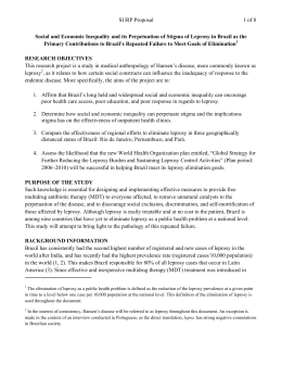
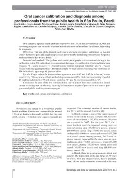
The volume of Iluid obtained from blisters at all times after induction ranged from
30-150 µl (median = 60; mean = 66); the
range and distribution of volume did not
diffcr significantly between the active patients and the controls. In blisters induced
in the skin of cured, control voluntecrs the
median total cellularity varied from 80-400
cells/mm 3 (ranging from 50-550 cells/mm 3 )
over all times studied (Fig. 1). Cellularity
was highcr during thc first 24 hr, as part of
the nonspecific inflammatory response to
blister induction (`'). No statistically significam difTerences were seen among patients
who had had diffcrent types of leprosy earlier.
To determine the reproducibility of cell
sampling using thc blister technique, two
blisters were aspirated at the same time from
a number of control patients. The paired
results gave a correlation coefTicient of 0.613
(p = 0.003) (Fig. 2).
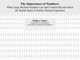
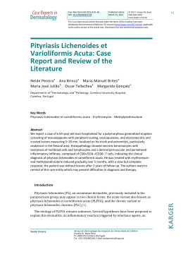
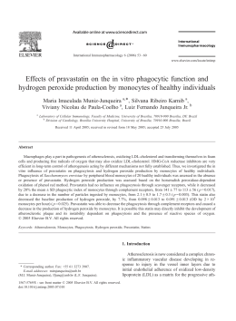
In active leprosy lesions, total cell counts
varied much more widely than in the controls, ranging from 1-2800 cells/mm 3 (Fig.
3). Median values for the difTerent immunopathologic types of leprosy were similar
at all times studied.
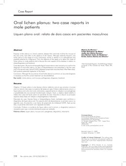
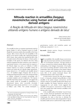
Mononuclear cells predominated in active lesions, with median percentages of
70%-91% over all times studicd (Fig. 4).
Polymorphs accounted for more than 30%
of the cells in blisters over some BL lesions
(3 patients) and LL lesions (2 patients), but
not in BB or BT lesions. Neither eosinophils
•
494^
International imanai of Lcprosv^ 1989
•
25
•
LL
■
!1L
•
BB-BT
500-
EE^O
400-^
20
°
x
.^
15
^I^I.
7
O
cn
ï
U
5
300-^
(-.)
^
•
O
H
200-
10
U
+^4.^Jku a
ó
U
N^
100-^♦
♦
..
^+
1
18-24
1
30
á6
1
48
2
1
72
1
96
ROURS AFTER BLISTER INDUCTION
FIG. 3. Total cell counts in blisters over active, uncomplicated leprosy lesions. Four blisters were induced
directly on active lesions which were classified by clinical and histologic criteria. Blisters were aspirated only
once, at different intervals after induction. For cach
type of lesion: point = mediais value; vertical bar =
range; N = numher of observations.
FIG. I.
Cellularitv of blisters in curvd, inactive control patients. Four blisters were induced (as dcscribed
in text) on the volar surface of the forearm in patients
with no evidence of active disease. Blisters were aspirated at different intervalsafter induction. Each point
= cell count from one blister; bar = mediais value.
8
6
4
2
1^23^4
FIG. 2. Correlation of cell counts in paircd blisters.
Four blisters induccd on the forcar] of curcd, inactive
patients were sampled in pairs at indicated intervals
after blister induction. Total cell counts were determined independently for each blistcr. Correlation coefficient for these paired samples is 0.613; slope of the
curve was detcrmined by linear regression.
4.1
•
•
*
^•
11 ^ •
24^30^48^72^96
Hours After Blister Induction
BLISTER 1 CELLS/CMM X 0.01 (X 0.01)
•
5
nor basophils were present in substantial
numbers.
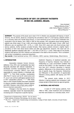
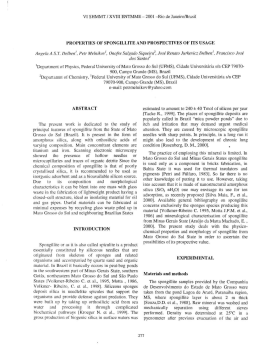
Among mononucicar cells, the median
ratio of T cells : monocytes (Leu4+ : NSE+
cells) remained between 0.5-1.5 for all types
of leprosy at all times studied, except for a
predominante of T cells at 48 hr in BT lesions (Fig. 5). This differencc was statistically significant (p < 0.005), and it is notable that this was observed in blisters with
similar total cell counts (Fig. 3), indicating
an absoluto as well as a relative increase in
T cells in BT lesions.
At 48 hr, the median heiper : suppressor
(H:S) (CD4:CD8) ratio was 3.3 in BT lesions
and 1.3 in BL and LL lesions, a statistically
significant differcnce (p < 0.005). The median H:S ratio ranged from 1.0-3.0 at other
times, but the range of values was also great
and at none of these times were the differences statistically significant (Fig. 6).
DISCUSSION
This study addrcsses the feasibility of using suction-induced blisters to evaluate the
Rangdaeng, et al.: Suction-induced Blisters^495
57, 2
B
A LL
BL
C. 68-BT
100
4-
Bo.
c0.
40
,
70.
1^
4,30^4tl^))^9e
HOURS •F TER BLISTER INDUCTION
HOURS AFTER BLISTER INDUCTION
HOURS ',TER BLISTER REDUC TION
FIG. 4. Percentage of mononuclear cells in blisters over different types of leprosy lesions. Percentage of
mononuclear cells (Iymphocytes and monocytes) was determined for blisters aspirated at different times after
induction on different types of leprosy lesions. Each point = one blister; horizontal bars = median values.
relative numbers and types of cells in the
cutaneous infiltrates of leprosy, with minimal trauma to the patient or the cells. Rebuck's "skin windows" (I") were the conceptual forerunner of this approach. Other
investigators attempted to apply Rebuck's
technique to leprosy ( 4 )), but the cutaneous
abrasion required is difkicult to standardize,
and meaningful data concerning mononuclear cells was obscured by the brisk polymorphonuclear responses induced by cover
slips. As noted by the originators of the blister technique ( 8 ), this method introduces no
foreign material of any kind and therefore
approximates more closely the conditions
of the intact skin. The suction uscd to induce the blisters does elicit a mild, nonspecific inflammatory response which peaks
during the first 24 hr in healthy volunteers
((r). In this study, samples were obtained from
24 hr onward to minimize the effects of this
nonspecific response.
The total cellularity of the blisters over
active lesions varies greatly, not unlike the
variations in cellularity observed in the lesions in histologic sections ("). The cellularity of blisters itself is therefore not a valuable indicator of the immunologic status of
the underlying lesion, although it is also notable that many blisters over active lesions
had many more cells than the highest num-
bers seen in blisters in cured, inactive patients. The marked kinetic changes which
may be seen with Chis technique during an
accelerating immune response (`') were neither expected nor observed in these lesions,
4
_
• LL
■ BL
• BT
.7.i 3 ■1
2_
1.
^^I
N+^6. 8, 3^5, 6. 4^5. 7, 3^3. 6. 4
24-30^48^ 72^ 96
HOURS AFTER BLISTER INOUCTION
FIG. 5. T-cell monocytc ratios in leprosy lesions.
Among cells obtained from blisters at dilferent times
after induction over leprosy lesions, total T-cell
numbers were determined using anti-Lcu4 antibody;
monocytes were identified by staining for nonspecific
estcrase. For cach type oflesion: point = median value;
vertical bar = range; N = number of observations.
Dilference between BT and BL-LL 48 hr after induction is statistically significant (p < 0.005).
496^
International Journal of Leproso ^ 1989
10
Li_ Eli_ BT
9
^
8
-
7
-
a
rx 6
-
5
-
4
-
o
P-
occ
N64^
á
N
w
x 3
A
A
0
á
/
-
k
Rj
7, 3
6, 3, 4
5,3, 2
4.5,5
24-30
48
72
96
HOURS AFTER BLISTER INDUCTION
Fio. 6. T-helper:suppressor ratios in uncomplicatcd leprosy lesions. Among cells obtained from blisters at different times after induction over leprosy lesions, T-helper and -suppressor phenotypes ‘vcre
determined by immunostaining with anti-Lcu3 and
OKT8 antibodies, respectivcly. For each type oflesion:
point = median value; vertical bar = range; N = number of observations. Differencc betwcen ItT and I3LLL 48 hr after induction is statistically significant (p
< 0.005).
since no antigenic stimulation was used to
induce an immune response in this study.
The result is, instead, a profile of tlie "baseline" status of different types of leprosy lesions.
The method shows an acceptable degree
of reproducibility from blister to blister, as
evidenced by the data on paired blister samples. Some of the scatter which is seen is
probably due to imperceptible differences
in trauma during blister induction, or to
variations in the quantity of rcd blood cells,
which is usually negligible but was not
quantitated in this study.
The high percentage of mononuclear cells
within the blisters is in agreement with the
histologic features of these lesions ("), and
indicates that the method is suitable for the
study of the cells involved in immunologic
responses in leprosy. The percentage of
polymorphs infrequently exceeded 25%–
30% of the total and was, in most instances,
far less. Other investigators have demonstrated that the introduction of very few
bacteria is sufftcient to induce an acute inflammatory response in a blister ( 8 ), and we
view the occasional occurrence ofnumerous
polymorphs in one blister (out of a set of
3-4 in a volunteer) as likely to be the result
of such contamination due to a crack or
puncture of the blister. Intcrestingly, however, among patients in whom 3-4/4 blisters contained over 30% neutrophils, the
majority developed a reaction (either type
1 or 2) soon thereafter. The extent and significance of this observation are under investigation.
Monocytes/macrophages were a major
cellular component in blisters over active
lesions, consistem with their prominent
place in all types of leprosy lesions. However, monocytes/macrophages werc also
numerous in blisters in cured, control patients, as they were in earlier studies of
healthy volunteers (" and Scollard, unpublished results). The activation status and
functional activity of the monocytes/macrophages may differ in these different situations, but these parameters were not assessed in this study.
In BT lesions, T-helper/inducer phenotypes outnumbered T-suppressor/cytotoxic
phenotypes at least 3 to 1 at 48 hr after
blister induction, in agreement with the patterns observed in biopsy studies (`' '6 ). Recruitment ofT cells finto the blister appeared
to continue after this time, probably nonspecifically, and the trend was no longer evident at 96 hr. The statistically significant
results at 48 hr, however, indicate that the
method does provide a sample of the eutaneous infiltrate, reflecting the status of the
lesion.
Phenotype alone does not indicate the
functional status of the cells. Preliminary
studies suggest that direct assessment of
some functional capabilities of blister exudate cells is possible, however, and further
work is in progress.
The blister technique has proved to be
very acceptable to patients, enabling multiple sequential studies of the same patient.
Another distinct advantage of this method
over biopsies is the ability to make more
precise determinations of the total number
of cells present in the blister—an accurate
57, 2^Rangdaeng, et al.: Suction-induced Blisters^497
denominator for quantitative studies of cell
subsets. The precise origin of every cell in
the blister is not known, and some cells may
be recruited finto the blister from dermal
capillaries rather than from the immunoinflammatory inlìltrate. Nevertheless, previous studies with purified protein derivative (PPD) skin tests have demonstrated that
the blister does reflect the immunologic activity in the dermis, as indicated by correlations between 48 hr induration and increased numbers of cells (`') and of soluble
interleukin-2 (IL-2) receptor (Tac peptide)
(' 3 ) in blisters induced over the PPD reaction. The differences in the H:S ratio in blisters over lepromatous versus tuberculoid lesions to a levei similar to that seen in biopsies
suggest that this is also the case with leprosy
lesions. Together, these results indicate that
suction-induced blisters of er a quantitative
reproducible method, minimally traumatic
and amenable to multiple sampling, with
which cells can be obtained which are representative of the cutaneous infiltrates in
leprosy lesions.
SUMMARY
The cellular contents of blisters induced
by suction over new, uncomplicated leprosy
lesions, and in the skin of cured, control
patients, have been examined with enzymeand immuno-histochemical staining over a
period of 4 days. The total cellularity of the
blisters varied over a vide range, not correlated with the type of leprosy. Mononuclear cells predominated at ali times studied, with nearly equal percentages of
monocytes and T lymphocytes. The T-helper: suppressor ratio was significantly greater in BT than in BL and LL lesions at 48
hr. Suction blisters offer a painless, quantitative, reproducible, multiple-sampling
method for obtaining edis from the cutaneous infiltrates of leprosy for phenotyping
or functional analysis.
RESUMEN
Utilizando técnicas enzimo- e inmuno-histoquímicas se examinó, durante 4 dias, el contenido celular de
Ias vesículas inducidas por succión sobre lesiones dérmicas no complicadas de casos nuevos de lepra y sobre
la piei de pacientes curados usados como control. La
celularidad total delas vesículas varió en forma amplia
} no correlacionó con ningún tipo de lepra en particular. En todos los tiempos estudiados predominaron
las células mononucleares y los porcentajes dc monocitos y linfocitos T fueron equivalentes. A las 48
horas, la relación de linfocitos T cooperadores: supresores fite significativamente mayor en los casos BT que
en las lesiones BL y LL. Las vesículas inducidas por
succión representan un método de muestreo múltiple,
indoloro, cuantitativo, y reproducible, adecuado para
oblener células de los infiltrados cutáneos de la lepra
y para cl análisis funcional y fenotípico de las mismas.
RESUME
On a examiné sur une période de 4 ans, au moyen
de méthodes de coloration histochimiques des enzymes et immunohistochimiques, les contenus cellulaires de vésicules produites par succion sur des lésions
neuves et non compliquées de lèpre, ainsi que dans la
peau de malades témoins guéris. Le contenu cellulaire
total des vésicules variait dans une large mesure, et
n'était pas en corrélation avec le type dc lepre. Les
cellules mononucléaires ont prédominé pendam toute
la période d'étudc, livrant des proportions à peu près
égales de monocytes. Le rapport des lymphocyles T
adjuvante et des suppresseurs était significativement
plus élevé après 24 heures dans les lésions de lépre
tuberculoide dimorphe que dans celles de lèpre lépromateuse dimorphe ou lépromateuse. Les vésicules dc
succion constituent une méthode sins douleur, quantitative, reproductible, et pouvant ctre répétée dans de
multiples échantillons, permettant d'obtenir des ccllules à partir des infiltrats cutanés de lèpre, en vuc de
leur analysc phénotypique ou fonctionnelle.
Acknowledgntents. This work was supported by PSTC
Grant No. 5.141 from USAID, and by a Research
Strengthening Grant from the WHO/TDR program to
the Research Instituto for Health Sciences, Chiang Mai
Univcrsity, Chiang Mai, Thailand.
We are most grateful to Atcha Masarat, Utaiwan
Utaipat, and Rawiwan Kurapakorn for their excellent
nursing and tcchnical assistance.
REFERENCES
1. BLOOM, B. R. Lcarning from leprosy: a perspective
on immunology and the Third World. J. Immunol.
137 (1986) i—ix.
2. BROWN, A. E., NELSON, K. E., MAKONKAWKEYOON,
S., VITHAYASAI, V., SCOLLARD, D. M. and BULLOCK,
W. E. A study of the immunological elfects of
cimetidine in patients with lepromatous leprosy.
Int. J. Lcpr. 53 (1985) 559-564.
3. BRowN, A. E., VITHAYASAI, V., SCOLLARD, D. M.,
NELSON, K. E., MOSES, V., SCIIAUr, V. and
MAKONKAWKEYOON, S. L'mphocyle transformation in lepromatous leprosy: a study of the influence of disease activity and symptom duration.
Southeast Asian J. Trop. Mcd. Public Hcalth 17
(1986) 104-110.
4. 13ULLOCK, W. E., Ho, M. F. and CHEN, M. J. Quantitative and qualitative studies of the local cellular
498^
International Journal qf Leprosl'^ 1989
exudativc response in !eprosy. J. IZeticuloendothel. Soc. 16 (1974) 259-268.
5. GARRIE, S. A. and LEVAN, N. Cell-window tcsts in
lepromatous Icprosy. (Letter) Lancei 1 (1970) 1 1 16.
6. KENNEY, R. T., RANGDAENG, S. and SCOLLARD, D.
M. Skin Mister 1mmonocytology: a new method
to quautify ccllular kinetics ! o vivo. ,I. Immunol.
Methods 97 (1987) 101-110.
7. KIISTALA, U. and MUSTAKALLIO, K. K. hz-vivo
separation of epidermis hy production of suction
blistcrs. Lances 1 (1964) 1444-1445.
8. Kiistala, U., Mustakallio, K. K. and RORSMAN, H.
Suction blistcrs in the study of ccllular dynamics
of inflammation. Acta Derm. Venereol. (Stockh.)
47 (1967) 150-153.
9. MODLIN, R. L., HOFMAN, F. M., TAYLOR, C. R.
and REA, T. 11. T lymphocytc subsets in the skin
lesions of paticnts with Icprosy. J. Am. Acad. Dermato!. 8 (1983) 182-189.
10. REIUCK, J. W. and CRGWLEY, .1. H. A method of
studying !eukocyte functions in vivo. Ann. N.Y.
Acad. Sei. 59 (1955) 757-805.
11. RIDLEY, D. S. and JOPLING, W. H. Classilication
of Icprosy according to immunity: a hve-group
system. Int. J. Lepr. 34 (1966) 255-273.
12. SCOLLARD, D. M. A revicw of immunopalhologic
studies of human leprosy lesions in sito. Hawaii
Med. J. 47 (1988) 54-59.
13. SCOLLARD, D. M., WAGNER, D. K. and NELSON,
D. L. Release of solubie intcrleukin-2 receptors
(Tac peptiele) in vivo during human immune responses to toberculio. ('h o. Immunol. Immunopathol. 46 (1988) 450-455.
14. SCIiWINN, C. P. and FERGUSON, C. T. Cytopreparatory techniqucs. In: C'ontt u 'ndium of Diagnostic Cytolo i . Wicd, G. L., Koss, L. G. and Reagan, J. W.. eds. Chicago: Tutorials of Cytology,
International Academy of Cytology, 1976, 4th ed.,
pp. 360-369.
15. SKINSNES, O. K. Thc immunological spectrum of
Icprosy. In: Leproerin Thcoryand Natiic•e. Cochrane, R. G. and Davcy, T. F., eds. Baltimore: Williams and Wilkins, 1964, pp. 156-182.
16. VAN VOORHIS, W. C., KAPLAN, G., SARNO, E. N.,
HORWITZ, M. A., STEINMAN, R. M., LEVis, W. R.,
NOGUEIRA, N., HAIR, L. S., GATTASS, C., ARRICK,
13. A. and COHN, Z. A. The cutancous inhltrates
ofleprosy: cellular characteristics and the predominam T-ccll phenotypes. N. Engl. J. Med. 307 (1982)
1593-1597.
17. WOOD, G. S. and WARNKE, R. Suppression of endogenous avidin-binding activity and its relevance
to biotin-avidin delection systems. J. Histochem.
Cytochem. 29 (1981) 1196-1204.
18. YAM, L. J., L1, C. Y. and CROSiY, W. H. Cytochemical identification of monocytes and granulocytes. Am. J. Clin. Pathol. 55 (1971) 283-290.
.
Download