Departamento de Profilaxia da Lepra (S. Paulo) and Centro Internacional de
Leprologia (Rio de Janeiro) — Brazil
SOME ASPECTS OF IMMUNITY IN LEPROSY AND THEIR
IMPORTANCE IN EPIDEMIOLOGY, PATHOGENESIS AND
CLASSIFICATION OF FORMS OF THE DISEASE. *
Based on 1529 Lepromin Tested Cases
A. ROTBERG
Sanatorio "Padre Bento" (D. P. L. S. Paulo)
and Centro Internacional de Leprologia
(Rio de Janeiro)
The influence of the phenomena of resistance in the pathogenesis of
leprosy was for a long time studied only with the observation of clinical
and epidemiological facts. Hence the theories based on environmental
factors and those related to sex, age, eating habits, individual
constitution and different debilities, in contradiction to the exclusive
action of the germ.
Man, however is a conjunction of varied factors and influences,
within with it is almost impossible to follow the track of conditions
leading up to resistance to infections. If the study is transferred
from that of man to man, to that of human group to group, the
difficulties still persist, because we shall never secure the variations of
* Presented before the International Leprosy Conference of Cairo, Egypt, March 1938.
** This study was made in diverse divisions of Leprosy Department of S. Paulo
State, Brazil (Director Dr. Salles Gomes Jr.) under the patronage of the
International Center of Leprology of Rio de Janeiro (Director Prof. Dr. Ed. Rabello).
I wish here to express my hearty thanks to all who contributed to it in any
way, especially to Drs. Salles Gomes Jr.; Lauro Souza Lima, Director of the Sanatorio
«Padre Bento»; Nelson de Souza Campos, Dermatologist of the Preventories; Manoel de
Abreu, Director of the Sto. Angelo Hospital-Colony and H. Cerruti, Histopathologist. I also
beg to thank my Professor in the Dermatologic Clinic of S. Paulo University. Prof. Dr.
J. Aguiar Pupo, for the interest he showed in this study.
— 46 —
one factor and have the others stationary. So, if in order to
study the influence of climate, we we could divide populations by different climatic zones, we would also find differences of race, eating
habits, general sanitation, as well as different conditions of life and
occupation. Because of the impassibility of experimental studies for
lack of animals receptive to leprosy, it is not surprising that no
theory of resistance to it could be established in its entirety, without
discussions, and even contestations. We only remember, as an
example, in disaccord with the accepted theories, the cases of initial
leprosy in adults, its incidence in individuals who have conserved
their bodily vigor, its non obligatory incidence in individuals debilitated in every way and in spite of their intimate contact with patients of open leprosy; and this in such numbers, as not to be
thought of as a simple law of average.
A new fact, however, has appeared and opened the way to
possibilities of study along this line; that is, the skin-reactions with
antigens prepared with materials of leproma. A careful and
systema t i c s t ud y o f t h es e rea c t io n s will b e d e stin ed to
p re sen t r esults of value in the epidemiology and the etiopathology
of leprosy, and to establish on a more scientific base the best
conditions for contagion or resistance, or the later evolution of the
disease in the infected individual.
SKIN REACTIONS IN LEPROSY
The first investigations of skin-reactions had in view the obtaining
of a test, capable of constituting a process of early diagnosis,
analogous to tuberculin for the infection of the bacillus of Koch.
The attempts of TEAGUES (1), NICOLLE (2), MANTOUX (3), MARCAOUX and
(4), with lepromatous antigens, or with leprolin of ROST,
brought no practical results, and were forgotten, as were also the
leprin of BABES (5), the glycerine and aqueous extracts of S CHOLTZ and
KLINGMÜLLER (6).
PAUTRIER
In some posterior researches facts were observed which brought
attention to the authors, and which value began to be given to their
real importance.
M UC H (7), K U L ES (8), B ER N UC CI (9), F ER RARI (52), M AR I ANI
(10, 11), M ONTANES (12), N EGRO (13), and A MBROGIO (14), noticed with their diverse intradermic reactions, the greater reactivity of
the forms considered resistant, both incipient and neural, contrasting
with the weak or non-reactivity of the mixed or nodular forms.
Having injected his antigen of leproma into the skin of 403
patients, MITSUDA (15) observed the
fugacity of reac-
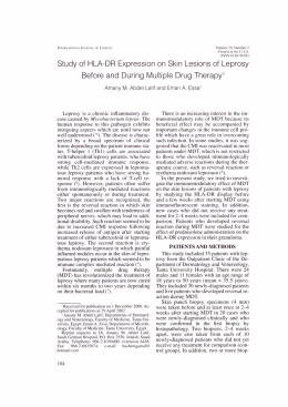
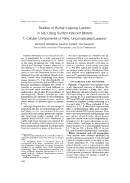
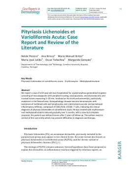
Fig. 1
Strong positive lepromin test, scar
formation.
Fig 2
Positive Lep r o mi n t e s t s i n a s a r c o i d t y p e o f
t u b e r c u l oi d l e p r o s y S t a n d a r d a n t i g e n a n d
di l u t i o n s 1 : 8 a n d 1 : 1 5 .
Fig. 3
S t r o n g , ul c e r a te d l e pr o m i n t e s t , in t u berculoid leprosy, with 1 : 1 0 di l u t i o n o f
the standard antigen.
— 49 —
tive phenomena in nodular cases, while in the neuro-macular
an initial hyperemic reaction was observed with papulous
infiltration in 2 weeks, persisting for a longer period still.
Identical reactions in healthy individuals.
From his observations, MITSUDA, concluded that healthy and the
patients with macular and neural leprosy presented great resistance
to infection, a resistance which the nodular ones "exhausted" in their
struggle against the germ.
The studies of MITSUDA were resumed by HAYASHI (16.17), who,
abandoning all attempts of diagnosis, established definitely the
divergence between the stage of the disease and the intensity of the
reaction, and insisted on its immunitary value, and its importance in
classification of clinical forms, preparing the recognition and use
which the reaction has in our days.
Similar conclusions, of great clinical and epidemiological
value, resulted from the work of BARGEHR (18, 19, 20) and DE
LANGEN (25), in which the technique of preparing the antigen varied,
being more concentrated, and its application by scarification
instead of the intradermic injection.
The reaction to lepromin is, however, sometimes very slow, and may
begin 3 or 4 weeks after the application. It is thus understood that a
simple deposit of the antigen, even highly concentrated, on the
scarification of the skin, from where it is rapidly eliminated, represents
a minimum introduction, which could only reveal an exaggerated reactivity of the organism, failing to indicate the medium grades of
resistance. Besides this, its dosage being very difficult, there have
not been many followers of BARGHER'S and DE LANGEN'S technique.
Modern researches took as a standard the techniques of MITSUDA and
HAYASHI, based on the intradermic injection of a fixed quantity of the
antigen indicated, as well as the criterion of reading also indicated by
these authors.
PERSONAL TECHNIQUE
Preparation of antigen: The technique of preparing the
antigen adopted by us is based on that of H AYASHI, with some
variations. Lepromas collected aseptically in sterile normal saline water are boiled in a water bath for one hour, after which
they are relieved of any piece of skin that they may have, cut
into small bits with scissors, ground in a mortar and weighed.
For each gram of the triturate prepare 20 cc, of the sterile
water which served for the first boiling, adding more water of
necessary. The mass of leproma is again triturated strongly,
with a little of this prepared water. After a short rest, the
murky liquid on the surface is sucked up with a fine Pasteur
pipette, and filtered through 4 layers of gauze, being received
— 50 —
in a balloon. New quantities of water sucked up will show mediocre turbidity, contrasting with the strong murkiness of the first
water.
The mass of leproma may now be discarded. The balloon
containing the antigen filtered by the gauze is put into the autoclave
for 15 minutes at a temperature of 120 0. To the contents is added
0,5% of carbolic acid, then distributed in sterile ampoules or tubes
of the insuline type.
Aspect — The material thus obtained presents a cloudy, milk-like
aspect, and when in repose it leaves a deposit at the bottom of
container. The bacterioscopic examination shows a great number
of Hansen's bacilli and globi in all fields. Since the bacillary count
is practically impossible, and consenquently also the titration of the
antigen, we obtained originally a great quantity of material with
which most of the tests were made.
Duration — The antigen has exceptionally conservative qualities.
We are even now getting excellent reactions from antigen which we
prepared in 1933 and has been kept in dark, rubber-covered glasses,
all experiments having been checked by our present antigen.
Denomination — The most varied names have been given to this
type of antigen. H AYASHI calls it «vaccine»; M UIR (22, 23) and the
authors from India adopted the term «leprolin». As we had occasion
to mention, (25) the term «leprolin» is not appropriate, since it
brings to mind the «tuberculin» process. Since there is not
presently any culture of Hansen's bacilli, such a process could not
be followed. A more appropriate term for the material prepared with
triturated leproma would be the designation used by BARGEHR,
«lepromin», leaving the term «leprolin» to the product of the
metabolism process, or to the toxins of Hansen's bacilli, when their
culture is obtained.
Application — A fine syringe of 1 cc. and a short needle are used
for injecting the lepromin into the skin, preferably in the front part
of the thigh, on account of the strong reactions that it may cause,
though, in certain cases, we have applied it in the arm. It is
advisable, generally, to inject 0,1 cc. This is rather difficult to
accomplish with an ordinary syringe, not only because of losses of
quantities in the syringe, but also due to accidental introduction
into the hypoderma. The most efficient method is to note the
diameter of the anemic papule which should measure 1 cm.,
corresponding approximately with the desired quantity.
Reading — Following the methods of H AYASHI , we begin
ours first observations by reading each reaction eight, sixteen and
twenty-four days after the injection. After some experiments, we
were convinced that the initial readings made on the eighth and
sixteenth days were not necessary. The cases of positive reaction
to lepromin generally reach the peak of reaction from the third to
the sixth week after the injection, this being an ideal period for
the
reading.
A
reading
taken
on
the
eighth
day
— 51 —
may give as positive a populous lesion which may represent a lesion
of trivial irritation in involution. We have often observed this even
in advanced cases of nodular leprosy. As a matter of fact there is no
necessity, usually, of successive readings of the same reactions, as
timely readings are quite sufficient to comprehend tardy reactions.
Our experience proved that a period of thirty days after the
injection is ideal for a routine reading, not overloking, however, a
checking-up later, in special cases, such as those presenting very
slow reaction, necrosis, etc.
Types of Reaction — The reactive lesion is elementarily a
nodule, raising up the skin, which at the surface turns reddish-violet
more or less intense. This aspect may vary in accordance with the
evolution of the lesion. Crusty formations are frequent as a
consequence of the particularly intense suppuration undergone by
the nodules of reaction; the scar aspects, from involution of a
nodular lesion (photo 1) ; the frankly ulcerous aspects giving
emission of serosity during a few months; and the deep nodules, and
deep infiltration without erythema on the surface.
Classification of Reactions — The original classifications were made
in accordance with HAYASHI: +++ for nodules with more than 1 cm.;
++ for those of one half cm. to 1 cm.; + for the infiltrations of less
than half a cm. next the reactions which are doubtful and finally
those which are totally negative. Having initiated the work with this
criterion of reading and classifying, we have continued it till today.
The verification of results, however, and their correlation with the
clinical forms and the histopathology, gave us the conviction that a
binary classification would be sufficient, in negative reactions (from
0 to 0,5 cm, including the reactions — and + of HAYASHI ) and
positives above 0,5 cm. including the ++ and +++ of H AYASHI and
the ++++ of other authors). The table X and the considerations
we expose about it justify our classification. Considering the
importance we assign to lepromin in distinction between «anergic» and
«allergic» cases, the intermediate dimension exactly of 0,5 cm.
should be considered «doubtful».
Involution — A reactive lesion may persist from a few weeks
to many months, resolving itself finally into a scar more or less
evident, which may sometimes characterize the intensity of a
previous reaction.
THE REACTION TO LEPROMIN IS A SPECIFIC REACTION
OF LEPROUS IMMUNITY
The clinic and bacterioscopy may affirm that a positive reaction to
lepromin occurs only in cases in which resistance of the organism to leprous
infection has been proved. As an example, let us take the extremes of
approximately 100% of positive reactions in tuberculoid leprosy and
approximately 0 % in nodular leprosy, according to our criterion of reading.
By
this
way
the
results
of
the
anthors,
not
— 52 —
always interpreted at the time, are explained and who, in search of a
test of allergy for diagnosis, the positiveness of which should be
frank in declared cases of leprosy, obtained instead the exact
contrary; that is, positive reactions in neural forms and in cases
of incipient macules; negative in nodular ones.
On the other hand, only antigens prepared with material of
leproma and rich in bacilli, in accordance with the technique of
MITSUDA-HAYASHI, MUIR and others, are capable of revealing this state of
immunity. Attempts at substituting the original antigen did not
give success, neither with other acid-fast bacilli, nor with the very
material of lepromas from which have been eliminated all bacilli by
filtration (16,17).
In an previous work (24) we have had occasion of reporting
what we have observed with the intradermic reactions to
tuberculin in healthy persons and in lepers, in which we have
searched previously for the reactivity to lepromin. Using the
synthetic tuberculin of Dorset, diluted to 1:10000, we observed the
results condensed as follows:
1. — Among healthy infants, lepromin + : 75,5 % negative
tuberculin reactions.
2. — Among healthy infants, lepromin — : 77,8 % negative
tuberculin reactins.
3. — Among lepers, lepromin + and —, identical indifference to
tuberculin reactions.
It becomes evident that the reactivity to tuberculin depends in no
way upon the reactivity to lepromin neither interferes with it in any
way. The anergic forms to lepromin present good coefficients of
positive tuberculin positive reaction, which proves that existent anergy
is not “general” for all the antigens, as BERNUCCI wishes; inversely, among
the lepromin-positives, numerous cases did not react to tuberculin, which is
contrary to AMBROGIO, for whom the allergic reactivity of leprosy is a form
of “general” hypersensitiveness to all external stimulants.
The substitution of antigens by cultures of acid-fast germs
obtained by various authors and announced as being Mycobacterium
leprae, or by the material of rat leprosy, rich in the Stefansky bacilli,
gave clearly positive reactions in the majority of individuals
tested. The mechanism by which these reaction was produced is not
explained, the authors generally thinking of local irritations produced
by the germ. It is known, however, that these positive reactions,
more precocious and of shorter evolution than the real lepromin test,
—53—
are constantly observed in all forms of the disease, even in lepromatous
cases. These reactions are other proofs of the specificity of the antigens of
leproma, which only exceptionally give positive reaction in cases of
bacillary leprosy.
THE REACTION TO LEPROMIN IS A SPECIFIC
REACTION OF ALLERGY
Admitted, with facts of observation, the immunitary nature of the
reaction to lepromin, some authors refuse to see in this reaction an
“allergic” response of the organism to the bacilli of Hansen. By the
definition, on account of allergic proofs can be taken only those which are
dependant on organic modifications caused by previous relations
between the organism and the causing agent which the antigen
represents.
Lacking a receptive animal to this proof, we cannot prove
experimentally that positive lepromin tests results only in a previously
infected organism. This forces us to conduct our reasoning with the
clinico-epidemiological observations.
An adult healthy man of endemic areas reacts generally positively to
lepromin. It is not within our reach to discover the initial lesion of
leprosy which will justify this reactional capacity and its allergic
nature. If this phase of primary inoculation exists, it escapes, at present,
at least, the known processes of investigation. Nothing analogous,
therefore, exists here to the initial anatomical and radiological lesion of
tuberculosis, which causes the allergic reactivity to tuberculin.
There still remains the resort to research into the reactivity to
lepromin in individuals in whom the epidemiological inquiry reveals
diverse conditions of contact with the bacilli of Hansen.
1st. — The observations of stronger and more frequent positive reactions
among healthy individuals living in leprosariums than among the
general healthy population in minor contact with the bacilli of Hansen,
make one suspect a prior infection which aborted and produced an
alteration in the manner of cutaneous reaction; that is, allergy.
It was already noted in the original observations of M ITSUDA that
the healthy, even without known contact with lepers, react to
lepromin. This reaction, however, was more intense in three nurses
who worked for ten years in the leprosarium. The studies of
BARGEHR are more suggestive from this point of view. The reaction
is negative in the healthy, who never had contact with a leper. The
positive reaction of the «contacts» is due, according to BARGEHR ,
to
the
formation
of
specific
antibodies
by
— 54 —
virtue of minimum previous infections. De LANGEN obtained
identical results, and gives the same interpretation. M UIR observed in healthy children of lepers stronger reactions than among
boys without contact, which «may be taken as an index of
augmented resistance against leprosy». In the children of Oshima,
M UNEUCHI (26) accentuated the existence of reactions, the
stronger, the longer the duration of the previous contact with the
sick parents. STEIN and STEPERIN (27) present very interesting and
elucidative results. In 49 healthy adults working in leprosariums
the frequency and intensity of positive reactions were directly
proportional to the intimacy of contact with the patients, varying
according to the professions — the doctors having the
strongest reactions: in the same profession the frequency and
intensity of reactions were directly proportional to the length of
service. Healthy individuals, free from contact with leprosy and
with lepromin negative reactions. after a certain time of service in
the leprosarium became lepromin-positive and this positiveness
became more defined and increased with the time of habitation.
2nd. — The final and probably decisive argument would be the
verification of the reactivity to lepromin in countries securely free
from leprosy. The allergic nature of the L. T. (lepromin test) would be
confirmed in these countries by a frequent negative result in contrast
to that observed in endemic countries. This is the condition which
MUIR imposes for considering, allergic the L. T.
A research of this nature is not within our reach, and could, for
example, only be done in certain European countries. But this
research has already been done, even though in a very small scale in
proportion to the importance of the subject, and from this we can get
certain data of value, whilst awaiting more extensive and informative
studies.
CUMMINS and WILLIAMS (28) inoculated with lepromin furnished by
MUIR 25 psychopaths hospitalised in London without any admissive
contact with leprosy. The peak of reaction was always observed on
the 8th day, reaching in but 6 times the maximum of 9,5 mm.,
declining soon after till the 22nd day, the diameter being then 3,5
to 4,5 mm. in 19 cases, and 6 mm. in the 6 cases above. In these
6 cases referred to having reached 6 mm. on the 22nd day, an
intradermal reaction with bacilli of Koch, made at the same time,
gave lesions of much greater diameter.
Comparing these results with the reactions which we call “positive”,
nodular, late and always superior to 5 mm. we are forced to believe
that in no case did CUMMINS and WILLIAMS obtain positive reactions.
D UBOIS (30) gives results obtained with a lepromin furnished
by VAN BRESEGHEM in 29 individuals who had never left Belgium.
Of these, 14 reacted with 0-2 mm., and 10 with 3-5 mm. In only
5 were observed reactions of 6-10 mm., sometimes with suppuration.
— 55 —
Based on these results, DUBOIS opposed the allergic nature of L. T.
because he encountered “numerous positive reactions”. In accordance
with our method of reading (negative up to 5 mm.) only in 5 cases
were the reactions positive.
In his recent work BONCINELLI (31) protests against the
hypothesis of the allergic nature of L, T., presenting 44 individuals, healthy and coming from zones not endemic of leprosy in
Italy, and in them, positive reactions were observed in 22 cases.
We are again in a position caused by disagreement in the methods of
reading the reactions by different authors. BONCINELLI includes among
“clear positive reactions”, 9 reactions of the type which he calls
“pompho-papulous”, with aspects analogous to the tuberculin reactions
and lasting on an average half a week-and which we would call negative.
We doubt still further in admitting as positive 9 other reactions which the
author calls “papulous”, persisting 2 to 3 weeks (he does not give
dimensions). There remain 4 nodular reactions, identical to our real
positive reactions.
Now, let us compare these results with those verified in healthy
inhabitants of endemic countries:
We have already seen that adults always react with «nodular» lesion to the intradermic injection of lepromin. Let us add
now the observations, to the same intent, of M UIR (9 strong
reactions in 10 cases); T AJIRI (32) 100% ; M ITSUDA 10 in 13;
F ERNANDEZ (33) 75 to 77% ; A DANT (35) and C HIYUTO, 67% in
children and 9 in 10 adults (36).
Our Observations in regard to reactions to lepromin in individuals not
affected by leprosy were made in healthy children of lepers.
isolated in the Preventories of São Paulo State, and in 144 adults, 55
being contacts of leprous patients, 19 suffering from various tro-
— 56—
pical diseases (leishmaniosis, blastomycosis) and 70 from pulmonarytuberculosis without known contact with cases of leprosy, according to a
previous work (51). Not including children, that will be studied apart,
we have the following graph according to the reading of H AYASHI . (See
tables at the end of work).
We shall point out that our reactions ++ and +++, refer to large
nodular formations, often with supuration and permanence of lesion for
several months before involution.
These results, as those of other authors who have worked in
endemic countries, contrast clearly with the weak and transitory
reactions, and in reduced number, of the non-endemic countries, and
bring a new contribution to the allergic nature of L. T.
APPLICATION OF THAT KNOWLEDGE TO
EPIDEMIOLOGICAL FACTS
Once proved the necessity of contact with the germ in order to
determine the positive L. T., we fall by analogy, into the same order of
ideas that gave to tuberculin the value of allergic and diagnostic test of
primary infection of tuberculosis, in spite of JADASSOHN’s (37)
admitting that the allergy in leprosy may be produced by bacillary
protoplasma, without pathogenicity.
As BARGEHR had supposed, with DE LANGEN and others, the
positiveness of the reactions seems to signify infection, to which the
organism reacts, with the immunity, and which manifests itself with an
allergic reaction. In leprosy the allergic reaction is equal to a reaction
of immunity. From this point of view leprosy approximates closely to
trichophytosis, in accordance with the animal experiments of B RUNO
B LOCH .
Granted the analogy with tuberculosis in reference to the general
infectivity of the adult man, we must see if there is equally a difference
in the behaviour to the tests in leprosy between the child and adult,
both healthy.
Among the authors who have studied the reaction to lepromin
in children, CHIYUTO observed a totality of negative reactions below
the first year of age; 52,9 % positive reactions up to 2 years; 66,6
% from 2 to 3 years, and 100 % positive reactions above 3 years.
MUIR observed positive reactions in children of lepers,
proportionally more frequent as the age increased. Identical
results were observed by TAJIRI (negative below 5 years; positive
above that age). FERNANDEZ observed 5,26% of positive reactions in
children of less than 2 years and 25 % in children less than 3,
who had had contact with lepers.
— 57 —
Our observations refer to 323 healthy infants, children of lepers,
isolated in the "Preventories" of the State of Sao Paulo. The distribution of
these infants in groups of 3 to 3 years provided us
the table 2, on which is based the graph above n. 2. (As previously, we call
negative reactions, those of less than 0.5 cm. in the 30th day of reading).
— 58 —
The analogies with tuberculosis are evident; but not all authors
believe that the positiveness of the reactions signify “infection”.
KLINGMULLER (38) studying and condensing the existing
bibliography (and in this he includes first J ADASSOHN )
shows that it could depend on a banal hypersensibility, or an
allergy produced by the simple «presence» of the germ, without any
pathogenic action.
It seems that this hypersensibility is not so banal, because it
presents itself with very particular aspects and is intimately correlated
with clinical and histological immunity, disappearing totally in even
precocious cases which tend to bacillary impregnation, in spite of these
cases continuing to react to many other antigens and external
stimulants.
As to the action of “presence”, it is d ifficu lt to believe that by
itself it is capable of effecting a profund modification of tissular reactivity
so as to show an allergic response of such prolonged evolution as
that to lepromin.
For these considerations we are inclined to admit the existence of
primary leprous lesions. We cannot indicate evidently the type and
location of lesion, which might perhaps be elucidated by observation
directed in this sense. The study of SERRA (39) revealed the presence of
acid-fast bacilli in the glands of numerous healthy individuals in
contact with open leprosy. SERRA holds that they are leprosy bacilli in a
“saprophytic” state: latent leprosy, as in the rat, waiting for a
predisposing cause for eclosion. The introduction of the germ would
be through the tonsils, where the germ would find a surrounding for
permanence and resistance, passing later to the general lymphatic
system.
Th e g en era l in f ec t i v i ty o f lep ro sy , w e adm i t, wo u ld be in
perfect accord with the observations of S ERRA, with the introduction of
bacilli of Hansen from the dust of environments of bacillary cases into
the lymphatic organs of the upper air passages and consequent
allergization of the receiver.
ALLERGY AND RELATION TO LEPROSY
The importance of the role of allergy and its variations in the
pathogenesis of leprosy, and in the mutations of its clinical forms has
been accentuated by many authors, among whom we may cite ARNING, LIE,
WADE, ROGERS, MUIR, RABELLO JR. We shall study here the conceptions of MITSUDA
and JADASSOHN.
— 59 —
In the original work of MITSUDA there is the following interpretation of
anergy found by him among nodular patients: “The nodular patients have
lost their immunity in the struggle against the germ”. Trerefore, MITSUDA
admits that the anergy to lepromin is a “consequence” of the aggravation of
the disease. Generalized natural immunity would be overcome, in the cases,
by infection, which would determine, with breaking of resistance, the negative
reaction to lepromin.
This hypothesis is in disagreement with the high prognostic value which
is attributed to L. T. in our days, since HAYASHI.
Our observations will contribute to the examination of the MITSUDA
hypothesis. If we admit this hypothesis we must expect that the more advanced
the disease, the smaller will be the individuals reacting positively. Therefóre, if we
allow 100% of positivity to lepromin among non-bacillary cases, and 0 % in
strongly bacillary cases we shall have, for example, 50 % of positive reactions as
an average among slightly bacillary patients. Or, again, 100% of reactions
progressively weaker, from non-bacillary to strongly bacillary.
Let us see the distribution of these cases according to the degree of bacillary
elemination (negative —, weak + and strong ++) (See table III).
Conclusion — There is no difference in reactivity between weak and
strong bacillary cases. One cannot accuse a slightly bacillary macular case of
having “caused” the almost total anergy, which is seen in the graph. Then,
anergy preceeds bacillary leprosy; it is not caused by leprosy. Anergy is already
present before any clinical manifestations; the nodular leprosy will not anergize
an individual, just as cannot anergize him a simple bacillary macule, which is the
majority of our cases. The opposite seems to be the real evolution: in an anergic
— 60 —
case the infection is revealed, for example, by the macule, sooner or later
bacillary, and perhaps even evolutive into leproma.
It remains to be seen if this anergy preceeds immediately the
declaration of bacillary leprosy; that -is, if a case which is allergic,
lepromin-positive, may turn into anergic before the invasion by the bacilli
of Hansen. The theories of the JADASSOHN school are all based on the
variability of allergy, conditioning clinical eventualities.
But once we come into contradiction with the accepted observations
of the prognostic value of L. T. and which assure that an individual who is
allergic is one, who is immune to disease at least to the bacillary types.
We are firmly convinced that a positive lepromin reaction is one of a
considerable stability. Even the patients debilitated by tropical diseases,
open tuberculosis, did not seem to be influenced in any appreciable manner
in the reactional capacity to the antigen of leproma. (Table I, Graph I).
The best proof, however, of allergic stability, and which approximates
the most of the experimental conditions, is the verification of the
evolution of tuberculoid forms. If the anergizations were possible in adults,
they should be also in cases of tuberculoid leprosy, recognized allergic; it
would be then verified the more or less frequent
transformations, of tuberculoid leprosy into bacillary forms. However
this transformation is very rare, doubtful and controverted. Let us see
now another division of our cases, not now by degree of bacillary
elimination, but by the type of lesion. The table IV refers only to three
types: the tuberculoid lesion, the erythematous or erythematohypochromic bacillary lesion, and the leproma and macula-leproma.
— 61 —
By the above graph we see there is no difference between the allergic
reactivity in No. 2 and No. 3 circles, which is another proof that there is
no gradual passage, which we should suppose if allergy was caused by
the advance of the disease.
We further see that the passage from a tuberculoid lesion to a simple
macule is extremely sharp and does not seem to indicate a gradual
passage from one type to another, with slow elimination of allergic
reactivity.
Conclusion — The anergy of a bacillary patient is not produced
"during" the sickness; as we also see it is not produced "before" the
illness, in an individual who has already been allergic.
CONGENITAL ORIGIN OF ANERGY
For what reason is the leper, even slightly bacillary, totally anergic? If
the allergic individual, lepromin-positive, does not turn anergic, leprominnegative, neither before nor after the appearance of the disease, we are
forced to admit that the anergic individual was always so, even from birth,
in spite of the contact with the bacillus, as infection shows. Leprous
anergy is the resultant of congenital incapacity to react with an immunoallergic condition to the infection by the bacillus of Hansen.
What are the conditions connected with this incapacity to react to the
bacillus, we do not know, and probably will not be known for some time.
It is already present at birth and seems to depend on exclusively
hereditary factors.
The controverted inheritance of predisposition takes on thus a new
objective aspect, which was already suspected by JADASSOHN, when he
stated: "there is undoubtedly an individual difference in the capacity of
allergization in contact with the bacillus of Hansen."
To avoid repetition we shall give this factor, or the conjunction of
factors, which gives capacity of allergization the name of "natural factor"
abreviated to Factor N.
Therefore, the individual not inheriting the factor N will not develop
allergy in contact with the bacillus, and will remain always anergic.
Among these anergic cases are the candidates to the bacillary forms of
leprosy, once there are accessory factors, as superinfections, organic
debilities, bad environment, etc. We will return to this point when we deal
on the pathogenesis of clinical forms.
— 62 —
THE QUESTION OF PREDISPOSITION OF INFANCY
When there appeared the first results of lepromin tests in infancy,
showing a large number of negative reactions (almost a totality
below 3 years of age) the authors valorized this negativity in
order to corroborate the frequency of infantile contamination with
leprosy. In fact, they said: negativity to lepromin is equal to receptivity to
leprosy.
— 63 —
We think that this wiew must be modified.
Let us look at Graph II. The dark parts of the columns indicate an
infection to which the organism answered with allergy. The corresponding
white parts do not indicate receptivity, but do indicate "absence of
infection", and tend to diminish in proportion to the increase in age and
probability of infection, exactly as in tuberculosis. The truly receptive
cases, or rather those, without the factor N., definitely anergic, are
confused in these white parts with the anergy of "non-infection", and in
extremely small proportion.
The examination of this table or graph, from another aspect; that is,
considering the dark parts as a sign of infection, shows that a majority of
infections are acquired in endemic countries before 16 years of age. This
does not mean "receptivity", but is simply the evidence that leprosy finds
facility in contaminating the new generations as they come up. The adult
who comes from non-endemic zones is affected with the same constancy
as in infancy, reacting or refusing to react with the immuno-allergy
according to his possession of factor N.
As to the greater frequency of "declared" leprosy in infancy, it might be
explained by the fact that latent leprosy generally finds sufficient
conditions to eclose in the period before adult age.
The graph V shows allergy compared in healthy individuals and lepers
of the same age. (Tables II and V).
While the line of positivity goes up rapidly with the age in healthy
infants, it is irregular among the sick children due to the existence of
allergic cases (tuberculoid in general) and anergic ones, (cases without
factor N., in which latent leprosy became external).
TYPE OF DISEASE AND AGE OF INFECTION
This confusion between "definite" anergy, due to lack of factor N; and
"accidental anergy" due to lack of opportunity to infection, was the reason
for MUIR’s suggestion.
MUIR observed that as the lepromin test is weak or negative
in infancy and strong in adults, we might logically expect the
leprous infection of children to tend always to cutaneous
bacillary forms, while the adult would give only the forms of
resistance, as for example neural negative leprosy. He proposed
the confirmation of this fact or its rectification.
This would be confirmed if in fact anergy in infancy was a
definite anergy, but we have seen that this is not the case. If a
— 64 —
child inherits factor N. which is generally the case, and sooner or later
has contact with leprosy, it will develop allergy, remaining totally
immune, or giving "allergic forms" of the disease, tuberculoid, etc. On the
contrary, in the adult with "definite" anergy, latent leprosy acquired in
infancy may exteriorize it self by any motive, assuming the characteristics
of bacillary leprosy.
Our cases may serve as a demonstration. On one side we place the
resistant forms to leprosy (tuberculoid and Boeck's sarcoid type) and on
the other side the more severe ones (lepromatous, macula-leproma),
investigating in each group the approximate age, in which the initial
lesion appeared. (Table VI).
We see that the older the age, the more frequent were the allergic
types of lesions that appeared but they exist equally in infancy (14,9%
below 10 yars). The bacillary lesions increased in the same manner, even
if less apparent, that above 26 years such bacillary lesions are shown in
27.4 % of the cases (against 50.7 % allergic). In infancy below 10 years we
have, however, 8.1% of bacillary forms against 14.9% of allergic forms.
STUDY OF NATURAL FACTOR N
The
factor
N
guarantees allergy
and
immunity.
Any
condition which is seen to be related to the presence of this factor
will
— 65 —
have extraordinary reach in the study of leprosy. At present we content
ourselves with admitting its congenity and heredity.
We will, nevertheless, attempt to correlate factor N with some data of
our cases.
AGE
There is no correlation between factor N and age. The common anergy
of infancy is not derived from lack of factor N but from lack of infection.
We have already established the distinction between these two types of
anergy.
COLOR
Nearly all our cases belong to the white race. It not possible to make a
statistical study comparing such diverse totals. We will say only that in 9
cases of colored patients we found 3 positive reactions (2 tuberculoids
and 1 pre-tuberculoid). In general lines it seems that factor N has no
relation to race.
SEX
The division of cases into sexes has been made separately.
1st. — among lepers in general.
2nd. — among lepers between 0 to 9, and 10 to 15 yars (to eliminate
errors due to difference of ages).
3rd. — among healthy children of lepers.
4th. — among tuberculous, non-lepers.
From this, table VII resulted.
Conclusion — There is no difference in sex as to conditions of
resistance and immunity to leprous infection.
NATIONALITY
Brazil is a country of immigration, continually receiving
immigrants from European countries. The influx of these elements
from regions where leprosy does not assume an endemic character
has been pointed out as one of the causes of increase of intensity of
leprous foci in Brazil, for lack of an atavic immunity which the
— 66 —
native of the country possesses to a high degree. The same is said of
descendants of these immigrants, though born in this country.
The discrimination of our cases by nationality presents thus a
particular interest. On one side were considered all individuals born in
European countries, on the other those born in Brazil (a large majority in
the State of São Paulo). Among these was made a new subdivision
according to the nationality of the parents, if Brazilians, foreigners or
Brazilian and foreigner. This division is:
1st. — among adult lepers;
2nd. — among minor lepers, under 15 years.
We have added a subdivision between healthy minors, children of
lepers. All being Brazilians we considered only the ascendency according
to family name (Table VIII).
— 67 —
Conclusion — There is no difference worth noting as to allergic
reactivity between natives and foreigners, or, considering natives,
between the descendants of the one or the other. The elevation of the
column of descendants of marriage of national with foreigner is
paradoxical, but refers to percentages based on relatively small
quantities, and so liable to error.
DEBILITATING DISEASES AND IMMUNITY TO LEPROSY
Debilitating diseases are generally accused of preparing the field for
the breaking out of leprosy or the unfavorable evolution of it this action
operating, according to some authors, as a perturbating influence
on the immunitary equilibrium, this should be reflected clearly in
the allergic response to lepromin. Ideal conditions of experimen-
— 68 —
tation would be the performance of L. T. in an individual securely allergic
as soon as this individual suffered any depression whatever in health. In
the impossibility of presenting observations of this type we must limit
ourselves to showing the reaction in adults, non-lepers, and suffering
various affections, preferably debilitating ones.
Our observations: These refer to 19 cases, 1 being lupus vulgaris
(reaction +++), 3 with malaria (all ++), 8 with leishmaniosis (1 +, 2 ++, 5
+++). More illustrative are the 7 other cases, all of "blastomycosis", a
disease of Brazil produced by the "Paracoccidioides", highly consumptive
and of a fatal prognosis in a relatively short time. Of these cases only
once there was a weak positive reaction (+). In the others it was frankly
nodular, 3 times ++ and 3 times +++. The sedimentation index of these 3
cases +++, was 87, 96 and 107, in one hour, by Westergreen's technique.
We publish apart (51) the seventy results of reaction to lepromin in
open cases of pulmonary tuberculosis, hospitalized. We found 3 negatives
reactions and 7 weak reactions. In the 60 remaining there formed typical
nodules, sometimes ulcerated, 27 ++ and 33 +++ (Table 1). Though
lacking a greater number of cases and sufficient control with totally
healthy individuals, we are led to believe that the allergy to lepromin is
resistant to debilities and organic modifications produced by various
intercurrent diseases, whose role in the breaking out and development of
leprosy seems should be reserved to anergic cases.
VELOCITY OF SEDIMENTATION OF RED CELLS AND
REACTION TO LEPROMIN
In 448 of our cases hospitalized in the Sanatorio "Padre Bento", we
could accompany the sedimentation index of the hematias, which in that
Sanatorium is taken weekly in the routine of the control of treatment,
according to the technique and reading of MUIR. For each patient there
were made 4 readings around the L. T. performance date.
The table IX will show S. I. in allergic and anergic cases to lepromin.
The average readings above 31 are observed almost solely in the anergic
group, and this is logical, for, in this group, are the most bacillary cases,
and the nodular forms.
We are interested especially in the high percentage of readings below
30 (77.3 % of the total), and better yet below 15 (45.7 % of the total)
among allergic cases. The low S. I. is considered an index of a good
organic disposition and humoral equilibrium. Neither of the two
necessitates, therefore, alteration for the case becoming anergic to
lepromin.
— 69 —
We see thus, that the positive lepromin reactions occur in even greatly
debilitated individuals, non lepers, and that the negative reactions may
occur in cases of leprosy, bacillary or not, even when the bodily vigor is
intact.
— 70 —
EPIDEMIOLOGICAL AND PROPHYLACTICAL
CONSEQUENCES
The base of natural resistance to leprosy is the factor N, the essential
of which has not yet been demonstrated. On the variations of this factor
one can build up theories which refer to the diffusion of leprosy among
diverse peoples and at different periods of history.
The paralization of leprosy in Europe, for example, should be due to
isolation, progressive elimination, and sterilization of cases without the
factor N, and who were victims of evolutive leprosy, as well as to the lack
of a favorable environment to its diffusion. Thus, the present population
of Europe would be composed, in our opinion, of a majority of "resistant"
individuals, and possessing the factor N. In contact with leprosy the
immuno-allergic condition is cleary developed. There is, however, a
minority without this factor, whose ascendants were also anergic but that
for whatever motive were neither infected nor sterilized. These cases
remain healthy whilst they are not in contact with leprosy. If contact
occurs, there is infection, without development of immunity. The infection
remains latent and awaits accidental causes for breaking out.
RECEPTIVITY OF FOREIGNERS
It is possible that these accidental causes of eclosion of latent leprosy
in anergic individuals may be more frequent among foreigners, less
adapted to the climate and environment than the native, justifying thus
greater incidence of "declared" leprosy among immigrants.
LEPROSY IS A HIGHLY CONTAGIOUS, BUT A HIGHLY
IMMUNIZATING DISEASE
The contagion of leprosy is very much more frequent than is generally
admitted. As with tuberculosis, the infection is general to the population,
where it is not recognizable except by the positivity to lepromin. The
clinically declared cases are due to unknown disturbances of the
biological equilibrium, in which probably debilities and superinfections
have their part, and may be bacillary or persistently negative, in relation
to allergic reactivity.
The guarantee of the human organism against the infection of leprosy
is assured by a ready and efficient immunitary response, which will
restrict the leprosy within the reduced limits of its known present
incidence.
— 71 —
INTIMATE AND PROLONGED CONTACT
The axiom of necessity of intimate and prolonged contact with a leper
for infection must be re-checked. The sporadic cases of leprosy which do
not present the usual existence of contact with open cases, can be
explained in that way. Such prolonged contact, however, seems to have
an important role as the cause of superinfections, acting principally on
anergic cases.
INFECTION IN ENDEMIC COUNTRIES OCCURS BY
PREFERENCE IN INFANCY
We have already seen, by the graphs, that infection is made in 70 % of
the cases before 16 years of age.
These data refer to children of lepers. That the same
observation can be made in general is proved by the works of
CHIYUTO and of MUIR, who observed a totality of positive
reactions in healthy children above 3 years of age, without
known contact with leprosy.
THERE IS NO GREATER SUSCEPTIBILITY IN INFANCY
This infection of children does not, however, represent any biological
susceptibility whatsoever, and depends only on the accidental fact of its
being the candidate to contagion, because the adult had been already
contaminated in his turn. The adult who did not receive his infection in
infancy, as those who come from European countries, are infected and
immunized with the same facility as infants.
HEREDITARY PREDISPOSITION
If the infection is so generalized how might we explain the greater
incidence among "contacts", compared with sporadic cases? There are
three plausible reasons, which frequently combine:
1st. —
Consanguinity, with probability of inheritance of predisposition; that is, absence of factor N, base of immunity.
2nd. —
Superinfections, straining the weakened defences in anergic
cases (acting also in allergic cases tuberculoid lesions).
3rd. —
Identity of social environment.
— 72 —
THE BACILLUS OF HANSEN IS THE AGENT OF INFECTION
The presence of the bacillus of Hansen in the lepromatous antigen of
MITSUDA-HAYASHI was considered, by the experiments of filtration, as
essential for the manifestation of positive reaction in allergic cases: it is
therefore, equally the cause of this allergy and of the infection. It does not
matter that it could be proved some other day that there is another cause
for the allergic reaction together with the bacillus and eliminated with it
by filtration: such other cause existing beside the bacillus in the antigen,
will exist also in the living leproma and in the nasal mucosa of the
bacillary case. If the bacillus is not the cause of infection, it is at least an
indicator of the presence of the infecting agent, and this is sufficient for
prophylactic guidance.
THE SUCCESS OF PROPHYLAXYS IN LEPROSY
The bacillary case, the "open case", is the case which should be
isolated. If we can imagine that in a determined region we could isolate all
the bacillary patients in one day we must admit that new cases would
continue to appear for a certain time. In effect the whole population of a
region is constituted in reality of individuals already infected, with latent
leprosy. The immune cases are protected. Among the anergic cases,
however, for different motives, leprosy may externize itself and assume
bacillary characteristics, and this at a very near or more remote period of
their lives.
The following generation, free from all infectious cases, already
isolated or dead, would be free from contagion and would return to the
conditions of virgin people to leprosy. The curve of incidence, so long a
plateau, even after isolation of all infectious cases, would tend to drop, by
virtue of absence of superinfections and would reach zero in another
generation.
THE SO-CALLED PERIOD OF INCUBATION
Another subject for verification would be the "time of incubation". Lacking
experimentation with animals or humans, this period of time was determined
by clinical-epidemiological observation, giving as initial and final terms those
moments when the individual had contact with a known case of leprosy and
the moment of eclosion of clinical or bacteriological symptoms. However, both
— 73 —
of these terms are, in our opinion, subjects of criticism. The real moment
of infection, almost always in man, is before the supposed moment, and
may date from infancy, in endemic countries, in spite of the nonexistence of open cases of leprosy in the immediate environment of the
individual. The final term, the eclosion, clinically or bacteriologically
manifested, is dependant on disturbances which do not represent in any
manner the true term of a "biological" incubation, Thus, for example, if
the individual is infected without immunization, leprosy may declare itself
in the first year or 2 or 3 yars later, by any intercurrence, this not
signifying "variations" of the period of incubation. The proper expression
would be "period of latency".
The observations of cases of leprosy in children under one year, the
existence of strong lepromin reactions reactions in such cases, since of
the tuberculoid form, as it was observed by SOUZA CAMPOS (40), leads us
to believe that the infective and immunitary "movements" of leprosy are
not so dilated as it is generally supposed. In these children we would
admit that the primary infection, the allergization and the eclosion of
tuberculoid manifestation are a process of months only.
LEPRA REACTION AND LEPROMIN TEST
It has been already attempted to explain lepra reaction as a
phenomenon of immunitary nature, representing the effort of the
organism against the infecting agent. The eruptive nodule would be thus
a real endogenic lepromin reaction, provoked by the resistance of the skin
to the bacillus thrown into circulation.
We will not enter into the discussion of lepra reaction; we merely
mention that erythema nodosum is a syndrome with occurs in numerous
infectious disuses where sepsis does not occur as explanation of the
phenomenon. Besides this, the interval wich goes from the administration
of a provocative, as iodine, to the appearance of the eruptive nodule is
much shorter than necessary for the formation of a real positive lepromin
reaction.
We shall mention finaly that out of our cases, 220 patients were in L.
R. the moment of the test or had already suffered it. The distribution of
the cases according to their allergic reaction to lepromin provided us the
table XI, the examination of which will show that a lepra reaction
can not be considered a process of "specific" defense against
leprosy. It is possible that we deal with a phenomenon wich
— 74 —
is allergic, or better parallergic, but the relation of this allergy to the true
specific allergy to lepromin cannot be demonstrated, at least by our
present methods of research.
FERNANDEZ observed 11,28% of positive reactions in cases of
L. R. and 53,93% in cases which never had L. R. We suppose
that the small percentage of positive L. T. in the L. R. could be
still further reduced if they had been considered negatives under
0,5 cm.
ALLERGY AND PATHOGENESIS OF LEPROSY
The relations of cutaneous tuberculosis to allergy, well studied, from
the clinical as well as from the experimental point of view, principally by
JADASSOHN's school revealed general biological facts which were applied
immediately to other infections. We have already seen that JADASSOHN
foresaw their application to the special pathology of leprosy.
Let us note rapidly the experiments of LEWANDOWSKY, which are the
homatogenous reproduction of Koch's phenomenon.
Injecting the bacillus of Koch into the heart of a normal
Indian pig we obtain in two weeks a papulo-squamouss eruption
which does not delay in transforming it self into a diffuse
dermatitis. Histopathology: diffuse infiltrations of polymorphonuclear leucocytes. No giant cells. Numerous bacilli in every
field.
The reinfection of this animal by hematogenous way gives in
24 hours a follicular tumefaction with erythema in the skin of
the abdomen, in 2 days a diffuse desquamation, in 10-14 days
red papules with clear centers with strongly adherent scales.
Histopathology: clearly circumscribed infiltrations, with many
epithelioid and giant cells. Caseosis and necrosis around the
arteries. Bacilli, very rare.
«Whenever bacilli grow unhindered in the body the organism
responds with non-specific inflammation. If, on the contrary, the
antibodies desintegrate the bacilli, reducing them to bacillary
albumen, there is produced tubercular or tuberculoid structure»
(Law of LEWANDOWSKY–JADASSOHN).
A Tuberculoid lesion is therefore a lesion of resistance to re-infection
or superinfection, resistance coincident with the allergic condition
produced by the primary infection.
— 75 —
PATHOGENESIS OF FORMS OF LEPROSY
We admit that the contamination by leprosy in endemic environments
is as prevalent as that by tuberculosis, and that this contamination is
revealed only, at least till today, by the positivity of the intradermic
reactions to lepromin. By analogy with the phenomena of KOCH and
LEWANDOWSKY, we shall admit that a re or superinfection of these
individuals already infected and allergic will produce a tuberculoid lesion.
Tuberculoid leprosy is the leprosy of re or superinfection of an allergic
individual. This reinfection may be exogenous, and determine the isolated
tuberculoid lesions at the level of the skin; the propagation through nerve
branches, always encountering an allergic resistance, will continue to
produce the manifestations of the tuberculoid type in the nerves,
sometimes with caseosis. They can also be endogenic, by hematogenic or
lymphogenic route, and determine the disseminated lesions of
tuberculoid structure, and the so-called "tuberculoid lepra reaction"
recently described by WADE (41), SCHUJMAN (42), FERNANDEZ (34).
FERNANDEZ referred recently to have found usually the bacilli of
Hansen in the recent lesions of «tuberculoid lepra reaction». In
agreement with RABELLO’s JR. opinion (43), we see that these
«tuberculoid reactions» resemble thus very much an endogenic,
hematogenic, lepromin-reaction.
A banal and bacillary inflammation would be, on the contrary, an
infection of a virgin case of leprosy; and this is the probable structure of
the supposed primary lesion. The allergy developed at the cost of this has
as immediate effect a "cure" of these same primary lesion and general
immunization of the organism, which passes now to react with a
tuberculoid lesion to new infections. But the first infection does not
always give origin to allergy, because, as we have seen above, there seems
to lack in certain individuals a basic element for the formation of the
immuno-allergic condition, and which we denominate the factor N. The
primary focal lesion will remain latent until the causes usually given as
favouring the breaking out of leprosy (exalted bacillary virulence, debility,
mal-nutrition, fatigue), and the bacillary overcharges, determine the
objective manifestations to the clinician or to the bacteriologist.
Thus we have by endogenic super-infections by the hematic or
lymphatic routes, the erythematous and erytheznato-dyschromic bacillary
macules, the diffuse leprosy (which is a general dissemination of bacilli
through teguments without the formation of identifiable lesions), the
exhantenuvtic-edematous
and
urticariform
macules,
the
— 76 —
brownish - yellowish, fulvous macules (macula-leproma) and the
lepromas.
Our cases will illustrate what we have just delineated. We will
not divide them by forms of disease in accordance with this or that
classification, but by "type" of lesion, on which there cannot be
theoretical discussions. In case of concomitance of several types of
lesions, the case was classified by the most severe lesion, the most bacillary.
Below are the types into which we have distributed all the cases studied,
and of which we will not make the systematic, but give only some
identifiying characteristics.
1.
— Leproma (of any type); L.
2. — Macula-leproma, Macules of yellow to chestnut tones, generally
infiltrated, lepromas "en nappe"); Ml.
3. — Diffuse leprosy. Leprosy of the skin without formation of visible lesions.
Even erythema may not be evident, and diagnosis is made by finding habitually
bacilli in scattered points of the tegume nt. Ed;
4. — Bacillary erythematous macule. Infiltrated or not, with in general diffuse
margins, sometimes figurated, of uniform color or with tendency to form rings. Me
+;
5. — Bacillary hypochromic macule. Non-pigmented macules with more or
less visible back-ground of erythema. Bacilli, present Mh+;
6. — Edematous urtificariform macule, of in general rapid appearance, Bacilli,
present Mu +;
7.
— Same without bacilli. Mu --;
8.
— Erythematous macule without bacilli. As in 4 without bacilli. Me -- ;
9.
— Hypochromic macule without bacilli. As in 5, without bacilli Mh -- ;
10. — Involuted macule, which nature and characteristics, present or anterior,
are not possible of determination. Faded. Mi;
11.
— Clinically tuberculoid macule. Tbc. cl.;
12. — Tuberculoid macule with histological confirmation (lupoid type, pure
tuberculoid) Tbc. hist.;
13. — Tuberculoid macule with histological confirmation (type sarcoid of
Boeck) Tbc. S. B.;
14.
— Atrophic macule, spontaneously scarred. M. atr. cic.;
15.
— Lesions in nerve trunks (amyotrophia, thickening) without apparent
lesions nor bacilli on the skin. N.
The division of these types of leprosy by lepromin reactivity in
accordance with HAYASHI's method of reading, provides us the Table X.
— 77 —
Examination of this table shows at once an interesting fact. The types
classified from 1 to 7 are represented among the ++ and +++ reactions, only
in very small quantities; they are anergic cases. This signifies that the
reaction + (under 0,5 cm.) behaves more or less like the negative reaction,
in regard to the frequency among anergic and bacillary cases and does not
seem to signify, at least at present, appreciable immunitary defense.
Between the reaction + and ++ we observe
an evident barrier. This barrier becomes more evident when we observe
the inverse phenomenon, in the types 11 to 14. The reactions are
distributed more or less equally in ++ and +++, falling practically to
zero in the column of weak reactions (+).
This is the motive
tabulated as negative
without destinction
several graphs of this
for giving as "anergic" the reactions +, which were
reactions, and "allergic" the reactions ++ and +++,
between them. This division was adopted in
work.
— 78 —
This graph justifies the considerations which we made in regard to
pathogenesis of the lesions of leprosy. There are, however, cases which
prove that it is not only allergy that governs the clinical modality of all
cases of leprosy.
ALLERGY DOES NOT ENTI RELY GOV ERN THE CLI NICAL
MANIFESTATIONS OF LEPROSY
1st. — There is among the healthy population of endemic zones a
proportion, though small, of individuals who, in spite of constant contact
with lepers, and probable infection, do not succeed in developing allergy,
because of hereditary factors, in our opinion. In spite of this, we
cannot say that these individuals will all become declared lepers. There
is evidently a natural non-allergic resistance, or such conditions of
health, vitality and resistance, as to hinder the efflorescence of the
disease.
2nd. — The examination of columns 1 to 7 of the graph X will show
that between equally anergic cases, one may remain in a state of simple
bacillary macule, while another may reach the state of advanced nodular
leprosy. In column 10 (involuted macules) there are cases of anergy as
well. The same general non-allergic resistance, enters here into action,
paralysing or rendering undeveloped such clinical manifestations.
3rd. — The observation of columns 12 to 14 will show that a pure
tuberculoid case of leprosy, one of the Boeck's sarcoid type and one with
atrophied macules are not distinguished from one another by variations in
allergy.
4th. — The infection of the nerves does not depend on allergy. Some
authors consider the neural form of leprosy to be eminently allergic
because the bacilli, encountering an allergic resistance at the skin, tend
to take shelter in the nerves. This interpretation does not explain:
A. The numerous anergic cases, lepromatous or not, with flagrant
infection of the nerves.
B. The numerous allergic cases, tuberculoid or not, without invasion
of the nerve trunks.
This part deserves a chapter by itself.
— 79 —
I NV AS ION OF THE NERVES
The clinical examination of our cases revealed the existence of
characteristic lesions of the nerves or of the neurotrophic type
(amyotrophies, thickening of nerves, perforating ulcer) in 221
patients, which represents 20,7% of the total studied. Of these, 99
were among the allergic and 122 among the anergic, in the
proportion of 24.9% and 18.3% respectively. There is, therefore, a
high percentage of anergic cases in which the infection attacked the
nerves, a proportion which represents 3/4 of the proportion of nerve
infection among the allergic cases and does not appear to indicate
that there is a dominant question of allergy in the formation of the
neural complications. The high allergic reactivity in pure neural leprosy
is explained by the frequent disappearance of the skin lesions in
allergic cases becoming uncharacteristic or unrecognizable; the disturbances produced by the nerve lesion, though inactive are, however,
conserved.
PATHOLOGY OF PURE NEURAL LEPROSY
Leprous immunity concerns not only the skin but also the nerves.
Cutaneous lesion of an allergic case is benign and tends to
tuberculoid and atrophic lesions. If for any motive there is infection of
the nerves, allergy manifests itself in the same form, with tuberculoid
structure, caseous degeneration, or simple infiltration tending to
cicatricial fibrosis.
The consequences of the fight against the germ are, however,
unequal. While at the level of the skin the destruction of the germ
may proceed in unobjective manners, in the case of the nerves it is
difficult not to feel the effects of the destruction or the compression of
the fibres, even when there is an organic victory against the infectious
agent.
This is the motive of the pure neural allergic forms.
In anergic cases, on the other hand, the infection attaining the
nerves by the same motive (therefore non-allergic) encounters the
same anergy which it encountered at the surface of the skin and a
lesion results, as in the skin, of bacillary type, with leprous infiltration,
with Virchow cells, and secondary compression of the nerves, with its
consequences. It is possible to admit that in a determined case, though
anergic, the cutaneous manifestations reduce to the most difficult
recognizable state for the reason of other non-allergic conditions of the
determination of the clinical forms, as we have seen above (bacillary
overcharges, natural resistance, environment etc.,). Clinically the case
— 80 —
presents itself as a pure form of neural leprosy, without lesions or
bacilli in the skin. The anergy, indicated by the antigen of MITSUDAHAYASHI, will reveal the true condition of the patient, demonstrating the
bacillary infiltrative neuritis.
Therefore pure neural leprosy can occur just as much in allergic
cases (the great majority) as in anergic ones.
PATHOGENESIS OF NON-BACILLARY MACULAR
LEPROSY
The graph X of the relations between the types of leprous lesions and
the degree of allergy, reveals that between the allergic group constituted
by tuberculoid macules, sarcoids and scars, and the anergic one
including the lepromas, bacillary macules, diffuse leprosy, etc. there
is a group of lesions which presents a certain allergic indifference:
there are the simple erythematous or erythemato-dyschromic, nonbacillary macules. The existence of many identical clinical types in
anergic and allergic cases makes one suspect immediately that such
types represent an initial aspect of lesions, the evolution and the
latter aspect of which will depend, in great measure, upon the reactive
condition of cutaneous allergy.
In case of persistent anergy, the macule infiltrates, becomes
bacteriologically positive, lepromatous, once there appears the
contributing and unknown factors already cited (bacillary
overcharges, fatigue, illness, etc.). In case of allergy these macules
become definitely abortive, assumming either the cicatricial aspect or not,
or presenting the clinical and histological characteristic of pure
tuberculoid lesions, or the sarcoid of Boeck type, as we have observed.
ALLERGIC REACTIVITY AND HISTOLOGY
The evolution
representation.
we
have
just
outlined
has
its
histological
The
tuberculoid
structure
has
been
recognized,
since
L EWANDOWSKY , as the histological representation of allergy. All
tuberculoid lesions of leprosy occur in allergic cases. The common
bacillary lesion or the frank lepromatous structure exists only in
anergic cases. The apparent exceptions of these rules will be studied
apart. (see "some doubts")
It now remains to investigate the reciprocal, that is, to see
whether in every allergic case the lesion is tuberculoid, or whether in
every anergic case the lesion is banal or bacillary.
— 81 —
As it would be expected, in view of the initial evolution outlined
above, this is not the case.
If we study the histology of two absolutely equal clinically and
bacteriologically macules, (hypochromic negative initial macules) one
from an allergic case and the other from an anergic one, we will
observe in the latter, banal infiltrations without any characteristics,
whilst in the former we will perceive a tendency to follicular disposition
of the infiltrations, sometimes even a frank pre-tuberculoid structure.
Often, however, there is no possible distinction between the
histological picture of an allergic case and an anergic one. The
histological identily accompanies the clinical. It is the neutral lesion
from which the pre-tuberculoid and tuberculoid lesions will originate if
there is allergy; bacillary lesions and leproma if anergy.
This study of histological evolution in function of allergy was but
recently begun by us and we cannot present definite results yet. It is
an open field for investigations.
SOME DOUBTS
The study of the reactions lo lepromin is recent and its technical
preparation, application, time for reading and interpretation of
results, are not fixed with uniformity, rendering comparison difficult
between the different authors. Variable are also the methods of
classification of the clinical forms, the appreciation of evolution, the
interpretation of the histological pictures, which together complicate
the study of the question still further. Therefore, there are sufficient
motives for the appearance of doubtful points.
1st. — The existing literature on the coexistence of bacillary lesions
and tuberculoid structures appears to be a contradiction to the conclusions
of studies like ours, where the tuberculoid structure is considered as a
form of high allergy. It is necessary to note in the first place that
those cases are extremely rare, and that in them the allergic reactivity
was not investigated; any explanation of these rare cases would have to
be based on their lepromin reaction, present and future, in their
evolution, summing up, in their biological sense.
For example: Among our cases there are two whose lesions reveal a
sarcoid structure and which, in spite of this, react slightly to lepromin.
We cannot yet say if these sarcoid lesions are the histological aspect
of weak and useless resistance of an organism almost anergic which
will soon succumb to the bacillary invasions ("passage" to the
lepromatous form, and, at a determined moment "coexistence" of lesions
of
both
types"),
or
if
they
represent
the
beginning
— 82 —
of resistance for belated appearance of allergic reactivity (infection and
invasion on the ante-allergic period?) or even still a small allergy,
but "sufficient" for definite resistance.
In cases of real tuberculoid leprosy, passing to the bacillary and
anergic form, there must be documents with complete studies of
reactivity to lepromin.
2nd. — The technique of application and reading of lepromin reactions
could be made uniform, with a conventional base. But the uniformization of
the antigens would be difficult because it would depend upon the bacillary
content, which is practically impossible to determine because of the more
or less enmeshing of the bacilli in globi. Perhaps one could make the
titration of the antigen by the provoked reaction in an individual with
previous known allergic reactivity.
We may note however that such differences, generally, slightly
marked, of antigens prepared accordingly to a certain technique,
would not have great inconvenience in practice, due to the very similar
response of the organism.
Thus, we have rarely secured a positive reaction with antigens
purposely concentrated in individuals with bacillary leprosy, already
recognized anergic by negative results to the standard antigens. On the
other hand, a highly allergic individual, continues generally to react strongly
to dilutions of the standard antigens. The photograph 2 shows reactions in a
case of sarcoid leprosy with the standard antigen and dilutions of 1: 8,
and 1:15. Even with the last we obtained a reaction of 10 mm. In a
case of tuberculoid leprosy with particularly intense lepromin reaction, a
new test with dilution of 1:10, gave the ulcerous lesion as a result, as
shown in photograph 3.
The question of uniformization of the antigen is brought out for the
need of a solution, to cases of intermediary grades of allergy, in which the
small differences of concentrations of the antigen could cause
erroneous classifications of a determined case. We have the
impression that in the eleven cases of table X of positive reactions
(++) among bacillary patients, the reaction could be reduced to + from
using an antigen a little more diluted, without altering the conjunct.
While this question is not solved, cases which react around the
borderline size from anergy to allergy (about 5 mm) should be estimated
with great prudence.
3rd. — We spoke a little while ago about the ante-allergic period, which
is the interval between infection and the appearance of allergy. We
have no idea of the duration of this period in leprosy,
— 83 —
nor of its pathological importance in this disease. We can possibly admit
the following: a child becomes infected, and the bacillus of Hansen
encounters such conditions in this organism that the infection shows itself
rapidly by the usual clinical or bacteriological symptoms, in less time than
necessary to the constitution of an allergic state. The appearance, after, of the
allergic state should bring modifications in the aspect of the lesions
(scaring or passing to a tuberculoid lesion of macules already bacillary,
perhaps even those observed cases of acute fusions of lepromas (?)).
LEPROMIN REACTION AND CLASSIFICATION OF
CLI NI CAL FORMS
All authors who have studied the reaction to lepromin in leprosy, have
related their results with various clinical forms of the habitual classifications.
Reviewing the bibliography, we can say that the lepromin
reactions were found positive in a large percentage of the pure
neural, macular, and incipient forms and negative in the bacillary,
cutaneous, nodular, mixed ones. Such were, for example, the
results of M UI R , H AS H I M O T O , K O M AT S U , N I T T O (44),
M U N EU C H I , D UBOIS , D EGOTT E (29), B HAT TACHERJI (45), R AO
(46), B ONCI N EL L I , S T EI N and S T EP ER I N , M O N T A N ÉS , S O U Z A
A R A U J O (47), N EGRO , A OKI (48), C UMMINGS -LYLE (49), T ISSEUIL ,
(40), F ERNANDEZ , and our own (25).
Our first proposal would be to study, as the above authors and
ourselves did, the results of the experiments with lepromin in the various
clinical forms of the disease, and search, for the stable factor, which would
be a clinical classification of cases, the fluctuations of this variable factor
represented by allergic tests.
However, this factor, which we have desired to suppose fixed, constitutes,
as we know, exactly one of the most discussed problems of leprology. The
classifications diverge from author to author and often on essential points,
causing great difficulty in the study of comparison and bibliographic
research. Still further, even if adopting a determined classification, it will
not always be easy to include all the studied cases in it, even with the aid
of the laboratory.
On the other hand, the constant handling of experiments with lepromin in
patients of leprosy since 1933, and continued observation of these cases
from the point of view of clinical evolution, histology and bacteriology,
have
made
the
value
of
the
L.
T.
so
im-
— 84 —
portant to us that we have come to mentally classify these patients
according to their allergic reactivity. We have thus gone, personally,
contrary to the initial proposition, tending to consider as a fixed
factor the allergic reactivity, the clinical manifestations varying in
accordance with it.
A primary division into the groups of Allergic and Ancrgic could
not be, however, the basic classification of the forms of leprosy, since we
would have to place side by side in the same allergic group a case of
tuberculoid leprosy of the skin, and another of pure neural leprosy with
mutilations, and, still further, a healthy adult.
Biologically there is nothing extraordinary in this fact, since
there would remain together only those non-bacillary forms of resistance; however, it seems to us an exaggeration to force in any way
a classification which must be clinical at first. We suppose we should
thus conserve the binary division into cutaneous leprosy and neural
leprosy, dominantly based on clinical manifestations, adding also
mixed leprosy.
Leprosy
Cu ta ne ous . .
C
Neural . . .
N
Mixed . . .
CN
HELP OF ALLERGY
As indispensable and necessary complement for a subdivision of
clinical forms, let us now also add the grade of allergic reactivity
specified in each case. This does not imply that the lepromin test
should be made obligatory in all patients. The nodular cases,
lepromatous, those with fulvous macules and the clearly bacillary
ones, are all considered at once as anergic. A possible allergic
reactivity, exceptional in these cases, would have merely a scientific
value, and not classificative. The tuberculoid forms, diagnosed
clinically or histologically are evidently classified as allergic cases.
The detailed study of allergic reactivity is necessary only in the
macular cases, as this is the critical point of the classifications. We
pretend having demonstrated that the group of erythematous and
erythemato-dyschromic macules occur equally with anergy as with
allergy, and that this reactivity influences greatly the evolution of
the lesion and the prognosis of the case. The study of this reactivity is
undoubtedly more important than the research of bacilli, very
aleatory and variable in these cases, and therefore not serving as a
basis
for
classification.
Still
further,
the
allergic
re-
— 85 —
activity always dominates the bacillary impregnation; an allergic case
is generally negative to bacterioscopy, and when positive, we can be
sure that we are dealing with rare bacilli, energetically fought and
candidates to the destruction (bacilli of tuberculoid lesions, etc.).
Inversely, an anergic case is generally bacillary; and if not, it is a
favorable soil for the spread of bacilli, which will not delay in appearing
under the influence of various factors (bacillary overcharges, fatigue,
debility, etc.). The bacterioscopical negativity, present in these anergic
cases, could be still due to hidden germs in internal tissues.
Therefore, as long as the clinical and bacterioscopical manifestations
are not clearly indicative of a reactive-allergic state, the research of this
state will be necessary with a L. T.
CUTANEOUS LEPROSY
Cutaneous leprosy includes all cases presenting specific
manifestations at the surface of the skin, characterized by the
present methods of research, clinical, histological and bacteriological.
Thus the term, "cutaneous leprosy" will not give the idea of gravity
such as obtained by the reading of the existing classifications. It
merely expresses the aspect of leprosy from the point of view of
dermatology, and includes as well the strongly bacillary lesions of
the leproma type, as the simple macules, whether bacillary or not, as
well as the lesions practically uninhabited of the tuberculoid
type.
These various types of cutaneous lesions, their histological structure and
bacillary content, are dependant on the allergic reactivity of the cutaneous
tissues.
The disturbances of sensibility at the surface of these lesions
are considered as the consequence of leprous processes at the local
terminations of the nerves, and do not influence the classification. In
the same way the manifestations of initial neuritis, which are not
characterized in any perfect manner (slight nerve thickenings,
doubtful amyotrophies) would not take out such cases of the cutaneous
form.
SUBDIVISION
The so-called macular form is included in the cutaneous. The
term "macule", is, however, too much extensive and needs
discrimination, because there are macular lesions which represent the
degree next to leproma, macular lesions, of extreme resistance, as
the atropho-cicatricial and tuberculoid, and the intermediate forms
— 86 —
(erythemato-dyschromic, etc.). The "macular" form expresses nothing
from the point of view of biology, because it represents the more widely
separated allergic conditions.
Therefore, we, have, in the cutaneous form: the type of "leproma",
which needs no description, the "macula leproma" type (ml) which is
the fulvous macule, strongly bacillary, prelepromatous; and the several
types of the various macules, "erythematous macule" (me),
"hypochromic macule" (mh) ; "diffuse leprosy" (ed).
The type me includes edematous macules which we described above.
Diffuse leprosy has no appreciable clinical characteristics and
identification is made by searching for the bacilli on various points of
the skin, healthy in appearance. The lepromin test revealing anergy in
cases suspected of leprosy and without visible lesions often gives trace of
a case of diffuse leprosy, which bacterioscopic examination should
confirm, even if one has to persist in them.
In case of erythematous and erythemato-dyschromic macules, if bacilli
are found, the anergic condition is "ipso facto" proved, and the
lepromin test may figure as complement. In case of bacillary negativity
(or an examination not being, possible) a L. T. is imperative, because
negative macules of this clinical types occur both in the allergic cases
as in the anergic ones (table X). We already know that sometimes
the histological examination, presenting a slightly different picture in
the one or the other, can be helpful. One cannot always have resource,
however, to biopsy, which, furthermore, does not give certainty in many
cases. The allergic reactivity is the only appreciable difference, and
here the study takes on the greatest interest because it is the only
index of probable later evolution of the lesion.
The following group within the cutaneous form is that of highly allergic
macules: the lepromin test is dispensable for the purpose of
classification, since these are the clinical and histological characteristics
of tuberculoid lesions (tb) or of spontaneous cicatricial lesions of
leprosy (ac).
ABSTRACT
The division of the cutaneous form into sub-types is made directly
into "elementary" types (leproma, hypochromic macule, etc.) adding
thereto the cutaneous reactivity (al-allergy, an-anergy). If there are
difficulties in classifying the subtypes, the indication of reactivity is
sufficient.
— 87 —
Examples:
Lepromatous leprosy, though with not clear neuritis:
Cl (allergy dispensed)
Erythematous macules. Here the indication of reactivity is necessary.
C me
al
an
Tuberculoid macules
C tb (allergy dispensed)
Macular case, in which it is difficult to distinguish clearly the
clinical type of lesion.
C m
an
al ( indication of allergy)
The occurrence of various types of cutaneous lesions, will be
represented by combinations of small letters.
A case of lepromas and erythematous macules
Cl me (allergy dispensable)
General outline:
MIXED LEPROSY
In case the infection has attained the nerves in such a manner that
its lesions became clinically unelusive (specially clear lesions of nerve
trunks, amyotrophies, lagophtalmus, perforating ulcer, etc.), the patient
is considered of mixed form, if the cutaneous lesions persist. In these
cases it is sufficient to add the letter "N" to the symbol of cutaneous
leprosy, and finally the allergic condition, if necessary.
— 88 —
Examples:
Case of tuberculoid lesion, with neuritis
C tb N (allergy dispensable)
Case of lepromas with amyotrophies
Cl N (allergy dispensable)
Case of erythematous macules with nerve thickenings
C me N
al
an
(indicate the allergy)
NEURAL FORM
Supposing now that the cutaneous manifestations are absolutely
absent, the case would be classified by its frank nerve lesions, or by its
disturbances of typically neural origin (especially neuritis,
amyotrophies, perforating ulcers) in the neural form.
We have no cutaneous manifestation to lead us objectively in the
perception of organic reactivity to the infection. The L. T. again
acquires its importance here, principally because we can not institute as
processes of routine the bacteriological or histological examination of the
nerve trunks.
In case of neural leprosy, the positive lepromin test, will indicate a
suspicion of a structure of resistance, the nature of which only biopsy will
solve (tuberculoid, colliquative, fibrous). If the test is negative we must
admit a structure of invasion (leproma of the nerves, bacillary
infiltration). The prognosis becomes unfavorable, and it seems that
we are authorized to believe that the lesions on the skin can appear
under various circumstances and with the characteristics of the anergic
lesions. The fact also may happen that repeated bacterioscopic
examinations of the apparently healthy skin will show that we are
dealing with a case of diffuse leprosy.
NOTE: These are all simple suggestions based on the importance of
allergy, and which can he adapted to a more complete classification. We
think that one only classification cannot include all cases that appear
in practice, without sacrifice of simplicity, and it will be sometimes
necessary to indicate a case by a whole term (lepra bullosa, secondary
neural case, arrested case, etc.) and perhaps by its degree of allergic
specific reactivity (lepromin positive, lepromin negative).
— 89 —
ABSTRACT AND CONCLUSIONS
The author describes the technique employed in the preparation of
lepromin, the types of reaction found, the criterion of reading, etc. He
considers as positive reactions those with more than 5 mm diameter in
the 30th day of reading.
Reactions were studied in 1529 individuals, both lepers and
healthy, with or no contact with leprosy; and the results are correlated
with age, sex, race, nationality, degree of bacillary elimination, types of
disease, debility of the body through various diseases, speed of
sedimentation of the red cells, lepra reaction, (present or prior to the
test).
Analyzing his results, together with the works of various authors,
about lepromin, in endemic and non-endemic zones, and comparing the
data with those of general pathology, in particular with tuberculosis, he
arrives at the following conclusions:
Epidemiology — Leprosy is a highly contagious but also highly
immunizating disease. The bacillary leper it the propagator of the infection.
However, close or prolonged contact with such bacillary cases is not
necessary to acquire the infection. Hence, in endemic countries a large
part of the population have been contaminated and immunized. Infection
occurs naturally at younger ages, not because of special receptivity in
infancy, but rather because adults were already contaminated in their
turn. (The author makes a distinction in childhood between "real" anergy
and anergy of "non-infection").
A predisposition to leprosy exists, which is represented by an
incapacity to react with the immuno-allergy to the bacillary invasion, and
which has no relation to age, sex, race, nationality or general conditions
of health. The greater ratio of cases of "declared" leprosy in familial foci is
due to probable inherited predisposition, to superinfections and to identity
of environment.
Clinic — The expression, "period of incubation" seems erroneous, and
should be substituted by "period of latency". A tuberculoid, sarcoid or
atropho-cicatricial leprosy is a manifestation of re-infection at the level of
the skin or of the nerve of an already infected and allergized individual. A
primary infection, with allergy, may possibly be made through the
upper air passages, in the regional lymphatic organs. An endogenic reinfection of this same individual, through hematogenic or lymphogenic
dissemination
from
the
primary
focus,
— 90 —
will have the so-called "tuberculoid lepra reaction" and the other cases with
multiple tuberculoid lesions as consequence.
In a definitely anergic case; that is, in the so-called "predisposed",
incapable of reacting sufficiently with allergy against the bacillary
invasion (not to be confused with the anergy of infancy, often due to the
absence of such invasion) the primary focus will remain latent. The
transformation into declared leprosy is made under conditions, yet
unknown, entering possibly into action the role of superinfections, debility,
various illness, inadaptation to climate and environment (foreigners!) etc.
In the anergic individual the clinical manifestation may be a simple
macule, bacillary or not, or an acute and extensive lepromatization. Therefore
the distinction between the one and the other is no longer made by the
action of the allergic condition, but rather by the influence of another kind
of resistance, probably connected with the above mentioned conditions
of superinfections, debility, inadaptation, etc. In the same way the degree
of allergy does not distinguish the cases amongst the allergic groups, the
tuberculoid, the sarcoid, the cicatricial and abortive lesions. Also the
invasion of the nerves seems to occur independently of the allergic
condition of the individual.
The existence of typical tuberculoid lesions is not probable in bacillary
cases. The cases mentioned in bibliography refer perhaps to "lesions of
tuberculoid structure", "en passage" to frank bacillary lesions, and
produced by a resistance lacking in completeness, and useless therefore
to the organism.
The lepra reaction (erythema nodosum) is a phenomenon independant
of specific leprous immunity.
Histology — The cutaneous lesion, absolutely incipient, presents an
uniform histological structure, just as much in allergic cases as in anergic
ones. However the evolution differs. In the first case, there will be
observed a gradual tranformation to a pre-tuberculoid lesion, or a simple
paralysation. In the second case, a gradual infiltration, a bacillary
invasion, a lepromatization, depending on the non-allergic factors just
mentioned. The same facts can happen by analogy in the nerve tissues.
Prophylaxis and therapeutics — Admitting general contamination, the
cases of declared leprosy will continue to appear even after isolation of all
bacillary patients, diminishing gradually by the inexistence of possibility
for superinfections. The following generation will be, however, in the
conditions of a virgin people of leprosy.
— 91 —
Therapeutics has no probability of altering the specific allergic
reactivity, but may guide by having in view the non-allergic factors of
resistance (involuted macules in anergic cases!). The question resolves
itself into studying and remedying the causes which, in determined
anergic cases, make a simple macule transform itself into a leproma; if
superinfection is one of the causes, as it seems, the consequences are
obvious and pertain to, prophylaxis.
Classification — The author, emphasing the importance of allergy in
the evolution and determination of the types of the disease, suggests its
employment in the classification of the forms of leprosy. He presents for
discussion a plan of classification with the following principal
characteristics:
1st. — Primary division into the large classic types: cutaneous, neural,
and mixed. He would include in the cutaneous types all cases with lesions
in the skin, from the leproma to the tuberculoid macule; in the neural
type, all the cases with appreciable lesions in the nerve trunks.
2nd. — Secondary division into elementary types (leproma, hypochromic
macule, diffuse leprosy, tuberculoid leprosy, cicatricial macule).
3rd. — Characterization of the allergic condition in doubtful cases, and
in types of lesions, which may exist both in allergic and in anergic cases:
(erythematous macule, erythemato-hypochromic macule, involuted
macule).
TABLE I
L. T. IN ADULTS, NON-LEPERS.
— 92—
T A B L E II
L . T . I N H E A L T H Y C H I L D R E N O F P A R E N TS A F F E C TE D BY
LEPROSY.
AGE
T A B L E III
L. T. AND DEGREE OF BACILLARY ELIMINATION
T A B L E IV
L. T. AND CLINICAL TYPE OF LESION
— 93 —
TABLE V
L. T. AMONG LEPERS, DISTRIBUTED BY AGES
TABLE VI
TYP E OF LESI ON I N RELATION TO AGE OF APPEARANCE
T
ABLE
VII
L. T. AND SEX
— 94 —
TABLE VIII
L.T. A ND NA TI ONALI TY , I NDI V I DUAL, AND AS CENDA NT
TABLE IX
L . T. AN D S EDI M EN T A TI O N I ND EX
— 95 —
TABLE X
L. T. AND TYPE OF LESION
TABLE XI
L. T. AND LEP RA REACTION
BIBLIOGRAPHY
1 — TEAGUES O. — The cutaneous reaction in leprosy. Preliminary report. The
Philippine Jour. of Sc. 4:323-327, 1909.
2 — NICOLLE CH. — Compt. Rend. de l'Acad. des Sc. 1907. In FERRARI, Arch, Ital. di
Derm, Sif. e Ven. 4:305, 330, 1929.
4 — MARCHOUE & PAUTRIER, in KLINGMÜLLER. Lepra, JADASSOHN. Vol. X/2,1930,
pgs. 614-618.
5 — BABES — In KLINGMÜLLER, op. cit.
6 — SCHOLTZ & KLINGMÜLLER — In KLINGMÜLLER op. cit.
7 — MUCH — In KLINGMÜLLER, op. cit.
— 96 —
8 — KULES G. — Ueber die Zuechtung der Leprabacillen u. Leprinbereitung.
Allrussicher Bundeskongress zur Leprabekampfung 16/XI/1928, in Zentralblatt f.
H. u. G. 35: 470, 1930.
9 — BERNUCCI - La reattivitá della cute ad antigeni tuberculinici e ad antigeni
aspecifici studiata in varie condizioni patologiche. Giorn. Ital. delle Mal. Ven,
65:1183-1204, 1924
10 — MARIANI G. — Nuove osservazioni sulle reazioni provocate sperimentalmente
con materiale lebbroso nell'uomo. Gior. Ital. di Derm. e Sif. 65:402, 1924.
11 — MARIANI G. — Osservazioni sopra una forma speciale di allergia cutanea nella
lebbra (Lepra tuberculoide sperimentale nell'uomo).
12 — MONTANES — Diagnostico precoz de la lepra por medio de las intradermorreacciones com emulsiones de bacilo de Hansen. Actas Dermo-Sifil, 25:159160, 1932.
13 — NEGRO - Alergia y immunidad en la lepra. Actas Dermo-Sif. 25: 211, 1932.
14 — AMBROGIO A. — Alcune osservazioni cliniche e sperimentali sui fenomeni
allergici nell’infezione lebbrosa. Pathologica, 25:524-531, 1933.
15 — MITSUDA K. — Les lépreux maculo-nerveux, d'une part, les tubereux d’autre
part, se comportent differément à la suite d'une inoculation d’émulsion de tubercle
lépreux. III Conférence Internationale de la lépre. Strasbourg 1923, pgs. 219-220.
16 — HAYASHI F. — Mitsuda's skin reaction in leprosy. International Jour. of
Leprosy 1:31-38, 1933.
17 — HAYASHI F. — Mitsuda’s skin reaction and leprosy classification (abstract)
s/d.
18 — BARGEHR P. — Spezifische Hautreaktionen bei Lepra. Zts. f. Immun. u.
experim. Therapie 47:529-531, 1926.
19 — BARGEHR P. Die Selbstheilung der Lepra. Muench. Mediz. Woch. 73:22092210, 1926.
20 — BARGEHR P. — Kuenstliche lepraspezifische Allergic and aktive
Immunisierung gegen Lepra. Zts. f. Immun. u. exper. Therapia 49:346-353, 1926.
21 — DE LANGEN C. D. — Specific skin-reactions in cases of leprosy-Mededeelingen
van den dienst der Volksgezondheid in Nederlandsch-Indie 18:113-119, 1929.
22 — MUIR E. — The leprolin test. Leprosy in India, 5:204-218, 1933.
23 — MUIR E. — Leprosy and tuberculosis. Leprosy Review, 8:117-122 1937.
24 — ROTBERG A. — Estudo sobre as cuti-reações tuberculinicas na lepra. Rev.
Bras, de Leprologia, 6: n.° 3, Set. 1938.
25 — ROTBERG A. Contribuição para o estudo das cuti-reações alérgicas na lepra
(Reação de Mitsuda-Hayashi) Tese de S. Paulo, 1934.
26 — MUNEUCHI T. — Uber die Mitsudasche Reaktion bei leprösen and bei noch
nicht an Lepra erkrankten Kindern. Jap Jour, of Derm. and Urol. Urol. 39:10,
1936.
27 — STEIN & STEPERIN — The specific allergy in lepers. The Urol. and Cut. Review,
38:860-863, 1934.
28 — CUMMINS L. D. & WILLIAMS E. M. — Cutaneous sensitivity to acidfast bacilli in
suspension. British Med. Jour., 1:702-703, 1934.
— 97 —
29 — DUBOIS A. & DEGOTTE — La réaction de Mitsuda dans le 1èpre. Bull. de la
Soc. de Path. Exotique, 27:802-805, 1934.
30 — DUBOIS A. — La réaction de Mitsuda (Note complementire) Bull. de la Soc. de
Path. Exotique, 29:649-651, 1936.
31 — BONCINELLI U. Ricerche ed osservazioni sulla reattivitá cutanea dei lebbrosi
alle cosidete «lepromine». Giorn. Ital. di Dermat. e Sifil. 78:630, 1937.
32 — TAJIRI I. —Über die Untersuchungsergebnisse der Mitsudaschen Reaktion bei
leprösen in «Aiseien». Jap. Jour. of Derm. a Urol., 38:119, 1935.
33 — FERNANDEZ J. M. M. — El leprolin test. Revista Argentina de DermatoSifilologia 18:108-128, 1934.
34 — FERNANDEZ J. M. M. — La Reaccion leprosa tuberculoide. Revista Brasileira
de Leprologia 5:419-463, 1937.
35 — ADANT M. — Quelques recherches sur la lèpre. Ann, de la Soc. Beige de Med.
Trop. 12:392, 1932.
36 — CHIYUTO S. — Leprolin test. Monthly Bull. Philippine Health Service, 12:300307, 1932.
37 — JADASSOHN J. — Lepra, in Kolle & Wassermann, Handbuch der pathogenen
Mikroorganismen, Vol. 5:11, pgs. 1063-1231, 1928.
38 — KLINGMÜLLER V. Die Lepra, Handbuch d. H. u. G. JADASSOHN, Vol. X/2 pg.
167-175, Berlin, 1930.
39 — SERRA A, — Ricerche biologiche sull’infezione leprosa. Gior. Ital. di Derm. e
Sif. 67:1109-1128, 1926.
40 — SOUZA, CAMPOS N. — Dois casos raros de evolução da lepra na infancia. Com.
á Soc. Paulista de Leprologia 1937. Revista Brasileira de Leprologia 5:557, 1937.
41 — WADE H. W. — Tuberculoid changes in leprosy. II. Lepra reaction in
tuberculoid leprosy. Intern. Jour of Leprosy, 2:279-292, 1934.
42 — SCHUJMAN S. — Reaccion leprosa tuberculoide. Revista Arg. de Dermatosif.,
19:411-435, 1935.
43 — RABELO JR. — Forma clinica e alergia de tecido em dermatologia tropical. BoI.
da Soc. Med. e Cir. de S. Paulo, 1934, pag. 111.
44 — HASHIMPTO T., KOMATSU H. & NITRO S. — IntradermaleReaktion bei Lepra mit
den Verschiedenen Säurefesten Bazillen and ihren Produkten, Jap. Jour. of D. a
Urol, 39:95, 1936.
45 — BHATTACHERJI K. — Experiments with leprolin. Leprosy in India 7:79-82,
1935.
46 — RAO G. R. — The leprolin test in early neural cases. Leprosy in India 7:75-77,
1935.
47 — SOUZA ARAUJO H. C. — A cuti-reação de Bargehr na lepra (nota previa)
Medicamenta 11:2-6, 1932.
48 — AOKI T. — Die frühzeitige Diagnose and Therapie der Lepra. Jap. Journ. of
Derm, and Urol., 31:137-146, 1931.
49 — CUMMINS S. LYLE — Intradermal tests with extract of leprous skin in cases of
leprosy and in non--lepers-Tubercle 11:299-303, 1930 in Zentralb. f. H. u G.
35:72, 1932.
51 — ROTBERG A. & FLEURY OLIVEIRA J. — A reação da lepromina na tuberculose.
3rd. Meeting of Leprosy Dept. S. Paulo State, Brazil, 1937.
52 — FERRARI — Arch Ital. di Derm. Sif. e Vet. 4:305-330, 1929.
Download