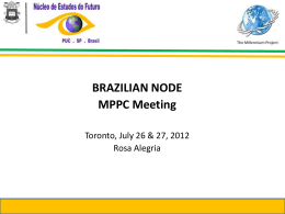
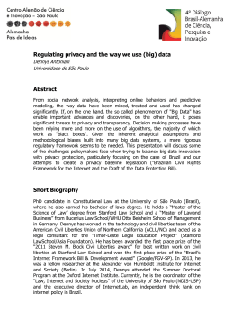
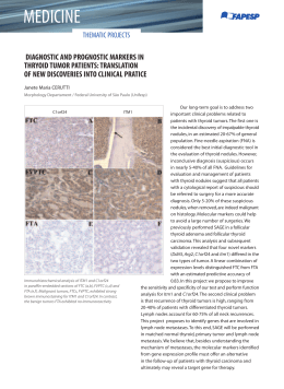
129 Artigo Original Detection of micrometastases in pN0 non-small cell lung cancer: an alternative method combining tissue microarray and immunohistochemistry* Detecção de micrometástases em câncer de pulmão não-pequenas células estádio pN0: um método alternativo combinando imunohistoquímica e análise em microsséries Maíra Rovigatti Franco1, Edwin Roger Parra2, Teresa Yae Takagaki3, Fernando Augusto Soares4, Vera Luiza Capelozzi5 Abstract Objective: To present an alternative method of detecting micrometastases in lymph nodes previously testing negative for non-small cell lung cancer (NSCLC) by routine hematoxylin-eosin staining. Methods: A total of 77 hilar and mediastinal lymph nodes resected from 18 patients with NSCLC were investigated for the presence of micrometastases using a combination of microarray analysis and immunohistochemistry. Results: Micrometastases were detected by identifying cytokeratin- and chromogranin-positive cells in lymph node microarrays. Of the 18 patients initially staged as pN0 through routine hematoxylin-eosin staining, 9 (50%) were restaged as N1, and the prognoses were re-evaluated in terms of histological and clinical parameters. The comparison of the survival curves revealed that survival was higher in the patients without micrometastases than in those with micrometastases. In addition, in the multivariate analysis adjusted for age, gender, histological type, and restaging, the presence of micrometastases proved to be an independent predictor of survival. Among patients who had been previously staged as pN0, the risk of death was found to be 7-times greater for those later diagnosed with micrometastases than for those in whom no micrometastases were identified. Conclusion: The combination of microarray analysis and immunohistochemistry might represent a low-cost and less time-consuming alternative for identifying occult micrometastases and predicting prognoses in surgically resected patients with pN0 NSCLC. Larger randomized, prospective studies are needed in order to determine the accuracy of this method. Keywords: Lung neoplasms; Microarray analysis; Chromogranin A; Survival analysis. Resumo Objetivo: Apresentar um método alternativo para detectar micrometástases em linfonodos previamente negativos para câncer de pulmão não-pequenas células (CPNPC) pela coloração de rotina com hematoxilina-eosina. Métodos: Setenta e sete linfonodos hilares e mediastinais ressecados de 18 pacientes portadores de CPNPC foram investigados para a presença de micrometástases associando-se análise em microsséries e imunoistoquímica. Resultados: Micrometástases foram detectadas após a identificação de células neoplásicas citoqueratina e cromogranina positivas em microsséries de linfonodos. Dos 18 pacientes inicialmente estadiados como pN0 pela coloração de rotina com hematoxilina-eosina, 9 (50%) foram reestadiados como N1, e o prognóstico foi reavaliado em função de parâmetros histológicos e clínicos. A comparação das curvas de sobrevida mostrou que os pacientes sem micrometástases tiveram maior sobrevida do que os portadores de micrometástases. Além disso, após a análise multivariada controlada para idade, sexo, tipo histológico e reestadiamento, a presença de micrometástases mostrou-se como um fator independente na sobrevida. Entre os pacientes que haviam sido previamente estadiados como pN0, o risco de morte mostrou-se 7 vezes maior para os que foram posteriormente diagnosticados com micrometástases do que para aqueles nos quais não foram identificadas micrometástases. Conclusão: A combinação da análise em microsséries com a imunoistoquímica pode representar um método alternativo de baixo custo e menos demorado para identificar metástases ocultas e prever o prognóstico em pacientes portadores de CPNPC pN0 cujos tumores foram cirurgicamente ressecados. São necessários estudos prospectivos randomizados com casuísticas maiores para determinar a acurácia desse método alternativo. Descritores: Neoplasias pulmonares; Análise em microsséries; Cromogranina A; Análise da sobrevida. * Study carried out in the Departments of Pathology and Thoracic Surgery of the University of São Paulo School of Medicine and in the Department of Pathology of the A.C. Camargo Hospital, São Paulo, Brazil. 1. Medical Student. Faculdade de Medicina da Universidade de São Paulo – FMUSP, University of São Paulo School of Medicine – São Paulo, Brazil. 2. PhD in Sciences. Faculdade de Medicina da Universidade de São Paulo – FMUSP, University of São Paulo School of Medicine – São Paulo, Brazil. 3. Assistant Professor in the Department of Thoracic Surgery. Hospital das Clínicas da Faculdade de Medicina da Universidade de São Paulo – HC/FMUSP, University of São Paulo School of Medicine Hospital das Clínicas – São Paulo, Brazil. 4. Head of the Department of Pathological Anatomy. A.C. Camargo Cancer Hospital, São Paulo, Brazil. 5. Associate Professor in the Department of Pathological Anatomy. Hospital das Clínicas da Faculdade de Medicina da Universidade de São Paulo – HC/FMUSP, University of São Paulo School of Medicine Hospital das Clínicas – São Paulo, Brazil. Correspondence to: Vera Luiza Capelozzi ou Edwin Roger Parra. Departamento de Patologia, Faculdade de Medicina da Universidade de São Paulo, Av. Dr. Arnaldo, 455, CEP 01246-903, São Paulo, SP, Brasil. Tel 55 11 3061-7427. E-mail: [email protected]/[email protected] Submitted: 27 April 2007. Accepted, after review: 6 June 2007. J Bras Pneumol. 2008;34(3):129-135 130 Franco MR, Parra ER, Takagaki TY, Soares FA, Capelozzi VL Introduction Since 2006, lung cancer has been the leading cause of death from all types of cancer among Brazilian men and the second leading cause among Brazilian women.(1) It has also been reported to be the leading cause of cancer death in Europe and the United States.(2,3) The tumor-node-metastasis (TNM) system for staging lung cancer is widely used as a guide for predicting prognosis. Identifying lymph node metastases, determining tumor grade, and qualifying metastatic status constitute the most accurate system currently available for predicting prognosis in patients undergoing complete surgical resection of the tumor. However, approximately 30% of patients with pathological stage I non-small cell lung cancer (NSCLC) experience tumor recurrence and die, despite complete surgical resection of the tumor.(4,5) This suggests that occult micrometastatic tumor cells, which are not detected using the methods currently available (clinical staging and conventional histopathological techniques such as hematoxylin-eosin staining), spread to regional lymph nodes or to distant mesenchymal organs prior to the time of surgery. For an accurate prediction of prognosis, lymph node status must be assessed, and micrometastases must be taken into account. The recent development of tissue microarray technology has enabled researchers to conduct retrospective studies using archival, formalin-fixed, paraffin-embedded tissues. This technology, which was developed by Kononen et al.,(6) allows simultaneous examination of hundreds of samples on a single microscope slide and has been used not only for detection of protein in tumor cells but also for quantification of gene expression.(7-10) The method allows minute tissue cylinders to be removed from selected areas in tissue blocks and subsequently fitted into empty ‘recipient’ paraffin blocks.(11) This technology includes multitumor microarrays (samples from multiple histological tumor types), progression microarrays (samples of different stages of tumor progression within a given organ), prognosis microarrays (samples for which clinical follow-up data are available), and cryomicroarrays (frozen samples that might be more suitable than formalin-fixed tissues for detection of RNA).(12,13) The advantages of this method are as follows: reagent costs, technical time, and variability of results are J Bras Pneumol. 2008;34(3):129-135 reduced; the immunostaining images can be stored digitally; and the hierarchical cluster analysis can be used in the interpretation of results.(9,14) In recent years, various authors have successfully employed the immunohistochemical staining method to detect micrometastatic tumor cells in lymph nodes,(15-17) bone marrow, and peripheral blood.(18,19) However, the cost of analyzing all lymph nodes by this method is quite high, since the detection of micrometastases, especially in patients with stage I tumors, requires that multiple sections be evaluated, which involves large-scale immunohistochemical staining. We postulate that an alternative method, combining tissue microarray analysis and immunohistochemistry, can reduce costs and be effective in detecting micrometastases in lymph nodes that have previously tested negative for NSCLC by routine hematoxylin-eosin staining. Therefore, the aim of this study was to present this alternative method, as well as to establish the relationship between stage change and prognosis in patients with completely resected NSCLC tumors. Methods This study was approved by the Ethics Committee of the University of São Paulo. The study sample comprised 18 patients (12 men and 6 women) under treatment in the Department of Pathology of the University of São Paulo School of Medicine Hospital das Clínicas (9 patients had squamous cell carcinoma, 8 had adenocarcinoma, and 1 had large cell carcinoma). Each of those 18 patients underwent complete tumor resection between 1992 and 2005. The mean age was 57 years (range, 37-83 years). All of the patients were clinically staged as T1-3N0M0, and all of the cases were considered potentially curable through surgical resection of the primary tumor and dissection of the hilar and mediastinal lymph nodes (systematic nodal dissection). Clinical staging included routine chest X-ray, bronchoscopy, computed tomography of the chest/upper abdomen, abdominal ultrasound, bone scan and positron emission tomography (the last used only in patients treated since 2004, when it first became available at our facility). In patients presenting lymph nodes with a short axis diameter of less than 1 cm, mediastinoscopy and lymph node biopsy were performed. The follow-up period ranged from 10 to 131 months. Detection of micrometastases in pN0 non-small cell lung cancer: an alternative method combining tissue microarray and immunohistochemistry Close follow-up evaluation was documented by requiring each family practitioner to complete a form regarding local relapse, distant metastases, and outcomes. Following the initial diagnostic evaluation, 6 of the 18 patients with NSCLC died due to local recurrence. Further details regarding these patients are summarized in Table 1. The tumor tissue used in the present study was derived from formalin-fixed pathological samples taken from resected lung specimens after routine pathological studies had been completed. The histological diagnosis and tumor classification were reviewed and verified by two pathologists in accordance with the 2004 World Health Organization guidelines.(20) On average, four lymph nodes were available from each patient, and all were examined in order to confirm the absence of metastases. The final pathological tumor staging identified 8 stage IA (T1N0M0) patients, 4 stage IB (T2N0M0) patients, and 5 stage IIB (T3N0M0) patients. From the 18 patients evaluated, a total of 77 hilar and mediastinal lymph nodes had been surgically removed. Those 77 lymph nodes were analyzed for the presence of micrometastases using a combination of tissue microarray technology and immunohistochemical staining. 131 An average of 7 tissue cylinders (each 1 mm in diameter) per lymph node were removed from the subcapsular lymph node area and subsequently fitted into empty slots in ‘recipient’ paraffin blocks.(21) Two recipient blocks were created for the study, eventually containing, respectively, 198 and 240 tissue cylinders. A total of 12 paraffin-embedded sections per block were submitted to immunostaining. Micrometastatic tumor cells were evaluated through immunohistochemical staining using the avidin-biotin complex immunoperoxidase technique. The antibodies used were anti-cytokeratin 7 (CK7, Clone OV-TL 12/30, 1:100 dilution; Dako, Glostrup, Denmark) and cytokeratin AE1/AE3 (Clone AE1 and AE3; Dako, Carpinteria; CA, USA; 1:320 dilution), which recognize most cytokeratins, including type 1 (acidic type) and type 2 (basic type). The chromogranin-A antibody (Clone DAK-A3; Dako, Glostrup, Denmark; 1:600 dilution), which recognizes neuroendocrine cells, was also used. The presence of cytokeratins or chromogranin A-positive cells in lymph node sections, which was determined using tissue microarray technology, was accepted as evidence of the presence of micrometastatic tumor cells, even when only a single cytokeratin or a single chromogranin A protein positive cell was detected. The specimens were examined and Table 1 - Clinicopathological characteristics of the patients. Number of patients presenting positive cells in immunohistochemical staining Variable CK7 p AE1/AE3 p Chromogranin A p Micrometastases (n = 18) (n = 18) (n = 18) (n = 18) All patients 5/18 (28%) 7/18 (39%) 6/18 (33%) 9/18 (50%) Gender Male 5/12 (41.7%) 0.063 6/12 (50%) 0.171 4/12 (33.3%) >0.99 7/12 (58.3%) Female 0/6 (0.0%) 1/6 (16.7%) 2/6 (33.3%) 2/6 (33.3%) Age (years) 1/9 (11.1%) 0.114 3/9 (33.3%) 0.629 3/9 (33.3%) 1.00 4/9(44.4%) ≤57 >57 4/9 (44.4%) 4/9 (44.4%) 3/9 (33.3%) 5/9 (55.6%) Histology Adenocarcinoma 1/8 (12.5%) 0.278 3/8 (37.5%) 0.684 1/8 (12.5%) 0.131 3/8 (37.5%) Squamous cell 4/9 (44.4%) 4/9 (44.4%) 4/9 (44.4%) 5/9 (55.6%) carcinoma Large cell 0/1 (0.0%) 0/1 (0.0%) 1/1 (100%) 1/1 (100%) carcinoma Initial pathological stage T1N0M0 0/4 (0.0%) 0.034 1/4 (25%) 0.163 0/4 (0.0%) 0.269 1/4 (25%) T2N0M0 3/12 (25%) 4/12 (33.3%) 5/12 (41.7%) 6/12 (50%) T3N0M0 2/2 (100%) 2/2 (100%) 1/2 (50%) 2/2 (100%) p 0.317 0.637 0.447 0.223 CK7: cytokeratin 7; AE1/AE3: cytokeratin AE1/AE3; T: tumor; N: nodal metastasis; and M: metastasis. J Bras Pneumol. 2008;34(3):129-135 132 Franco MR, Parra ER, Takagaki TY, Soares FA, Capelozzi VL checked by three pathologists who were blinded as to patient data, including outcomes. Brownish cytoplasmic staining was indicative of positivity for CK7, AE1/AE3, or chromogranin A, which, taken together with positive cell morphology, was seen as evidence of the presence of neoplastic cells metastasized to the lymph node. Data regarding the patients were obtained through a retrospective review of the medical records. The association between clinicopathological data and micrometastatic status was analyzed using a contingency table. Statistical significance was evaluated using the chi-square test or Fisher’s exact test. Kaplan-Meier curves were generated, and the final multivariate analysis was performed using the Cox proportional hazards model. All statistical procedures were performed using the Statistical Package for the Social Sciences program, version 10.0 (SPSS Inc., Chicago, IL, USA). The level of significance was set at p < 0.05.(22) Results Using immunohistochemical staining with CK7, AE1/AE3, and chromogranin A protein, single tumor cells and small clusters of tumor cells were seen in the tissue cylinders of the lymph node specimens (Figure 1). Cells staining positive for CK7, AE1/AE3, or chromogranin A were found in 12 (0.23%), 24 (0.46%), and 16 (0.30%) of the samples, respectively. Of the 77 lymph nodes included in the study, 9 (12%) were CK7 positive, 14 (18%) were AE1/AE3 positive, and 10 (13%) were chromogranin A positive. When the immunohistochemical staining for cytokeratin was used in conjunction with that a 50 Mm c b 50 Mm 50 Mm Figure 1 - Immunohistochemistry in pN0 lymph nodes resected from patients with non-small cell lung cancer. Note brownish cytoplasmic staining for cytokeratin 7 (a), cytokeratin AE1/AE3 (b) and chromogranin A (c), which, taken together with the cell morphology, was considered indicative of neoplastic cells metastasized to the lymph node. Magnification: ×400. J Bras Pneumol. 2008;34(3):129-135 Detection of micrometastases in pN0 non-small cell lung cancer: an alternative method combining tissue microarray and immunohistochemistry for chromogranin A, micrometastases were found in 22 lymph nodes from 9 (50%) of the 18 patients. Restaging of the nodal status was performed based on the combination of cytokeratin and chromogranin immunohistochemical staining. Among the patients staged as pN0 based on routine hematoxylin-eosin staining, 9 (50%) were restaged as N1. The clinical and morphological data were re-evaluated for associations as well as for recurrence and survival. The presence of CK7-positive micrometastatic tumor cells in the pN0 lymph nodes was significantly associated with pathological stage (p < 0.05), as can be seen in Table 1, which also shows the distribution of the remaining clinical characteristics of the patients. No significant associations were found for AE1/AE3 or chromogranin A immunostaining in the pN0 lymph nodes. The Kaplan-Meier survival curves presented in Figure 2 show that survival was lower among the patients presenting chromogranin A-stained 133 micrometastatic tumor cells in the lymph nodes than among those without such micrometastases, and this difference was of borderline significance (p = 0.05). Considering survival in a multivariate analysis adjusted for age, gender, histological type, and restaging, the survival rate was 7-fold higher in the absence of micrometastases for patients younger than 57 years with adenocarcinoma. The relative risks for these predictors are shown in Table 2. The paraffin blocks containing the lymph node sections were submitted to serial sections with the objective of increasing the surface area of the sections for diagnosis. Twelve 3- to 4-µm thick sections were obtained from the two blocks. These new sections presented broader areas for examination, totaling 142.2 mm2 per lymph node. Table 3 shows the comparison between tissue microarray analysis and the standard method for lymph node analysis in terms of advantages and cost. Discussion 1.1 Probability of survival 1.0 0.9 0.8 0.7 0.6 0.5 0 20 40 60 80 100 120 Survival in months after biopsy 140 Chromogranin A Present Absent Figure 2 - Kaplan-Meier plots of survival probability versus follow-up period in months in patients with pathological stages N0 and N1. The group presenting no chromogranin A-immunostained lymph node cells is represented by the upper curve, and the group presenting chromogranin A-immunostained lymph node cells is represented by the lower curve. Lung cancer is currently thought to arise from the accumulation of various genetic changes, such as mutations and deletions. Recent advances in molecular biology and genetics have produced new diagnostic and treatment possibilities for clinical oncology. The detection of micrometastases has potentially significant implications for prognosis, primarily in patients with NSCLC.(23) Pathological tumor staging represents the most accurate system currently available for predicting prognosis in patients who have undergone radical tumor resection. However, the average 5-year survival rate for patients with completely resected stage I NSCLC tumors is only approximately 70%,(4,5,23) and approximately 30% of such patients experience recurrence. This finding suggests that occult micrometastases exist prior to the time of surgery. The rate of this occurrence is clearly underestimated by current clinical staging and conventional histopathological methods. By combining tissue microarray technology and immunohistochemical staining, we identified occult micrometastatic tumor cells in pN0 lymph nodes in 50% of the patients with completely resected NSCLC tumors. In addition, in the univariate and multivariate analyses, the patients with lymph node micrometastases had a poorer prognosis than did the patients without such micrometastases. The J Bras Pneumol. 2008;34(3):129-135 134 Franco MR, Parra ER, Takagaki TY, Soares FA, Capelozzi VL Table 2 - Multivariate Cox regression analysis of prognostic factors. Variable n Relative risk Gender Male 12 3.557 Female 6 1 Age (years) 9 5.440 ≤57 >57 9 1 Histology Adenocarcinoma 8 0.077 Squamous cell carcinoma 9 0.000 New stage N0 9 7.100 N1 9 1 95% confidence interval 0.451-28.042 p 0.228 1.116-26.511 0.036 0.011-0.542 0.010 1.190-42.381 0.032 prognostic impact was independent of the TNM staging system. We have demonstrated that, in 50% of the patients with NSCLC evaluated in the present study, the combined use of tissue microarray technology and immunohistochemical staining to detect occult tumor cells in regional lymph nodes changed the staging status. Tissue microarray technology has potential applications in the practice of diagnostic histopathology.(24) Using the combination of tissue microarray analysis and immunohistochemistry, pathologists are now able to perform unprecedented large-scale analyses. The advantages of this combined use are significant: it allows a large number of samples, which are taken from a limited amount of archival tissues, to be assessed simultaneously for numerous markers and be processed under identical conditions; it presents an excellent concordance with conventional methods; it reduces costs; and it is less time-consuming.(25) We used CK7 and AE1/AE3 cytokeratin antibodies as well as chromogranin A antibody as the major markers for micrometastases in the lymph nodes. Using CK7, we detected micrometastases in 28% of the patients, compared with 39% for AE1/AE3 and 33% for chromogranin A. The diagnostic efficiency was increased by combining tissue microarray technology and immunohistochemical staining. Furthermore, detection of micrometastases using chromogranin A was significantly associated with poor survival, which makes this antibody a potentially useful marker in clinical and pathological practice. Considering survival in a multivariate analysis adjusted for age, gender, histological type, and restaging, the probability of survival was 7-fold higher in the absence of micrometastases for patients younger than 57 years with adenocarcinoma, underscoring the importance of detecting micrometastases in all lymph nodes resected from patients with lung cancer. Our study showed the value of combining tissue microarray technology and immunohistochemical staining to detect occult micrometastatic tumor cells in lymph nodes. However, although this combination might represent a low-cost and less time-consuming method of predicting recurrence and prognosis in patients with completely resected stage I NSCLC tumors, our study has limitations. First, the patient sample was small. Second, there have been no other studies comparing this technique with the gold standard methods typically employed to detect micrometastases and with which our findings might be compared. In conclusion, larger randomized, prospective studies are needed in order to determine the accu- Table 3 - Benefits and cost. Method Stage pN0 (%) Standard 0 Microarray 50 Paraffin blocks (n) 77 2 J Bras Pneumol. 2008;34(3):129-135 Lymph node (n) 77 77 Cost (US$) 5351.50 834.00 Paraffin sections (n) 1 12 Surface analyzed (mm2) 12 142.2 Detection of micrometastases in pN0 non-small cell lung cancer: an alternative method combining tissue microarray and immunohistochemistry racy of and validate the combined use of tissue microarray technology and immunohistochemical staining as an alternative method of detecting occult micrometastatic tumor cells in pN0 NSCLC lymph nodes. Acknowledgments We would like to express our gratitude to Carlos Ferreira Nascimento, Esmeralda Miristene Eher, and Sandra de Morais Fernezlian for their skillful technical assistance. The present study received financial support from the Brazilian Conselho Nacional de Desenvolvimento Científico e Tecnológico (CNPq, National Council for Scientific and Technological Development), the Fundação de Amparo à Pesquisa do Estado de São Paulo (FAPESP, Foundation for the Support of Research of the State of São Paulo; grant no. 2001/14566-9), and the Laboratório de Investigação Médica 05 (LIM 05, Medical Investigation Laboratory 05) of the University of São Paulo School of Medicine Hospital das Clínicas. References 1. Instituto Nacional do Câncer [Homepage on the Internet]. Brasilia: Ministério da Saúde. [cited 07 Jul 20] Estimativa da Incidência e Mortalidade por Câncer no Brasil. Available from: www.inca.gov.br/estimativa/2008 2. Janssen-Heijnen ML, Gatta G, Forman D, Capocaccia R, Coebergh JW. Variation in survival of patients with lung cancer in Europe, 1985-1989. Eur J Cancer. 1998;34(14):2191-6. 3. Wingo PA, Ries LA, Rosenberg HM, Miller DS, Edwards BK. Cancer incidence and mortality, 1973-1995: a report card for the U.S. Cancer. 1998;82(6):1197-207. 4. Martini N, Bains MS, Burt ME, Zakowski MF, McCormack P, Rusch VW, et al. Incidence of local recurrence and second primary tumors in resected stage I lung cancer. J Thorac Cardiovasc Surg. 1995;109(1):120-9.. 5. Strauss GM, Kwiatkowski DJ, Harpole DH, Lynch TJ, Skarin AT, Sugarbaker DJ. Molecular and pathologic markers in stage I non-small-cell carcinoma of the lung. J Clin Oncol. 1995;13(5):1265-79. 6. Kononen J, Bubendorf L, Kallioniemi A, Bärlund M, Schraml P, Leighton S, et al. Tissue microarrays for highthroughput molecular profiling of tumor specimens. Nat Med. 1998;4(7):844-7. 7. Henshall S. Tissue microarrays. J Mammary Gland Biol Neoplasia. 2003;8(3):347-58. 8. Simon R, Sauter G. Tissue microarrays for miniaturized highthroughput molecular profiling of tumors. Exp Hematol. 2002;30(12):1365-72. 9. Mobasheri A, Airley R, Foster CS, Schulze-Tanzil G, Shakibaei M. Post-genomic applications of tissue microarrays: basic 135 research, prognostic oncology, clinical genomics and drug discovery. Histol Histopathol. 2004;19(1):325-35. 10. Packeisen J, Korsching E, Herbst H, Boecker W, Buerger H. Demystified... tissue microarray technology. Mol Pathol. 2003;56(4):198-204. 11. Tawfik El-Mansi M, Williams AR. Validation of tissue microarray technology using cervical adenocarcinoma and its precursors as a model system. Int J Gynecol Cancer. 2006;16(3):1225-33. 12. Russo G, Zegar C, Giordano A. Advantages and limitations of microarray technology in human cancer. Oncogene. 2003;22(42):6497-507. 13. Takahashi M, Yang XJ, Sugimura J, Backdahl J, Tretiakova M, Qian CN, et al. Molecular subclassification of kidney tumors and the discovery of new diagnostic markers. Oncogene. 2003;22(43):6810-8. 14. Simon R, Mirlacher M, Sauter G. Tissue microarrays in cancer diagnosis. Expert Rev Mol Diagn. 2003;3(4):421-30. 15. Chen ZL, Perez S, Holmes EC, Wang HJ, Coulson WF, Wen DR, et al. Frequency and distribution of occult micrometastases in lymph nodes of patients with non-small-cell lung carcinoma. J Natl Cancer Inst. 1993;85(6):493-8. 16. Passlick B, Izbicki JR, Kubuschok B, Thetter O, Pantel K. Detection of disseminated lung cancer cells in lymph nodes: impact on staging and prognosis. Ann Thorac Surg. 1996;61(1):177-82; discussion 183. 17. Izbicki JR, Passlick B, Hosch SB, Kubuschock B, Schneider C, Busch C, et al. Mode of spread in the early phase of lymphatic metastasis in non-small-cell lung cancer: significance of nodal micrometastasis. J Thorac Cardiovasc Surg. 1996;112(3):623-30. 18. Ohgami A, Mitsudomi T, Sugio K, Tsuda T, Oyama T, Nishida K, et al. Micrometastatic tumor cells in the bone marrow of patients with non-small cell lung cancer. Ann Thorac Surg. 1997;64(2):363-7. 19. Krüger W, Krzizanowski C, Holweg M, Stockschläder M, Kröger N, Jung R, et al. Reverse transcriptase/polymerase chain reaction detection of cytokeratin-19 mRNA in bone marrow and blood of breast cancer patients. J Cancer Res Clin Oncol. 1996;122(11):679-86. 20. Travis WD. Pathology and Genetics of Tumours of the Lung, Pleura, Thymus and Heart. World Health Organization Classification of tumours. Lyon: IARC Press; 2004. 21. Bubendorf L, Nocito A, Moch H, Sauter G. Tissue microarray (TMA) technology: miniaturized pathology archives for highthroughput in situ studies. J Pathol. 2001;195(1):72-9. 22. Statistical Package for the Social Sciences (SPSS). Version 10.0. [Computer program]. Chicago: SPSS Inc.; 2002. 23. Nesbitt JC, Putnam JB Jr, Walsh GL, Roth JA, Mountain CF. Survival in early-stage non-small cell lung cancer. Ann Thorac Surg. 1995;60(2):466-72. 24. van de Rijn M, Gilks CB. Applications of microarrays to histopathology. Histopathology. 2004;44(2):97-108. 25. Jacquemier J, Ginestier C, Charafe-Jauffret E, Bertucci F, Bege T, Geneix J, et al. [Small but high throughput: how “tissue-microarrays” became a favorite tool for pathologists and scientists] [Article in French] Ann Pathol. 2003;23(6):623-32. J Bras Pneumol. 2008;34(3):129-135
Baixar