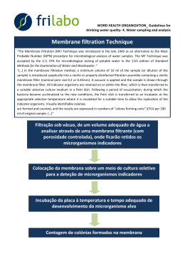
MEDICINE
THEMATIC PROJECTS
OCULAR SURFACE RECONSTRUCTION:
BASIC, CLINICAL AND SURGICAL ASPECTS
Rubens Belfort MATTOS Júnior
Ophthalmology Departament / Federal University of São Paulo (Unifesp)
Epithelial expansion from a limbal explant
cultivated ex vivo (x40)
Our study includes the following objectives: (i) to
evaluate the ultra-structural, biochemical and molecular
aspects of the amniotic membrane colonized ex vivo by
corneal epithelium; (ii) to analyze the alterations of the
ocular surface by different specialized diagnostic methods;
(iii) to evaluate new treatments for dry eye; (iv) to evaluate
the efficiency of reconstructive surgery of the ocular
surface by using amniotic membrane associated or not
with limbal transplant in Ocular Surface Disorder. Briefly
we present the methods used for each protocol: scanning
transmission electron microscopy in fresh amniotic
membrane preserved by two different methods;
quantification by ELISA – in both preserved and fresh
amniotic membrane – of growth factors (recombinant
human epithelial growth factor (EGF), fibroblastic growth
factor (FGF-4, FGF-basic), hepatocyte growth factor (HGF),
keratinocyte growth factor (KGF), beta-transformant growth
factor (TGF-b)), interleukins (IL-4 e IL-10) and postglandins
(PGE2); identification and quantification of growth factors
and cytokines mRNA’s in the human amniotic membranes
by PCR techniques, "Northern blot" and "Western blot";
use of the amniotic membrane in experimental cicatricial
keratoconjunctivitis in rabbits; ultra-structural analysis
of cell proliferation (immunofluorescence with Ki-67),
apoptosis (morphological method with Hoechst 33342)
and immunohistochemical/FACS to determine the
expression of the epithelial.
MEDICINE
SUMMARY OF RESULTS
TO DATE AND PERSPECTIVES
MAIN PUBLICATIONS
This project includes the development of 15 research projects
[5 basic research (PE), and 10 clinical research (PCC)]. The
resources provided by FAPESP allowed us to install the
Advanced Ocular Surface Laboratory (CASO), the first cell biology
lab in Brazil focused on the development of cell therapy
technology for ocular surface reconstruction.
Obtained results to date: Ultra-structure characterization,
biochemical and molecular aspects of the human amniotic
membrane. The evaluation of the efficacy of amniotic
membrane and limbal stem cell transplantation for ocular surface
reconstruction. Graft survival was observed with a cumulative
survival of 33% after a mean follow-up time of 33 months.
Increase in postoperative visual acuity was observed in 60.6%
of the operated eyes during this period. Marked impact on graft
survival was observed for patients with Stevens-Johnson
syndrome, dry eye, keratinization, eyelid abnormalities, and
allogeneic conjunctival limbal transplantation (independently of
HLA compatibility) (p<.05). Preoperative dry eye was the most
important prognostic parameter for surgical outcome (p<.001).
We are also completing three important prospective,
comparative studies evaluating the efficacy of amniotic membrane
in the treatment of pterygia, bullous keratopathy and corneal
and scleral thinning.
We developed at the CASO the technique for amniotic
membrane colonization with human limbal epithelial stem cells
expanded ex vivo. In our hands, the best method includes the
use of fragments of limbal tissue (and not cell suspension) over
EDTA deepithelialized human amniotic membrane. We are also
evaluating different cell lines (conjunctival epithelia and immature
dental pulp stem cells) in order to compare the survival and
possibility for clinical application.
We started to transplant these ex vivo limbal epithelial
cultivated amniotic membranes in patients with total limbal
stem cell deficiency with reasonable results in the short-term.
More cases are now scheduled to be operated in order to obtain
a better evaluation of the procedure and survival of the
transplanted cells.
We developed the technique of impression cytology as a
diagnostic tool for limbal stem cell deficiency and other diseases.
We are now standardizing immunohistochemical analysis with
different cell and proliferation markers.
We are in the process of evaluating, through a double blind
controlled study, the efficacy of topical use of 0.05% cyclosporine
A in the treatment of dry eye secondary to Sjögren`s syndrome.
Our thematic project is a multi-disciplinary project of
the Federal University of Sao Paulo (UNIFESP) supported by
FAPESP, with the participation of another two national and
two international Universities (University of Nottingham, UK,
and McGill University, Montreal, Canada).
Gomes JAP, Santos MS dos, Cunha MC, Mascaro
VLDM, Barros JN, Sousa LB de. 2003. Amniotic
Membrane Transplantation for Partial and Total
Limbal Stem Cell Deficiency Secondary to Chemical
Burn. Ophthalmology. 110: 466-73.
Gomes JAP, Santos MS dos, Ventura ÂS, et al. 2003.
Amniotic Membrane with Living Related Corneal
Limbal / Conjunctival Allograft for Ocular Surface
Reconstruction in Stevens-Johnson Syndrome.
Arch Ophthalmol. 121: 1369-74.
Dua HS, Gomes JAP, King AJ, Maharajan VS. 2004.
The Amniotic Membrane in Ophthalmology. Surv
Ophthalmol. 49: 51-77.
Barros JN, Mascaro VLDM, Gomes JAP, et al. 2004.
Avaliação da presença de células caliciformes na
córnea humana. Arq Bras Oftalmol. 67: 121-5.
Gomes JAP, Romano A, Santos MS, Dua HS. 2005.
Amniotic membrane use in ophthalmology. Curr
Opin Ophthalmol. 16: 233-40.
Santos MS, Gomes JAP, Hofling-Lima AL, Rizzo LV,
Romano AC, Belfort Jr R. 2005. Survival analysis of
conjunctival limbal grafts and amniotic membrane
transplantation in eyes with total limbal stem cell
deficiency. Am J Ophthalmol. 140: 305-6.
Santos MS, Gomes JAP, Hofling-Lima AL, Rizzo LV,
Romano AC, Belfort Jr R. 2006. Survival analysis of
conjunctival limbal grafts and amniotic membrane
transplantation in eyes with total limbal stem cell
deficiency. Am J Ophthalmol. 141: 599-600.
Melo GB, Gomes JAP, Glória MA, Martins MC,
Haapalainen EF. 2007. Avaliação morfológica de
diferentes técnicas de desepitelização da membrana
amniótica humana. Arq Bras Oftalmol. 70: 407-411.
Pena JDO, Melo GB, Gomes JAP, Haapalanien EF,
Komagome CM, et al. 2007. Análise ultraestrutural e
de fatores de crescimento de diferentes métodos
de preservação da membrana amniótica utilizada
em cirurgia ocular. Arq Bras Oftalmol. 70:756-62.
Cristovam PC, Glória MA, Melo GB, Gomes JAP. 2008.
Importância do feeder layer 3T3 para estabelecer
cultura de células epiteliais do limbo obtidas de rimas
córneo-esclerais. Arq Bras Oftalmol. Submetido.
Rubens Belfort MATTOS Júnior
Universidade Federal de São Paulo (Unifesp)
Departamento de Oftalmologia
Rua Botucatu, 822 – Vila Clementino
04023-062 – São Paulo, SP – Brasil
+55-11-5085-2010
[email protected]
Download