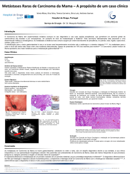
UNIVERSIDADE FEDERAL DE PERNAMBUCO
CENTRO DE CIÊNCIAS BIOLÓGICAS
MESTRADO EM BIOQUÍMICA E FISIOLOGIA
CARCINOMA DUCTAL INVASIVO MAMÁRIO ESPORÁDICO: UMA
ABORDAGEM HISTOQUÍMICA E IMUNOHISTOQUÍMICA
MOACYR JESUS BARRETO DE MELO RÊGO
ORIENTADOR: PROF.DR.EDUARDO BELTRÃO
Recife, 2009.
2
MOACYR JESUS BARRETO DE MELO RÊGO
CARCINOMA DUCTAL INVASIVO MAMÁRIO ESPORÁDICO: UMA
ABORDAGEM HISTOQUÍMICA E IMUNOHISTOQUÍMICA
Dissertação
cumprimento
apresentada
para
parcial
o
das
exigências para obtenção do título de
Mestre em Bioquímica e Fisiologia
pela
Universidade
Pernambuco.
Recife, 2009.
Federal
de
3
Rego, Moacyr Jesus Barreto de Melo
Carcinoma ductal invasivo mamário esporádico: uma abordagem
histoquímica e imunohistiquímica/ Moacyr Jesus Barreto de Melo Rego. –
Recife: O Autor, 2009
53 folhas: il., fig., tab.
Dissertação (mestrado) – Universidade Federal de Pernambuco.
CCB. Departamento de Bioquímica e Fisiologia, 2009.
Inclui bibliografia
1. Histoquímica com lectinas. 2. Carcinoma ductal invasivo. 3.
Neoplasia mamária. 4. Proteína p53 I Título.
577.1
CDU (2.ed.)
UFPE
572
CDD (22.ed.)
CCB – 2009- 68
4
ÍNDICE ANALÍTICO
LISTA DE FIGURAS
LISTA DE TABELAS
RESUMO
ABSTRACT
1 INTRODUÇÃO
10
1.1 CÂNCER: UMA VISÃO GERAL
10
1.2 A MAMA E O CÂNCER DE MAMA
12
1.3 LECTINAS E SUA APLICAÇÃO NA HSTOQUÍMICA
16
1.4 p53 E SUA APLICAÇÃO COMO MARCADOR IMUNOHISTOQUÍMICO
18
2 OBJETIVOS
22
2.1 OBJETIVOS GERAIS
22
2.2 OBJETIVOS ESPECÍFCOS
22
3 ARTIGO CENTÍFICO
23
4 CONCLUSÕES
41
5 REFERÊNCIAS BIBLIOGRÁFICAS
42
ANEXO I
5
AGRADECIMENTOS
A Deus, a quem devo tudo na minha vida.
Aos meus pais, Moacir e Fernanda e as minhas irmãs Andressa e Alessandra, que a cada dia
despertam e ressaltam o que há de melhor em mim enquanto ser humano, e a todos os
demais da minha família, eternos encorajadores.
Ao Prof. Dr. Eduardo Isidoro Carneiro Beltrão orientador sempre presente e incentivador.
Obrigado por acreditar em mim nesses cinco anos de jornada científica.
A todos do grupo Biomarcadores no Câncer, minha família laboratorial.
Minha gratidão a Carmelita Cavalcanti, Luiz Alves, Carolina Fonseca, Rossil Hassan,
Mário Ribeiro, Tereza Cartaxo e Alexandre Alves, pois sem a ajuda de vocês não teria
chegado aqui.
A Marina Ferraz Cordeiro, primordial para realização dessa dissertação, IC dedicada e
responsável. Obrigado por toda ajuda e também pelas risadas.
A todos os meus amigos de todas as horas e de toda a vida, Cleviton Monteiro, Victor
Hugo, Eduardo Lundgren, Marcelle Mota, Gabriela Souto, Paula Hirakawa, Amanda
Pessoa, Mirella Soares, Petra Barros, Clefersson Ferreira, Jofre Neto, vocês são essenciais.
Bem como a família “dos Fantinis” (Dona Dora, André, Tobias e Michele) pelos momentos
inesquecíveis.
6
LISTA DE FIGURAS
INTRODUÇÃO
Figura 1: Capacidade de migração de células neoplásicas malignas como indicação do grau
de malignidade tumoral (Fuster e Esko, 2005)
08
Figura 2: Número de artigos científicos e patentes utilizando lectinas em métodos de
diagnóstico entre 1986 e 2006 (Mullican et al, 2007)
14
ARTIGO CIENÍFICO
Figure 1: Lectin histochemistry of invasive ductal carcinoma. a) Con A staining ductal cells
in poor-differentiated (white arrow) but not in well-differentiated (black arrow) tumours in
the same biopsy (100x), b) PNA staining atipical mitosis (arrows) (200x), c) Heterogeneous
staining pattern of UEA-I was observed in cytoplasm and plasmatic membrane (200x). 2830
7
LISTA DE TABELAS
INTRODUÇÃO
Tabela 1: Estimativa para o ano de 2008 das taxas brutas de incidência de câncer de mama
por 100.000 mulheres (INCA, 2006).
12
ARTIGO CIENTÍFICO
Table 1: Clinical and histopatological characteristics of patents used on this study.
27
Table 2: Lectin histochemistry of neoplasic (IDC) and normal tissues of breast.
31
8
RESUMO
O câncer de mama é a neoplasia mais freqüente e a maior causa de morte de câncer em
mulheres no mundo. Dentre as várias neoplasias que acometem esse órgão o carcinoma
ductal invasivo (CDI) representa de 65% a 80% de todos os carcinomas, o qual apresenta
grande heterogeneidade fenotípica e genotípica. Por causa dessa heterogeneidade a procura
por biomarcadores das neoplasias mamárias vem aumento nos últimos anos. Nesse sentido
lectinas, (glico)proteínas reconhecedoras de carboidratos, vem sendo utilizadas como
tradutoras de glicocódigos e biomarcadores de várias neoplasias. Esse trabalho objetivou
analisar o perfil de carboidratos do CDI através da histoquímica com lectinas e
correlacionar essa expressão com parâmetros clínicos e histopatológicos (idade, tamanho
do tumor, variante histológica e expressão de p53). Oitenta e oito biópsias de CDI e 20
biópsias de mastoplastia redutora e bordas livres de tumor, utilizadas como controles
normais foram obtidas no Hospital das Clínicas da UFPE. Para a histoquímica com lectinas,
cortes de 4µm foram tratados com tripsina 0,1%, metanol-peróxido de hidrogênio 0,3% e
incubadas com as lectinas conjugadas a peroxidase (horseradish peroxidase – HRP),
Concanavalin A, Con A-HRP, Ulex europeus I, UEA-I-HRP e Peanult Aglutinin, PNAHRP, especificas para glicose/manose, L-fucose e D-galactose, respectivamente. A
imunohistoquímica para o p53 foi realizada através da técnica da estreptavidina-biotinaperoxidase. Ambas as reações foram reveladas com diaminobenzidina (DAB)-H2O2,
contra-coradas com hematoxilina e avaliadas em microscopia óptica. Houve uma
associação entre a marcação das lectinas, Con A (p<0,001), PNA (p<0,001) e UEA-I
(p<0,001), nos dois grupos de estudo, normal e CDI. A prevalência da positividade foi
significativamente maior no grupo com câncer.A proteína p53 mutante foi detectada em
34,1% dos casos. Morfologicamente PNA reconheceu mitoses atípicas e, UEA-I e Con A
endotélio vascular e todas as lectinas marcaram as células neoplásicas. Não houve
correlação significativa entre a marcação das lectinas e os parâmetros clínicos e
histopatológicos (p>0,005). Os resultados indicam que ocorre uma maior disponibilidade
de glicose/manose, L-fucose e D-galactose no CDI, sugerindo a utilização das lectinas
UEA-I, PNA e ConA como ferramentas histopatológicas na diferenciação dos tecidos
normais e portadores dessa malignidade.
Palavras-chave: histoquímica com lectinas, carcinoma ductal invasivo, p53.
9
ABSTRACT
Breast cancer is the most frequent neoplasia and the main cause of cancer death in women
worldwide. Among the several types of mammary gland neoplasia Invasive Ductal
Carcinoma (IDC) represents 65% to 80% of all carcinomas. The major problem concerning
this neoplasia is the great genotypic and phenotypic heterogeneity. This feature explains the
increasing search for biomarkers of breast cancer in recent years. Among these probes
lectins, (glycol)protein that recognizes carbohydrates, have been used as decipherers of cell
glycocodes and biomarkers in cancer. This study aimed to evaluate the carbohydrate profile
in IDC by lectin histochemistry and to correlate it to clinical and histopathological
parameters such as age, tumor size, histological variant and p53 expression. Eighty-eight
biopsies of IDC, and 20 biopsies of reductional mastoplasty were obtained at the Clinic
Hospital of the Federal University of the State of Pernambuco. Sixty-eight ICD free borders
were used as normal controls besides the reductional mastoplasty. For lectin histochemistry
tissue sections (4μm) were treated with trypsin (0.1%), methanol-hydrogen peroxide (0.3%)
and than incubated with horseradish peroxidase (HRP) conjugated lectins (Concanavalin A,
Con A-HRP, Ulex Europeans I, UEA-I-HRP, Peanult Aglutinin, PNA-HRP; specific for
glucose/mannose, D-galactose and L-fucose respectively. p53 immunohistochemistry was
performed using the estreptavidin-biotin-peroxidase technique. Both reactions were
revealed with diaminobenzidine (DAB)-H2O2, counter-stained with haematoxylin and
evaluated on optical micrsocopy. There is a significant association between lectins Con A
(p<0.001), PNA (p<0.001) and UEA-I (p<0.001) in the two study groups, normal and IDC.
The prevalence of positivity was significantly higher in the group of cancer. Mutant protein
of p53 was detected in 34,1% of CDI biopsies. Morphologically PNA recognized atypical
mitosis and Con A and UEA-I vascular endothelium. All lectins of this study recognize
neoplastics cells. There is no association with lectins and clinical parameters (p>0.005).
Results indicated that there is a higher disponibility of glucose/mannose, L-fucose and Dgalactose residues in IDC when compared to normal tissue indicating these lectins as
biomarkers for this malignancy.
Key-words: Lectin histochemistry, Invasive Ductal Carcinoma, p53
10
1 INTRODUÇÃO
1.1 CÂNCER: UMA VISÃO GERAL
O câncer, segundo a Sociedade Americana do Câncer (2008), é definido por um
grupo de doenças caracterizadas pelo crescimento descontrolado e disseminação de células
anormais. É uma das principais causas de morte mundial, geralmente excedido apenas por
doenças cardiovasculares, sendo uma doença genética que afeta uma em cada três pessoas
no planeta (De Meija et al., 2003). O grau de malignidade do câncer está relacionado à sua
habilidade em desarranjar a arquitetura tecidual, invadir fronteiras de tecidos adjacentes e
mestastizar para órgãos distantes (Figura 1).
Na carcinogênese ocorrem mudanças na expressão de proteínas celulares que
levam à alterações nas vias de sinalização induzindo, frequentemente, mudanças no
microambiente tumoral, causando inflamação e resultando em mudança no perfil das
proteínas teciduais que podem ser secretadas (Kopf e Zharhary, 2007). A carcinogênese
inicia-se a partir de uma alteração no DNA celular ou da ativação anormal de genes
presentes em células sadias. Tais genes coordenam a proliferação, a diferenciação, a morte
por apoptose e outras funções celulares primordiais. A expressão amplificada de proteínas
reguladas por tais genes pode determinar a progressão de alterações celulares levando aos
estados neoplásicos malignos (Conde et al., 2005).
No que se refere às funções bioquímicas, as moléculas de adesão e vias de
transdução dos sinais para o crescimento tumoral são alterados durante a progressão
tumoral. As proteínas normais codificadas pelos proto-oncogenes (genes que regulam a
proliferação celular) desempenham funções iniciais nas células durante o desenvolvimento
das neoplasias. Dentre estas moléculas, destacam-se os fatores de crescimento, proteínas
envolvidas na recepção e transdução de sinais localizados na superfície celular (Guo et al.,
2006).
11
Figura 1: Capacidade de migração de células neoplásicas
malignas como indicação do grau de malignidade tumoral.
(Fusker e Esko, 2005)
Os diferentes tipos de carboidratos na superfície celular apresentam uma importante
função no controle de vários processos fisiológicos e patobioquímicos no organismo
(Phipps et al., 2005). Mudanças quantitativas e/ou qualitativas nos componentes lipídicos e
protéicos da célula e organelas celulares também são evidentes durante o desenvolvimento
dos processos patológicos (Astoul et al., 2000).
12
O mecanismo de reconhecimento molecular na superfície da célula, o que a torna
capaz de reconhecer células semelhantes e assim interagir com as mesmas mantendo a
homeostase, é afetado quando células normais sofrem desdiferenciação. O resultado é um
crescimento e divisão descontrolados (Shekhar et al., 2004). Estudos utilizando tumores
realizados por Hirschefelt & Thonsen na década de 30 foram os primeiros a demonstrar
alterações bioquímicas nas células tumorais, o que foi posteriormente confirmado como
padrões de glicosilação aberrantes associados ao tumor (Dabelsteen et al., 1992).
Com o advento da tecnologia de anticorpos monoclonais e o avanço das pesquisas
com a histoquímica com lectinas, descobriram-se vários epítopos "tumor-específicos"
dentre os quais carboidratados. Correlações significativas entre certos tipos de glicosilação
alterada e o prognóstico de tumores experimentais em animais e tumores humanos
aumentam o interesse sobre essas mudanças bioquímicas específicas (Beltrão et al., 2001;
Melo-Junior et al., 2006).
1.2 A MAMA E O CÂNCER DE MAMA
A mama é um apêndice cutâneo modificado que descansa bilateralmente sobre o
músculo peitoral na parte superior anterior da parede torácica. A mama feminina tem entre
6 e 10 sistemas ductais entrelaçados e sustentados por um estroma fibroso denso e adiposo.
Cada sistema ductal se origina do mamilo e se ramifica sucessivamente até originar a
unidade terminal ducto-lobular. Essa unidade terminal é formada por pequenos ácinos que
se originam de um único terminal ductal, cada grupamento de ácinos compreende um único
lóbulo que é sustentado por um tecido conjuntivo especializado, hormonalmente
responsivo, denominado estroma intralobular (Yoder et al, 2007).
O câncer de mama é o câncer mais comum em mulheres do mundo inteiro
apresentando-se como uma das doenças com a maior heterogeneidade genotípica,
fenotípica e clínica (Rakha et al, 2008). Essa heterogeneidade torna-se explicita quando
indivíduos com o mesmo estágio da doença e diagnósticos patológicos semelhantes podem
ter cursos cínicos muito diferentes (Lajos, 2008). Sabe-se que o tecido mamário normal é
formado por vários grupos celulares, o grupo de células tronco multipotente
indiferenciadas, caracterizadas pela capacidade de diferenciação em inúmeras linhagens
13
celulares e as células progenitoras mamárias, um grupo de células derivadas de células
tronco com capacidade limitada de diferenciação. No final do desenvolvimento essa
linhagem celular se diferencia nas células mioepitelial, ductal e alveolar (Wicha et al,
2006).
A partir desses dados surgem duas hipóteses para carcinogênese mamária. O
modelo histoclástico propõe que inicialmente ocorre uma transformação maligna
ocasionada por multações múltiplas em uma célula seguida de uma posterior expansão
clonal. Já o modelo hierárquico ou hipótese do câncer das células tronco defende que a
transformação maligna ocorre em um subconjunto normal de células tronco e progenitoras
que tiveram desregularão da auto-renovação na formação de novas células (Melchor e
Benitz, 2008).
O estágio final da carcinogênese, a transição do carcinoma limitado pela membrana
basal dos ductos e lóbulos (carcinoma in situ) ao carcinoma invasivo, é o estágio menos
compreendido. As funções gênicas específicas necessárias à invasão são de difícil
identificação (Porter et al., 2004). O desequilíbrio cromossomal ocorre com os ganhos ou
perdas em múltiplos locus, como em lesões hiperplásicas que progridem para carcinoma
ductal in situ até carcinoma ductal invasivo. Não obstante, as mudanças cromossômicas
compartilhadas com ambos os carcinomas ductal in situ e adjacente ao carcinoma ductal
invasivo demonstram sua relação clonal e evolucionária (Menezes et al., 2006).
Ligados aos fatores genéticos, estudos epidemiológicos conduzidos em diferentes
populações determinaram que fatores de risco como idade, localização geográfica, situação
socioeconômica e eventos reprodutivos (idade da menarca e da menopausa, infertilidade,
gravidez e amamentação) estão associados a uma maior incidência de carcinomas de mama
(Dumitresco & Cotarla, 2005).
Dentre os tumores malignos da mama, o carcinoma ductal invasivo (CDI)
representa o maior grupo, constituindo cerca de 65 a 80% dos carcinomas mamários, são
caracterizados pela formação tubular que invade o parênquima adjacente. Vinte por cento
dos CDIs são categorizados em um subtipo específico, como tubular, medular, mucinoso,
papilar e micropapilar apresentando características citológicas e histológicas únicas, porém,
80% dos CDIs não possuem subclassificação. A grande quantidade de semelhanças
histológicas dos carcinomas, bem como da doença mamária proliferativa atípica, traduz-se
14
nas manifestações externas de dúzias ou centenas de alterações biológicas. Atualmente,
nenhuma alteração genética ou funcional comum pode ser encontrada em cada tipo de
câncer de mama. A maioria das alterações reportadas ocorre em somente um subconjunto
de carcinomas e, em geral, em combinações altamente variáveis com outras alterações
(Mirtrunen e Hirvonen, 2003) o que impulsiona os estudos em biomarcadores para esses
tipos de câncer (Yoder et al., 2007).
Os CDIs são graduados baseados em três características histológicas: atipia nuclear,
nível de atividade mitótica, e extensão da formação tubular, cada uma dessas características
recebem valores numéricos de 1 a 3 para o cálculo do grau (“score”) de Bloom-Richardson.
O CDI bem-diferenciado ou grau I, possui 3 a 5 pontos, o CDI intermediário ou grau II
com 6 a 7 pontos e o CDI pouco diferenciado ou grau III possui de 8 a 9 nove pontos
(Bloom e Richardson, 1957).
O carcinoma ductal In situ (CDIS) tem uma taxa de progressão para CDI de 43%,
estimada por oito estudos independentes (Leonard e Swain, 2004).Além disso um risco
aumentado de recorrência de CDI foi associado ao grau de atividade nuclear moderado ou
alto podendo levar a metástase (Kerlikowske et al., 2003). O grau nuclear é utilizado como
um determinante importante para a decisão do diagnóstico e posterior tratamento (Chapman
et al., 2007. A maioria dos cânceres invasivos de mama provavelmente desenvolve-se
durante longos períodos de tempo e podem culminar em formas metastáticas (Dantas et al.,
2003). Entretanto o padrão e ritmo de metástase para o CDI é bastante imprevisível, em
geral eles metastatizam para os linfonodos axilares primariamente. Subsequentemente
podem surgir tumores secundários nos ossos, pulmão, e fígado; o cérebro é um local
incomum correspondendo a menos de 5% dos pacientes (Meisner et al., 2008).
O câncer de mama ainda pode ser classificado como esporádico ou familiar. O
esporádico está geralmente associado com mutações somáticas que geram diferentes tipos
moleculares com diferentes padrões clínicos. No câncer de mama familiar o portador possui
uma mutação hereditária em um dos genes de susceptibilidade de câncer de mama (BRCA1
e BRCA2). Em contraste com a homogeneidade da mutação BRCA1/2 no câncer de mama
familiar é observada uma heterogeneidade tanto em carcinomas familiares como
esporádicos baseados nas técnicas de imunohistoquímica (Melchor et al., 2008).
15
A Organização Mundial da Saúde estima que, por ano, ocorram mais de 1.050.000
novos casos de câncer de mama em todo o mundo, o que o torna o câncer mais comum
entre as mulheres (WHO, 2006). Tem-se observado, em alguns países desenvolvidos, como
os Estados Unidos, Canadá, Inglaterra, Holanda, Dinamarca e Noruega, um aumento da
incidência do câncer de mama acompanhado de uma redução da mortalidade por esse
câncer, o que está associado à detecção precoce por meio da introdução da mamografia
para rastreamento e à oferta de tratamento adequado. Em outros países, como no caso do
Brasil, o aumento da incidência tem sido acompanhado do aumento da mortalidade, o que
pode ser atribuído, principalmente, a um diagnóstico e instituição de terapêutica adequada
tardias (INCA, 2006).
As estimativas para o ano de 2008 indicavam uma ocorrência de 472.050 casos
novos de câncer no Brasil segundo o INCA (2006). O tipo mais incidente no sexo feminino,
com exceção do câncer de pele (não melanoma), seria o de mama (49.000 casos). Estes
valores correspondem a um risco estimado de 52 novos casos a cada 100 mil mulheres
acompanhando o mesmo perfil da magnitude observada no mundo. Dados demonstraram
que no Brasil, em 2006, foram informadas 36.744 internações e 2.344 mortes de mulheres
por neoplasia maligna da mama (Datasus, 2007). A cada ano, cerca de 22% dos novos
casos de câncer em mulheres são de mama. Dados sobre a incidência de câncer de mama
por regiões e capitais apresentam-se resumidos na Tabela 1.
Tabela 1: Estimativa para o ano de 2008 das taxas brutas de incidência de câncer de mama
por 100.000 mulheres.
Estados
Taxa bruta
Capital
Taxa bruta
Rio grande do sul
80,50
Porto Alegre
119,72
São Paulo
72,52
São Paulo
95,30
Rio de Janeiro
92,77
Rio de Janeiro
120,3
Distrito Federal
51,11
Minas Gerais
42,46
Belo Horizonte
64,97
Paraná
56,16
Curitiba
76,02
Santa Catarina
52,03
Florianópoles
62,45
Bahia
24,92
Salvador
50,87
Ceará
35,65
Fortaleza
49,64
Pernambuco
44,82
Recife
87,90
Fonte: Instituto Nacional do Câncer (INCA)/Ministério da Saúde-2006.
16
1.3 LECTINAS E SUA APLICAÇÃO NA HISTOQUÍMICA
A variação na expressão de carboidratos nos vários processos metabólicos, de
desenvolvimento e de reconhecimento celular, possibilita o uso das lectinas como
marcadores estruturais revelando a organização das superfícies celulares e mudanças
durante envelhecimento e patologias (Beltrão et al., 1998, 2001, 2003; Campos et al., 2006;
Melo-Junior et al., 2006). O interesse no uso de lectinas como reagentes para diagnóstico
biomédico tem aumentado significativamente no novo milênio. No entanto, os dados exatos
sobre e como lectinas são aplicadas na rotina de diagnóstico tem sido ainda muito
limitados. Este fato é corroborado pela presença de poucas opiniões elaboradas sobre este
tema, que é atualmente restrito a um numero pequeno de artigos e publicações (Gemeiner et
al., 2008). Entretanto o numero de artigos sobre a utilização de lectinas em diagnóstico bem
como a solicitação de patente sobre essas proteínas vem aumento consideravelmente no
últimos anos como pode ser observado na Figura 1 (Mullican et al., 2007).
Lectinas são uma classe de proteínas de origem não imunológica, de distribuição
ubíqua na natureza e que reconhecem carboidratos, livres ou ligados às superfícies celulares
através de sítios de ligações a carboidratos, nos quais a hidrofobicidade é a principal força
de interação (Thom et al., 2007). Elas despertam as atenções a mais de 140 anos (Mitchell,
1860; Stillmark, 1888). Conforme descrito na literatura especializada (Ambrosi et al., 2005;
Gabius et al., 2004; Sharon e Lis, 2003, 2004) essas biomoléculas percorreram um longo
caminho desde a sua primeira detecção em plantas ao seu atual estágio, como moléculas de
reconhecimento com excitantes funções e aplicações (Gemeiner et al., 2008) como o uso da
ricina (Ricinus comunis) e a abrina (Abricus precatorium) por Paul Ehrlich como modelo
antigênico em estudos imunológicos já em 1890 (Sharon e Lis, 2003, 2004).
Número de patentes e artigos
17
Ano
Figura 2: Número de artigos científicos e patentes utilizando lectinas em métodos de
diagnóstico entre 1986 e 2006 (Mullican et al., 2007).
Atualmente, o número de lectinas identificadas e caracterizadas é estimado em
aproximadamente 1000. Sharon (2007) em seu panorama de evolução conclui que o
reconhecimento por lectinas em tecidos animais é sem dúvida, um dos principais métodos
desenvolvidos pela glicobiologia durante a última metade do século XX. É previsto que o
tempo de vasta aplicação de lectinas como ferramentas de diagnóstico e terapêutica ainda
esta por vir (Ambrosi et al., 2005; Gabius et al., 2004).
Diferenças quantitativas e qualitativas entre a lectina e seu carboidrato específico
foram observadas em numerosas glicoproteínas. Assim, lectinas são marcadores celulares
para carboidratos e, em um modelo simplificado conhecido como mecanismo de Fischer de
"chave-e-fechadura" (Fischer, 1894), elas representam uma chave, ou "o molho de chaves"
(Hardy, 1997) para o glicocódigo (Ambrosi et al., 2005; Gabius et al., 2002, 2004). Em
outras palavras, enquanto os açúcares estejam aptos a manter as informações e funções
18
biológicas, as lectinas são capazes de decifrar seu código, sendo, portanto, "decifradoras"
do glicocódigo (Kuno et al., 2005).
Lectinas já foram identificadas em células animais, sendo algumas solúveis no
citosol, nucleoplasma ou líquidos de corpo, e muitas outras presentes na membrana
plasmática. As lectinas da membrana estão envolvidas tanto no transporte celular como
também no reconhecimento célula-célula (Duverge, 2003). A interação lectina-caboidrato
ocorre através de uma porção limitada da molécula protéica. Este segmento estrutural
denominado domínio de reconhecimento ao carboidrato (do inglês Carbohydrate
Recognition Domain) consiste em um domínio globular com uma alta conservação de
resíduos de aminoácidos (Loris et al., 1998; Santos, 2001).
Em histoquímica as lectinas, conjugadas à peroxidase, isotiocianato de fluoresceína,
éster de acridina e criptato, têm sido intensamente utilizadas para caracterização de
superfícies celulares como sondas na detecção de mudanças na glicosilação e na expressão
de carboidratos, que ocorrem durante a embriogênese, crescimento e doença (Campos et
al., 2006). Nesta última as lectinas têm sido empregadas no auxílio de diagnósticos
histopatológicos, para o mapeamento dos estágios de diferenciação e desdiferenciação,
nível de malignidade e capacidade de metástases em tecidos da cavidade oral (Rego et al.,
2005); cérebro humano (Beltrão et al., 2003); mama (Beltrão et al., 1998, 2001; Korourian
et al., 2008); pele (Melo-Junior et al., 2006), pulmão (Thoma et al., 2007) entre outros.
1.4 p53 E SUA APLICAÇÃO COMO MARCADOR IMUNOHISTOQUÍMICO
Os avanços nas investigações das alterações moleculares que ocorrem nas células
submetidas à transformação maligna ajudam a desvendar os mecanismos da ocorrência e
progressão dos tumores. Neste sentido, a identificação da expressão de moléculas
específicas associadas ao processo carcinogênico tem conduzido ao conhecimento de um
crescente número de marcadores moleculares que demonstram relação com as
características das neoplasias malignas. Estes marcadores complementam os parâmetros
clínicos e histológicos tradicionalmente utilizados para identificar o grupo dos pacientes de
alto risco que possuem um curso clínico mais agressivo da doença. A caracterização de
neoplasias através de marcadores moleculares específicos pode esclarecer as variações
19
clínicas individuais de cada paciente, ajudando em estimativas mais confiáveis sobre o
prognóstico da doença e orientando as condutas terapêuticas (Schliephake, 2003).
O gene p53 foi inicialmente concebido como um possível oncogene devido a sua
capacidade de transformar fibroblastos (Lane e Crawford, 1979). As anormalidades ou a
inativação do gene p53 têm sido apontadas como os defeitos genéticos mais comuns no
câncer humano (Moll et al., 2001). O p53 é um gene supressor tumoral em mamíferos,
localizado em 17p13.1, que controla respostas cruciais ao estresse celular desenvolvidas
pelos muitos tipos de danos ao DNA (por exemplo a radiação UV e a hipóxia) e
estimulações oncogênicas, que conduzem à sua ativação (Moll et al., 2001). Este gene
recebe esse nome porque codifica uma proteína nuclear de 53 kd (Sirvent et al., 2001),
tendo sido primeiramente identificado através da associação nuclear de sua proteína com o
vírus tumoral SV40 (Lane e Crawford, 1979).
A seqüência gênica do gene p53 apresenta-se altamente conservada entre as
diferentes espécies, possuindo aproximadamente 20 kb, 11 exons e 393 códons. A região
com propriedade de ativação da transcrição está localizada na extremidade N-terminal de
seu produto gênico, proteína p53 (Soussi e May, 1996).
A complexidade gênica deste gene aumentou recentemente, quando Bourdon e
colaboradores (2005) estabeleceram que o gene humano p53 pode codificar pelo menos
nove diferentes isoformas de proteínas (p53, p53β, p53γ, Δ133p53, Δ133p53β, Δ133p53γ,
Δ40p53, Δ133p53β e Δ133p53γ), que podem ser originadas a partir de dois sítios distintos
de transcrição, além de também serem influenciadas por promotores e trocas (“splicins”)
alternativos. Além disso, as isoformas da proteína p53 possuem diferentes localizações
intra-celulares, sugerindo possíveis atividades biológicas específicas (Bourdon, 2007).
Algumas investigações já confirmaram a detecção e a expressão anormal de algumas
isoformas da proteína p53 na leucemia mielóide aguda (Anensen et al., 2006), em
neuroblastomas (Goldschneider et al., 2006) e nos tumores de cabeça e pescoço (Boldrup et
al., 2007).
O gene p53 controla o ciclo celular de células normais, regulando pelo menos dois
estágios na resposta às injúrias ao DNA: as transições G1/S e G2/M (Kastan et al., 1992).
Quando ocorrem danos ao DNA, a ativação do p53 pode estimular a parada do ciclo celular
nestes estágios, a apoptose, ou, em algumas circunstâncias, a senescência, prevenindo por
20
meio disso a formação de tumores (Hibi et al., 2000; Moll et al., 2001). O retardo na
progressão do ciclo celular permite que o DNA lesado seja reparado antes da célula entrar
na fase S ou na fase M. No entanto, se o dano ao DNA for muito extenso, dificultando o
reparo, a célula entra no processo de apoptose (Fisher, 2001). Por outro lado, nos casos de
inativação do p53, as células podem progredir para a fase S sem reparo do DNA danificado,
resultando em instabilidade genômica e permitindo o acúmulo de novas mutações (Moll et
al., 2001).
A função do p53 no controle do ciclo celular na fase G2 foi demonstrada por Shen e
colaboradores (1996), investigando células em diferentes fases de seu ciclo, antes da
ativação de p53. A análise por citometria de fluxo revelou, nesses casos, que a indução
deste gene em células sincronizadas na fase G1/S fazia com que estas parassem na fase G2.
Portanto, a forma selvagem de p53 pode bloquear a progressão do ciclo celular tanto na
fase G1 quanto em G2, dependendo do momento em que ocorre sua superexpressão.
De acordo com Borresen-Dale (2003), a perda das funções do p53 tem como
conseqüência a replicação de um molde de DNA anormal, o surgimento de células tumorais
com mutações em p53 que tenham também uma alta freqüência de mutações em outros
genes, além da expressão alterada de genes alvos do p53. A inibição da síntese de DNA
durante a indução de genes responsáveis pelo reparo do DNA pode ser explicada pelo
conceito de que o p53 é o guardião molecular do genoma (Kannan et al., 2000).
A proteína p53 selvagem tem uma meia-vida curta, de aproximadamente seis a 20
minutos, enquanto sua forma alterada tem meia vida de até seis horas. A eventual detecção
de uma pequena quantidade da forma selvagem de p53 é o resultado de um processo
dinâmico entre expressão, estabilização e degradação desta proteína. Mediadores positivos
e negativos regulam precisamente a estabilidade e a atividade da p53. A estabilização da
proteína p53, com aumento da sua expressão, pode ocorrer em resposta a vários agentes que
causam dano ao DNA, acarretando mudanças conformacionais, ativação e acúmulo nuclear
dessa proteína (Hupp et al., 2000). A alta concentração desta proteína nas células tumorais
ocorre devido à formação de um complexo com outros tipos de proteínas, como o produto
do MDM2 e proteínas de choque térmico, essa interação aumenta a meia-vida da p53 em
muitas horas, permitindo a detecção de seu acúmulo nuclear (Finlay et al., 1988)
21
Em condições normais, a detecção da p53 selvagem por Imunohistoquímica (IHQ)
pode ocorrer em algumas células em resposta ao dano no DNA (Borresen-Dale, 2003).
Segundo Geisler e colaboradores (2001), a correlação entre o acúmulo nuclear de p53
medido IHQ e mutações no gene p53 detectadas por sequênciamento é menor que 75%. A
razão para isso é que nem todas as mutações produzem uma proteína estável e outras
podem resultar em proteínas defeituosas (truncadas) que podem não ser detectadas por
IHQ. Outras vezes, no entanto, proteínas não mutadas, porém ligadas a outra proteína
celular (formando compostos inativos), podem dar um resultado positivo somente na IHQ.
Muitos estudos relatam atualmente a correlação entre a expressão da proteína p53 com o
câncer de mama, em modelos experimentais (Debies et al, 2008), como possível alvo para
terapias do câncer (Vazquez et al, 2008), como valor prognóstico (Von Minckwitz, 2008) e
como marcador imunohistoquímico da evolução de carcinoma in situ para o carcinoma
invasivo (Menezes et al., 2006).
22
2 OBJETIVOS
2.1 GERAL
Analisar o perfil imunohistoquímico para p53 e de glicoconjugados de biópsias
mamárias de pacientes diagnosticadas com carcinoma ductal invasivo (CDI).
2.2 ESPECÍFICOS
- Avaliar o perfil de p53 de pacientes portadoras de CDI através da imunohistoquímica;
- Analisar o perfil de L-fucose, D-galactose e glicose/manose das biópsias utilizando a
histoquímica com lectinas;
- Correlacionar os achados histoquímicos, imunohistoquímicos com dados clínicos (idade e
tamanho do tumor) e histopatológicos (invasão linfonodal e variante histológica).
23
3 ARTIGO CIENTÍFICO
EXPLORING THE GLYCOCODE OF INVASIVE DUCTAL CARCINOMA WITH
LECTIN HISTOCHEMISTRY
LECTIN HISTOCEMISTRY OF INVASIVE DUCTAL CARCINOMA
Original paper
Moacyr J. B. M. Rêgo1; Marina F. Cordeiro1; Rebeca M.V.A. Lima1; Carmelita L.B.
Cavalacanti1; Eduardo I. C. Beltrão1,2*.
1 Laboratório de Imunopatologia Keizo Asami (LIKA), Recife, Brazil;
2 Departamento de Bioquímica, Centro de Ciências Biológicas, Universidade Federal de
Pernambuco (UFPE), Recife, Brasil.
* Correspondence to Dr. Eduardo Beltrão. Universidade Federal de Pernambuco,
Laboratório de Imunopatologia Keiso Azami – LIKA/UFPE, Av. Prof. Moraes Rego, s/n,
Campus Universitário, Recife, Pernambuco, Brasil. 50670-490.
E-mail: [email protected]
(a ser submetido à Pathology & Oncology Research)
24
ABSTRACT
Breast cancer is the most frequent neoplasia as well as the main cause of death from cancer
among women and invasive ductal carcinoma represents the largest group, of breast
carcinomas. The major problems of these lesions is the great genotypic and phenotypic
heterogeneity.
Carbohydrates
mediate
cell-cell
adhesion
and
differenciation/desdifferentiation of many tumours. This work aimed to evaluate lectins as
biomarkers and decipherers of glycocode providing useful information for diagnoses
providing useful information for pathologists. Eighty-eight biopsies of patients diagnosed
as invasive ductal carcinoma were evaluated with lectin hisochemistry using PNA, UEA-I
and Con A, specific to D-galactose β(1Æ3) N-acetylgalactosamine, L-fucose and
glucose/mannose, respectively, and expression of p53 using immunohistochemistry.
Lectins staining was compared to clinical and histopathologic parameters (tumour size,
histological grade, lymph node invasion and age at biopsy). Results indicated a significant
association between Con A (p<0,001), PNA (p<0,001) and UEA-I (p<0,001) and the two
groups studied (normal and IDC tissues) where their staining was higher in cancer group.
However only 34,1% of biopsies were positive for p53. Morphologically PNA also
recognized atypical mitosis and Con A recognized also vascular endothelium besides
neoplastic cells. Results suggest that the expression of carbohydrates were up-regulated in
IDC tissues and lectin histochemistry is indicated as auxiliary tool for diagnoses of IDC.
Key words: invasive ductal carcinoma, glycocode, lectin histochemistry, p53
25
ABREVIATIONS LIST
Con A
DAB
IDC
PNA
UEA-I
BSR
Canavalia ensiformes aglutinin
Diaminobenzidina
Invasive Ductal Carcinoma
Peanout aglutinin
Ulex europeus aglutinin
Bloom-Scarff-Richardison
26
INTRODUCTION
Breast cancer is the most frequent neoplasia as well as the main cause of death from
cancer among women. Since the 1990 there has been an increasing incidence of 0.5% per
year. This growth rate allows estimating around 1.4 million new cases by 2010 (1). Among
the malignant tumors of breast, invasive ductal carcinoma (IDC) represents the largest
group, contributing to about 65 to 80% of breast carcinomas (2). Breast cancers are
neoplasias, which present a great clinical, phenotypic and genotypic heterogeneity (3). This
heterogeneity becomes explicit when patients with the same stage of disease and similar
pathological diagnoses may have very different clinical courses (4).
Advances in investigations of the molecular changes occurring in cells under
malignant transformation help us to understand the mechanisms of the occurrence and
progression of tumors (3). The search for specific molecules associated with carcinogenic
process has led to a growing number of neoplasic biomarkers which in association with
clinical and traditional histological parameters have been used to identify high risk patients
groups, which have a more aggressive clinical course of disease (4, 5).
Carbohydrates have an enormous potential for encoding biological information. The
combination of these molecules, in glycoproteins and glycolipids, dots the outer surface of
all cells and serve as cellular identification tags to the surrounding world (6). The variation
in the expression of carbohydrates in many metabolic processes, such as development and
cell recognition, allows the use of lectins as structural biomarkers revealing the
organization of cell surfaces and changes during aging and diseases (7). Lectins are
structurally diverse carbohydrate-binding (glycol)proteins of non-immune origin (8) that
agglutinate cells and recognize carbohydrates in oligosaccharides and glycoconjugates (9).
27
Thus, lectins are natural cell-recognition probes for carbohydrates and such ability has been
explored by their use as biomedical diagnostic tools in the new millennium (10) of a broad
spectrum of diseases. All based on their ability to decipher the glycocode on cell surface.
Lectins are the key or "the bunch of keys" to the sugar code (11, 12). In other words, whilst
sugars are able to carry the biological information, lectins are capable of deciphering their
code, the glycocode (13).
Tumour lectinology has so far shown cytochemical and histochemical differences
between normal and transformed tissues such as mammary (7, 14), brain (15), lung (16)
and within a single class of tumour (17). This work aimed to evaluate the use of
Concanavalin A (Con A), Ulex europeus I (UEA-I) and Peanut agglutinin (PNA) lectins in
histochemistry for the characterization of the glucose/mannose, L-fucose and D-galactose
profile in the cells of tumours of breast invasive ductal carcinoma and their correlation with
clinical parameters and immunohistochemistry for p53.
p53 is a tumour suppressor gene in mammals witch controls key responses to cell
stress caused by DNA damage and oncogene stimulation (18). This gene gets its name
because encodes a nuclear protein of 53kd that can be indentified by immunohistochemistry
(19).
28
MATERIALS E METHODS
Eighty-eight biopsies diagnosed as sporadic invasive ductal carcinoma of the breast were
obtained from the Tissue Bank of the Clinic Hospital at the Federal University of the State
of Pernambuco – UFPE, Northeast Brazil. Patients’ age and tumour characteristics were
obtained from the Pathology data base at UFPE and were summarized in table 1. Twenty
biopsies of reductional mastectomy and sixty-six tumour negative margins were used as
normal tissue (control).
Lectin histochemistry: sections (4µm) of specimens were deparaffinized in xyline and
hydrated in graded alcohol (100 - 70%). Slices were treated with a 0.1% (w/v) trypsin
solution for 2 min at 37°C and with a 0.3% (v/v) methanol-H2O2 solution for 30 min at
25°C and then incubated with lectins conjugated to horseradish peroxidase, HRP (Con AHRP, UEA-I-HRP and PNA-HRP at 50μg/mL). All solutions were prepared with 10mM
phosphate buffer solution, pH 7.2 containing NaCl 150mM (PBS). Washings between each
protocol step were carried out with this buffer. Peroxidase was visualized with a solution
of diaminobenzidine-H2O2, for 5-8 min. Tissues were counter-stained with haematoxylin
and evaluated by light microscopy. Lectin binding inhibition assays were developed
incubating the lectin with its corresponding specific sugars, methyl-α-D-mannoside for Con
A, L-fucose for UEA-I and D-galactose for PNA (100 to 500mM) prior to tissue
incubation. Following steps were as described above.
29
Immunohistochemitry: Sections (3μm) of specimes were deparaffinized in xyline and
hydrated in graded alcohol (100 - 70%). Antigenic sites were thermally recovered in
steamer with citrate buffer at pH 6.0 than incubated in methanol-hydrogen peroxide
solution (1:1) for 30min and immune-assayed for p53 (1:100) with the streptavidin-biotin
peroxidase (DAKO). Negative controls were carried out omitting the primary antibody
(anti-p53). Any nuclear positivity was reported as positive.
Statistical analysis: SPSS 11.0 software for Windows statistical package was used and
included a description of the measured variables with mean (SD) for quantitative variables
and percentages for qualitative ones. The bivariate analysis of association between
qualitative variables was made with chi-squared test. The comparison of the mean values of
quantitative variables between groups was made with ANOVA. The significance level was
set at a p value < 0.05.
RESULTS
Patient population and tumor characteristics
Table1 summarizes tumor sizes, histologic grade, p53 expretion,.metastasis and age of
diagnosis in breast cancer patients whose tumous were analyzed in this estudy. The size,
mesured or stimated, was further catecorizaed as 1(≤1), 2(>1 and ≤2),3(>2 and ≤3), 4(>3
and≤4) and 5(>4) the age of pacients diagnosed with IDC ranged to 35 to 83 years with
54,3 years of age as the median
30
Table 1: Clinical and histopathological characteristics of patients used in this study
Factor
Category
Number
Percentage
Tumour size (cm)
1
10
11,36
2
33
37,5
3
14
15,9
4
18
20,45
5
13
14,77
Histological grade
SBR I
38
40,9
SBR II
14
43,18
SBR III
36
15,9
p53
Negative
58
65,9
Positive
30
34,1
Age
Minimum
83
Maximum
35
Median
54,3
Lymph node invasion Yes
17
19,31
No
71
80,68
Morphological description
In normal tissues none of lectins used recognized their specific carbohydrates in
ductal cells of mammary gland. However UEA-I showed positive staining in the apical
membrane of luminal cells. In IDC tissues Con A stained ductal cells with a homogeneous,
diffuse and cytoplasmatic pattern (Figure 1a), few tissue samples also presented membrane
staining. This lectin did not recognize inflammation area differing from stroma which was
positive stained. In addiction Con A recognized vascular endothelium in IDC. For PNA
staining was observed in the nuclear membrane and cytoplasm. Atypical mitoses were
stained by PNA (Figure 1b) while stroma and inflammation area were not. In UEA-I
positive cases L-fucose residues in poor-differentiated cells were recognized (Figure 1c)
and its adjacent stroma was negative. As Con A UEA-I stained vascular endothelium too.
31
Figure 1a: Lectin histochemistry of invasive ductal carcinoma. Con A staining ductal cells
in poor-differentiated (white arrow) but not in well-differentiated (black arrow) in the same
biopsy (100x).
32
Figure 1b: Lectin histochemistry of invasive ductal carcinoma. PNA staining atipical
mitosis (arrows) (200x).
33
Figure 1c: Lectin histochemistry of invasive ductal carcinoma. Membrane and
cytoplasmatic staining pattern of UEA-I (200x).
Statistical analysis of lectin histochemistry
In this series of patients tumours were classified in five groups Con A, PNA and
UEA-I positive groups, triple negative and triple positive groups. Table 1 summarizes
tumour related factors and lectin histochemistry staining evaluated in this series. Table 2
depicts the lectin histochemistry staining of neoplasic and normal breast tissues. Lectin
histochemistry of neoplasic and normal tissues showed that neoplasic tissues showed a
significant association with tissue malignant transformation Con A (p<0,001), PNA
(p<0,001) and UEA-I (p<0,001) when compared to their normal counterpart. Lectin
staining was abolished with specific carbohydrates at 300 mM.
34
For the mean age at diagnosis we have found no significant differences among
lectin groups. But we found that patients with triple negative lesions were significantly
younger than women with triple positive (45.41 vs. 56.54; P < 0.01). Eight per cent of
tumours were triple negative for the lectins used (Con A, PNA and UEA-I) while 29.5%
were triple positive for the same lectins. We have found no significant association with
lectins and age (Con A, P = 0.539; PNA, P = 0.449; UEA-I, P = 0.881) or p53 expression
(Con A, P = 0.541; PNA, P = 0.553; UEA-I, P = 0.234).
Table 2: Lectin histochemistry of neoplasic (IDC) and normal
tissues of breast.
Neoplasic
Normal
Tissue
Tissue
Lectin
p value
n
%
n
%
Con A
Positive
67
76,1
0
0,0
Negative
21
23,9
88
100,0 <0,001
PNA
Positive
46
52,3
0
0,0
Negative
42
47,7
88
100,0 <0,001
UEA-I
Positive
51
58,0
8
9,1
Negative
37
42,0
80
90,9 <0,001
DISCUSSION
An important area of cancer research is to search for biomarkers for a very early
detection of the disease, thus avoiding the cancer spread and metastasis (20). In the posgenomic era other molecules begin to prominence in the research for biomarkers with
diagnosis and prognostic value, such as carbohydrates since evidence has been gathered
35
that the cellular protein glycosylation pattern is influenced by several physiological
changes, such as occurrence of a disease (12, 21). At that point where the genome is
deciphered, the deciphering of glycocode has just beginning. The versatility of these
molecules to encode biological information is shown when two identical monosaccharides
can link to form 11 different disaccharides (6, 22).
Our results shown a significant expression of D-galactose β(1Æ3) Nacetylgalactosamine, L-fucose and Glucose/Branched Mannose in IDC patients. This
changes in the expression of carbohydrates and less than or greater expression of these may
be involved with the emergence of tumor metastases and angiogenesis (23). L-fucose over
expression was detected to and fucosylated glycans have been implicated in the
pathogenesis of several human diseases. Many examples of altered glycosylation in cancer
involve fucose-containing oligosaccharides (24).
Our study can not establish a statistically association between lectin staining and
tumor size, age, lymph node invasion and histological grade (BSR). These results are in
accordance with previous studies using lectins and other biomarkers in breast tissue (14, 25,
26). When we compared the lectin histochemistry with immunohistochemistry for p53
similar results are observed for both groups, negative and positive p53. These findings
indicated a no association between p53 and lectin staining. p53 is expressed at low levels
under unperturbed conditions. However, the p53 pathway is activated by any cellular stress
that alter the normal cell-cycle progression or can induce mutations of the genome leading
to the transformation of a normal cell into a cancerous cell. p53 prevents the multiplication
of damaged cells that are more likely to contain mutations and exhibit abnormal cellular
growth than undamaged cells (27). The positive and negative immunostaining for p53 on
36
tumour section in our study should be interpreted with cautious. Bourdon (2007) (28)
drawn the attention that commonly available p53 antibodies can detect some p53 isoforms,
but do not identify them specifically. Such observation may explain our results regarding
p53 positivity and negativity since all samples were previously diagnosed as IDC.
Lectin histochemistry is a tool in pathology which enables us to reach the
carbohydrate profile of cell in normal and pathologic development. Changes in
glycosylation are commonly observed in human carcinomas and may contribute to the
malignant phenotype downstream of certain oncogenic events. Recent studies emphasize
the importance of understanding the structure of carbohydrates present in tumour tissues for
two clinical needs. In one aspect, antibodies against unique carbohydrate in tumourassociated antigens, especially those secreted into blood, may provide new and better
biomarkers. Secondly, glycans uniquely expressed on cancer cells provide targets for novel
cancer therapy. The levels of glycosyltransferase activities are altered in cancer cells and
hence target structures can be selected on an enzymatic basis considering the specificity of
the particular enzymes involved in the assembly of these complex carbohydrate structures
(20).
The altered accessible content of glucose/mannose in IDC has already been
observed by us (29). We observed that among normal, fibrocystic disease (intraductal
hyperplasia and fibroadenoma) and IDC breast tissues undergo changes regarding their
saccharide in membrane glycoconjugates.
Recent advances in glycotechnology will find number carbohydrates in
glycoconjugates as tumour markers. This search is to create a cell identification tag and as
well as to answer one of the most important issues in cancer, why specific sugar(s)
change(s) its(their) content in cell to increase their ability of surviving.
37
In summary, our results support the hypothesis that altered glycosylation of the
membrane-bound glycoconjugates in IDC evidenced by lectin histochemistry can be used
as a potential signature in cancer.
ACKNOWLEDGEMENT
Authors thank to CNPq and FACEPE for financial support.
REFERENCES
1. Parkin DM, Bray F, Farlay J et al (2005) Global cancer statistics. CA Cancer J Clin
55:74-108.
2. Yoder BJ, Wilkinson EJ et al (2007) Molecular and Morphologic Distinctions between
Infiltrating Ductal and Lobular Carcinoma of the Breast. The Breast J. 13(2): 172-179.
3. Rakha EA, El-Sayed ME, Reis-Filho JS et al (2008) Expression profiling technology: its
contribution to our understanding of breast cancer. Histopathology 52: 67–81.
4. Pusztai L (2008) Current Status of Prognostic Profiling in Breast Cancer. The Oncologist
13:350–360.
5. Von Minckwitz G, Sinn HP, Raab G et al (2008) Clinical response after two cycles
compared to HER2, Ki-67, p53, and bcl-2 in independently predicting a pathological
complete response after preoperative chemotherapy in patients with operable carcinoma of
the breast. Breast Cancer Res. 10(2):R30.
6. Nangia-Makker P, Conklin J, Hogan V, Raz A. Carbohydrate-binding proteins in cancer,
and their ligands as therapeutic agents. Trends in Molecular Medicine 2002; 8(4): 187-192.
7. Campos LM, Cavalcanti CLB, Lima-Filho JL et al (2006) Acridinium ester conjugated
to lectin as chemiluminescent histochemistry marker. Biomarkers, 11(5):480-484.
38
8. Kennedy JF, Palva PMG, Correia MTS et al (1995) Lectins, versatile proteins of
recognition: a review. Carbohyd Pol. 24: 219-30.
9. Sharon N (2007) Lectins: carbohydrate-specific reagents and biological recognition
molecules. J Biol Chem. 282:2753-64.
10. Gemeiner P, Mislovičová D, Tkáč J et al (2009) Lectinomics. II. A highway to
biomedical/clinical diagnostics. Biotechnol Advanc. 27(1):1-15
11. Ambrosi M, Cameron NR and Davis BG. (2005) Lectins: tools for the molecular
understanding of the glycocode. Org Biomol Chem. 3:1593-608.
12. Gabius HJ, Siebert HC, Andre S et al (2004). Chemical biology of the sugar code.
Chembiochem. 5:741-64.
13. Kuno A, Uchiyama N, Koseki-Kuno S, et al (2005). Evanescent-field fluorescenceassisted lectin microarray: a new strategy for glycan profiling. Nat Methods. 2:851-6.
14. Korourian S, Siegel E, Kieber-Emmons T, Monzavi-Karbassi B. Expression analysis of
carbohydrate antigens in ductal carcinoma in situ of the breast by lectin histochemistry.
BMC cancer 2008; 8:136.
15. Beltrão EIC, Medeiros PL, Rodrigues OG et al (2003) Parkia pendula lectin as
histochemistry marker for meninge tumor. Eur J Histochem. 27(2):139-142.
16. Thoma I, Schult-Kronefeld O, Burkholder I et al (2007) Lectin Histochemistry of
metastatic adenocarcinomas of the lung. Lung Cancer. 56:391-397.
17. Gabius HJ, Vehmeyer K, Gabius S et al (1988) Clinical application of various plant and
endogenous lectins to leukemia.Blut. 56(4):147-52.
39
18. Moll UM, Erster S, Zaika A. (2001) p53, p63 and p73--solos, alliances and feuds
among family members. Biochim Biophys Acta.1552(2): 47-59.
19. Sirvent JJ, Fortuño-Mar A, Olona M, Orti A. (2001) Prognostic value of p53 protein
expression and clinicopathological factors in infiltrating ductal carcinoma of the breast. A
study of 192 patients. Histol Histopathol.16(1): 99-06.
20. Chandrasekaran, N et al. (2006) The pattern of glycosyl- and sulfotransferase activities
in cancer cell lines: a predictor of individual cancer-associated distinct carbohydrate
structures for the structural identification of signature glycans.
Carbohyd Research,
341:983-994.
21. Kelly LS, Birken S, Puett D. (2007) Determination of hyperglycosylated human
chorionic gonadotropin produced by malignant gestational trophoblastic neoplasias and
male germ cell tumors using a lectin-based immunoassay and surface plasmon resonance.
Mol Cell Endocrinol. 2;260-262:33-9.
22. Sharon N and Lis H (2004) History of lectins: from hemagglutinins to biological
recognition molecules. Glycobiol. 14:53R-62R.
23. Kannagi R, Izawa M, Koike T, Miyazaki K, Kimura N (2004) Carbohydrate-mediated
cell adhesion in cancer metastasis and angiogenesis. Can. Sci. 95: 377–384.
24. Becker DJ, Lowe JB (2003) Fucose: biosynthesis and biological function in mammals.
Glycobiol. 13(7):42-53.
25. Muños M, Fernández-Aceñero J, Martín S, Schneider J (2008) Prognostic significance
of molecular classification of breast invasive ductal carcinoma. Arch Gynecol Obstet. DOI:
10.1007/s00404-008-0867-1.
40
26. Baynes C, Healey CS, Pooley KA et al. (2007) Common variants in the , ATM,
BRCA1, BRCA2, chek2, TP53 cancer susceptibility genes are unlikely to increase breast
cancer risk. Breast C Research. 9:R27 (DOI: 10.1186/bcr1669).
27. Bourdon JC, Fernandes K, Murray-Zmijewski F, et al. (2005) p53 isoforms can regulate
p53 transcriptional activity. Genes Dev. 19(18): 2122-37.
28. Bourdon JC (2007) p53 and its isoforms in cancer. Brit J Cancer, 97:277-282.
29. Beltrão EIC, Correia MTS, Figueredo-Silva J et al. (1998) Binding evaluation de
isoform Cratylia mollis lectin to human mammary tissues. Appl Biochem Biotechn.
74:125-134.
41
4 CONCLUSÕES
•
Há uma diferença significativa na expressão de L-fucose, D-galactose e
glicose/manose nas células neoplásicas dos pacientes com IDC em relação ao
controle normal;
•
Há uma expressão de p53 mutante em apenas 34,1% dos casos não sendo
estatisticamente significante entre os grupos analisados;
•
Não foi observada uma correlação estatística entre invasão linfonodal, tamanho do
tumor e variante histológica com a marcação das lectinas;
•
os pacientes triplo negativos para as lectinas foram significativamente mais jovens
que os triplo positivos;
•
Não há correlação entre marcação IHQ para p53 e os parâmetros clínicos e
histopatológicos analisados;
•
Não há correlação entre marcação da p53 e a marcação das lectinas;
•
Con A pode ser utilizada com biomarcador das células pouco-diferenciadas em
pacientes com áreas tumorais bem-diferenciadas e pouco diferenciadas;
•
PNA pode ser um possível biomarcador para mitose atípica;
•
Con A, PNA e UEA-I apresentaram-se como sondas histoquímicas para CDI.
42
5 REFERÊNCIAS BIBLIOGRÁFICAS
Ambrosi M, Cameron NR and Davis BG. Lectins: tools for the molecular understanding of
the glycocode. Org Biomol Chem. 3:1593-608; 2005.
Anensen N, Oyan AM, Bourdon JC, Kalland KH, Bruserud O, Gjertsen BT. A distinct p53
protein isoform signature reflects the onset of induction chemotherapy for acute myeloid
leukemia. Clin Cancer Res. 12(13): 3985-92; 2006
ASTOUL, C.H., et al. Accessibility of the high-mannose glycans of glyprotein gp120 from
human immunodeficiency virus type 1 probed by in vitro interaction with mannose-binding
lectins. Biochem. Biophys. Res. Commun. 274(2): 455-460, 2000
Beltrão E.I.C. ; Correia M.T.S.; Figueredo-Silva J.; Coelho L.C.B.B. Binding evaluation de
isoform Cratylia mollis lectin to human mammary tissues. Appl Biochem Biotechn. v. 74,
p. 125-134, 1998.
Beltrão E.I.C. ; Medeiros P.L. ; Rodrigues O.G. ; Figueredo-Silva J. ; Valença M.M. ;
Coelho L.C.B.B. ; Carvalho-Jr L.B. Parkia pendula lectin as histochemistry marker for
meninge tumor. Eur J Histochem. v 27,n. 2 p. 139-142, 2003.
Beltrão, E.I.C, Cabral T.A, Figueredo-Silva J, Coelho, L.C.B.B.; Carvalho Jr. L.B.
Infiltrating ductal mammary carcinoma: a lectin histochemistry study. Anais Faculdade
Medicina da UFPE. 46(1):32-35, 2001
Bloom e Richardson. Histological grading and prognosis in breast cancer, a study of 1409
cases of wich 395 have been followed for 15 years. 1957
Boldrup et al, Espression of p53 isoforms in squamous cell carcinoma of the head and
neck. 2007
43
Borresen-Dale AL. TP53 and breast cancer. Hum Mutat. 21(3): 292-00; 2003
Bourdon JC, Fernandes K, Murray-Zmijewski F, Liu G, Diot A, Xirodimas DP, et al. p53
isoforms can regulate p53 transcriptional activity. Genes Dev. 19(18): 2122-37; 2005
Bourdon JC. p53 and its isoforms in cancer. Br J Cancer. 97(3): 277-82; 2007
Campos, L.M.; Cavalcanti, C.L.B.; Lima-Filho, J.L.; Carvalho-Junior, L.B.; Beltrão, E.I.C.
Acridinium ester conjugated to lectin as chemiluminescent histochemistry marker.
Biomarkers, Sep-Oct, V.11, n. 5, 480-484, 2006
Chapman JA, Miller NA, Lickley HL, Qian J, Christens-Barry WA, Fu Y, Yuan Y,
Axelrod DE. Ductal carcinoma in situ of the breast (DCIS) with heterogeneity of nuclear
grade: prognostic effects of quantitative nuclear assessment.. BMC Cancer, 7:174-181,
2007.
Conde, D.M.; Terresan, R.Z.; Kashimoto, e.; Carvalho, L.E.C.; Filho, C.C. Fibroadenoma
in axillary supernumerary breast: case report. São Paulo Med. J. v. 123, n. 5, p. 253-5,
2005.
Dabelsteen, E.; Clausen, H.; Mandel, U. Carbohydrate changes in Squamous cell
carcinomas. Apmis. (suppl. 27) v. 100, p. 130-138, 1992.
Dantas, K.A.; Santos, G.C.; Filho, O.G. Sistemas de graduação para carcinoma: estudo
comparativo da concordância cito-histológica. Rev Bras Gine Obst. v. 25, n. 2, 2003
Datasus, 2007. Disponível em: http://w3.datasus.gov.br/datasus. Acesso em 12 nov. 2008.
Debies MT, Gestl SA, Mathers JL, Mikse OR, Leonard TL, Moody SE, Chodosh LA,
Cardiff RD, Gunther EJ.Tumor escape in a Wnt1-dependent mouse breast cancer model is
44
enabled by p19Arf/p53 pathway lesions but not p16 Ink4a loss. J Clin Invest. 118(1):5163: 2008.
De Mejia EG, Bradford T, Hasler C. The anticarcinogenic potential of soybean lectin and
lunasin. Nutr Rev. 61(7): 239-46; 2003
Dumitresco, R.G.; Cotarla, I. Understanding breast câncer risk — where do we stand in
J Cell Mol Med.v. 9, n. 1, p. 208-21, 2005.
Duverger, E; , Frison N, Roche AC, Monsigny M Carbohydrate-lectin interactions assessed
by surface plasmon resonance. Biochimie. (85): 167–179, 2003.
Finlay CA, Hinds PW, Tan TH, Eliyahu D, Oren M, Levine AJ. Activating mutations for
transformation by p53 produce a gene product that forms an hsc70-p53 complex with an
altered half-life. Mol Cell Biol. 8(2): 531-9; 1988
Fisher DE. Pathways of apoptosis and the modulation of cell death in cancer. Hematol
Oncol Clin North Am.15(5): 931-56; 2001
Fischer E. Einfluss der Konfiguration auf die Wirkung der Enzyme. Ber Dtsch Chem
Ges.27:2985-93; 1894
Fusker M.M and Esko J..D. The sweet and sour of cancer: glycans and therapeltic targets.
Nature reviews. 5:526-542; 2005
Gabius HJ, Andre S, Kaltner H and Siebert HC. The sugar code: functional lectinomics.
Biochim Biophys Acta. 1572:165-77; 2002.
Gabius HJ, Siebert HC, Andre S, Jimenez-Barbero J and Rudiger H. Chemical biology of
the sugar code. Chembiochem. 5:741-64; 2004
45
Geisler S, Lonning PE, Aas T, Johnsen H, Fluge O, Haugen DF, et al. Influence of TP53
gene alterations and c-erbB-2 expression on the response to treatment with doxorubicin in
locally advanced breast cancer. Cancer Res. 61(6): 2505-12; 2001
Gemeiner P, Danica Mislovičová, Ján Tkáč, Juraj Švitel, Vladimír Pätoprstý,
EvaHrabárová, Grigorij Kogan, Tibor Kožár. Lectinomics. II. A highway to
biomedical/clinical diagnostics. Biotechnology Advances. In press, 2008
Goldschneider D, Horvilleur E, Plassa LF, Guillaud-Bataille M, Million K, Wittmer-Dupret
E, et al. Expression of C-terminal deleted p53 isoforms in neuroblastoma. Nucleic Acids
Res. 34(19): 5603-12; 2006
Guo HB, Nairn A, Harris K, Randolph M, Alvarez-Manilla G, Moremen K, Pierce M.
Loss of expression of N-acetylglucosaminyltransferase Va results in altered gene
expression of glycosyltransferases and galectins. FEBS Letters, 582:527-535, 2008.
Hardy BJ. The glycosidic linkage flexibility and time-scale similarity hypotheses.
Theochem-Journal of Molecular Structure. 395:187-200; 1997.
Hibi K, Trink B, Patturajan M, Westra WH, Caballero OL, Hill DE, et al. AIS is an
oncogene amplified in squamous cell carcinoma. Proc Natl Acad Sci USA. 97(10): 54627; 2000
Hupp TR, Lane DP, Ball KL. Strategies for manipulating the p53 pathway in the treatment
of human cancer. Biochem J. 352 Pt 1: 1-17; 2000
INCA - Instituto Nacional de Câncer Brasil. Ministério da Saúde. Secretaria de Atenção à
Saúde. Coordenação de Prevenção e Vigilância. Incidência de câncer no Brasil. Rio de
Janeiro: INCA, 94p, 2006. Disponível em: http:// www.inca.gov.br /2006. Acesso em: 28
nov. 2008.
46
Kannan K, Amariglio N, Rechavi G, Givol D. Profile of gene expression regulated by
induced p53: connection to the TGF-beta family. FEBS Lett.470(1): 77-82; 2000
Kastan MB, Zhan Q, el-Deiry WS, Carrier F, Jacks T, Walsh WV, et al. A mammalian cell
cycle checkpoint pathway utilizing p53 and GADD45 is defective in ataxiatelangiectasia.Cell. 71(4): 587-97;1992
Kerlikowske K, Molinaro A, Cha I, Ljung BM, Ernster VL, Stewart K, Chew K, Moore
DH 2nd, Waldman F.Characteristics associated with recurrence among women with
ductal carcinoma in situ treated by lumpectomy. . J. Nat. Cancer Inst, 95:1692-1702,
2003.
Kopf E, Zharhary D. Antibody arrays--an emerging tool in cancer proteomics. Int J
Biochem Cell Biol.39(7-8):1305-17; 2007
Korourian, S. Eric Siegel, Thomas Kieber-Emmons, Behjatolah Monzavi-Karbassi.
Expression analysis of carbohydrate antigens in ductal carcinoma in situ of the breast by
lectin histochemistry. BMC cancer; 8:136. 2008
Kuno A, Uchiyama N, Koseki-Kuno S, Ebe Y, Takashima S, Yamada M, et al. Evanescentfield fluorescence-assisted lectin microarray: a new strategy for glycan profiling. Nat
Methods. 2:851-6; 2005
Lajos P. Current Status of Prognostic Profiling in Breast Cancer. The Oncologist, 13:350–
360. 2008.
Lane DP, Crawford LV. T antigen is bound to a host protein in SV40-transformed cells.
Nature. 278(5701): 261-3; 1979
Leonard, G.D; Swain, S.M. J. Ductal carcinoma in situ, complexities and challenges. Nat
Cancer Inst, 96:906-920, 2004.
47
Loris R, Hamelryck T, Bouckaert J, Wyns L. Legume lectin structure. Biochim Biophys
Acta. 3;1383(1):9-36; 1998.
Meisner, L.W.A, M. Houman Fekrazad, Melanie E. Royce, MD. Breast Disease: Benign
and Malignant. Med Clin N Am. 92; 1115–1141. 2008.
Melchor and Javier Benítez. An integrative hypothesis about the origin and development of
sporadic and familial breast cancer subtypes. Carcinogenesis vol.29 no.8 pp.1475–1482,
2008.
Melchor L, Honrado E, García MJ, Alvarez S, Palacios J, Osorio A, Nathanson KL,
Benítez J. Distinct genomic aberration patterns are found in familial breast cancer
associated with different immunohistochemical subtypes.Oncogene, 27, 3165–3175. 2008
Melo-Júnior M.R.; Araújo-Filho J.L.S.; Patu, V.J.R.M.; Machado, M.C.F.P.; Beltrão,
E.I.C.; Carvalho-Jr, L.B. Digital image analysis of skin neoplasms evaluated by lectin
histochemistry: potential marker to biochemical alterations and tumour differential
diagnosis. J Bras Patol Med Lab. v. 42, n. 6, p. 455-460, 2006.
Menezes, M.V.M.; Cestari, A.L.O.; Almeida, O.; Alvarenga, M.; Pinto, G.A.; Gurgel,
A.S.C.; Souza, G.A.; Zeferino, L.C. Protein expression of c-erbB-2 and p53 in normal
ducts, ductal carcinoma in situ and invasive carcinoma of the same breast. São Paulo Med
J. v. 124, n. 3, p. 121-124, 2006.
Mitchell SW Researches upon the venom of the rattlesnake: With an investigation of the
anatomy and physiology of the organs concerned: Smithsonian Contributions to
Knowledge, p. 145. 1860.
48
Mirtrunen, K.; Hirvonen, A. Molecular epidemiology of sporadic breast cancer The role of
polymorphic genes involved in oestrogen biosynthesis and metabolism. Mutation
Research v. 544, p. 9–41, 2003.
Moll UM, Erster S, Zaika A. p53, p63 and p73--solos, alliances and feuds among family
members. Biochim Biophys Acta.1552(2): 47-59; 2001
Mullican M. CAS Search Service Report # 1971632 for Lectins used as Diagnostics,
Columbus, OH, USA. 2007.
Phipps, S.; Yang, T.H.J.; Habib, F.K.; Reuben, R.L.; Mcneill, S.A. Measurement of tissue
mechanical characteristics to distinguish between benign and malignant prostatic disease.
Urology. v. 66, p. 447–450, 2005.
Porter, D.; Lahti-Domenici, J.; Keshaviah, A.; Bae, Y.K. et al. Molecular markers in ductal
carcinoma in situ of the breast. Mol Cancer Res. v. 1, p. 362, 2004.
Rakha E.A, M E El-Sayed, J S Reis-Filho1 & I O Ellis. Expression profiling technology: its
contribution to our understanding of breast cancer. Histopathology 52, 67–81. 2008.
Rêgo, M.J.B.M., Cavalcanti, C.L., Carvalho-Jr L.B., Sobral A.P.V., Beltrão E.I.C. Lectin
histochemistry of Mucoepidermoid Carcinoma. Oral Oncology Supplement. 1(1): 178179, 2005.
Santos CF, Mann K, Farias CM, Del Sol FG, , Grangeiro TB, Nagano CS, Cavada BS,
Calvete JJ. The amino-acid sequence of the glucose/mannose-specific lectin isolated from
Parkia platycephala seeds reveals three tandemly arranged jacalin-related domains. Eur J
Biochem. 268(16):4414-22; 2001.
Schliephake H. Prognostic relevance of molecular markers of oral cancer--a review. Int J
Oral Maxillofac Surg. 32(3): 233-45; 2003
49
Sharon N. Lectins: carbohydrate-specific reagents and biological recognition molecules. J
Biol Chem. 282:2753-64; 2007
Sharon N and Lis H. Lectins. Dordrecht ; Boston:Kluwer Academic Publishers.2003.
Sharon N and Lis H. History of lectins: from hemagglutinins to biological recognition
molecules.Glycobiology. 14:53R-62R; 2004
Shen Z, Pardington-Purtymun PE, Comeaux JC, Moyzis RK, Chen DJ. Associations of
UBE2I with RAD52, UBL1, p53, and RAD51 proteins in a yeast two-hybrid system.
Genomics. 37(2): 183-6; 1996
Shekhar, M.M.; Nangia-maakker, N.; Tait, L.; Raz, M.F. Alterations in galectin-3
expression and distribution correlate with breast cancer progression: functional analysis of
galectin-3 in breast epithelial– endothelial interactions. Am. J. Pathol. v. 165, p. 1931–
1941, 2004.
Sirvent JJ, Fortuño-Mar A, Olona M, Orti A. Prognostic value of p53 protein expression
and clinicopathological factors in infiltrating ductal carcinoma of the breast. A study of 192
patients. Histol Histopathol.16(1): 99-06; 2001
Sociedade Americana do Câncer. Cancer Facts and figures 2008. Disponível em:
http://www.cancer.org/downloads/STT/2008CAFFfinalsecured.pdf
Acessado
em:
15/07/2008
Soussi T, May P. Structural aspects of the p53 protein in relation to gene evolution: a
second look. J Mol Biol. 260(5): 623-37; 1996
Stillmark H. Uber rizin, ein giftiges ferment aus Samen von Ricinis communis L., und
ainigen anderen Euphorbiaceen. Dorpat (Tartu):1888.
50
Yoder. Brian J, Edward J. Wilkinson, and Nicole A. Massoll. Molecular and Morphologic
Distinctions between Infiltrating Ductal and Lobular Carcinoma of the Breast. The Breast
Journal, Volume 13 Number 2, 2007 172–179.
Thoma I.; Schult-Kronefelda O.; Burkholderbi.; Goerna M.; Andritzkya B.; Blonskic K.;
Kuglerd C.; Edlerb L.; Bokemeye C..Lectin histochemistry of metastatic adenocarcinomas
of the lung. L Cancer. v 56, p. 391-397,2007.
Vazquez A, Bond EE, Levine AJ, Bond GL. The genetics of the p53 pathway, apoptosis
and cancer therapy. Nat Rev Drug Discov. 7(12):979-87; 2008.
Von Minckwitz G, Sinn HP, Raab G, Loibl S, Blohmer JU, Eidtmann H, Hilfrich J, Merkle
E, Jackisch C, Costa SD, Caputo A, Kaufmann M. Clinical response after two cycles
compared to HER2, Ki-67, p53, and bcl-2 in independently predicting a pathological
complete response after preoperative chemotherapy in patients with operable carcinoma of
the breast. Breast Cancer Res. 10(2):R30. 2008.
Wicha,M.S. Liu S, Dontu G.. (Cancer stem cells: an old idea—a paradigm shift. Cancer
Res., 66, 1883–1890; discussion 1895–1886. 2006.
WORLD
HEALTH
ORGANIZATION
(WHO),
2006.
Disponível
em:
http://www.who.int/en/. Acesso em 05 nov. 2008.
Wang H, Ng TB, Liu Q. A novel lectin from, the wild mushroom Polyporus adusta.
Biochem Biophy Res. Com. 307:535-539, 2003.
51
ANEXO I
Instruções para autores da revista Pathology And Oncology Research
52
Online Manuscript Submission
Springer now offers authors, editors and reviewers of Pathology & Oncology Research the option of
using our fully
web-enabled online manuscript submission and review system. We request authors to submit
manuscripts online to the journal‘s editorial office. Our online manuscript submission and review
system offers authors the option to track the progress of the review process of manuscripts in real
time. Manuscripts should be submitted to:
http://pore.edmgr.com
The online manuscript submission and review system for Pathology & Oncology Research offers
easy and
Straight forward log-in and submission procedures. This system supports a wide range of
submission file formats: for manuscripts – Word (preferred), WordPerfect, RTF, TXT and LaTex; for
figures – TIFF (preferred), GIF, JPEG, EPS, PPT, and Postscript.
NOTE: By using the online manuscript submission and review system, it is NOT necessary to
submit the manuscript also in printout + disk.
In case you encounter any difficulties while submitting your manuscript on line, please get in touch
with the responsible Editorial Assistant by clicking on “CONTACT US” from the tool bar.
Electronic figures
Electronic versions of your figures must be supplied. For vector graphics, EPS is the preferred
format. For bitmapped graphics, TIFF is the preferred format. The following resolutions are optimal:
line figures - 600 - 1200 dpi; photographs - 300 dpi; screen dumps - leave as is. Colour figures can
be submitted in the RGB colour system. Font-related problems can be avoided by using standard
fonts such as Times Roman, Courier and Helvetica.
Language
We appreciate any efforts that you make to ensure that the language is corrected before
submission. This will greatly improve the legibility of your paper if English is not your first language.
pore.edmgr.com
Manuscript Presentation
The journal’s language is English. British English or American English spelling and terminology may
be used, but either one should be followed consistently throughout the article. Leave adequate
margins on all sides to allow reviewers’ remarks. Please double-space all material, including notes
and references. Number the pages consecutively with the first page containing:
- running head (shortened title)
- article type
- title
- author(s) full name(s)
- affiliation(s)
- full address for correspondence, including telephone and fax number and e-mail address
Original papers must be structured as follows: Introduction, Materials and Methods, Results,
Discussion
53
Abstract
Please provide a short abstract of a minimum of 100 , up to 250 words. The abstract should not
contain any undefined abbreviations or unspecified references.
Key words
Please provide 5 to 10 key words or short phrases in alphabetical order.
Abbreviations
Abbreviations and their explanations should be collected in a list.
Article types
Original Papers, Reviews, Brief Communications, Case Reports, and Book Reviews will be
considered for publication.
Symbols and units
Système International (SI) units should be used throughout.
Section headings
First-, second-, third-, and fourth-order headings should be clearly distinguishable but not
numbered.
Appendices
Supplementary material should be collected in an Appendix and placed before the Notes and
Reference sections.
Notes
Please use endnotes rather than footnotes. Notes should be indicated by consecutive superscript
numbers in the text and listed at the end of the article before the References. A source reference
note should be indicated by means of an asterisk after the title. This note should be placed at the
bottom of the first page.
Cross-referencing
References should be cited in the Vancouver style, i.e. numbered in the order in which they appear
in the text and arranged in numerical order in the reference list.
Acknowledgements
Acknowledgements of people, grants, funds, etc. should be placed in a separate section before the
References.
References
1. Journal article:
Smith J, Jones M Jr, Houghton L et al (1999) Future of health insurance. N Engl J Med 965:325–
329
2. Journal issue with issue editor:
Smith J (ed) (1998) Rodent genes. Mod Genomics J 14(6):126–233
3. Journal issue with no issue editor:
54
Mod Genomics J (1998) Rodent genes. Mod Genomics J 14(6):126–233
4. Book chapter:
Brown B, Aaron M (2001) The politics of nature. In: Smith J (ed) The rise of modern genomics, 3rd
edn. Wiley, New York
5. Book, authored:
South J, Blass B (2001) The future of modern genomics. Blackwell, London
6. Book, edited:
Smith J, Brown B (eds) (2001) The demise of modern genomics. Blackwell, London
7. Chapter in a book in a series without volume titles:
Schmidt H (1989) Testing results. In: Hutzinger O (ed) Handbook of environmental chemistry, vol
2E. Springer, Berlin Heidelberg New York, p 111
8. Chapter in a book in a series with volume title:
Smith SE (1976) Neuromuscular blocking drugs in man. In: Zaimis E (ed) Neuromuscular junction.
Handbook of experimental pharmacology, vol 42. Springer, Berlin Heidelberg New York, pp593–
660
9. Proceedings as a book (in a series and subseries):
Zowghi D et al (1996) A framework for reasoning about requirements in evolution. In: Foo N,
Goebel R (eds) PRICAI'96: topics in artificial intelligence. 4th Pacific Rim conference on artificial
intelligence, Cairns, August 1996. Lecture notes in computer science (Lecture notes in artificial
intelligence), vol 1114. Springer, Berlin Heidelberg New York, p 157
10. Proceedings with an editor (without a publisher):
Aaron M (1999) The future of genomics. In: Williams H (ed) Proceedings of the genomic
researchers, Boston, 1999
11. Proceedings without an editor (without a publisher):
Chung S-T, Morris RL (1978) Isolation and characterization of plasmid deoxyribonucleic acid from
Streptomyces fradiae. In: Abstracts of the 3rd international symposium on the genetics of industrial
microorganisms, University of Wisconsin, Madison, 4–9 June 1978
12. Paper presented at a conference:
Chung S-T, Morris RL (1978) Isolation and characterization of plasmid deoxyribonucleic acid from
Streptomyces fradiae. Paper presented at the 3rd international symposium on the genetics of
industrial microorganisms, University of Wisconsin, Madison, 4–9 June 1978
13. Patent:
Name and date of patent are optional
Norman LO (1998) Lightning rods. US Patent 4,379,752, 9 Sept 1998
14. Dissertation:
Trent JW (1975) Experimental acute renal failure. Dissertation, University of California
15. Institutional author (book):
International Anatomical Nomenclature Committee (1966) Nomina anatomica. Excerpta Medica,
Amsterdam
16. Non-English publication cited in an English publication:
Wolf GH, Lehman P-F (1976) Atlas der Anatomie, vol 4/3, 4th edn. Fischer, Berlin. [NB: Use the
language of the primary document, not that of the reference for "vol" etc.!]
17. Non-Latin alphabet publication:
The English translation is optional.
Marikhin VY, Myasnikova LP (1977) Nadmolekulyarnaya struktura polimerov (The supramolecular
structure of polymers). Khimiya, Leningrad
55
18. Published and In press articles with or without DOI:
18.1 In press
Wilson M et al (2006) References. In: Wilson M (ed) Style manual. Springer, Berlin Heidelberg New
York (in press)
18.2. Article by DOI (with page numbers)
Slifka MK, Whitton JL (2000) Clinical implications of dysregulated cytokine production. J Mol Med
78:74–80. DOI 10.1007/s001090000086
18.3. Article by DOI (before issue publication with page numbers)
Slifka MK, Whitton JL (2000) Clinical implications of dysregulated cytokine production. J Mol Med
(in press). DOI 10.1007/s001090000086
18.4. Article in electronic journal by DOI (no paginated version)
Slifka MK, Whitton JL (2000) Clinical implications of dysregulated cytokine production. Dig J Mol
Med. DOI 10.1007/s801090000086
19. Internet publication/Online document
Doe J (1999) Title of subordinate document. In: The dictionary of substances and their effects.
Royal Society of Chemistry.Available via DIALOG. http://www.rsc.org/dose/title of subordinate
document. Cited 15 Jan 1999
19.1. Online database
Healthwise Knowledgebase (1998) US Pharmacopeia, Rockville. http://www.healthwise.org. Cited
21 Sept 1998
Supplementary material/private homepage
Doe J (2000) Title of supplementary material. http://www.privatehomepage.com. Cited 22 Feb 2000
University site
Doe J (1999) Title of preprint. http://www.uni-heidelberg.de/mydata.html. Cited 25 Dec 1999
FTP site
Doe J (1999) Trivial HTTP, RFC2169. ftp://ftp.isi.edu/in-notes/rfc2169.txt. Cited 12 Nov 1999
Organization site
ISSN International Centre (1999) Global ISSN database. http://www.issn.org. Cited 20 Feb 2000
Figures
All photographs, graphs and diagrams should be referred to as a ‘Figure’ and they should be
numbered consecutively (1, 2, etc.). Multi-part figures ought to be labelled with lower case letters (a,
b, etc.). Please insert keys and scale bars directly in the figures. Relatively small text and great
variation in text sizes within figures should be avoided as figures are often reduced in size. Figures
may be sized to fit approximately within the column(s) of the journal. Provide a detailed legend
(without abbreviations) to each figure, refer to the figure in the text and note its approximate location
in the margin. Please place the legends in the manuscript after the references.
Tables
Each table should be numbered consecutively (1, 2, etc.). In tables, footnotes are preferable to long
explanatory material in either the heading or body of the table. Such explanatory footnotes,
identified by superscript letters, should be placed immediately below the table. Please provide a
caption (without abbreviations) to each table, refer to the table in the text and note its approximate
location in the margin. Finally, please place the tables after the figures legends in the manuscript.
Proofs
56
Proofs will be sent to the corresponding author by e-mail. Your response, with or without
corrections, should be sent within 72 hours.
Offprints
Twenty-five offprints of each article will be provided free of charge. Additional offprints (both hard
copies and PDF files) can be ordered by means of an offprint order form supplied with the proofs.
Page charges and colour figures
No page charges are levied on authors or their institutions. Colour figures are printed free of
charge.
Copyright
Authors will be asked, upon acceptance of an article, to transfer copyright of the article to the
Publisher. This will ensure the widest possible dissemination of information under copyright laws.
Permissions
It is the responsibility of the author to obtain written permission for a quotation from unpublished
material, or for all quotations in excess of 250 words in one extract or 500 words in total from any
work still in copyright, and for the reprinting of figures, tables or poems from unpublished or
copyrighted material.
Additional Information
Additional information can be obtained from:
Cristina dos Santos, PhD
Pathology & Oncology Research
Springer Science Business+Media B.V.
P.O. Box 17
3300 AA Dordrecht
The Netherlands
E-mail: [email protected]
Please visit our site at:
57
Baixar