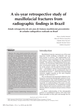
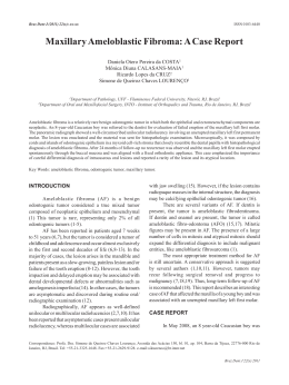
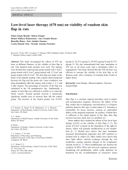
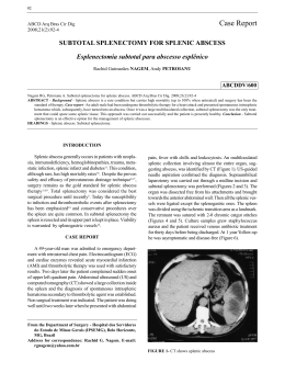
Med Oral Patol Oral Cir Bucal 2007;12:E56-9.
�
Oral Leiomiosarcoma
Primary leiomyosarcoma of the mandible. A case report
João de Jesus Viana Pinheiro 1, Sérgio de Melo Alves 2, Edson Okuda 3, Waldir Antonio Jorge 4, Ruy Gastaldoni Jaeger 5,
Ney Soares de Araújo 6
(1) DDS, PhD, Universitary Hospital João de Barros Barreto, Department of Oral Pathology, School of Dentistry, Federal University
of Pará
(2) DDS., MS. Department of Estomatology, School of Dentistry, University Center of Pará
(3) DDS, Oral and Maxillofacial Surgery Clinic, São Paulo Tatuapé Hospital
(4) DDS, PhD, Department of Stomatology, School of Dentistry, São Paulo University
(5) DDS, PhD, Department of Histology and Embryology, Institute of Biomedical Sciences, São Paulo University
(6) DDS, PhDe Department of Oral Pathology, School of Dentistry, São Paulo University
Correspondence:
Dr. João de Jesus Viana Pinheiro
Av. Almirante Wandenkolk, 1243, 1006
Umarizal. Belém-Pará-Brazil – ZIP Code: 66050-030
E-mail: [email protected]
Pinheiro JJV, Alves SM, Okuda E, Jorge WA, Jaeger RG, Araújo NS.
Primary leiomyosarcoma of the mandible. A case report. Med Oral
Patol Oral Cir Bucal 2007;12:E56-9.
Received: 1-10-2005
Accepted: 20-05-2006
© Medicina Oral S. L. C.I.F. B 96689336 - ISSN 1698-6946
Indexed in:
-Index Medicus / MEDLINE / PubMed
-EMBASE, Excerpta Medica
-SCOPUS
-Indice Médico Español
-IBECS
ABSTRACT
The aim of this article is to describe a case of leiomyosarcoma of the mandible with immunohistochemical analysis that
was useful in making the final diagnosis.
A 40-year-old woman was referred to the Stomatology Clinic of São Paulo Tatuapé Hospital, , for evaluation of a lesion
on the left side of the mandible. This lesion presented a fast growth in the last 6 months. Intraoral examination showed
a firm, fixed, red colored mass measuring, approximately 60-mm in diameter. No lymph nodes involvement was found.
The radiographic examination showed a lytic lesion showed ill-demarcated radiolucent with facial and lingual cortical
bone destruction. Microscopic examination of the mandibular lesion showed a neoplasm composed by interlacing
fascicles of spindle-shaped cells. Most of the cells presented a blut-ended elongated shape. A marked cellular pleomorphism was observed, represented by cells with irregular shape and abundant eosinophilic cytoplasm. Nuclei were large,
hyperchromatic, either vacuoled or cigar-shape. The cytoplasm of the cells stained red with Masson’s trichrome stain.
Neoplastic cells expressed vimentin, smooth-muscle actin, HHF-35 and desmin. These findings were consistent with
the diagnosis of leiomyosarcoma.
Key words: Imunohistochemical, leiomyiosarcoma, mandible.
INTRODUCTION
Leiomyosarcoma makes up 6 to 7% of all soft tissue
sarcomas. This neoplasm of smooth muscle is more common
in older adult population and most often arises in the uterus,
gastrointestinal tract, and the retroperitonial region(1).
Women are more affected than men because of the uterine
association. The prognosis is poor, with a hight percentage
of recurrence or metastasis. The most common sites of metastasis include the lungs, bone, brain and the lymph node.
This neoplasm, is a extremely rare mesenchymal lesion in the
oral cavity, with unusual bone location because of paucity
of smooth muscle in that site(2). However, the definitive
histologic diagnosis of leiomyosarcoma is difficult because
of the similarity with other spindle-cell sarcomas(1). The
immunohistochemical analysis for cytoesqueletal proteins
such as actin, vimentin, desmin, HHF-35 and ultrastructural examination has been widely used for the diagnosis of
leiomyosarcoma(3).
The aim of this article is to describe a case of leiomyosarcoma of the mandible with immunohistochemical analysis
that was useful in making the final diagnosis.
CASE REPORT
A 40-year-old woman was first seen with a lesion on the
left mandible. This lesion had demonstrated rapid growth
during the last 6 months. Intra oral examination showed a
E56
Med Oral Patol Oral Cir Bucal 2007;12:E56-9.
�
firm, fixed, red colored mass measuring 60-mm in diameter.
No lymph nodes involvement was found.
A biopsy was performed under local anesthesia. The specimen was fixed in formalin, parafin embedded and stained
by hematoxylin-eosin. A diagnosis of an inflammatory
chronical process with desmoplasia of the connective tissue
was made. Due to the clinical appearance of the lesion, it
was suggested that the biopsy should be repeated, in order
to obtain a more representative sample. Unfortunately,
the patient returned one month later, with a pronounced
enlargement of the left side of the mandible with depressed
area, (Fig. 1A). No fistula or discharge were observed and
all the teeth in the region were vital. At this point the patient
reported pain. In addition, the computed tomographic imaging showed a lytic, ill-demarcated radiolucency lesion with
facial and lingual cortical bone perfuration. The medular
bone was destroyed, there was calcification into tumor masss
and expansion of soft tissue. The Poorly defined margins
suggest a malignant tumor. (Fig. 1 B).
The rapid onset of a painful left mandibular swelling, lacking
signs of infection or inflamation, along with radiographic
features of facial and lingual bone destruction suggested
malignancy. For a patient at this age and sex, primary and
metastatic malignancies in mandibular bone should be considered. The differential diagnosis could include soft tissue
sarcomas, osteosarcoma and condrosarcoma.
The patient was admitted at São Paulo Tatuapé Hospital
for an emergency surgery. Another incisional biopsy was
performed.
Oral Leiomiosarcoma
Microscopic examination of the mandibular lesion showed a
neoplasm composed of interlacing fascicles of spindle-shaped cells . Most of the cells were blunt-ended and elongated
shaped. A marked cellular pleomorphism was observed,
represented by cells with irregular shape and abundant
eosinophilic cytoplasm. Nuclei were large, hyperchromatic,
either vacuolated or cigar-shaped (Fig. 1C). Mitotic figures
could occasionally be identified (less than of 2 per 10 high
power fields). The tumor exhibited hemangioperycitomalike areas. Few giant cells were present. Necrotic areas were
noted. Immunohistochemistry was carried out through the
streptoavidin biotin method (LSAB, Dako Corporation Carpinteria CA USA ). The antibodies used were anti-vimentin
(1:200 Tris-HCl), anti-smooth-muscle actin (1:50 Tris-HCl),
anti-HHF35 (1:50 Tris-HCl), anti-desmin (1:100 Tris-HCl),
anti-S100 protein (1:400 Tris-HCl), anti-CD-34 (1:50 TrisHCl), h-caldesmon (1:100 Tris-HCl). All antibodies were from
Dako (Dako NS, Glostrup, Denmark), except the anti-CD34,
purchased from Novocastra (Novocastra, Newcastle, UK).
Neoplastic cells expressed vimentin, HHF-35, desmin, h-caldesmon and smooth-muscle actin (Fig. 1D). These findings
were consistent with the diagnosis of leiomyosarcoma.
The hemimandibullectomy was the chosen treatment for the
present case. The patient has been followed for 11 months
and no local recurrence has been observed. The cervical
and submandibular regions remain free of palpable lymph
nodes. Systemic examinations revealed no other site affected
by this malignant neoplasm. The neoplasm in question was
a primary leyomiosarcoma of the mandible.
Fig. 1. (A) Facial view of patient shows depressed area visible below the left commissure and
swelling on the left side of mandible.
(B) Coronal CT image demonstrates an ill-demarcated radiolucency with facial, lingual cortical
bone destruction. Poorly defined margins suggest a malignant tumor.
(C) Most cells presented as blunt-ended, elongated shaped, with a marked pleomorphism,
represented by cells with irregular shaped and abundant eosinophilic cytoplasm. Nuclei were
large hyperchromatic, either vacuolated or cigar-shaped. (hematoxylin-eosin stain, original
magnification x 400).
(D) Photomicrograph showing positive immunoreaction for smooth-muscle actin in the
tumor cells (streptoavidin-biotin stain, original magnification x 630).
E57
© Medicina Oral S.L. Email: [email protected]
Med Oral Patol Oral Cir Bucal 2007;12:E56-9.
�
DISCUSSION
The leiomyosarcoma has been divided into three main
groups based on geographic occurrence: 1) retroperitoneal
and intra-abdominal; 2) cutaneous and subcutaneous; and
3) vascular. Each group has certain clinical and biological
differences, althought, they are histologically similar. The
site of the leiomyosarcoma is more important for prognosis
than its histological features. Those arising in a superficial
cutaneous localization have the best prognosis (1).
Intraoral leiomyosarcoma occurs predominantly in males
with a ratio of 2:1 and may present at any age, (2) ranging
from 1 to 88 years (3-5). The mean age of the 10 previously
described patients with leiomyosarcoma arising from jaw
bone was 43.8 years (6). The patient of the present case is a
woman of 40 years with a primary lesion at the mandible.
Farman (7) found only 5 oral cases among 7,748 benign
smooth-muscle tumors of the whole body. In a review of
the literature, in 1977, by Farman and Key (8), 11 cases of
primary leiomyosacomas in the jaws or in the oral cavity
were located. A total of 30 cases of primary leiomyosarcoma in the oral cavity have been described during the last
75 years (6, 9-13). The intraoral locations of these tumors
were as follows: cheek (5), mandible (8), gingiva (4), maxilla
(4), floor of the mouth (3), tongue (3), soft palate (2), hard
palate mucosa (1) (7, 13).
In the oral cavity, the jaw bones have been the most prevalent
location for this tumor, accounting for 45% of all reported
cases. Including the current case, only 9 cases have been
reported in the mandible (7, 13). There is no distinct clinical
feature that may suggest the presence of oral leiomyosarcoma. These tumors can appear as a painless, well-circunscribed mass, firmly adherent to the surrounding tissues (3).
Histopathologically, this neoplasm is characterized by sheets
of sweeping, alternating bundles and fascicles of densely
packed spindle cells with abundant fibrillar eosinophilic
cytoplasm and indisctinct cytoplasmic borders. The nucleus
is usualy centrally located and blunt-ended, squared-off or
cigar-shaped. Occasional cells have perinuclear vacuoles
(1,9,14,15). In some cases, nuclear hyperchromasia, and
pleomorphism are pronounced. Both typical and atypical
mitosis, as few as one or as many as 20 per high-power
field populate the tumor (9,11). The diagnosis of soft
tissue tumors of mesenchymal origin can be very difficult
and necessitates differentiation from a number of other
tumors (16,17). Masson’s trichrome staining and immunohistochemial evaluation for muscle antigens are helpful in
diagnosis separating leiomyosarcoma from other sarcomas
(18). Positive reactions for desmin, vimentin, smooth-muscle actin and h-caldesmon have been demonstrated in this
neoplasm (3, 19-21).
The case presented fulfilled these criteria and confirmed the
diagnosis of leiomyosarcoma.
Surgical resection, hemimandibullectomy or hemimaxillectomy, partial maxillectomy, are the prefered initial treatments for cases of leiomyosarcoma primary in the jawbones
(15, 13). Regional lymph node investigation is recommended
if there is clinical evidence of lymphadenopathy (21).
Oral Leiomiosarcoma
A hemimandibullectomy was the treatment of the choice
in the present case. The patient has been following by 11
months and no local recurrence has been observed. The
cervical and sumandibular regions remain free of palpable
lymph nodes. Systemic examinations revealed no other site
affected by this malignant neoplasm.
To summarize our report, we should emphasize the importance of an early diagnosis on the survival of the patient.
This diagnosis can only be achieved if a representative
fragment of the lesion is obtained. In addition, immunohistochemistry was very important to establish a final diagnosis
of leiomyosarcoma.
REFERENCES
1. Enzinger FM, Weis SW, eds. Soft Tissue Tumors: St. Louis, MO, Mosby
Year Book Inc; 1995. p. 491-510.
2. Schenberg ME, Slootweg PJ, Koole R. Leiomyosarcoma of the oral
cavity. Report of four cases and review of the literature. J Craniomaxillofac
Surg 1993;21:342-7.
3. Izumi K, Maeda T, Cheng J, Saku T. Primary leiomyosarcoma of the
maxila with regional lymph node metastasis: Report of a case and review
of the literature. Oral Surg Oral Med Oral Pathol Oral Radiol Endod
1995;80:310-9.
4. Freedman PD, Jones AC, Kerpel SM. Epithelioid leiomyosarcoma of
the oral cavity: Report of two cases and review of the literature. J Oral
Maxillofac Surg 1993;51:928-32.
5. Haedicke G, Kaban LB. Smooth-muscle tumors of the oral cavity. Plast
Reconstr Surg 1988;81:264-9.
6. Carter LC, Aguirre A, Boyd B, DeLacure MD. Primary leiomyosarcoma
of the mandible: Report of a case and review of the literature. Oral Surg
Oral Med Oral Pathol Oral Radiol Endod 1999;87:477-84.
7. Farman AG. Benign smooth-muscle tumours. S Afr Med J 1975;
49:1333-408.
8. Farman AG, Key S. Oral leiomyosarcoma: Report of a case and review
of the pertainnig to smooth-muscle tumors of the oral cavity. Oral Surg
Oral Med Oral Pathol 1977;43:402-9.
9. Bass B, Archard H, Sussman R, Stern M. Saunders V. Clinicopathologic
conferences, case 62: expansile radiolucent lesion of the mandible. J Oral
Maxillofac Surg 1986;44:799-803.
10. Nishi M, Mimura T, Senba I. Leiomiosarcoma of the maxila. J Oral
Maxillofac Surg 1987;45:64-8.
11. Poon CK, Kwan P-C, Yin N-T. Leiomyosarcoma of gingiva: Report of a
case and review of the lierature. J Oral Maxillofac Surg 1987;45:888-92.
12. Mindel RS, Calcaterra TC, Ward PH. Leiomyosarcoma of the head
and neck: A review of the literature and report of two cases. Laryngoscope
1975;85:902-10.
13. Karlis V, Zaslow M, Minkowitz G, Glickman RS. Leiomyosarcoma
of the mandible: Report of a case and literature review. J Oral Maxillofac
Surg 1996;54:1127-30.
14. Allen CM, Neville B, Damm DD, Marsh W. Leiomyosarcoma metastatic to oral region: Report of three cases. Oral Surg Oral Med Oral
Pathol 1993;76:752-6.
15. Kratochvil FJ, MacGregor SD, Budnick SD, Hewan-Lowe K, Allsup
H. Leiomyosarcoma of the maxila: Report of a case and review of the
literature. Oral Surg Oral Med Oral Pathol 1982;54:647-55.
16. Lloria-Benet M, Bagan JV, Lloria de Miguel E, Borja-Morant A,
Alonso S. Oral leiomyoma: a case report. Med Oral 2003;8:215-9.
17. Sanchez Jimenez J, Dean Ferrer A, Alamillos Granados F, Ruiz Masera JJ, Villar Pastor C, Garcia Lopez A, Penalba Manegold M. Adult
rhabdomyoma in the masticatory area. New case presentation and review
of the literature. Med Oral 2001;6:64-8.
18. Lack EE. Primary leiomyosarcoma of adrenal gland. Case report
with immunohistochemical and ultrastructural study. Am J Surg Pathol
1991;15:899-905.
19. Lo Muzio L, Favia G, Mignogna MD, Piattelli A, Maiorano E. Primary
intraoral leiomyosarcoma of the tongue: An immunohistochemical study
and review of the literature. Oral Oncol 2000;36:519-24.
E58
Med Oral Patol Oral Cir Bucal 2007;12:E56-9.
�
Oral Leiomiosarcoma
20. Mesquita RA, Migliari DA, De Souza SOM, Alves MR. Leiomyosarcoma of the buccal mucosa: A case report. J Oral Maxillofac Surg
1998;56:504-7.
21. Watanabe K, Kusakabe T, Hoshi N, Saito A, Suzuki T. h-Caldesmon
in leiomyosarcoma and tumours with smooth muscle cell-like differentiation: its specific expression in the smooth muscle cell tumor. Hum Pathol
1999;30:392–6.
E59
© Medicina Oral S.L. Email: [email protected]
Download