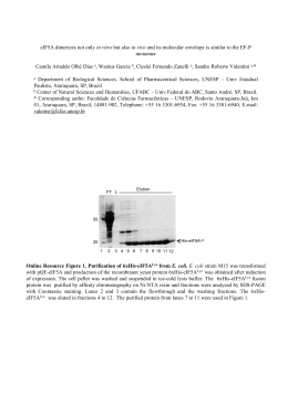
Molecular & Biochemical Parasitology 125 (2002) 103 /112
www.parasitology-online.com
Characterization and comparative functional analysis in yeast of a
Schistosoma mansoni Rho1 GTPase gene
Túlio Marcos Santos, Carlos Renato Machado, Glória Regina Franco, Sérgio Danilo
Junho Pena *
Laboratório de Genética Bioquı́mica, Departamento de Bioquı́mica e Imunologia, Instituto de Ciências Biológicas, Universidade Federal de Minas
Gerais, Caixa Postal 486, Belo Horizonte CEP 30161-970, MG, Brazil
Received 7 May 2002; received in revised form 3 September 2002; accepted 3 September 2002
Abstract
Low-molecular weight GTP-binding proteins (LMWGPs) of the Ras superfamily are believed to play a role in Schistosoma
mansoni female development and egg production. Here we describe the characterization of a novel S. mansoni gene (SMRHO1 ),
highly homologous to Rho-type LMWGPs from several other organisms and encoding a polypeptide with 193 amino acids and an
estimated molecular mass of 21.8 kDa. SMRHO1 complemented a Saccharomyces cerevisiae rho1 null mutant strain even in
restrictive temperature and calcium concentration, in contrast with the human RHOA GTPase that was not able to provide
complementation in such conditions. Comparison of the amino acid sequence of the a3-helix loop7 regions of the two proteins
allowed the identification of the proline 96 and threonine 100 amino acid residues of human RHOA as the most probable
determinants of the complementation differences. We generated SMRHO1 mutants (smrho1E97P , smrho1L101T and smrho1E97P,
L101T
) by site directed mutagenesis and reproduced the conditional lethality phenotype at high temperature, providing strong
evidence that the related amino acid positions (Gln101 and Ile105) in the Rho1 GTPase are indeed important for regulation of the cell
wall synthesis performed by this protein in yeast. The observation that specific amino acid positions seem to be important for the
different functions performed by the Rho GTPases leads to the idea that SMRHO1 might be a useful target in the development of
new anti-schistosomiasis drugs, although it does share high sequence homology with the human RhoA GTPase.
# 2002 Elsevier Science B.V. All rights reserved.
Keywords: Schistosoma mansoni ; Complementation; Yeast; Rho GTPase; Site directed mutagenesis
1. Introduction
Schistosoma mansoni is a digenetic trematode that
causes schistosomiasis, a disease that afflicts more than
200 million people worldwide [1]. Since 1992, the S.
Abbreviations: GDP, guanine diphosphate; GTP, guanine
triphosphate; LMWGPs, low-molecular weight GTP-binding
proteins; SMRHO1 , Schistosoma mansoni Rho1 GTPase gene;
SMRHO1, S. mansoni Rho1 GTPase protein; smrho1 , S. mansoni
Rho1 GTPase mutant gene; UPGMA, unweighted pair group with
arithmetic mean.
Note: The nucleotide sequence reported in this paper has been
submitted to the GenBankTM/EMBL Data Bank with the accession
number AF140785.
* Corresponding author. Tel.: /55-31-3284-8000; fax: /55-313227-3792
E-mail address: [email protected] (S.D.J. Pena).
mansoni Genome Project has contributed to the discovery of close to 10 000 new genes of this parasite [2].
The study of important genes for S. mansoni survival
and pathogenicity is the first step for the development of
new drugs and vaccines for schistosomiasis control.
Following penetration into the human host skin as
cercariae, the parasites mature into adult male and
female forms that live in constant copulation in the veins
of the portal system and ovulate for the duration of their
lives. There is evidence of involvement of low-molecular
weight GTP-binding proteins (LMWGPs) of the Ras
superfamily in the S. mansoni female maturation process
and egg production [3,4]. This is of great interest for
schistosomiasis control because inflammatory reactions
triggered by the eggs, and not by the worms themselves,
are the cause of liver fibrosis, the most serious pathological lesion of the disease.
0166-6851/02/$ - see front matter # 2002 Elsevier Science B.V. All rights reserved.
PII: S 0 1 6 6 - 6 8 5 1 ( 0 2 ) 0 0 2 1 8 - 9
104
T.M. Santos et al. / Molecular & Biochemical Parasitology 125 (2002) 103 /112
The Ras superfamily members (Ras, Rho, Rab, Sar1/
Arf, and Ran) are proteins capable, through the binding
and hydrolysis of GTP, to create a switch between an
active GTP-bound conformation and an inactive GDPbound conformation [5,6]. The ‘on/off’ activity of the
LMWGPs is controlled by several regulatory proteins:
the guanine nucleotide exchange factors (GEFs) promote the exchange of the GDP for GTP; the GTPaseactivating proteins (GAPs) accelerate the hydrolysis of
the GTP to GDP; and the GDP dissociation inhibitors
(GDIs) inhibit the dissociation of the GDP from the
GTPase [7]. LMWGPs receive upstream signals through
their regulators and transduce signals to a multitude of
effector molecules, while remaining in the GTP-bound
form [8 /10]. In this fashion, the members of the Ras
superfamily perform important regulatory roles, controlling a variety of cellular activities, such as cell cycle,
gene expression, cytoskeletal reorganization, vesicle
trafficking, nucleocytoplasmic transport and microtubule organization [6,11/14].
It has been shown that a reduction of isoprenoid endproducts synthesis by mevinolin, an inhibitor of hydroxymethylglutaryl-CoA reductase, was capable of decreasing
egg
production
and
blocking
the
schistosomiasis pathology in infected mice [4] as well
as causing loss of parasite viability at higher drug
concentrations [15]. Similar effects were observed in
schistosome females as the result of prenyltransferase
activity inhibition by BZA-5B [16]. This is important
because the blocking of prenylation of a 25 kDa group
of GTP-binding proteins from schistosomes using BZA5B led to the speculation that LMWGPs could be
playing a role in the egg laying mechanism [17]. These
observations stimulated the search for LMWGPs that
might be associated with the regulation of signal
transduction pathways in S. mansoni. A gene encoding
a RAB-related GTP-binding protein was cloned from S.
mansoni genomic DNA using degenerate oligonucleotide primers in an attempt to identify LMWGPs possibly
involved in female egg production [16]. The in vitro
translation product of this SMRAB gene is geranylgeranylated and the recombinant protein binds GTP. Using
an immunochemical approach, Schussler et al. (1997)
showed that the Ras, GAP and MAP kinases displayed
sexual and developmental regulation in schistosome
extracts [3]. The involvement of these molecules in the
female maturation process was not unexpected, since the
Ras, GAP and MAP kinases participate in the same
signal transduction pathways known to regulate cell
proliferation events. More recently, a schistosome
cDNA encoding a gene highly similar to K-Ras from
various organisms was isolated and characterized [18].
This S. mansoni K-Ras homologue gene is overexpressed in females in comparison with male worms and
it has been detected in all developmental stages of the
parasite. The recombinant SMRAS protein is farnesy-
lated in vitro and the native protein was identified by
immunocytolocalization associated with the worm subtegument.
As part of the S. mansoni Genome Project, we
isolated from a cercariae library a cDNA clone highly
homologous to the Rho-type LMWGP genes from
several organisms [19]. We report here the complete
cDNA and genomic sequences of this gene. Sequence
alignments, molecular phylogenetic analysis, and complementation of a Saccharomyces cerevisiae rho1 null
mutant strain allowed us to identify it as a Rho-type
LMWGP (SMRHO1 ), the first gene of this class to be
identified in S. mansoni .
The complementation of yeast containing a rho1
deletion with SMRHO1 occurred even in restrictive
conditions of temperature and calcium concentration
and was much more complete than the only conditional
complementation previously observed, with the human
RHOA GTPase [20]. Comparison of the a3-helix loop7
amino acid sequences of the S. cerevisiae RHO1, human
RHOA and SMRHO1 GTPases, allowed identification
of Pro96 and Thr100 amino acid residues in human
RHOA as the most probable determinants of the
difference in the complementation ability of SMRHO1
and RHOA GTPases. Indeed, when we used site
directed mutagenesis to replace Glu97 and Leu101
residues of SMRHO1 by Pro and Thr, respectively,
SMRHO1 became only capable of conditional complementation of the S. cerevisiae rho1 null mutant strain.
Consequently, although highly homologous to human
RHOA, the S. mansoni Rho1 GTPase may prove to be
an interesting target for the development of new antischistosomiasis drugs.
2. Materials and methods
2.1. Cloning procedures and DNA sequencing
The proofreading Pfu DNA polymerase (Stratagene)
was used in PCR amplifications for subcloning. Amplification products used in subcloning or transformation
procedures were previously purified using the Wizard
PCR-Prep kit (Promega). Cloning into pUC18 vector
was done using the SureClone Ligation Kit (Amersham
Biosciences). Plasmid purifications were done by alkaline lysis using the Wizard Plus SV Miniprep kit
(Promega). DNA sequencing reactions were prepared
using Thermo Sequenase Sequencing Kit (Amersham
Biosciences) with fluorescent M13 primers and carried
out in an A.L.F. DNA sequencer (Amersham Biosciences).
T.M. Santos et al. / Molecular & Biochemical Parasitology 125 (2002) 103 /112
2.2. cDNA sequencing
The SMC0220R cDNA clone containing the S.
mansoni Rho GTPase gene was obtained by random
selection from a S. mansoni cercariae library as described previously [19]. Initially the 5? and 3? ends of the
SMC0220R cDNA clone was sequenced using M13
primers. Subsequently, the PTV3 (5?-TGA TGT TCC
CAT TGT CTT AG-3?) and PTV4 (5?-AAA TCA AAT
CTC AGA CAA TT-3?) primers were designed to
amplify the internal region of the insert. The 291 bp
internal fragment amplified was subcloned into pUC18
vector and sequenced on both strands. In order to
obtain a reliable full-length sequence, the ends of the
insert were amplified using the M13 reverse/PTV5 (5?CTA AGA CAA TGG GAA CAT CA-3?) and PTV6
(5?-TGT CTA GAT TTG ATT TTC CG-3?)/M13
universal primer pairs. The generated fragments of 527
and 234 bp, respectively, were subcloned into pUC18
and sequenced on both strands.
2.3. Genomic sequencing
We used the SMRHOF (5?-GGC AAC AAT GGC
GAG TG-3?) and PTV4 primer pair to amplify the
genomic sequence of S. mansoni Rho GTPase gene. The
PCR amplification generated a fragment of about 700
bp. This fragment was excised from the agarose gel,
purified using Gene Clean kit (Bio 101), cloned into
pUC18 vector, and sequenced on both strands. In order
to obtain a reliable sequence, the genomic clone was
PCR amplified into two distinct fragments using the
SMRHOF/PTV5 and PTV3/PTV4 internal primer pairs.
The generated fragments of 417 and 324 bp were
subcloned into pUC18 and sequenced on both strands.
2.4. Sequence analyses
Sequence alignments and open reading frame (ORF)
search were done using the DNASIS program (Hitachi).
Homology searches were performed using BLAST programs [21] for sequence similarity search at NCBI.
Alignments of amino acid sequences and UPGMA
analysis were performed with the software DIALIGN
2.1 [22].
2.5. Yeast strains and media
All yeast strains employed are listed in Table 1. The
diploid strain BY4727/36 was obtained crossing by
replica platting BY4727 with BY4736 strain [23], both
of them obtained from ATCC. YPD rich medium was as
described [24]. YNB minimal medium was purchased
from Difco and 2% glucose or 2% galactose was
included as carbon source when required.
105
Table 1
Yeast strains used in this study
Strain
Genotype
BY4727
MATa his3D200 leu2D0 lys2D0 met15D0
trp1D63 ura3D0
BY4736 MATa ade2D::hisG his3D200 met15D0 trp1D63
ura3D0
BY4727/ MATa/MATa his3D200 met15D0 trp1D63
36
ura3D0
SPYD
MATa/MATa his3D200 met15D0 trp1D63
ura3D0 Drho1::HIS3
CMY
MAT? ade2D::hisG? his3D200 met15D0 trp1D63
ura3D0 leu2D0 lys2D0 Drho1::HIS3
pYEDP (SMRHO1 )
Reference
[23]
[23]
This
study
This
study
This
study
2.6. Cloning on yeast galactose-inducible overexpression
vector
The S. mansoni Rho GTPase gene was amplified from
the SMC0220R cDNA clone using SMRHOF and
SMRHOR (5?-TTA GAG AAC AGT GGA GAT
CA-3?) ORF flanking primers and cloned into pUC18.
The S. cerevisiae RHO1 gene, containing its promoter
(0.2 kb) and terminator (0.4 kb) regions, was obtained
by direct amplification from yeast colony using primers
RHOSACF (5?-ACA TGA GAT CTA CGC AGA
TCA-3?) and RHOSACR (5?-CAT CGA TCA TTC
CTC TGA GTA-3?). The amplified fragment (1226 bp)
was previously digested with Dra I in order to remove
the promoter region, cloned into pUC18 and sequenced
on both ends. Thereafter, the fragments containing
SMRHO1 and RHO1 were recovered from pUC18 by
digestion with Bam HI/Kpn I and cloned into the same
restriction sites of the pYEDP60-2-ADE2-URA3GAL10-CYC1 plasmid (a kind gift of Dr Francisco
Nóbrega, UNIVAP, São Paulo) for overexpression in
yeast under control of GAL10-CYC1 promoter induced
by galactose [25].
2.7. Yeast genetic manipulation methods
Transformations were done with 1 mg of purified
linear DNA or 0.5 mg of plasmid DNA by LiAc/SSDNA/PEG procedure [26]. The diploid S. cerevisiae
strain SPYD (Table 1) containing one of the RHO1
alleles disrupted was obtained by the PCR-mediated
gene disruption method [23] using the diploid strain
BY4727/36. The 60 mer primers RHO1HIS3F (5?-ATG
TCA CAA CAA GTT GGT AAC AGT ATC AGA
AGA AAG CTG GCT TCA TTC AAC GTT TCC
CAT-3?) and RHO1HIS3R (5?-GCT AGA AAT ATG
AAC CTT CCA ACA AAA CTG AGG TTG GAG
GAG TAT CAT ACT GTT CGT ATA-3?) were used to
amplify the HIS3 auxotrophic marker gene cloned into
pDIS plasmid (a kind gift of Dr Francisco Nóbrega,
106
T.M. Santos et al. / Molecular & Biochemical Parasitology 125 (2002) 103 /112
UNIVAP, São Paulo). The 1243 bp amplified product
contained at both ends 40 bp of sequence of the S.
cerevisiae RHO1 was purified and transformed into
BY4727/36 diploid strain for disruption of one of the
RHO1 gene copies through a homologous recombination event. Disrupted mutants were selected on plates of
YNB-glucose containing the required nutrients except
histidine. PCR amplification of a 788 bp hybrid fragment containing part of RHO1 and HIS3 genes using
RHOSACF and HIS311 (5?-AAC CCT ATA CCT
GTG TGG A-3?) primers, confirmed the insertion of
the auxotrophic marker in place of one of the RHO1
genomic copies in the yeast clones (data not shown). The
SPYD strain was transformed with the S. mansoni Rho
and yeast Rho1 genes cloned into pYEDP galactoseinducible plasmid and the clones were selected on YNBglucose containing the required nutrients. The cells
transformed with pYEDP (SMRHO1) were induced to
sporulate in order to obtain rho1 null mutant cells
complemented with the S. mansoni Rho GTPase gene.
Sporulation of the diploid cells in liquid medium was
induced as described [23]. The sporulated cultures were
treated with NovoZym (Sigma) to digest the ascus wall,
vortexed vigorously for 2 min to release spores from the
asci and subsequently heated at 58 8C for 15 min to kill
any remaining vegetative cells [24]. Spores dilutions were
spread on YNB-galactose plates supplemented with the
required nutrients. The rho1 null mutant strain CMY
(Table 1), complemented by SMRHO1 gene galactoseinducible overexpression, was confirmed by negative
PCR amplification of RHO1 gene using RHOSACF/
RHOSACR primers, amplification of SMRHO1 gene
using SMRHOF/SMRHOR primers, and amplification
of a 788 bp hybrid fragment, containing part of HIS3
and RHO1 genes, using RHOSACF/HIS311 primers
(data not shown). The PCR products were also
sequenced.
2.8. Site directed mutagenesis of SMRHO1
Megaprimer PCR method [27] was used to generate
the smrho1E97P , smrho1L101T single mutants and the
smrho1E97P, L101T double mutant of SMRHO1 gene.
SMRHOE97P (5?-AGT TTT GCA AAC ATT CCA
GA-3?), SMRHOL101T (5?-GG ACA CCA GAA ATC
CGT C-3?) and SMRHO97P101T (5?-T CCA GAA
AAA TGG ACA CCA GA-3?) primers, containing the
mutations E97P, L101T, and E97P-L101T (highlighted
in bold), respectively, were used in a first PCR round
with SMRHOR primer to produce the megaprimers.
SMRHOF primer and the purified megaprimers were
used to generate SMRHO1 mutant copies in a second
PCR round. SMRHO1 mutant copies were cloned into
pUC18 and sequenced on both strands to confirm the
presence of the mutations. The 0.2 kb RHO1 promoter
region was PCR amplified from yeast colony using
PROMORHOF (5?-CTG AAG CTT ACC AGC AGG
AAT TCG-3?) and PROMORHOR (5?-CAG
CTG CAG CTT TCT AGT ATA ATT-3?) primers
containing Hind III and Pst I sites (showed underlined),
respectively. The pYTS plasmid was constructed introducing the amplified RHO1 promoter region into
Hind III/Pst I gap of the pYEplac112 plasmid [28]
(a kind gift by Dr José Miguel Ortega, UFMG, Belo
Horizonte). Subsequently, pYTS1, pYTS2, pYTS21,
pYTS22, and pYTS23 plasmids were constructed introducing RHO1 , SMRHO1, smrho1E97P , smrho1L101T
and smrho1E97P, L101T , respectively, into Bam HI/Kpn I
gap, positioned immediately downstream of the PstI site
of pYTS. These constructions and pYTS were transformed into CMY strain and the transforming clones
were selected at 23 8C in YNB-glucose plates containing only leucine, lysine, and methionine. In these
conditions, the galactose-inducible overexpression of
SMRHO1 is inhibited and the yeast only express the
wild type and mutant genes cloned into the pYTS
plasmids under control of the RHO1 promoter. The
presence of wild type and mutant gene copies in the
different mutant clones was confirmed by sequencing of
the corresponding PCR products.
3. Results
3.1. cDNA sequence analyses
The partial sequences obtained after sequencing the
subclones were aligned and produced a 1016 bp
consensus that represents the full-length SMC0220R
cDNA clone. We observed, however, that the insert was
a chimera of two cDNA molecules joined by the
adapters used in the cercarial library construction. The
actual cDNA molecule encoding the S. mansoni Rho
GTPase homologous gene represented 841 bp of this
sequence (GenBank accession number AF140785). The
most significant ORF found was 579-nt long and
encoded a putative polypeptide of 193 amino acids
with an estimated molecular mass of 21.8 kDa. The
cDNA also contained a long 3?-untranslated portion,
which is a known feature of S. mansoni transcripts. We
named this gene ‘SMRHO1 ’ for S. mansoni homologue
of the Rho1 and RhoA-type LMWGPs. In Fig. 1A, we
show the alignment of the amino acid sequence of
SMRHO1 with the Rho GTPases from 11 other species.
A noteworthy feature was the insertion of a serine
residue very close to the N-terminal end, which has not
been observed in any of the other Rho GTPase
homologs examined (Fig. 1A). Another interesting
element was the presence of the polar amino acid
aspartate (position 191) substituting for the first (generally aliphatic) amino acid of the C-terminal prenylation motif CAAX (Fig. 1A). Apart of these two
T.M. Santos et al. / Molecular & Biochemical Parasitology 125 (2002) 103 /112
107
Fig. 1. (A) Amino acid sequence alignment of S. mansoni (Sm ) Rho1 GTPase (SMRHO1) with the Rho GTPases from 11 other species. Identical
amino acids among the sequences are indicated by asterisks and (o) mark amino acid differences observed only in SMRHO1. The functional regions
of the GTPase active site are highlighted in light gray. Prenylation site is in black. Switch I and II regions are indicated underlined above the
sequences. %I, identity; %S, similarity; O, amino acid alignment overlap. (B) Phenogram of Sm Rho1 GTPase with the other Rho GTPases. Ap ,
Aplysia californica ; Xl , Xenopus laevis ; Hs , Homo sapiens ; Hp , Hemicentrotus pulcherrimus ; Dm , D. melanogaster ; Mm , Mus musculus ; Gg , Gallus
gallus ; Ce , Caenorhabditis elegans ; Cf , Canis familiaris ; Do , Discopyge ommata ; Sp , S. pombe ; Sc , S. cerevisiae ; Fn , F. neoformans ; Ca , C.
albicans .
peculiarities, the putative amino acid sequence of
SMRHO1 contained all other consensus sequences
conserved among the Rho-type GTPases [5,29,30], and
was highly homologous to all the LMWGPs (Fig. 1A).
A phenogram constructed using the UPGMA algorithm
revealed two major clusters: the first (lower main branch
in Fig. 1B) corresponding to the yeasts (S. cerevisiae,
Schizosaccharomyces pombe , Filobasidiella neoformans
108
T.M. Santos et al. / Molecular & Biochemical Parasitology 125 (2002) 103 /112
Fig. 2. Complementation of a yeast rho1 null mutant strain by galactose-inducible overexpression of SMRHO1 . (A) Growth in agar plate at 30 8C
of the wild type strain BY4736 containing the empty plasmid pYEDP and of the rho1 null mutant strain CMY complemented by galactose-inducible
overexpression of SMRHO1 . The media used were: YNB-galactose, YNB-galactose with 300 mM CaCl2 and YPD rich medium containing only
glucose as carbon source for galactose-inducible overexpression of SMRHO1 inhibition. (B) Growth at 37 8C of the strains BY4736(pYEDP) and
CMY in YNB-galactose and YPD rich medium containing only glucose as carbon source for galactose-inducible overexpression of SMRHO1
inhibition.
and Candida albicans ) and the second (upper main
branch in Fig. 2B) belonging to multicellular organisms.
Within the latter, the Schistosome Rho GTPase constituted as an isolated branch (Fig. 1B).
3.2. Genomic sequence analyses
Partial sequences obtained from the genomic subclones were aligned to produce a 722 bp consensus
genomic sequence that contained an ORF identical to
the SMRHO1 and three small introns (GenBank
accession number AF140785). The first intron has 32
bp and is located between switch I and II domains of the
protein. The second intron, of 33 bp, is in the middle of
the ORF. The last intron is also 33 bp long and is
located in the SAK motif of the protein, which
participates in interactions with the guanine of GTP
molecule in the active site. All introns interrupt the
protein frame. The canonical donor/acceptor splice sites
are conserved in all exon/intron junctions.
3.3. Complementation of a S. cerevisiae rho1 null mutant
strain by SMRHO1 galactose-inducible overexpression
The rho1 null mutation in yeast is lethal. For this
reason, we obtained first a heterozygous diploid strain
with one of the RHO1 alleles deleted (SPYD strain,
Table 1), which was viable at both 30 and 37 8C (data
not shown). To investigate the complementation phenotype of SMRHO1 gene in yeast cells, we transformed
the SPYD strain with a plasmid (pYEDP) containing
the S. mansoni Rho1 GTPase gene under control of a
galactose-inducible promoter GAL10. After sporulation
of the SPYD cells containing the plasmid
pYEDP(SMRHO1 ), we could then successfully select
on plates containing galactose a yeast rho1 null mutant
haploid strain (CMY, Table 1) complemented by overexpression of the SMRHO1 gene. This complementation could be shown to be due to the presence of
SMRHO1 gene, since CMY cells died in YPD rich
medium containing glucose as carbon source, in which
the expression of the S. mansoni GTPase gene in
pYEDP was inhibited (Fig. 2A and B). The SMRHO1
gene was able to complement the yeast rho1 null mutant
strain CMY even under temperature and osmotic stress
conditions such as at 37 8C and in presence of 300 mM
CaCl2 (Fig. 2A and B). The S. mansoni Rho1 gene
overexpression
in
a
yeast
wild-type
strain
{BY4736[pYEPD(SMRHO1 )]} did not produce significant alterations in the growth curve of these cells if
compared with the yeast RHO1 gene overexpression in
the same wild-type strain {BY4736[pYEPD(RHO1 )]}
(Fig. 3). However, the rho1 null mutant strain CMY,
which is complemented by SMRHO1 overexpression,
showed cell enlargement (data not shown) and also a
delay in the growth in the same fashion as wildtype strain overexpressing RHO1 gene {SPYD
[pYEPD(RHO1 )]} (Fig. 3). It occurred probably due
to an arrest in the cell cycle, as previously reported for
RHO1 overexpression in yeast [31]. The wild-type strain
containing only the plasmid {BY4736(pYEPD)} was
used as growth control.
3.4. Comparison among the sequence of the a3-helix
loop7 regions and functional complementation in yeast
performed by SMRHO1 and other Rho GTPases
The a3-helix loop7 had been previously defined as the
region of the human RHOA molecule responsible for its
inability to complement yeast rho1 null mutants at
37 8C or in the presence of 300 mM CaCl2 [20].
Drosophila melanogaster RhoA has its a3-helix loop7
T.M. Santos et al. / Molecular & Biochemical Parasitology 125 (2002) 103 /112
109
Fig. 3. Growth curves of the rho1 null mutant strain CMY complemented by galactose-inducible overexpression of SMRHO1 and of the wild type
strain BY4736, transformed with the plasmids pYEDP, pYEDP(RHO1 ) or pYEDP(SMRHO1 ). The cells were grown at 30 8C in YNB-galactose
liquid medium supplemented with the required amino acids, and counted at the indicated times.
region identical to human RHOA and it also cannot
complement yeast rho1 null mutants at 37 8C or in the
presence of 300 mM CaCl2 [32]. In order to try identify
the exact amino acid residues responsible for the
complementation differences seen between S. mansoni
and human Rho GTPase genes, we aligned the sequence
of S. cerevisiae RHO1 a3-helix loop7 with the corresponding regions of C. albicans RHO1, human RHOA,
and SMRHO1 GTPases (Fig. 4). The C. albicans RHO1
protein was included in the alignment because it fully
complements S. cerevisiae rho1 null mutants under any
culture conditions [33]. The C. albicans sequence
showed very few amino acid alterations in relation to
S. cerevisiae RHO1, none with apparent functional
importance to complementation [33]. However, amino
acid differences of the human RHOA and SMRHO1 a3helix loop7 regions, in comparison with S. cerevisiae
rho1 , were in greater number, determining occasionally
significant hydropathic changes (Fig. 4). The discrepancies of the amino acid constitution of the a3-helix loop7
regions of the Rho GTPases and the ability of each one
to complement S. cerevisiae rho1 null mutants suggest
that proline 96 and threonine 100 at a3-helix would be
the best candidate amino acids responsible for the
conditional complementation by human RHOA.
3.5. The phenotypic effects of the smrho1E97P ,
smrho1L101T , and smrho1E97P, L101T mutants in yeast
To test the importance of the proline 96 and threonine
100 residues for the conditional complementation of
human RHOA , we used site directed mutagenesis to
obtain SMRHO1 containing E97P, L101T, and E97PL101T (the discrepancy in residue numbers is due to the
serine insertion in SMRHO1 N-terminal). The three
mutant SMRHO1 genes, the wild type SMRHO1 and
the yeast RHO1 genes were cloned into the pYTS
plasmid under control of yeast Rho1 promoter. After
transformation of these plasmids into rho1 null mutant
strain CMY, we examined their phenotypes in YNBglucose plates, in which the galactose-inducible
SMRHO1 overexpression was, thus, inhibited. CMY
cells containing only the empty pYTS plasmid were
unable to grow under any of the tested conditions, while
the cells expressing SMRHO1 and RHO1 wild-type
genes under control of the RHO1 promoter showed a
normal growth even at 37 8C, as expected (Fig. 5).
However, the smrho1E97P, L101T double mutant was
unable to grow at 37 8C stress temperature without the
presence of 0.5 M sorbitol as osmotic stabilizer (Figure 6
A and B). The single mutant smrho1E97P grew less well
at 37 8C (Fig. 5A) but grew normally at this temperature in the presence of 0.5 M sorbitol (Fig. 5B). The
other single mutant, smrho1L101T , showed less deleterious effects in growth when compared with wild type
genes (Fig. 5A and B).
4. Discussion
Fig. 4. BLASTP alignment of S. cerevisiae (Sc ) Rho1 a3-helix loop7
with the corresponding regions of C. albicans (Ca ) Rho1, Homo
sapiens (Hs ) RhoA, and S. mansoni (Sm ) Rho1 GTPases. Asterisks
indicate identical amino acids, conservative amino acid changes are in
light gray and non-conservative amino acid changes are in black. %I,
identity; %S, similarity.
We here describe, the characterization of the first Rho
GTPase gene (SMRHO1 ) isolated from S. mansoni ,
encoding a protein that is highly homologous to the
Rho-type LMWGPs of several species (Fig. 1A). The
Rho-type LMWGPs characteristic motifs [5,29,30] are
conserved in SMRHO1, except for the CAAX prenyla-
110
T.M. Santos et al. / Molecular & Biochemical Parasitology 125 (2002) 103 /112
Fig. 5. Conditional complementation of the yeast rho1 null mutant strain CMY by expression of SMRHO1 mutants under control of RHO1
promoter. The CMY strain was transformed with the following plasmids (1) pYTS(vector), (2) pYTS1(RHO1 ), (3) pYTS2(SMRHO1 ), (4)
pYTS21(smrhoE97P ), (5) pYTS22(smrhoL101T ) and (6) pYTS23(smrhoE97P, L101T ). The picture shows the transformant growth after 3 days of
incubation at 30 or 37 8C in (A) YNB plates containing glucose but not galactose, to suppress the SMRHO1 overexpression and (B) YNB-glucose
plates supplemented with 0.5 M sorbitol as osmotic stabilizer.
tion domain that has a polar amino acid substituting the
first aliphatic residue. This amino acid substitution may
lead to the lack of prenylation in this protein. Further
experiments need to be done to verify if there are other
genes of this family coding for a protein with the
conserved prenylation motif CAAX. Another peculiarity found in the SMRHO1 deduced amino acid sequence
was the insertion of a serine residue close to its Nterminal, which has not been observed in any other
Rho-type LMWGP (Fig. 1A). As a consequence of such
sequence particularities, SMRHO1 occupies in an UPGMA phenogram a isolated branch within the cluster of
Rho-type GTPases already identified in other multicellular organisms (Fig. 1B).
Additionally, we demonstrated in this work that the
SMRHO1 homology (62% identity) to S. cerevisiae
RHO1 is sufficient to complement yeast RHO1 GTPase
functions, even when overexpressed in a knock out
strain (Fig. 2A). The complementation was adequate to
permit survival and growth even under stress conditions
such as in the presence of 300 mM CaCl2 or at 37 8C
(Fig. 2). In S. cerevisiae , RHO1 is a regulatory protein
required for cell cycle control [34] and also mediates bud
growth by controlling polarization of the actin cytoskeleton and cell wall maintenance [35 /37]. Consequently,
SMRHO1 can apparently perform all these RHO1
biological functions. However, we observed some phenotypic alterations, such as cell cycle arrest (Fig. 3) and
cell enlargement (data not shown) caused by SMRHO1
overexpression in the rho1 null mutant strain CMY
similarly to those previously reported for yeast RHO1
overexpression [31]. In contrast, the growth was appar-
ently normal when SMRHO1 was overexpressed in the
wild-type strain BY4736 (Fig. 3), although enlarged cells
were present yet (data not shown). Such results suggest a
RHO1 dominant effect over the SMRHO1 gene indicating that the S. manson i GTPase is not able to
perform perfectly all the RHO1 gene functions. This can
be explained by defective molecular interactions of the
SMRHO1 with some of the distinct RHO1 regulatory
and effector proteins, due to differences existent among
the heterologous and native GTPases.
The functional effect caused by amino acid sequence
differences can be addressed by comparing the results of
SMRHO1 and human RHOA complementation studies
in S. cerevisiae rho1 null mutants. In contrast to the
complete complementation provided by SMRHO1 , human RHOA overexpression is lethal and its expression
under control of the yeast RHO1 promoter produces
phenotypes sensitive to high temperatures such as 37 8C
[20]. However, in the presence of 0.5 M sorbitol this
temperature-sensitive phenotype was suppressed, providing the first evidence that one of the functions
performed by the S. cerevisiae RHO1 should be the
maintenance of the yeast cell osmotic integrity [20].
Indeed, RHO1 has been identified as an essential
regulatory subunit of 1,3-b-glucan synthase, the multienzymatic complex that synthesizes 1,3-b-linked glucan,
a major structural component of the yeast cell wall [35 /
37].
Analyses of human RHOA /yeast RHO1 chimeras
demonstrated that the a3-helix loop7 region of the
RHOA molecule was determinant of the conditional
complementation phenotype observed [20]. A simple
T.M. Santos et al. / Molecular & Biochemical Parasitology 125 (2002) 103 /112
comparison of this region in C. albicans [33], human [20]
and schistosome Rho GTPases led us to identify proline
96 and threonine 100 as candidate residues for the
functional difference between human RHOA and
SMRHO1 during heterologous expression in yeast
(Fig. 4). The presence of a proline in position 96 of
the human protein may determine a conformational
alteration in the a3-helix, reducing its interaction
capacity with effector molecules of S. cerevisiae . It is
well-known that proline can disrupt a-helices in watersoluble proteins [38,39] and, thus, influence the native
structural conformation and protein function and interactions. For instance, the mutation of a glutamic acid to
proline disrupts the interaction between cancer-related
papillomavirus E6 protein and E6AP cellular protein,
providing the first evidence that the E6-binding motif of
the E6AP must be helical when bound to E6 [40].
Similarly to proline, threonine is also common in bend
regions of kinked alpha helices [39]. The presence of a
threonine residue in the position 100 of RHOA could,
therefore, produce a conformational difference that
would have functional repercussions.
Our results with the smrho1E97P , smrho1L101T , and
smrho1E97P, L101T mutants confirmed the importance of
proline 96 and threonine 100 for the temperaturesensitive phenotype seen in yeast rho1 null mutant
complemented by the human RHOA GTPase. This
was most evident when we observed the phenotypic
effect of complementation with the smrho1E97P and
smrho1E97P, L101T mutants. The double mutation had a
particularly drastic effect onto the capacity of cell wall
synthesis regulation under high temperature (Fig. 5A),
probably due to an important conformational alteration
induced in the a3-helix. However, the single replacement
of a leucine by a threonine at position 101 did not have
such a severe phenotypic effect at 37 8C (Fig. 5A).
Recently, a set of high temperature-sensitive RHO1
mutants were generated by random PCR and arbitrarily
divided in two groups (rho1A and rho1B) according to
their functional defects [41]. It is interesting to observe
that the smrho1E97P and smrho1L101T mutants produced
in our work are located near to the W104R and E102K
mutations grouped as rho1B. This group is made up
specifically of mutants that showed defects in activation
of the 1,3-b-glucan synthase, the RHO1 effector enzyme
responsible for the yeast cell wall synthesis.
In conclusion, even though the Rho GTPases from
different species are small molecules that share a high
degree of homology, subtle amino acid differences in
certain regions can determine their interaction specificity
with the multiple regulatory and effector proteins of
each organism. Hence, there seems to be a direct
relationship between specific regions of Rho-type
LMWGPs and the different functions performed by
these proteins. This is in good agreement with previous
observations made on S. cerevisiae RHO1 [34,41]. The
111
SMRHO1 gene may, thus, provided an attractive target
for the development of new antischistosomiasis drugs, in
spite of its high sequence identity with human RHOA
gene.
The Schistosome Genome Project has already generated sequences of many genes and requires intensive
efforts on functional genomics [2]. Nevertheless, such
endeavor can be restricted by the technical difficulties of
parasite genetic manipulation. The use of S. cerevisiae
and other model organisms constitutes a powerful tool
for investigation of Schistosome gene function, as
demonstrated in this work.
Acknowledgements
The authors thank Dr Vasco Azevedo for initial
directions on SMRHO1 sequencing, Dr José Miguel
Ortega by the use of his laboratory facilities for yeast
cultivation, Dr Carlos Rosa for helpful insights on yeast
cell biology, and Kátia Barroso for carrying out
automated DNA sequencing. This investigation received
financial support from the following sources: PRPqUFMG, PADCT, CNPq, UNDP/WORLD BANK/
WHO Special Program for Research and Training in
Tropical Diseases (TDR No: 940325 and 940751),
USAID/HOH (No 264.01.01.04).
References
[1] Chitsulo L, Engels D, Montresor A, Savioli L. The global status
of schistosomiasis and its control. Acta Trop 2000;77:41 /51.
[2] Franco GR, Valadão AF, Azevedo V, Rabelo EM. The schistosoma gene discovery program: state of the art. Int J Parasitol
2000;30:453 /63.
[3] Schussler P, Grevelding CG, Kunz W. Identification of Ras, MAP
kinases, and a GAP protein in Schistosoma mansoni by immunoblotting and their putative involvement in male /female interaction. Parasitology 1997;115:629 /34.
[4] VandeWaa EA, Mills G, Chen GZ, Foster LA, Bennett JL.
Physiological role of HMG-CoA reductase in regulating egg
production by Schistosoma mansoni . Am J Physiol 1989;257:618 /
25.
[5] Bourne HR, Sanders DA, McCormick F. The GTPase superfamily: conserved structure and molecular mechanism. Nature
1991;349:117 /27.
[6] Takai Y, Sasaki T, Matozaki T. Small GTP-binding proteins.
Physiol Rev 2001;81:153 /208.
[7] Sasaki T, Takai Y. The Rho small G protein family-Rho GDI
system as a temporal and spatial determinant for cytoskeletal
control. Biochem Biophys Res Commun 1998;245:641 /5.
[8] Machesky LM, Hall A. Rho: a connection between membrane
receptor signaling and the cytoskeleton. Trends Cell Biol
1996;6:304 /10.
[9] Ridley AJ. Signalling by Rho family proteins. Biochem Soc Trans
1997;25:1005 /10.
[10] Aspenstrom P. Effectors for the Rho GTPases. Curr Opin Cell
Biol 1999;11:95 /102.
112
T.M. Santos et al. / Molecular & Biochemical Parasitology 125 (2002) 103 /112
[11] Kaibuchi K, Kuroda S, Amano M. Regulation of the cytoskeleton and cell adhesion by the Rho family GTPases in mammalian
cells. Annu Rev Biochem 1999;68:459 /86.
[12] Hirai A, Nakamura S, Noguchi Y, Yasuda T, Kitagawa M,
Tatsuno I, Oeda T, Tahara K, Terano T, Narumiya S, Kohn LD,
Saito Y. Geranylgeranylated rho small GTPase(s) are essential for
the degradation of p27Kip1 and facilitate the progression from
G1 to S phase in growth-stimulated rat FRTL-5 cells. J Biol Chem
1997;272:13 /6.
[13] Hu W, Bellone CJ, Baldassare JJ. RhoA stimulates p27(Kip)
degradation through its regulation of cyclin E/CDK2 activity. J
Biol Chem 1999;274:3396 /401.
[14] Bar-Sagi D, Hall A. Ras and Rho GTPases: a family reunion. Cell
2000;103:193 /200.
[15] Chen GZ, Foster L, Bennett JL. Antischistosomal action of
mevinolin: evidence that 3-hydroxy-methylglutaryl-coenzyme a
reductase activity in Schistosoma mansoni is vital for parasite
survival. Naunyn-Schmiedeberg’s Arch Pharmacol 1990;342:477 /
82.
[16] Loeffler IK, Bennett JL. A rab-related GTP-binding protein in
Schistosoma mansoni . Mol Biochem Parasitol 1996;77:31 /40.
[17] Chen GZ, Bennett JL. Characterization of mevalonate-labeled
lipids isolated from parasite proteins in Schistosoma mansoni .
Mol Biochem Parasitol 1993;59:287 /92.
[18] Osman A, Niles EG, LoVerde PT. Characterization of the Ras
homologue of Schistosoma mansoni . Mol Biochem Parasitol
1999;100:27 /41.
[19] Santos TM, Johnston DA, Azevedo V, Ridgers IL, Martinez MF,
Marotta GB, Santos RL, Fonseca SJ, Ortega JM, Rabelo EM,
Saber M, Ahmed HM, Romeih MH, Franco GR, Rollinson D,
Pena SDJ. Analysis of the gene expression profile of Schistosoma
mansoni cercariae using the expressed sequence tag approach.
Mol Biochem Parasitol 1999;103:79 /97.
[20] Qadota H, Anraku Y, Botstein D, Ohya Y. Conditional lethality
of a yeast strain expressing human RHOA in place of RHO1.
Proc Natl Acad Sci USA 1994;91:9317 /21.
[21] Altschul SF, Madden TL, Schäffer AA, Zhang J, Zhang Z, Miller
W, Lipman DJ. Gapped BLAST and PSI-BLAST: a new
generation of protein database search programs. Nucleic Acids
Res 1997;25:3389 /402.
[22] Morgenstern B. DIALIGN 2: improvement of the segment-tosegment approach to multiple sequence alignment. Bioinformatics
1999;15:211 /8.
[23] Brachmann CB, Davies A, Cost GJ, Caputo E, Li J, Hieter P,
Boeke JD. Designer deletion strains derived from Saccharomyces
cerevisiae S288C: a useful set of strains and plasmids for PCRmediated gene disruption and other applications. Yeast
1998;14:115 /32.
[24] Rose MD, Winston F, Hieter P. Methods in yeast genetics.
Plainview: Cold Spring Harbor Laboratory Press, 1990.
[25] Renaud JP, Cullin C, Pompon D, Beaune P, Mansuy D.
Expression of human liver cytochrome P450 IIIA4 in yeast. A
[26]
[27]
[28]
[29]
[30]
[31]
[32]
[33]
[34]
[35]
[36]
[37]
[38]
[39]
[40]
[41]
functional model for the hepatic enzyme. Eur J Biochem
1990;194:889 /96.
Gietz RD, Schiestl RH, Willems AR, Woods RA. Studies on the
transformation of intact yeast cells by the LiAc/SS-DNA/PEG
procedure. Yeast 1995;11:355 /60.
Sarkar G, Sommer SS. The ‘megaprimer’ method of site-directed
mutagenesis. Biotechniques 1990;8:404 /7.
Gietz RD, Sugino A. New yeast-Escherichia coli shuttle vectors
constructed with in vitro mutagenized yeast genes lacking six-base
pair restriction sites. Gene 1988;74:527 /34.
Kahn RA, Der CJ, Bokoch GM. The ras superfamily of GTPbinding proteins: guidelines on nomenclature. FASEB J
1992;6:2512 /3.
Self AJ, Paterson HF, Hall A. Different structural organization of
Ras and Rho effector domains. Oncogene 1993;8:655 /61.
Espinet C, de la Torre MA, Aldea M, Herrero E. An efficient
method to isolate yeast genes causing overexpression-mediated
growth arrest. Yeast 1995;11:25 /32.
Sasamura T, Kobayashi T, Kojima S, Qadota H, Ohya Y, Masai
I, Hotta Y. Molecular cloning and characterization of Drosophila
genes encoding small GTPases of the rab and rho families. Mol
Gen Genet 1997;254:486 /94.
Kondoh O, Tachibana Y, Ohya Y, Arisawa M, Watanabe T.
Cloning of the RHO1 gene from Candida albicans and its
regulation of b-1,3-glucan synthesis. J Bacteriol 1997;179:7734 /
41.
Drgonova J, Drgon T, Roh DH, Cabib E. The GTP-binding
protein Rho1p is required for cell cycle progression and polarization of the yeast cell. J Cell Biol 1999;146:373 /87.
Cabib E, Drgonova J, Drgon T. Role of small G proteins in yeast
cell polarization and wall biosynthesis. Annu Rev Biochem
1998;67:307 /33.
Delley PA, Hall MN. Cell wall stress depolarizes cell growth via
hyperactivation of RHO1. J Cell Biol 1999;147:163 /74.
Abe M, Nishida I, Minemura M, Qadota H, Seyama Y,
Watanabe T, Ohya Y. Yeast 1,3-b-glucan synthase activity is
inhibited by phytosphingosine localized to the endoplasmic
reticulum. J Biol Chem 2001;276:26923 /30.
Barlow DJ, Thornton JM. Helix geometry in proteins. J Mol Biol
1988;201:601 /19.
Kumar S, Bansal M. Geometrical and sequence characteristics of
a-helices in globular proteins. Biophys J 1998;75:1935 /44.
Be X, Hong Y, Wei J, Androphy EJ, Chen JJ, Baleja JD. Solution
structure determination and mutational analysis of the papillomavirus E6 interacting peptide of E6AP. Biochemistry
2001;40:1293 /9.
Saka A, Abe M, Okano H, Minemura M, Qadota H, Utsugi T,
Mino A, Tanaka K, Takai Y, Ohya Y. Complementing yeast rho1
mutation groups with distinct functional defects. J Biol Chem
2001;276:46165 /71.
Download