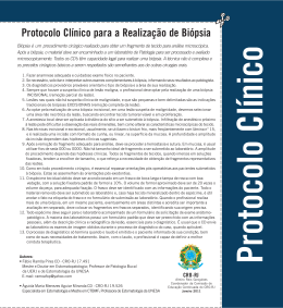
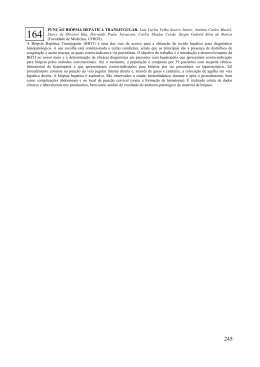
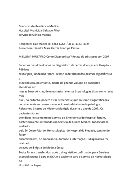
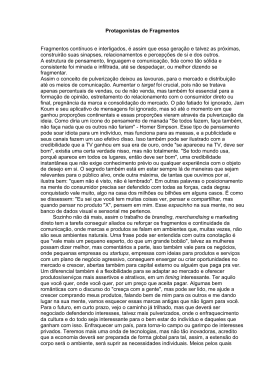
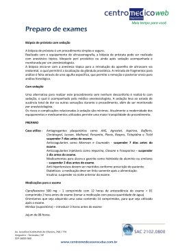
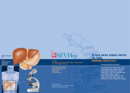
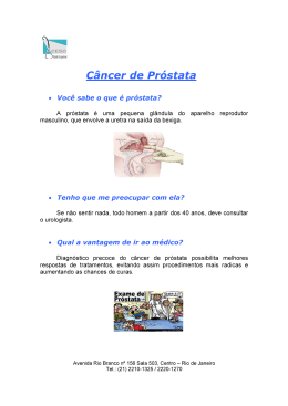
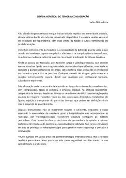
LUIZ EDISON SLONGO
ANÁLISE DOS FRAGMENTOS POSITIVOS DE BIÓPSIA NA
PREDIÇÃO DA EXTENSÃO LOCAL DO CÂNCER DE PRÓSTATA
Tese apresentada ao Curso de PósGraduação em Clínica Cirúrgica do
Setor de Ciências da Saúde, Universidade Federal do Paraná, como requisito
parcial à obtenção do título de Doutor.
Orientador: Prof. Dr. Renato Tambara Fº.
Coordenador: Prof. Dr. Jorge Eduardo
Fouto Matias
CURITIBA
2006
TERMO DE APROVAÇÃO
LUIZ EDISON SLONGO
ANÁLISE DOS FRAGMENTOS POSITIVOS DE BIÓPSIA NA PREDIÇÃO DA EXTENSÃO
LOCAL DO CÂNCER DE PRÓSTATA
Tese aapresentada como requisito parcial para obtenção do grau de Doutor
no Curso de Pós-Graduação em Clínica Cirúrgica, Setor de Ciências da Saúde da
Universidade Federal do Paraná, pela seguinte banca examinadora:
Orientador:
Prof. Dr Renato Tambara Filho
Departamento de Ciências da Saúde, UFPR
Banca examinadora
Prof. Dr. Luiz Carlos de Almeida Rocha
Prof. Dr. Sergio Ossamu Ioshii
Prof. Dra. Regina de Paula Xavier Gomes
Prof. Dr. Luiz Sergio Santos
Prof. Dr. Marco Aurélio Freitas Rodrigues
Curitiba,
26 maio de 2006
ii
AGRADECIMENTOS
Ao Professor Doutor RENATO TAMBARA FILHO, amigo e orientador. O meu eterno
agradecimento pelo exemplo, incentivo, compreensão, e preciosa contribuição
acadêmica na elaboração desta pesquisa.
Ao Professor Titular Doutor LUIZ CARLOS DE ALMEIDA ROCHA, amigo, chefe e
líder, pela oportunidade de ascender na carreira universitária.
Ao Doutor MÁRIO CESAR SUGISAWA, colaborador. Pelo incansável trabalho na
busca do aprimoramento da biópsia prostática.
Aos Professores Doutores LUIZ FERNADO BLEGI TORRES e SERGIO OSSAMU
IOSHII, colaboradores. Pela abnegação na avaliação histológica do protocolo
pesquisado.
Aos amigos e companheiros de trabalho Drs. NEY DE ALMEIDA FARIA NETO,
ROGÉRIO DE FRAGA, FERNANDO CESAR KOLESKI, JOSE MAURICIO FREHSE,
OSNY SILVESTRI e ALISSON FUCIO. Pelo incentivo, colaboração e fraternal apoio.
Aos colegas Drs. ROGERIO RIBAS, LUIZ SERGIO SANTOS, MANOEL ANTONIO
GUIMARÃES, THADEU BRENNY FILHO, AGENOR FERREIRA DA SILVA FILHO,
FERNANDO LORENZINI, SIDNEY BREVIGLIERI, REGINA DE PAULA XAVIER
GOMES e LUIZ GONZAGA DE FIGUEIREDO MOURA. Pela abnegação, espírito
científico e de luta na dedicação ao ensino médico e assistência aos necessitados
pacientes do Hospital de Clínicas – UFPR.
Ao Professor Doutor JORGE E. F. MATHIAS, Coordenador do Programa de PósGraduação em Clínica Cirúrgica do Departamento de Cirurgia da Universidade
Federal do Paraná. Pelo aprendizado e oportunidade de evolução acadêmica.
iii
À Roseli minha amada esposa
Aos nossos queridos filhos Julio, Helena e Claudia
Aos meus saudosos pais, Guerino Slongo e Maria Bonotto Slongo
“in memoriam”
Aos meus irmãos:
Os de sangue
Os de fé
iv
Aqui hoje terminam estas viagens
nas quais me acompanhastes
através da noite e do dia
e do mar e do homem.
De tudo quanto vos disse
vale muito mais a vida.
Pablo Neruda
v
SUMÁRIO
LISTA DE FIGURAS E GRÁFICO ...............................................................................
viii
LISTA DE TABELAS ...................................................................................................
ix
LISTA DE ABREVIATURAS ...........................................................................................
x
RESUMO .....................................................................................................................
xi
ABSTRACT .................................................................................................................
xii
1 INTRODUÇÃO .........................................................................................................
1
1.1 OBJETIVOS ............................................................................................................
4
2 REVISÃO DA LITERATURA ...................................................................................
5
3 PACIENTES E MÉTODO ........................................................................................
9
3.1 CARACTERÍSTICAS DA CASUÍSTICA ................................................................
10
3.2 BIÓPSIA DE PRÓSTATA.........................................................................................
11
3.2.1 ESTUDO HISTOLÓGICO DAS BIÓPSIAS DE PRÓSTATA..................................
13
3.3 PROSTATECTOMIA RADICAL RETROPÚBICA ..................................................
13
3.3.1 ESTUDO HISTOLÓGICO DOS ESPÉCIMES CIRÚRGICOS .............................
14
3.4 METODOLOGIA ESTATÍSTICA.............................................................................
16
4 RESULTADOS ........................................................................................................
18
4.1 ANÁLISE EXPLORATÓRIA DAS CARACTERÍSTICAS DA CASUÍSTICA.............
19
4.1.1 Idade...................................................................................................................
19
4.1.2 Exame Digital da Próstata...................................................................................
19
4.1.3 Antígeno Prostático Específico............................................................................
19
4.1.4 Estudo Ultra-Sonográfico Transretal ...................................................................
19
4.1.5 Estadiamento Clínico ..........................................................................................
20
4.2 RESULTADOS HISTOPATOLÓGICOS DA BIÓPSIA DE PRÓSTATA .................
20
4.2.1 Diagnóstico Histológico ......................................................................................
20
4.3 RESULTADOS HISTOPATOLÓGICOS DO PRODUTO DA PROSTATECTOMIA..
22
4.4 ANÁLISE ESTATÍSTICA COMPARATIVA ...............................................................
24
4.4.1 Exame Digital da Próstata .....................................................................................
25
4.4.2 Antígeno Prostático Específico..............................................................................
25
4.4.3 Ultra-Som Transretal .............................................................................................
26
4.4.4 Escore de Gleason na Biópsia ..............................................................................
26
4.4.5 Percentagem de Fragmentos Positivos ................................................................
27
4.4.6 Estudo do Fragmento com Maior Acometimento pela Neoplasia .......................
27
4.4.7 Estudo do Comprometimento Neoplásico da Margem Externa da Biópsia .........
28
4.4.8 Estudo da Infiltração Angiolinfática na Biópsia de Próstata ................................
28
4.4.9 Estudo da Infiltração Perineural na Biópsia de Próstata .....................................
29
4.4.10 Estudo das Duas Variáveis Conjuntas .............................................................
29
4.4.11 Estudo dos 47 pacientes com até 20% dos fragmentos positivos para neoplasia e com o fragmento mais comprometido até 70% em relação a margem
cirúrgica do espécime .......................................................................................
30
5 DISCUSSÃO ...........................................................................................................
31
6 CONCLUSÃO ..........................................................................................................
37
REFERÊNCIAS .......................................................................................................
39
APÊNDICE ..................................................................................................................
46
vii
LISTA DE FIGURAS
FIGURA 1 FIGURA 2 FIGURA 3 -
FIGURA 4 -
PRÓSTATA ESQUEMATIZADA E LOCAIS DAS PUNÇÕES SISTEMÁTICAS .............................................................................................
11
CORTE TRANSVERSAL DA PRÓSTATA. LOCAIS DAS PUNÇÕES
NA ZONA PERIFÉRICA E NA ZONA DE TRANSIÇÃO .......................
11
FRAGMENTO COM MARGEM PERIFÉRICA CORADA PELO NANQUIM VERDE SEPARADAMENTE PARA ESTUDO INDIVIDUALIZADO ...................................................................................................
FRAGMENTOS ACONDICIONADOS SEPARADAMENTE ..................
FIGURA 5A - PRÓSTATA PINTADA PELO NANQUIM AZUL. CONE DISTAL PARA
ESTUDO DA MARGEM APICAL ..........................................................
FIGURA 5B- CORTES DA PRÓSTATA EM FATIAS DE 4MM PARA ESTUDA DA
MARGEM CIRCUNJACENTES...............................................................
FIGURA 6 -
12
12
15
15
MARGEM CIRÚRGICA COMPROMETIDA. TINTA NANQUIM PINTA
OS ÁCINOS COM CÂNCER ................................................................
16
PSEUDOCÁPSULA PROSTÁTICA TRANSFIXADA PELA NEOPLASIA COM INFILTRAÇÃO DO TECIDO PERIGLANDULAR ..................
16
GRÁFICO 1 - CORRELAÇÃO ENTRE O NÚMERO DE FRAGMENTOS POSITIVOS
E A PORCENTAGEM DO FRAGMENTO MAIS COMPROMETIDO ....
21
FIGURA 7 -
viii
LISTA DE TABELAS
TABELA 1 -
ESTRATIFICAÇÃO CONFORME ESCORE DE GLEASON NA BIÓPSIA DE PRÓSTATA .............................................................................
20
ESTRATIFICAÇÃO DO ESCORE DE GLEASON NO ESPÉCIME
CIRÚRGICO ........................................................................................
23
TABELA 3
RESULTADO DO EXAME DE TOQUE RETAL ....................................
25
TABELA 4 -
CLASSIFICAÇÃO DOS PACIENTES POR PSA ..................................
25
TABELA 5 -
RESULTADO DO EXAME DE ULTRA-SOM PARA AUXÍLIO DIAGNÓSTICO NO CÂNCER DE PRÓSTATA ............................................
26
CLASSIFICAÇÃO DOS PACIENTES QUANTO AO ESCORE DE
GLEASON NA BIÓPSIA ......................................................................
26
CLASSIFICAÇÃO DOS PACIENTES POR PORCENTAGEM DE
FRAGMENTOS POSITIVOS ................................................................
27
TABELA 2-
TABELA 6 TABELA 7TABELA 8 -
TABELA 9 -
CLASSIFICAÇÃO DOS PACIENTES POR PORCENTAGEM DE
ACOMETIMENTO NEOPLÁSICO NO FRAGMENTO MAIS COMPROMETIDO ...............................................................................................
RESULTADO DO ESTUDO DA MARGEM PERIFÉRICA NA BIÓPSIA
27
28
TABELA 10 - CLASSIFICAÇÃO DOS PACIENTES QUANTO A INFILTRAÇÃO
ANGIOLINFÁTICA NA BIÓPSIA ..........................................................
28
TABELA 11 - CLASSIFICAÇÃO POR APRESENTAÇÃO OU NÃO DE INFILTRAÇÃO PERINEURAL NA BIÓPSIA ........................................................
29
TABELA 12 - DISTRIBUIÇÃO DOS PACIENTES POR PORCENTAGEM DE FRAGMENTOS POSITIVOS NA BIÓPSIA E FRAGMENTO COM MAIOR
COMPROMETIMENTO .......................................................................
TABELA 13 - DISTRIBUIÇÃO DOS PACIENTES COM ATÉ 20% DOS FRAGMENTOS POSITIVOS PARA CÂNCER E PORCENTAGEM DO
FRAGMENTO COM MAIOR COMPROMETIDO ATÉ 70% EM RELAÇÃO A MARGEM CIRÚRGICA ............................................................
ix
39
30
LISTA DE ABREVIATURAS
CAP
CÂNCER DE PRÓSTATA
PSA
ANTÍGENO PROSTÁTICO ESPECÍFICO
TNM-2002
ESTADIAMENTO CLÍNICO DO AMERICAN JOINT COMMITTEE ON
CANCER AND INTERNATIONAL UNION AGAINST CANCER
T1c
TUMOR IMPALPÁVEL, NÃO IDENTIFICADO POR IMAGEM
DIAGNOSTICADO POR BIÓPSIA DEVIDO ELEVAÇÃO DO PSA
T2a
TUMOR CONFINADO QUE COMPROMETE ATÉ METADE DE UM LOBO
PROSTÁTICO
T2b
TUMOR CONFINADO QUE COMPROMETE MAIS DA METADE DE UM
LOBO PROSTÁTICO
T2c
TUMOR CONFINADO
PROSTÁTICOS
T3a
T3b
TUMOR COM EXTENSÃO EXTRA-CAPSULAR UNI OU BILATERAL
T4
TUMOR QUE INVADE EXTRUTURAS ADJACENTES OU FIXO
QUE
COMPROMETE
OS
DOIS
TUMOR COM EXTENSÃO PARA VESÍCULA(S) SEMINAL(IS)
X
E
LOBOS
RESUMO
INTRODUÇÃO: A possibilidade de cura cirúrgica do câncer de próstata (CaP), com
mínimas sequelas, encontra-se na dependência da extensão do tumor. O preciso
conhecimento pré-operatório da extensão local da neoplasia tem sido um desafio
aos pesquisadores. Na década passada os métodos preditivos da extensão final do
CaP utilizando antígeno prostático específico (PSA), escore de Gleason e o
estadiamento clínico, foram exaustivamente investigados. Nos últimos anos vários
autores têm procurado estabelecer relação entre os achados histológicos da biópsia
e a extensão local da neoplasia. OBJETIVOS: Este estudo tem por finalidade: I)
avaliar a capacidade para predição da extensão local do CaP através da avaliação
histopatológica padronizada dos fragmentos de biópsia sistemática por agulha; II)
determinar as características dos pacientes que apresentaram os melhores
resultados cirúrgicos quando submetidos à prostatectomia radical retropúbica.
PACIENTES E MÉTODOS: Cem pacientes com diagnóstico de câncer confinado à
próstata (estadiamento clínico T1 e T2) foram estudados prospectivamente. Todos
foram submetidos à biópsia sextante estendida (12 ou 14 fragmentos) com análise
histopatológica padronizada para se determinar: a) percentagem de fragmentos
positivos; b) percentagem de infiltração neoplásica no fragmento mais acometido; c)
status da margem periférica nos fragmentos. Subsequentemente os pacientes foram
submetidos à prostatectomia radical retropúbica, onde se procurou preservar os
feixes vásculo-nervosos. Os espécimes cirúrgicos receberem avaliação
histopatológica para se estabelecer a extensão local da neoplasia ─ estadiamento
patológico ─ e as condições das margens cirúrgicas. RESULTADOS: A dosagem do
PSA médio foi 6,87 ng/ml; o estadiamento clínico correspondeu a T1c=50%,
T2a=17%, T2b=28% e T2c=5% dos casos e o escore de Gleason médio na biópsia
foi 5,76. Dentre os 100 pacientes, 54 apresentaram até 2 fragmentos positivos, em
67 o fragmento mais comprometido pela neoplasia não ultrapassava a 70% e em 11
as margens periféricas nos fragmentos de biópsia foram positivas. A extensão
extraprostática da neoplasia foi encontrada em 17% e margens positivas no
espécime cirúrgico em 29% dos casos. Nos pacientes com doença intraprostática,
59% apresentaram até 20% dos fragmentos positivos e nos pacientes com doença
extraprostática 71% apresentaram mais que 20% dos fragmentos positivos
(p=0,026). Dos 47 casos com até 20% dos fragmentos positivos e até 70% de
comprometimento neoplásico no fragmento mais acometido, apenas 8,51%
revelaram doença com extensão extraprostática e em 19,15% as margens cirúrgicas
foram positivas. O estudo das margens periféricas nos fragmentos de biópsia não foi
estatisticamente significativo. CONCLUSÕES: I) A variável com maior probabilidade
em predizer a extensão local ─ intra ou extraprostática ─ do CaP foi a percentagem
de fragmentos de biópsia positivos. II) Os pacientes com até 20% dos fragmentos
positivos e até 70% de comprometimento neoplásico no fragmento mais acometido
obtiveram melhores resultados cirúrgicos quando submetidos a prostatectomia
radical retropúbica.
Palavras chave: Próstata; Neoplasia prostática; Biópsia sextante estendida;
Extensão local do tumor: Prostatectomia radical retropúbica.
xi
ABSTRACT
ANALYSIS OF POSITIVE NEEDLE BIOPSY CORES TO PREDICT THE LOCAL
EXTENSION OF PROSTATIC CANCER.
BACKGROUND: The possibility of surgical cure for prostate cancer with minimal
sequelae depends on the cancer extension. Adequate preoperative information about
the local extension of tumor has been a challenge for the researchers. Over the last
decade, there was an overwhelming amount of investigation on predictive methods to
accurately stage prostate cancer utilizing PSA levels, Gleason score and the clinical
staging. Over the last years, several authors have tried to establish the relationship
between histological biopsy findings and pathological staging aiming at optimizing the
treatment. OBJETIVES: Analyze the cancer-positive biopsy cores from the
systematic prostate biopsy and to determinate: I) the variable that would be more
precisely predict the local extension of tumor, II) patient’s features related to the best
surgical outcome when submitted to nerve-sparing radical retropubic prostatectomy.
METHODS: This prospective series included 100 patients who underwent prostate
needle biopsy (sextant, extended to 12 or 14 cores), with standard histological
evaluation. All cases were diagnosed as clinical confined prostate cancer (T1 and
T2). The percentages of cancer-positive biopsy cores and of neoplasic infiltration in
the most compromised fragment and the peripheral margins status of the core-biopsy
were studied. The patients underwent nerve-sparing radical retropubic
prostatectomy. The surgical specimens were studied in a standard hystopathologycal
system to establish the neoplasm extension and the surgical margins status.
RESULTS: The mean PSA was 6.87 ng/ml. The clinical staging T1c, T2a, T2b and
T2c were 50%, 17%, 28% and 5%, respectively. The mean Gleason score in the
biopsies was 5.76. Among the patients, 54% presented with up to two positive
fragments; in 67% of the cases, the most compromised fragment didn’t go beyond
70% and in 11% of the cases, the peripheral margins of the cores were positive for
malignancy. Extraprostatic extension of the neoplasm was found in 17% of the
surgical specimens and positive surgical margins were found in 29% of the cases.
Among the patients with intraprostatic disease, 59% presented with up to 20% of
positive fragments, and among those with extraprostatic disease, 71% presented
with more than 20% of positive fragments (p=0,026). Of the 47 patients with up to
20% of positive fragments, and up to 70% of neoplasm involvement in the most
compromised fragment, 8,51% and 19,15% revealed extraprostatic extension
disease and positive surgical margins, respectively. The peripheral margin status on
cores was not statistically significant.
CONCLUSIONS: I) The best predictive variable in identifying the local extension of
tumor was the percentage of cancer-positive biopsy fragments. II) Patients with up to
20% of positive fragments for cancer and with up to 70% of neoplasm infiltration in
the more compromised fragment had the best surgical outcomes after undergoing to
nerve-sparing radical retropubic prostatectomy.
Key words: Prostate; Prostatic neoplasm; Sextant extended needle biopsy; Local
extension of tumor; Nerve-sparing retropubic radical prostatectomy
xii
Download