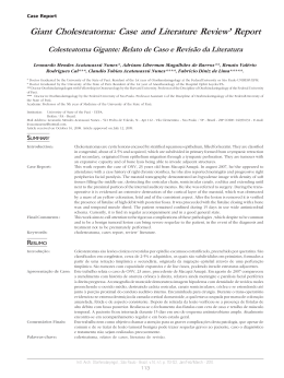
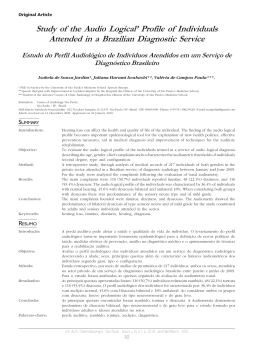
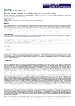
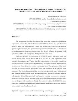
Case Report
Primary Cholesteatoma of the Bilateral External Acoustic
Meatus
Colesteatoma Primário de Meato Acústico Externo Bilateral
Luciana Almeida Moreira*, Raquel Chartuni Pereira Teixeira*, Carlos Eduardo Guimarães de Salles**,
Cláudia Inês Guerra de Sousa Silva**, Daniel Mochida Okada***.
* Resident Physician of Otorhinolaryngology and Head and Neck Surgery at HSPE-SP (R3).
** Otorhinolaryngologist.
*** Otorhinolaryngologist. Assistant Doctor of Otology at HSPE-SP
Institution:
Hospital do Servidor Público Estadual de São Paulo.
São Paulo / SP - Brazil
Mail address: Luciana Almeida Moreira - Rua Borges Lagoa, 933 - Apto. 73 - Vila Clementino - São Paulo / SP - Brazil - Zip code: 04038-032 - Telephone: (+55 11)
8684-5502 - E-mail: [email protected]
Article received on February 5, 2009. Approved on May 10, 2009.
SUMMARY
Introduction:
Objective:
Case Report:
Final Comments:
Keywords:
The cholesteatoma of the external acoustic meatus is an uncommon pathology. Most series in the
literature describe secondary cases, with a few reports of primary cholesteatoma. It is characterized
by the erosion of the external acoustic meatus bone portion by proliferation of the adjacent squamous
tissue.
To report an uncommon case of primary cholesteatoma of the bilateral external acoustic meatus.
Female patients aged 20 years old with ear ache for 3 years, associated to hypacusis and otorrhea to
the left. Without a previous history of otologic pathologies. Upon otoscopy, there could be noticed
bilateral erosion from the external acoustic meatus with a large amount of epidermal debris. The
computed tomography confirmed cholesteatoma of the bilateral external acoustic meatus and the
patient was submitted to modified radical tympanomastoidectomy to the right.
The primary cholesteatoma of the external acoustic meatus is an uncommon pathology that is part of
the differential diagnosis of chronic otalgia and otorrhea. The evaluation of its extension must be done
with computed tomography and surgery is the choice treatment.
cholesteatoma, bilateral external meatus, primary, bilateral.
RESUMO
Introdução:
Objetivo:
Relato do Caso:
Comentários Finais:
Palavras-chave:
O colesteatoma de meato acústico externo é uma patologia rara. A maioria das séries na literatura
descreve casos secundários, com poucos relatos de colesteatoma primário. Caracteriza-se pela erosão
da porção óssea do meato acústico externo por proliferação de tecido escamoso adjacente.
Relatar um caso raro de colesteatoma primário de meato acústico externo bilateral.
Paciente de 20 anos, sexo feminino, com otalgia há 3 anos, associada à hipoacusia e otorreia à
esquerda. Sem história prévia de patologias otológicas. À otoscopia, observava-se erosão bilateral do
meato acústico externo com grande quantidade de debris epidérmicos. A tomografia computadorizada
evidenciou colesteatoma de meato acústico externo bilateral, sendo a paciente submetida à
timpanomastoidectomia radical modificada à direita.
O colesteatoma primário de meato acústico externo é uma patologia rara, fazendo parte do diagnóstico
diferencial de otalgia e otorreia crônicas. A avaliação da sua extensão deve ser feita com tomografia
computadorizada e o tratamento de eleição é a cirurgia.
colesteatoma, meato acústico externo, primário, bilateral.
Intl. Arch. Otorhinolaryngol., São Paulo - Brazil, v.15, n.1, p. 96-98, Jan/Feb/March - 2011.
96
Primary cholesteatoma of the bilateral external acoustic meatus.
Moreira et al.
INTRODUCTION
The external auditory canal cholesteatoma (EACC)
is an uncommon disease that affects 1 per 1000 patients
with otologic complaints (1). It is characterized by the
erosion of the external auditory canal (EAC) bone portion
by proliferation of the adjacent squamous tissue (2). It may
be classified into primary or secondary trauma, surgery,
inflammatory process or obstruction of the EAC (5, 6).
There are several series in the literature describing secondary
EACC, but a few reports of primary EACC (2, 3). In a recent
article, PERSAUD et al. reported the first case of bilateral
primary EACC in a black young man (4). We reported an
uncommon case of bilateral primary EACC in a patient aged
20 years old.
CASE REPORT
L.C.B.S., student, female sex, 20 years old, from São
Paulo, reported bilateral monotone otalgia for 3 years,
associated to hypacusis and otorrhea at the left side (Figure
1). She reported periods of worsening of the earache,
especially associated to water entry at the EAC. There was
no prior record of other affections, trauma or otologic
surgeries. Upon otoscopy, she had erosion from both EAC
with a large number of epidermal debris that prevented the
viewing of the tympanic membrane (Figure 2). The tonal
audiometry (TA) confirmed a light bilateral conductive
loss. Temporal bones computed tomography (CT)
confirmed bilateral EACC with a stronger bone erosion of
the EAC right posterior wall (Figures 3 and 4). Radical
tympanomastoidectomy was performed and modified at
the right side with production of microbox and interposition
of the mastoid cortical bone between the clamp head and
the temporal fascia. Upon surgery, we noticed erosion of
the EAC posterior wall with a large amount of descamative
material There was a good postoperative evolution with
reduction of the gap at the right side of the AT that had no
signals of the lesion recurrence. The patient still expects
contralateral intervention.
Figure 1. Preoperative tonal audiometry. - Observed by
bilateral conductive loss, stronger at the left side.
Figure 2. Otoscopy at the left side. - Presence of epidermal debris
in the external acoustic canal, with erosion of the posterior wall.
Figure 3. Computed tomography of temporal bones in coronal
cut - left side - Enlarging of the auditory conduct and presence
of material with density of soft parts.
Figure 4. Computed tomography of temporal bones in axial
cut - left side - Well pneumatized and airy mastoid, confirming
the pathology is not originated from the middle ear.
Intl. Arch. Otorhinolaryngol., São Paulo - Brazil, v.15, n.1, p. 96-98, Jan/Feb/March - 2011.
97
Primary cholesteatoma of the bilateral external acoustic meatus.
Figure 5. Postoperative tonal audiometry. - Observed a
maintenance of the aerial-osseous gap.
Moreira et al.
The treatment of EACC is based on the type of
lesion and intensity of the symptoms. Patients with light
symptoms, small and circumscribed lesions, with a high
surgical risk or that refuse the surgery may be clinically
treated with frequent aspirations under microscopy and
otologic drops when required. The surgical treatment is
prescribed in the other cases to prevent progression of the
erosion and complications. The surgical procedure is
determined by the extension of the osteonecrosis, erosion
and assumption by the surgeon (3).
FINAL COMMENTS
DISCUSSION
The first description of the EACC was made by
TOYNBEE in 1850, but the precise definition of this disease
was obtained by PIEPERGERDES et al., in 1980, when the
differentiation between EACC and keratosis obturans was
made (2). The KO is defined as the accumulation of keratin
produced by exfoliation of the EAC skin. In the other hand,
the EACC is characterized by the erosion of the bone
portion of the EAC from the adjacent squamous tissue (2).
The differential diagnosis must also include neoplasms,
Panner disease and malignant external otitis (1). The
clinical profile is generally characterized by light and
constant otalgia, sporadic otorrhea and pruritus in the EAC.
The occurrence of hypacusis is due to the obstruction of
the EAC by the keratin debris. Alterations of the tympanic
membrane are not part of the profile (1).
The EACC etiology remains obscure, and there is the
attempt to classify it as primary or secondary to surgery,
trauma, inflammatory process, congenital stenosis or
obstruction of the EAC (5, 6).
As regards to the primary or spontaneous EACC,
there are several theories that seek to explain its etiology.
One of these states that such affection could appear from
a small trauma onto the EAC that would lead to periostitis
and further infiltration/erosion by the adjacent squamous
tissue (3). The other one reports alterations to the epithelial
migration of the EAC’s skin and to the cerumen produced
by it would lead to the appearing of the EACC (6).
The EACC is an uncommon affection that is part of
the differential diagnosis of chronic otalgia and otorrhea.
The treatment is especially surgical and the follow up is
clinical and exceptional. The evaluation of the lesion and
surgical planning must be done after performance of CT of
temporal bones. The most common surgery is modified
radical mastoidectomy.
BIBLIOGRAPHICAL REFERENCES
1. Anthony PF, Anthony WP. Surgical treatment of external
auditory canal cholesteatoma. Laryngoscope, 1982. 92(1):705.
2. Piepergerdes MC, Kramer BM, Behnke EE. Keratosis
obturans and external auditory canal cholesteatoma.
Laryngoscope. 1980, 90(3):383-391.
3. Vrabec JT, Chaljub G. External canal cholesteatoma. Am
J Otolaryngol. 2000, 21(5):608-14.
4. Persaud R, Singh A, Georgalas C, Kirsch C, Wareing M. A
new case of synchronous primary external ear canal
cholesteatoma. Otolarynol Head Neck Surg. 2006,
134(6):1055-6.
5. Naim R, Linthicum Jr. F, Shen T, Bran G, Hormann K.
Classification of the external auditory canal cholesteatoma.
Laryngoscope. 2005, 115(3):455-60.
6. Holt JJ. Ear canal cholesteatoma. Laryngoscope. 1992,
102(6):608-13.
Intl. Arch. Otorhinolaryngol., São Paulo - Brazil, v.15, n.1, p. 96-98, Jan/Feb/March - 2011.
98
Download