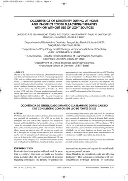
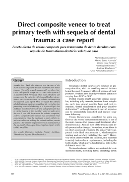
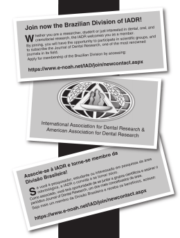
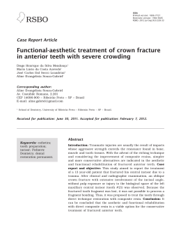
ISSN:
Printed version: 1806-7727
Electronic version: 1984-5685
RSBO. 2012 Oct-Dec;9(4):416-20
Original Research Article
Evaluation of the effectiveness of the tooth
whitening treatment associated with the
immersion in coloring solutions
Fabiano Carlos Marson¹
Karina Maria Salvatore de Freitas1
Giovani de Oliveira Correa1
Camila Paula Tonin Bressan1
Cléverson O. Silva1, 2
Corresponding author:
Fabiano Carlos Marson
Av. São Paulo, n. 172, sala 721
CEP 87030-040 – Maringá – PR – Brasil
E-mail: [email protected]
1
2
Ingá School – Maringá – PR – Brazil.
State University of Maringá – Maringá – PR – Brazil.
Received for publication: November 28, 2011. Accepted for publication: June 11, 2012.
Keywords:
tooth whitening; coloring;
immersion.
Abstract
Introduction: Tooth whitening or dental bleaching is a cosmetic
procedure that has established itself in Dentistry; however, the
staining because of the ingestion of some types of food may cause
several factors leading to the failure of this treatment. Objective:
This study aimed to evaluate in vitro the influence of the immersion
of whitened teeth in solutions with a high degree of pigmentation
on the efficacy of the bleaching performed with 16% hydrogen
peroxide. Material and methods: Fifty-six human teeth were selected,
bleached for 4 hours a day during 14 days and randomly divided
into 7 groups (n = 8). The groups G1/G2 and G3 were immersed
in coloring solutions immediately (IM) after bleaching (AP): G1
– tooth whitening + coffee (IM), G2 – tooth whitening + cola-based
soft drink (IM) and G3 – tooth whitening + red wine (IM); G4/and
G6 were immersed in the solutions for 2 hours (AP), G4 – tooth
whitening + coffee (AP), G5 – tooth whitening + cola based soft
drink (AP), G6 – tooth whitening + red wine and (AP) G7 – control.
After bleaching with an immersion time of 5 minutes, with the aid
of a digital spectrophotometer, the final color (FC) was measured 24
hours after the end of the bleaching treatment (day 15). Results: The
results for ANOVA showed no statistical differences in all groups.
Conclusion: There was no influence on the effectiveness of tooth
whitening immersed in coloring solutions.
RSBO. 2012 Oct-Dec;9(4):416-20 –
417
Introduction
Material and methods
The beauty standards presented by the media,
based on white and well aligned smiles with the
need to look good for acceptance in society, are
causing that the cosmetic procedures are the
most requested procedures in the dental office,
and functional procedures end up getting on low
priority [3, 13]. Among these procedures, tooth
whitening, which besides providing fast results and
being easily implemented, is considered minimally
invasive [12].
To succeed in the bleaching treatment is
necessary to identify the etiologic factor of the
darkening. The natural color of the teeth is altered
by extrinsic and intrinsic agents. As extrinsic agents,
it can be cited: excessive consumption of tobacco,
certain medications, chromogenic bacteria and dyes
from the diet, such as coffee, tea, wine, mate and some
soft drinks [21]. Intrinsic alterations have several
causing sources, such as malformation of dental
tissues, dental trauma, certain systemic medications
(tetracycline) and iatrogenic procedures [19].
The bleaching for vital teeth can be accomplished
by using two techniques: home bleaching and inoffice whitening. Basically, what differs one technique
from another is the time of whitening and the
concentration of the bleaching agents [13]. In the
home technique, low concentrations of the bleaching
agent are used, among which one can opt by the
carbamide peroxide or hydrogen peroxide. In the
in-office technique, highest concentrations of the
bleaching agent are used, and consequently, there is a
smaller time interval. The home bleaching technique
has been well accepted since it was described by
Haywood and Heymann, in 1989 [8].
In the bleaching process, the bleaching agents
act by releasing the oxygen into the dental structures
in a reaction so-called oxidation-reduction. This
process is directly related to the concentration
of the bleaching agent and the time in which the
enamel is in contact with the agent [1]. The dental
permeability associated with the low molecular
weight allows the oxygen to diffuse through the
enamel and dentin. On the other hand, the pigments
have a high molecular weight and longer chains that
in the bleaching process are broken into smaller
molecular chains [4, 9].
By starting the bleaching treatment, the patients
receive several recommendations, including dietary
restrictions, because there is not a given space of time
after the end of treatment so that we can introduce a
diet rich in foods and beverages with high potential
of pigmentation without any interference in the result.
Therefore, the aim of this study was to assess the
efficacy of the bleaching treatment associated with
the immersion in coloring solution.
This project was submitted and approved by the
Ethical Committee in Research of the Ingá School
under protocol number 0217.0.362.000-11.
Seventy-three human teeth, premolars and
molars free of caries and restorations, were stored
into 37% formaldehyde solution just after their
extraction. Next, the teeth underwent crown and
root scaling and planing through Robinson brush
and pumice. With the use of a spectrophotometer
(VITA Easyshade Compact), 56 teeth were selected
to meet the inclusion criteria: shade A3 or higher
saturation. The specimens were randomly selected
in six experimental groups and one control groups
(figure 1):
Figure 1 – Specimens embedded in dental wax plate
A f ter t he a ssi g n ment of t he g roups, a n
impression with alginate of each wax plate with
the respective teeth embedded was taken to obtain a
dental cast to construct an acetate tray in a vacuum
laminator (VH Equipamentos, Araraquara, Brazil).
To make the process easier, each tray was identified
with the number of the group it was assigned. The
color was determined through the parameters of the
international CIElab system (L* a* b*), in which L*
indicates the luminosity (its mean ranged from 0
– black to 100 – white), and a* and b* correspond
to the shade: a* represents the saturation at the
red-green axis and b* at the blue-yellow axis,
enabling the specificity of any color. With the use
of the digital spectrophotometer the initial values
of L*, a* and b* were determined through three
readings in the medium third of the buccal surface
of each tooth to obtain the mean of the initial color
of each tooth to be evaluated (CI).
After these separations, the teeth were submitted
to the bleaching process for 14 consecutive days.
They were in touch with the bleaching agent for 4
hours per day. This was accomplished by the 16%
carbamide peroxide gel (Whitegold Home, Dentsply,
Petrópolis, Brazil), applied through the tray onto
the buccal surface. After the determined period,
the trays were removed from the plates and washed
under running water.
Marson et al.
418 – Evaluation of the effectiveness of the tooth whitening treatment associated with the immersion in coloring solutions
The groups were divided according to the coloring solution and the moment of the bleaching
immersion, as follows (table I):
Table I – Division of the groups according to the solution used
Group
Solution
Immersion into the solutions
Time of immersion
G1
Coffee
Immediately after bleaching
5 minutes
G2
Cola-based soda
Immediately after bleaching
5 minutes
G3
Red wine
Immediately after bleaching
5 minutes
G4
Coffee
2 hours after bleaching
5 minutes
G5
Cola-based soda
2 hours after bleaching
5 minutes
G6
Red wine
2 hours after bleaching
5 minutes
G7
Control
Only bleaching
Control
The samples were immersed into the coloring
solutions during 5 minutes (once per day), through
vibrating movements, with the aid of a vibrator
device (VH Equipamentos, Araraquara, Brazil).
Next, the samples were removed and carefully
washed i n runni ng water a nd i mmersed i n
artificial saliva (Medfórmula, Maringá, Paraná,
Brazil). The saliva composition was as follows:
KH2PO4 (25 mM) 100 ml + Na2HPO4 (24 mM)
100 ml + KHCO3 (150 mM) 100 ml + NaCl
(1.0 mM) 100 ml + MgCl2 (0.15 mM) 100 ml
+ CaCl2 (1.5 mM) 100 ml + Citric acid (0,002
mM) 6 ml.
At every 24 hours, the bleaching procedures
and the immersion in the solutions were performed.
The exposure to the coloring solutions occurred
during 5 minutes only once per day always after
the tooth bleaching.
Elapsed 24 hours after the treatment ending,
a new color analysis was performed at the same
place of the first analysis to determine the final
color (CF). The difference between CI and CF (ΔE)
was calculated according to the follow equation:
ΔE = [(Δa)² + (Δb)² + (ΔL)²]1/2. The values obtained
from ΔE were statistically evaluated by ANOVA and
Tukey test, with a level of significance of 5%.
Graph 1 – Initial (before bleaching) and final (after
bleaching and immersion) means for the coordinate L*
Graph 2 – Initial and final means for the coordinate a*
Results
The color alteration was statistically analyzed
through the coordinates of L*, a* and b*, in all
groups. In graphs 1, 2 and 3 are shown the means
of each coordinate for each group evaluated. All the
groups were bleached and compared regarding to
the initial and final results.
Graph 3 – Initial and final means for the coordinate Δb*
RSBO. 2012 Oct-Dec;9(4):416-20 –
The values of ΔL*=L*final- L*initial, Δa*=a*finala*initial and Δb*=b*final-b*initial were converted to
obtain ΔE*= [(ΔL*)2 + (Δa*)2 + (Δb*)2] 0.5. ANOVA
was applied to observe if there was statistical
significance among the analyzed groups. There
was no statistical differences among groups ΔE
(p = 0.2332) (table II). Also, there is no significant
statistically difference when the values were
individually analyzed ΔL (0.3026), Δa (0.3709) and
Δb (0.0747).
Table II – Mean and standard deviation values of the
groups
Groups
ΔE and sd
G1
15.36±1.01
14.90±0.83
14.98±1.05
15.99±1.37
14.59±2.50
16.12±1.07
15.39±1.17
G2
G3
G4
G5
G6
G7
Thus, based on the results presented, the
coloring solutions had no significant influence on
the process of tooth whitening.
Discussion
The aim of this study was to investigate in
vitro if the contact of the tooth after being whitened
through the home technique with 16%carbamide
peroxide with high potential coloring solutions
jeopardize the results of the bleaching treatment.
It is highlighted that for this procedure, the intake
of food and/or beverages that have a high content
of dye should be avoided. The use of food dye is
contraindicated during the bleaching treatment.
However, most patients ultimately did not follow
this recommendation.
In the literature there are few studies related
to this subject, most studies used bovine teeth
for analysis. Bovine teeth have lower permeability
compared to human teeth. According to Schmalz
et al. [18], the permeability of bovine teeth becomes
half of that found in human teeth. One possible
explanation is that the density of the bovine dentinal
tubules is greater [17]. Therefore, in this study,
human teeth were used for clinical simulation.
For this study, we selected three solutions with a
high degree of pigmentation: coffee, red wine and
cola-based soda. These solutions are among the
most cited drinks in the literature as the causes
419
of extrinsic staining, however, they can become
as intrinsic stains when they penetrate into the
enamel microporosities [3]. Caneppele et al. [5]
conducted a study with the same solutions, and
Teo et al. [20] also included black tea. Cardoso et
al. [6] used only coffee, Attin et al. [2] used only
black tea and in the study of Ley et al. [11], they
used red wine.
In this process, the immersion time in the
coloring solution was 5 minutes to simulate the
time consumption in the general population. In
the study of Caneppele et al. [5] the immersion
totalized10 minutes daily, divided into two moments.
In the study of Cardoso et al. [6] the specimens
were immersed into coffee, 5 times per day during
60 seconds. Attin et al. [2] left the specimens
immersed into black tea for10 minutes. These
aforementioned authors corroborate this study by
the fact that no significant statistically differences
were found between the color of the specimens.
However, further studies are necessary, since in the
studies cited the immersion time was relatively low.
A longer immersion time means, in practice, larger
or excessive consumption of substances containing
coloring agents, which can be a determining factor
in tooth staining. One observation is the work of
Téo et al. [20]: the samples remained in contact with
the coloring solutions for about 1 hour. However,
this does not occur in practice, and as a result,
all substances showed high potential for staining.
The immersion in artificial saliva containing
calcium and phosphate ions after bleaching and
immersion procedures was done to simulate the
oral environment and thereby remineralize the teeth
[16], likely to several other studies [2, 5, 6, 11].
The measurement of the color of the teeth
was carried out by the spectrophotometry method
which provides accurate data, compared to visual
evaluation, which is executed by comparing the
color of the tooth with resin or porcelain scales.
The color determination was carried out through
the CIE lab system (L*a*b*), in which L* means
luminosity and a* and b* indicates the color and the
saturation [7]; the mean of these measurements is
∆E, in which the statistical test was performed.
The medium third was used as a parameter for
the color reading because it best represents the color
of the teeth; the cervical third is more darkened
and the incisal third more translucent. Differences
in color among these regions are clinically and
statistically significant [20].
Although the results of this study show that
the dyes from the diet influenced on the results
obtained at the end of the bleaching treatment, it
is necessary to observe that the dietary restrictions
still must be recommended by the dentists so that
Marson et al.
420 – Evaluation of the effectiveness of the tooth whitening treatment associated with the immersion in coloring solutions
there is not an intake excess of this type of diet by
the patients, since in this study we used a minimum
time of 5 minutes and these pigments were part of
extrinsic changes of the color of the teeth.
Conclusion
10. Joiner A. Tooth colour: a review of the
literature. J Dent. 2004;32:3-12.
11. Ley M, Wagner T, Bizhang M. The effect of
different fluoridation methods on the red wine
staining potential on intensively bleached enamel
in vitro. Am J Dent. 2006 Apr;19(2):80-4.
The staining solutions in this present study did
not interfere in the bleaching treatment.
12. Marson FC, Sensi LG. Clareação de dentes
vitalizados através da técnica de consultório. Rev
Dental Press Estét. 2007 Apr;4(2):60-71.
References
13. Marson FC, Sensi LG, Araújo FO, Andrada MAC,
Araújo E. Na era do clareamento dentário a laser
ainda existe espaço para o clareamento caseiro?
Rev Dental Press Estét. 2006 Jan;3(1):89-98.
1. Antón ARS, Lima MJP, Araújo RPC. Dentifrício
peróxido de hidrogênio: ação clareadora? Rev
OdontoCiênc. 2009 Apr-Jun;24(2):161-7.
2. Attin T, Manolakis A, Buchalla W, Hannig
C. Influence of tea on intrinsic colour of
previously bleached enamel. J Oral Rehabil. 2003
May;30(5):488-94.
3. Baratieri LN, Maia E, Andrada MAC, Araújo E.
Caderno de dentística: clareamento dental. 1. ed.
São Paulo: Santos; 2005.
4. Ben-Amar A, Liberman R, Gorfil C, Bernstein Y.
Effect of mouthguard bleaching on enamel surface.
Am J Dent. 1995 Feb;8(1):29-32.
5. Caneppele TMF, Souza AC, Valera MC, Pagani
C. Influência da embebição dental em substâncias
com corantes na eficácia do clareamento dental
com peróxido de carbamida a 16%. Arq Odontol.
2009 Oct-Dec;45(4):171-7.
6. Cardoso PC, Ferreira IA, Gondo R, Vieira LCC,
Baratieri LN. Influence of coffee on the resulting
shade of tooth bleaching. J Dent Res. 2005
Mar;84:9-12.
7. Cardoso PC, Godoy FS, Oliveira MT, Baratieri
LN. Influência do tempo de aplicação de um gel
clareador à base de peróxido de carbamida a 10%
na cor dos dentes – um estudo in vitro. Ciênc
Odontol Bras. 2007 Jan-Mar;10(1):78-83.
14. Marson FC, Sensi LG, Arruda T. Efeito do
clareamento dental sobre a resistência adesiva do
esmalte. RGO. 2008 Jan-Mar;56(1):33-7.
15. O’Brien WJ, Hemmendinger H, Boenke KM,
Linger JB, Groh CL. Color distribution of three
regions of extracted human teeth. Dent Mater. 1997
May;13(3):179-85.
16. Rodrigues JA, Basting RT, Serra MC, Rodrigues
Jr AL. Effects of 10% carbamide peroxide bleaching
materials on enamel microhardness. Am J Dent.
2001 Apr;14(2):67-71.
17. Schilke R, Lisson JA, Bauss O, Geurtsen
W. Comparison of the number and diameter of
dentinal tubules in human and bovine dentine by
scanning electron microscopic investigation. Arch
Oral Biol. 2000 May;45(5):355-61.
18. Schmalz G, Hiller KA, Nunez LJ, Stoll J,
Weis K. Permeability characteristics of bovine
and human dentin under different pretreatment
conditions. J Endod. 2001 Jan;27(1):23-30.
19. Sulieman MA. An overview of tooth-bleaching
techniques: chemistry, safety and efficacy.
Periodontol 2000. 2008 Oct;48(1):148-69.
8. Haywood VB, Heymann HO. Nightguard vital
bleaching. Quintessence Int. 1989;20(3):173-6.
20. Téo TB, Takahashi MK, Gonzaga CC, Lopes
MGK. Avaliação, após clareamento, da alteração
de cor de dentes bovinos imersos em soluções com
elevado potencial de pigmentação. RSBO. 2010
Oct-Dec;7(4):401-5.
9. Haywood VB, Heymann HO. Nightguard vital
bleaching: how safe is it? Quintessence Int. 1991
Jul;22(7):515-23.
21. Touati B, Nathanson D, Miara P. Odontologia
estética e restaurações cerâmicas. São Paulo:
Santos; 2001.
Download