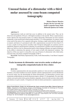
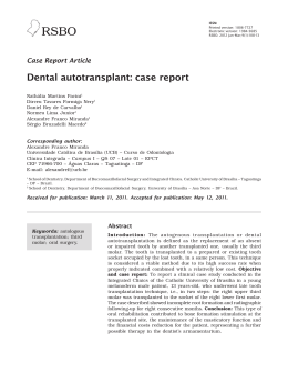
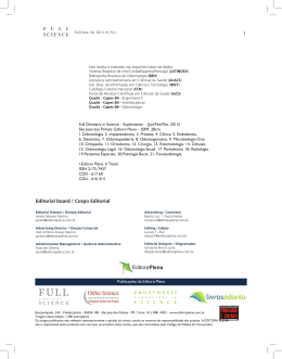
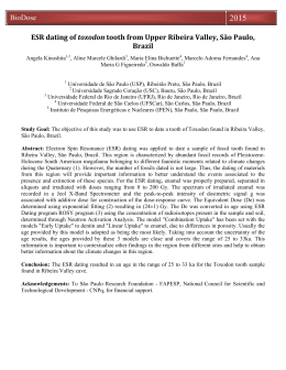
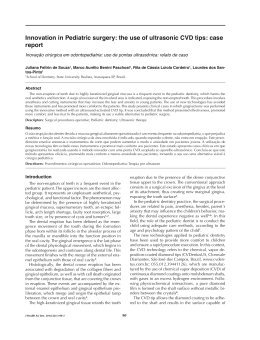
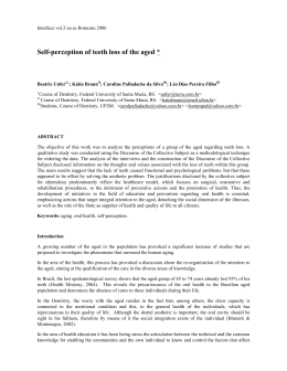
ISSN:
Printed version: 1806-7727
Electronic version: 1984-5685
RSBO. 2012 Jul-Sep;9(3):328-33
Case Report Article
Functional-aesthetic treatment of crown fracture
in anterior teeth with severe crowding
Diego Henrique da Silva Mendonça1
Mario Lúcio da Costa Azevedo1
José Carlos Dal Secco Leandrini1
Aline Evangelista Souza-Gabriel1
Corresponding author�:
Aline Evangelista Souza-Gabriel
Av. Costábile Romano, 2.201
CEP 14096-900 – Ribeirão Preto – SP – Brasil
E-mail: [email protected]
1
School of Dentistry, University of Ribeirão Preto – Ribeirão Preto – SP – Brazil.
Received for publication: June 30, 2011. Accepted for publication: February 7, 2012.
Keywords: esthetics;
tooth preparation;
incisor; Pediatric
Dentistry; dental
restoration permanent.
Abstract
Introduction: Traumatic injuries are usually the result of impacts
whose aggressive strength exceeds the resistance found in bone,
muscle and tooth tissues. With the advent of the etching technique
and considering the improvement of composite resins, simpler
and more conservative alternatives are indicated in the aesthetic
and functional rehabilitation of fractured anterior teeth. Case
report and objective: This study aimed to report the treatment
of a 13 year-old patient that fractured his central incisor due to a
trauma. After clinical and radiographic examination, an oblique
crown fracture with extensive involvement of the incisal angle,
without pulp exposure or injury to the biological space of the left
maxillary central incisor (tooth #21) was observed. Because the
fractured tooth fragment was lost, it was not possible to process a
fragment bonding. Thus, it was proposed to treat the tooth through
direct technique restoration with composite resin. Conclusion: It
can be concluded that the aesthetic and functional rehabilitation
with direct composite resin is a viable option for the conservative
treatment of fractured anterior teeth.
RSBO. 2012 Jul-Sep;9(3):328-33 –
329
Introduction
Case�������
report
Tooth trauma is a frequent emergency situation
at dental offices [19]. The high rates of violence, car
accidents, and greater participation of children in
sports activities have contributed to consider tooth
trauma as an increasing public health problem
[20].
Tooth trauma lesions comprise from a simple
enamel fracture to the total loss of the tooth. Enamel
and dentin fractures without pulp exposure do
not require an emergency treatment because their
prognosis is favorable even for late treatment. Crown
fractures with pulp exposure, intrusive luxation,
concussion, subluxation and primary tooth trauma
are considered injuries of moderate severity and
demand an early treatment [15, 19].
There is a predominance of tooth trauma in
male subjects, especially at school and growing
age, as a consequence of falls, fights, sports or car
accidents, trauma due to objects and child abuse [5,
19]. Children showing anterior open bite, marked
overjet, inappropriate lip coverage in additional to
inappropriate lip sealing are more prone to trauma
than children with normal occlusion [4, 5, 10, 20].
Social-economical factors have not been associated
to more occurrences of tooth trauma lesions [4].
Both the physical and the psychological damage
are present in tooth trauma. The lesions in the
anterior teeth may result in unfavorable effects on
the function and affect the self-steam, the behavior
and the professional success, especially if there is
the loss of the permanent tooth structure [4, 18,
20-22]. Avulsion, root and bone fractures have been
considered as the most serious acute situations and
should also receive an early treatment [18, 19].
Emergency situations involving the head and
neck frequently are dramatic experiences for both
the parents and the children. In several times,
the early treatment is not performed because of
either the lack of knowledge of the parents or by
the fact that the emergency care is provided by
hospitals, medical offices or public health centers.
All these factors together result in the delaying of
the dentist’s evaluation, affecting the treatment
prognosis [4, 19].
This limitation of the knowledge on the proper
procedures regarding to tooth traumas, mainly
avulsions, reinforces the need of education on not
only the initial treatment but also on the preventive
measures [4].
The aim of this study was to report the treatment
of a 13-year-old male patient who suffered a trauma
on the central incisor due to a fall.
A male patient was referred to the dentistry
clinic of the University of Ribeirão Preto (short
Unaerp) to restore a fractured anterior tooth.
During the anamnesis, the patient reported that
a fall had caused the tooth fracture. The clinical
and radiographic examinations revealed that the
left maxillary central incisor (tooth #21) showed
an extensive oblique crown fracture involving the
incisal angle (Class IV), without pulp exposure or
involvement of the biological space; also the tooth
was crowded and labially rotated (figures 1A, 1B,
1C and 1D).
Additionally the patient reported that the trauma
occurred one year ago, and the tooth fragment was
lost. Therefore, it was not possible to perform a
fragment bonding. The treatment proposed was to
restore the tooth through direct composite resin
technique.
At the first appointment, alginate impressions
(Jeltrate, Dentsply, Petrópolis, RJ, Brazil) were
executed to obtain study casts (dental stone, type
IV – Durone IV (Dentsply, Petrópolis, RJ, Brazil),
which were mounted in semi adjustable articulator
to analyze the occlusion, perform the diagnostic
waxing, and construct a silicone palatal guide
(Perfil, Vigodent, Bonsucesso, RJ, Brazil).
At the second appointment, a prophylaxis was
performed and tooth color selected through Vitta
Classical scale, resulting in the shade A1 for both
the enamel and dentin.
The patient was submitted to anesthesia and
absolute isolation by adapting a clasp (#212R)
because the tooth was crowded and labially rotated
(figure 2A).
Following, a bevel was constructed with the
aid of a #1190F bur (KG Sorensen, Barueri, SP,
Brazil) at high speed (figure 2B). The silicone
guide was tried on and adapted and the bonding
technique protocol was initiated: enamel etching
for 30 seconds, washing, drying, application and
light-curing of the adhesive system (Adper Single
Bond 2, 3M ESPE, Sumaré, SP, Brazil). Then,
the silicone guide was positioned on the palatal
surface of the tooth and an increment of composite
resin was applied (T neutral shade - Opallis, FGM,
Joinville, SC, Brazil) to restore the palatal enamel,
followed by light-curing for 30 seconds. After the
construction of the palatal surface, a second layer
of resin (DA1 shade – Opallis, FGM, Joinville,
SC, Brazil) was inserted to restore the dentin.
At this stage, the anatomical characteristics were
reproduced. The last layers of composite resin
330 –
Mendonça� et al.
Functional-aesthetic treatment of crown fracture in anterior teeth with severe crowding
(EA1 shade – Opallis, FGM, Joinville, SC, Brazil) were inserted and sculpted with the aid of a brush
for composite resin reproducing the texture of the labial surface.
After the removal of the absolute isolation, the restoration finishing was executed, and the occlusion
was adjusted through finishing burs. At the following appointment, the restoration polishing was
performed through abrasive discs (Sof-Lex Pop-On, 3M ESPE, St. Paul, MN, USA), abrasive rubber
points (Enhance, Dentsply, Petrópolis, RJ, Brazil), felt discs with abrasive paste, and silicon carbonate
brush (Astrobrush, Ivoclar Vivadent, Amherst, NY, USA). A smooth, bright surface was obtained and
an aesthetic, improved, more harmonious outcome was achieved. The treatment met the patient’s
expectation. After 18 months of following-up, the tooth is asymptomatic and the patient is undergoing
orthodontic treatment.
Figure 1 – A) Frontal view of the occlusion. Tooth #21 showing large oblique crown fracture; B) Left lateral view; C)
Maxillary occlusal view; D) Periapical radiograph of the central incisors
Figure 2 – A) Frontal view of the absolute isolation of the operative field ; B) Bevel preparation through #1190F bur
(KG Sorensen) at high speed
RSBO. 2012 Jul-Sep;9(3):328-33 –
331
Figure 3 – A) Silicone palatal guide adapted and insertion of composite resin increment; B) Insertion of opaque
composite resin increment for construction of the artificial dentin; C) Frontal view – treatment concluded; D) Final
aspect of the restoration after repolishing, 6-month following-up
Discussion
The restorations in fractured anterior teeth are
aesthetic-functional solutions for the consequences
of a tooth trauma. They are also required by the
patients to improve their appearance, providing
greater comfort and psychosocial well-being [6, 13].
In the case report presented here, the construction
of a direct composite resin restoration was the
most viable and efficient treatment choice, mainly
because of the good clinical performance of this
material, which exhibits an easy handling and
large variety of color shades, resulting in an almost
imperceptible restoration [13].
The mechanical resistance of fractured teeth
is compromised, because they show an irreversible
loss of tooth structures. Several direct restorative
systems have been employed, displaying different
performance in enamel and dentin [9].
The treatment of the fractured central incisor
reached results as the maintenance and integrity of
the tooth, as well as the recovering of the patient’s
self-steam, due to the improvement of tooth aesthetic
and consequently of the patient’s emotional state.
Tooth trauma is very common during childhood
and adolescence. The crown fractures exhibited
the highest percentage of all traumatic injuries
in permanent dentition (26% to 76%), followed by
luxations; the most affected area is the crown of the
teeth, mainly the enamel [16, 22]. In either primary
or permanent teeth, the trauma is frequently a severe
aesthetic, functional and psychological problem, and,
it may be considered as an emergency situation,
not only by the tooth problems and their future
consequences, but also by the child and parents’
emotional involvement [1, 12].
Camargo and Guedes-Pinto reported that in
children at 7 to 13 years-old, 59.2% of the 167
tooth fractures found were in enamel and dentin.
The most common type of crown fracture line is
oblique (69.3% of the cases), followed by horizontal
(28.9%) and vertical (1.8%) [12]. In the case presented
here, the patient presented a large oblique crown
fracture without pulp exposure in tooth #21.
Studies have indicated that the incidence of
tooth trauma is higher in males than females,
at 2:1 ratio [4, 5, 10, 20, 22]. In the school and
teenage age range, the traumas occur because of
several accidents, such as falls; strikes; bicycle,
car and sports accidents, among others. Also, the
child and teenager abuse should be emphasized
[12]. Additionally, the highest occurrence of tooth
traumas is verified at home, during school vacations,
when children are freer and play more [12].
The t reat ment of t he pat ient present i ng
tooth trauma basically comprises: controlling
of the parents’ anxiety, brief history in cases of
332 –
Mendonça� et al.
Functional-aesthetic treatment of crown fracture in anterior teeth with severe crowding
emergency care, trauma history, assessment of
general symptoms, fast behavior guidance of the
child, clinical examination, cleaning of the area,
radiographic examination, diagnosis, and treatment
planning; all information should be registered in
the patient’s file [12].
In cases of crown fracture, either the tooth
fragment bonding or the tooth restoration through
composite resin is the most adequate immediate
treatment for this type of trauma. The restoration
of a tooth undergoing a trauma and showing a
crown fracture involving the enamel and dentin is
performed through the restoration by light-cured
composite resin employing the enamel etching and
adhesive systems [1, 6, 8].
The immediate care of the traumatized teeth
and the future of the oral health of the patient
demand not only the emergency treatment, but also
an appropriate long-term following-up. Because of
the multidisciplinary involvement required for the
treatment of these cases, the general dentist is the
most appropriate professional to perform it [15].
Therefore, the dentist must be prepared to
solve the problem from both the therapeutic and
emotional standpoint. Consequently, the dentist
must have cont rol of t he situat ion, show ing
knowledge, serenity and security to both the patient
and parents. The professional should establish an
adequate treatment planning, minimizing further
sequelae and providing a higher probability of the
traumatized tooth maintenance until the patient
reaches the adult age [6, 12, 13].
In the case presented here, the aestheticfunctional treatment of the fractured tooth (#21),
employing the direct composite resin technique,
obtained a good outcome and reestablished the
physical and mainly psychological health of the
patient.
Conclusion
It can be concluded that the aesthetic-functional
rehabilitation with direct composite resin is a viable
option for the conservative treatment of fractured
anterior teeth.
References
1. Andreasen JO. Essentials of traumatic injuries
to the teeth. 1. ed. Copenhagen: Munksgaard;
1991.
2. Avelar FM, Penido CVSR, Cruz RA, Penido
SMMO. Colagem homógena de fragmento dentário
em incisivo central superior permanente – relato
de caso clínico. ����������������������
RFO. 2009;14(1):66-70.
3. Bendo CB, Paiva SM, Torres CS, Oliveira
AC, Goursand D, Pordeus IA et al. Association
������������
between treated/untreatred traumatic dental
injuries and impacto on quality of life of Brazilian
schoolchildren. Health Qual Life Outcomes.
2010;8(114):1-8.
4. Bonini GAVC, Marcenes W, Oliveira LB,
Sheiham A, Bönecker M. Trends in the prevalence
of traumatic dental injuries in Brazilian preschool
children. Dental Traumatol. 2009;25(6):594-8.
5. Cavalcanti AL, Bezerra PKM, De Alencar CRB,
Moura C. Traumatic anterior dental injuries in
7 – to 12 year-old Brazilian children. Dental
Traumatol. 2009:25(2):198-202.
6. Conceição EN. Dentística saúde e estética.
2. ed. Porto Alegre: Artmed; 2007.
7. De Rossi M, De Rossi A, Queiroz AM, Nelson-Filho
P. Management of complex dentoalveolar trauma:
a case report. Braz Dent J. 2009;20(3):259-62.
8. Fidel RAS. Complicated crown fracture: a case
report. Braz Dent J. 2006;17(1):83-6.
9. Garcia RN, Alvarez AEG, Dias CE, Mazaro
MA, Firmo T, Stuker H et al. Avaliação da
resistência de união de sistemas restauradores
contemporâneos em esmalte e dentina. RSBO.
2011;8(1):60-7.
10. Goenka P, Marwah N, Dutta S. Biological
approach for management of anterior tooth
trauma: triple case report. J Indian Soc Pedod
Prev Dent. 2010;3(28):223-9.
11. Góes KKH, Ribeiro ED, Lima Júnior JL,
Silva Neto JM. Avaliando os traumatismos
dentoalveolares: revisão de literatura. Rev Cir
Buco-Maxilo-Facial. 2005;5(1):21-6.
12. Guedes-Pinto AC, Wanderley MT, Bonini GAVC,
Cadioli IC, Prokopovitch I. Odontopediatria. 8. ed.
São Paulo: Santos; 2010.
RSBO. 2012 Jul-Sep;9(3):328-33 –
13. Melo LL. Traumatismo alvéolo-dentário. 1. ed.
Série EAP-APCD, v. 9. São Paulo: Artes Médicas;
1998.
14. Melo REVA, Silva MBL, Vitor CMA, Luna LA,
Firmo ACB. Traumatismo dentoalveolar. Int J
Dent. 2003;2(2):266-72.
15. Oliveira FAM, Gerhardt MO, Orso VA,
Oliveira VR. Traumatismo dentoalveolar: revisão
de literatura. Rev Cir Buco-Maxilo-Facial.
2004;4(1):15-21.
16. Poi WR, Pignatta LMB, Ricieri CB, Panzarini SR,
Sonoda CK. Tratamento de urgência em traumatismo
dento-alveolar. RGO. 2005;53(3):181-5.
17. Rezende FMC, Gaujac C, Rocha AG, Peres
MPSM. �������������������������������������
A prospective study of dentoalveolar
trauma at the Hospital das Clínicas, São
Paulo University Medical School. Clin Sci.
2007;62(2):133-8.
333
18. Sakai VT, Magalhães AC, Pessan JP, Silva SMB,
Machado MAAM. Urgency
�������������������������������
treatment profile of 0
to 15 year-old children assisted at urgency dental
service from Bauru Dental School, University of
São Paulo. J
����������������������������������
Appl Oral Sci. 2005;13(1):340-4.
19. Sanabe ME, Cavalcante LB, Coldebella CR, Abreue-Lima FCB. Urgências em traumatismos dentários:
classificação, características e procedimentos. Rev
Paul Pediatr. 2009;27(4):447-51.
20. Traebert J, Almeida ICS, Garghetti C, Marcenes
W. Prevalência, necessidade de tratamento e
fatores predisponentes do traumatismo na
dentição permanente de escolares de 11 a 13 anos.
Cad Saúde Pública. 2004;20(2):403-10.
21. Turkistani J, Hanno A. Recent trends in the
management of dentoalveolar traumatic injuries
to primary young permanent teeth. Dental
Traumatol. 2011:27(1):46-54.
22. Vasconcellos RJH, Oliveira DM, Porto GG,
Silvestre H, Silva E. Ocorrência de traumatismo
dental em escolares de uma escola pública do Recife.
Rev Cir Buco-Maxilo-Facial. 2003;3(4):9-12.
Download