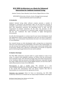
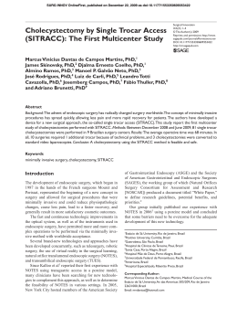
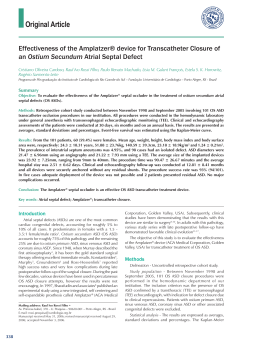
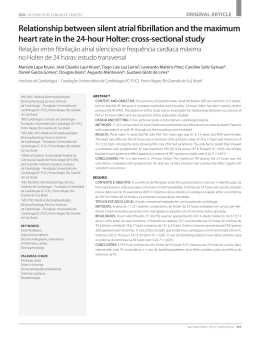
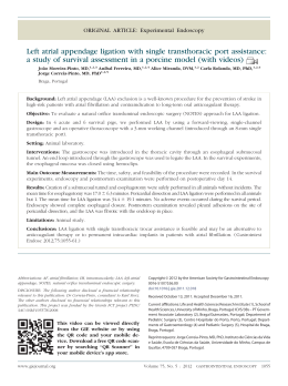
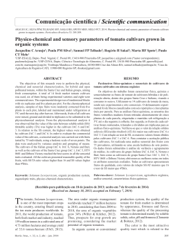
MULTIMEDIA
Rev Bras Cir Cardiovasc 2012;27(3):488-90
Robotic assisted minimally invasive surgery for
atrial septal defect correction
Cirurgia minimamente invasiva robô assistida na correção da comunicação interatrial
Robinson Poffo1, Alex Luiz Celullare2, Renato Bastos Pope3, Alisson Parrilha Toschi3
DOI: 10.5935/1678-9741.20120083
RBCCV 44205-11414
Descriptors: Heart septal defects, atrial. Surgical
procedures, minimally invasive/methods. Robotics.
Descritores: Comunicação interatrial. Procedimentos
cirúrgicos minimamente invasivos/métodos. Robótica.
CHARACTERIZATION OF PATIENT
The characteristics of the patient are: female, 24 years
old, 55 kg. She came to our department with complaints of
fatigue and palpitations. She denied any associated disease
or medication use. On physical examination, the patient was
eutrophic, eupneic at rest, without edema. No alterations of
pulmonary auscultation and cardiac auscultation revealed a
sinus rhythm, with pulmonary systolic murmur with fixed
splitting of 2nd heart sound. Resting blood pressure (BP)
was: 100/70 millimeters of mercury (mmHg).
The chest radiograph showed normal heart size and
increased pulmonary vasculature. Echocardiography
revealed a normal left atrial dimension (LA) of 2.9 centimeters
(cm) and left ventricular (LV) diameter within normality (LV
Systolic Diameter: 2.5 cm - LV Diastolic Diameter: 4.2 cm )
and normal myocardial thickness. The other cavities were
also normal. The presence of interatrial comunication (IAC)
secundum ostium type showed the Doppler that the shunt
with unidirectional flow of LA to the right. The ejection
fraction was estimated at 72% (Simpson). The pulmonary
artery pressure was estimated at 40 mmHg and increased
pulmonary blood flow: 1.5 cm /second (cm/s).
The electrocardiogram showed sinus rhythm with right
bundle branch block.
After discussing the clinical case, the surgery to repair
the IAC was indicated. In a conversation with the patient
after the explanation of the techniques type available to
the IAC and also signed informed consent, she chose the
robot-assisted minimally invasive approach [1].
Surgical correction consisted of atrial septal defect with
a bovine pericardial patch. The duration of extracorporeal
circulation (EC) was 63 minutes (min) and aortic clamping,
38 min.
The patient was extubated in the operating room; the
postoperative bleeding was 340 milliliters (ml), length of
ICU stay of 14 hours, had a great postoperative evolution
and was discharged on the 2nd day after surgery. On the
discharge day, the echocardiogram showed a normal
ventricular function and an intact atrial septum, with well
positioned patch without residual shunt.
THE VIDEO PERTINENT TO THE TEXT IS PUBLISHED ON
THE JOURNAL WEBSITE: http://www.rbccv.org.br/video/v27n3
Study carried out at Albert Einstein Hospital, São Paulo, SP, Brazil.
1. Master in Surgery from the Federal University of Paraná,
Coordinator of the Center for Minimally Invasive Cardiac Surgery
and Robotics of the Albert Einstein Hospital, São Paulo, SP,
Brazil.
2. Cardiovascular Surgeon at the Center for Minimally Invasive
Robotic Cardiac Surgery, Albert Einstein Hospital, São Paulo, SP,
Brazil.
3. Cardiovascular Surgeon at Albert Einstein Hospital, São Paulo,
SP, Brazil, and Hans Dieter Schmidt Hospital, Joinville, SC, Brazil.
488
Correspondence Address:
Robinson Poffo
672/01 Albert Einstein Avenue, - Block A-1, room 421 - São Paulo,
SP, Brazil – Zip code: 05652-901
E-mail: [email protected]
There is no conflict of interest in this work.
Article received on July 25th, 2012
Article accepted on August 30th, 2012
Poffo R, et al. - Robotic assisted minimally invasive surgery for atrial
septal defect correction
Abbreviations, Acronyms & Symbols
LA
IAC
EC
cm
cm/s
RICS
Fr
l/min
AAL
MAL
RML
min
ml
mmHg
BP
PTFE
Left Atrium
Interatrial Communication
Extracorporeal Circulation
Centimeters
Centimeters/seconds
Right Intercostal Space
French
Liters/minute
Anterior Axillary Line
Midaxillary Line
Right Midclavicular Line
Minutes
Mililiters
Millimeters of Mercury
Blood Pressure
Polytetrafluoroethylene
DESCRIPTION OF SURGICAL TECHNIQUE
The patient was intubated using Robert Shaw probe
intubation, for selective lung ventilation and positioned
with the right chest elevated at 20 °, with the arm alongside
the body. Disposable pads for external cardiac defibrillation
were placed in the region of the right scapula and
anterolateral hemithorax. We performed the passage of
nasopharyngeal thermometer and transesophageal
transducer [2].
After central venous puncture through the right internal
jugular vein and placement of double-lumen catheter, the
same jugular vein was punctured in its proximal portion,
and, through the Seldinger technique, a Bio-Medicus ®
arterial cannula (Medtronic, Inc.) No. 17 French (Fr) was
introduced. The puncture was guided by ultrasound in right
internal jugular vein and the cannula located in the region
of the superior vena cava.
Initially the markings were made for surgical access
(Figure 1). After preparing the skin, sterile transparent plastic
adhesive was applied across the exposed area (3M Steri
Drape®). Following the markings, three incisions were made
in the right mammary groove: (1) a more anterior one of 0.8
cm to place an 8mm trocar to the right atrial retractor located
between right midclavicular (RML) and the anterior axillary
lines (AAL), (2) the second one measuring 1.2 cm for the
introduction of a 12 mm trocar to the optics located 1 cm
above the AAL and the third incision (3) for the working
trocar measuring 2 cm, located after the AAL. The trocar
into the right atrial retractor was introduced into the chest
through the 5th right intercostal space (RICS) and the other
two entered the chest cavity through the 4th RICS. Then,
two more 0.8 cm incisions were made for the trocars and
8mm for the robot arms, the first (4) into two RICS near the
Rev Bras Cir Cardiovasc 2012;27(3):488-90
AAL, and another (5) at the 6th RICS, 2 cm after the AAL. A
sixth incision (C) of 0.5 cm was made at the midaxillary line
for introducing the transthoracic aortic clamp. CO2 was
continuously blown into the operative field at a rate of 3
liters / minute (l / min) through the trocar to the optic, which
had a side entrance.
Fig. 1 - Preoperative Marking and locations for introduction: 1.
Right atrial retractor 2. Optics, 3. Working trocar 4. Left arm of
the robot 5. Right-arm of the robot, C. transthoracic aortic clamp.
HL: Hemiclavicular line, AAL: anterior axillary line
After systemic heparinization, EC was established by
cannulation of the femoral vessels, and the skin was incised
in the right inguinal groove and the femoral vessels were
cannulated using the Seldinger technique under direct
vision. For arterial cannulation, Bio-Medicus cannula ®
No 19 Fr (Medtronic, Inc.) was used, and for venous line,
Bio-Medicus ® multistage femoral cannula No. 21 Fr
(Medtronic, Inc). For perfect positioning of the cannulas,
we used transesophageal echocardiography. It was then
initiated EC. Vacuum-assisted venous drainage was used.
The patient was maintained at 32°C.
Before the introduction of the trocars, the right lung
was selectivated. The trocar was located to the optics and
a micro-camera was introduced. The right hemithorax was
inspected and, subsequently, other trocars were inserted.
Approximation was conducted with DaVinci robotic system
(Intuitive Surgical Inc., Sunnyvale, CA) and connected to
the trocars (Figure 2). The clamps used in the surgical
procedure were specific for this robotic system and
consisted of: large needlecases, Cardiere tweezers, DeBakey
tweezers, scissors and dynamic atrial retractor. Under optical
vision, the pericardium was opened 2 cm parallely and
anteriorly to the phrenic nerve. This incision extended from
the superior to the inferior vena cava. The pericardium was
pulled by two stiches, which were exteriorized through the
chest wall using a retractor / hook. Both vena cava were
dissected and tied with heart ribbon.
489
Poffo R, et al. - Robotic assisted minimally invasive surgery for atrial
septal defect correction
Fig. 2 - External aspect of the operative field: DaVinci® robotic
system connected to trocars
Through the second RICS, transthoracic aortic clamp
(Chitwood clamp - Fehling, Inc.) was introduced in the
midaxillary line (MAL). By using thoracoscopy, the
ascending aorta was clamped and punctured with a 30cm
metal needle(Geister, Inc.) for administration of hypothermic
antegrade cardioplegia (6°C) with (Custodiol ®)HTK
solution. At the puncture site, a purse-string suture was
performed with polytetrafluoroethylene (PTFE) (Gore-tex
® - CV-3) wire. The opening of the right atrium was
performed parallelly to the atrial septum and removal done
with the aid of a specific robotic surgical retractor.
From the opening of the right atrium until its closure,
the CO2 insufflation flow rate of 3 liters / min was maintained,
with the goal of reducing the possibility of air embolism [3].
With the introduction of optics in the right atrium, IAC
ostium secundum type was visualized with some remnants
membranes, which was resected. Using measurements
acquired by three-dimensional echocardiography, a
compatible pericardial patch was made to fit the hole. The
suture patch on IAC edges was performed continuously
with PTFE (Gore-tex ® - CV-4) wire. Deaeration maneuvers
of left chamberwere performed before completing suturing
the pericardial patch with lung inflation. The right atrium
was also closed through continuous suture in two layers
of PTFE (Gore-tex ® - CV-4) wire. Both vena cava were
untied. The purse-string suture in the ascending aorta was
left open in the puncture site for cardioplegia, so that
residual air of the ascending aorta could be evacuated.
After appropriate deaeration, and checked by
transesophageal echocardiography, the aorta was
unclamped and the patient rewarmed.
After weaning from EC, another transesophageal
echocardiogram was performed to demonstrate that the
490
Rev Bras Cir Cardiovasc 2012;27(3):488-90
bovine pericardium was well located and absence of residual
shunt. The femoral vessels were decannulated and heparin
reversed. Concomitantly, the right internal jugular vein was
decannulated and achieved hemostasis by compression.
After review of hemostasis, the pericardium was closed by
2-0 braided polyester sutures. The chest drain was
exteriorized through the trocar orifice of the right arm of the
robot and placed inside the pericardial sac. It was kept in
negative aspiration of 20 mmH2O.
The accesses were closed in layers, first the muscle and
then the subcutaneous tissue with poligalactina 910 (Vicryl
Plus ® - Ethicon) 2-0 and 3-0 wires. Intradermal stiches
were used for skin sutures with polyglycolic acid (PGA
Monocryl ® - Ethicon) 4-0 colorless cutting needle. The
remaining holes with less than 1 cm were closed using simple
stitches made by nylon 5-0 (Figure 3). The dressings were
performed with Opsite (Smith & Nephew Plc ®).
Fig. 3 - Final surgical aspect of the operative field
REFERENCES
1. Argenziano M, Oz MC, Kohmoto T, Morgan J, Dimitui J,
Mongero L, et al. Totally endoscopic atrial septal defect
repair with robotic assistance. Circulation. 2003;108(Supp
1):II191-4.
2. Poffo R, Pope RB, Selbach RA, Mokross CA, Fukuti F, Silva
Junior I, et al. Cirurgia cardíaca videoassistida: resultados de
um projeto pioneiro no Brasil. Rev Bras Cir Cardiovasc.
2009;24(3):318-26.
3. Poffo R, Pope RB, Toschi AP, Mokross CA. Plastia valvar
mitral minimamente invasiva videoassistida: abordagem
periareolar. Rev Bras Cir Cardiovasc. 2009;24(3):425-7.
Download