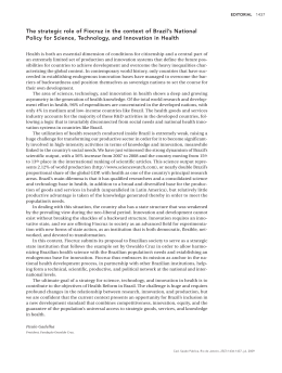
International Journal for Parasitology xxx (2012) xxx–xxx
Contents lists available at SciVerse ScienceDirect
International Journal for Parasitology
journal homepage: www.elsevier.com/locate/ijpara
Current Opinion
On the pathogenesis of Plasmodium vivax malaria: Perspectives from
the Brazilian field
Fabio T.M. Costa a,⇑,1, Stefanie C.P. Lopes a,1, Letusa Albrecht a,1, Ricardo Ataíde b,1, André M. Siqueira c,d,1,
Rodrigo M. Souza b,e,1, Bruce Russell f,1, Laurent Renia f,1, Claudio R.F. Marinho b,⇑,1,
Marcus V.G. Lacerda c,d,⇑,1
a
Departamento de Genética, Evolução e Bioagentes, Instituto de Biologia, Universidade Estadual de Campinas (UNICAMP), Campinas, SP, Brazil
Departamento de Parasitologia, Universidade de São Paulo (USP), São Paulo, SP, Brazil
c
Fundação de Medicina Tropical Dr. Heitor Vieira Dourado, Gerência de Malária, Manaus, AM, Brazil
d
Universidade do Estado do Amazonas, Manaus, AM, Brazil
e
Centro Multidisciplinar, Universidade Federal do Acre (UFAC), Campus Floresta, Cruzeiro do Sul, AC, Brazil
f
Singapore Immunology Network, Agency for Science, Technology and Research, Singapore, Singapore
b
a r t i c l e
i n f o
Article history:
Received 3 May 2012
Received in revised form 18 August 2012
Accepted 21 August 2012
Available online xxxx
Keywords:
Malaria
Plasmodium vivax
Clinical complications
Cytoadhesion
Pregnancy
Pathogenesis
Amazon
Brazil
a b s t r a c t
Life-threatening Plasmodium vivax malaria cases, while uncommon, have been reported since the early
20th century. Unfortunately, the pathogenesis of these severe vivax malaria cases is still poorly understood. In Brazil, the proportion of vivax malaria cases has been steadily increasing, as have the number
of cases presenting serious clinical complications. The most frequent syndromes associated with severe
vivax malaria in Brazil are severe anaemia and acute respiratory distress. Additionally, P. vivax infection
may also result in complications associated with pregnancy. Here, we review the latest findings on severe
vivax malaria in Brazil. We also discuss how the development of targeted field research infrastructure in
Brazil is providing clinical and ex vivo experimental data that benefits local and international efforts to
understand the pathogenesis of P. vivax.
Ó 2012 Australian Society for Parasitology Inc. Published by Elsevier Ltd. All rights reserved.
1. Introduction
The assumption that vivax malaria is only associated with
‘benign’ (uncomplicated) syndromes has been challenged by a
number of authoritative reviews on the pathogenesis of
Plasmodium vivax (Mendis et al., 2001; Anstey et al., 2009; Mueller
et al., 2009). Certainly, there have been well-documented reports
of severe vivax in Brazil (Alexandre et al., 2010; Siqueira et al.,
2010; Lanca et al., 2012). Unfortunately, the increased awareness
⇑ Corresponding authors. Address: Departamento de Genética, Evolução e
Bioagentes, Instituto de Biologia, Universidade Estadual de Campinas – UNICAMP,
P.O. Box 6109, 13083-970 Campinas, SP, Brazil. Tel.: +55 19 3521 6594; fax: +55 19
3521 6276 (F.T.M. Costa), Departamento de Parasitologia, Instituto de Ciências
Biomédicas – ICB II, Universidade de São Paulo – USP, Av. Prof. Lineu Prestes 1374,
Sala 44/40, São Paulo, SP 05508-900, Brazil. Tel.: +55 11 3091 7209 (C.R.F. Marinho),
Fundação de Medicina Tropical Dr. Heitor Vieira Dourado, Av. Pedro Teixeira 25,
Manaus, Amazonas 69040-000, Brazil. Tel.: +55 92 2127 3430 (M.V.G. Lacerda).
E-mail addresses: [email protected], [email protected] (F.T.M. Costa),
[email protected] (C.R.F. Marinho), [email protected] (M.V.G. Lacerda).
1
All authors equally contributed to this work.
of severe syndromes associated with vivax malaria has not yet
been matched by data to provide solid answers to why such cases
occur. The lack of a robust, reproducible and long-term in vitro
culture method has restricted the study of P. vivax pathobiology
in humans to endemic areas with well-equipped laboratories
(Suwanarusk et al., 2004; Handayani et al., 2009; Carvalho et al.,
2010; Chotivanich et al., 2012). In this opinion article, we discuss
the relevance of an adequate research infrastructure for the development of reliable in vivo and ex vivo functional assays in Brazilian
endemic areas where infected patients can easily access hospitals
with adequate facilities and personnel, such as the cities of Manaus
(Amazonas State), Cruzeiro do Sul (Acre State) and Porto Velho
(Rondônia State). In addition, recent observations regarding the
pathogenesis of P. vivax disease are discussed and key questions
are addressed.
2. Epidemiology of vivax malaria in Amazonian Brazil
In Brazil, malaria transmission is largely confined to the Amazon region (99.8% of reported cases). The National Malaria Control
0020-7519/$36.00 Ó 2012 Australian Society for Parasitology Inc. Published by Elsevier Ltd. All rights reserved.
http://dx.doi.org/10.1016/j.ijpara.2012.08.007
Please cite this article in press as: Costa, F.T.M., et al. On the pathogenesis of Plasmodium vivax malaria: Perspectives from the Brazilian field. Int. J. Parasitol. (2012), http://dx.doi.org/10.1016/j.ijpara.2012.08.007
2
F.T.M. Costa et al. / International Journal for Parasitology xxx (2012) xxx–xxx
Program dictates all major control policies, which are followed by
all states and municipalities. Due to the dramatic enhancement of
the control measures, the number of malaria cases reported in
Brazil has dropped almost 2-fold, from 635,646 cases in 1999, to
less than 310,000 cases in 2009, when 85% of the reported cases
were vivax malaria (DATASUS-SIVEP-Malária (http://portalweb04.saude.gov.br/sivep_malaria/default.asp). Despite these major advances achieved during the last decade, Brazil still reports
50–60% of the malaria cases in the Americas (WHO, 2011).
Although Brazil’s population has become more urbanised, many
inhabitants of the Amazon remain in riverine communities or recently colonised rural settlement areas; both in direct contact with
rainforest and associated malaria risk. Thus, malaria remains an
important public health issue for Brazil. Aside from the direct costs
associated with uncomplicated and severe vivax malaria, this disease is also responsible for a more insidious socioeconomic burden.
One specific example is the diminished school performance of
Amazonian children associated with P. vivax infection (Vitor-Silva
et al., 2009). The combination of rapidly occurring economic
changes in the Amazon and the diversity of sociological and environmental settings present a complex interaction of several risk
factors imposing relevant barriers for malaria control.
Although peaks in Amazonian malaria transmission are correlated with seasonal rainfall patterns, enough basal malaria occurs
to ensure a steady enrolment of patients in clinical trials year
round. Most cases occur in the outskirts of major cities, where migrants with low or no immunity to malaria live in crowded places
with little effective sanitation; such conditions promote outbreaks
(Ferreira Goncalves and Alecrim, 2004; Gil et al., 2007). In contrast,
people living in riverine communities are commonly exposed to
malaria infection from a very young age and they tend to develop
a sustained level of clinical ‘immunity’ by the age of 21–26 years
(Alves et al., 2002).
The epidemiology of malaria in recently colonised rural agroindustrial settlements has a pattern with features of open-cast
mines and city outskirts (Camargo et al., 1994). The number of malaria attacks in the Amazon is consistently decreasing, with a
marked reduction in the proportion of Plasmodium falciparum cases
(Oliveira-Ferreira et al., 2010), making Brazil one of the few endemic areas in which P. vivax malaria is highly predominant. Certainly, in Manaus, P. falciparum malaria was reported in only 0.8%
of the cases in 2011. Universal access to diagnostic health facilities
throughout the Amazon region, resulting in 60% of patients being
diagnosed by optical microscopy and treated within 48 h of presentation of symptoms, and the introduction of artemether/lumefantrine as the first-line of treatment for P. falciparum, have
contributed to the decrease in malaria cases since 2007 (OliveiraFerreira et al., 2010). This unique setting allows P. vivax immunity
to be evaluated in isolation without a significant confounding
influence from sequential or mixed P. falciparum infections. The
characteristics that make P. falciparum relatively easy to control
(e.g., late gametocyte production and lack of hypnozoites) may result in P. vivax becoming the dominant malaria species in many endemic areas. The emergence of chloroquine-resistant P. vivax in
Brazil and the lack of primaquine effectiveness (which are still
the first-line drugs for vivax infection in Brazil) is a cause for
considerable concern and will strengthen the dominance of this
malaria species in the Amazon (de Santana Filho et al., 2007).
3. The development of tropical health research facilities in the
Amazon
In the Amazon region, every patient with a febrile illness has
free access to malaria diagnosis and the appropriate antimalarial
treatment, which by law are not sold in general pharmacies.
Additionally, there is a robust databank system available on-line
(SIVEP-Malária (http://portalweb04.saude.gov.br/sivep_malaria/
default.asp). Brazil has a socialised health system (Sistema Único
de Saúde – SUS), providing well-equipped tertiary care centres
throughout several urban centres in the Amazon (Fig. 1). Centres
such as the Tropical Medicine Foundation Dr. Heitor Vieira Dourado Hospital (FMT-HVD) in Manaus and the Cruzeiro do Sul Maternity Ward (CSMW) in Cruzeiro do Sul, exemplify the type of
hospitals in which research on P. vivax malaria clinical severity,
drug resistance and pathogenesis can be effectively accomplished.
FMT-HVD has 150 hospital beds, many of which are available
for clinical trials, treats 30% of the malarial cases reported in Manaus and serves as the referral hospital for complicated cases. The
CSMW admitted more than 2,000 pregnant women in 2009, with
an estimated prevalence of plasmodial infections of 8.7% and with
a predominance of P. vivax (75.2%) (Vale, S.d.C.N., 2011. Malária em
gestantes no município de Cruzeiro do Sul pertencente à região
amazônica brasileira. Ph.D. Thesis, Universidade de São Paulo, Brazil). Historically, Cruzeiro do Sul has been the municipality within
Acre state with the highest number of malaria cases (87% in 2009).
In 2011, a hospital dedicated to women and newborn health (the
Women’s and Children’s Hospital of Juruá) was inaugurated and
coupled to the Gestacional Clinical Malaria Research Centre and
the Infectious Diseases Research Laboratory. These well-equipped
laboratories in Manaus and Cruzeiro do Sul enable studies on P. vivax immunopathogenic mechanisms by facilitating the collection
of P. vivax-infected erythrocytes (Pv-iE) from infected patients,
with or without complications, from pregnant women’s peripheral
blood and from P. vivax-infected placentas. Collection of human
tissues and parasites allows the performance of functional (e.g.,
cytoadhesion, short-term culture and invasion) and molecular
(e.g., genetic sequencing and gene expression) assays. Indeed, we
believe that long-term partnerships with local, national and global
research institutes will strengthen personnel capacity in situ,
improve training of graduate and undergraduate students and
enhance research quality.
4. Plasmodium vivax-attributable clinical complications and
related pathogenesis
The incidence of clinical complications in vivax malaria has
been attributed to a variety of factors, such as: transmission intensity, the presence of other endemic or non-communicable disease
host characteristics (gender, age and genetic background) and drug
resistance. Anaemia and respiratory distress are the most common
complications reported from vivax endemic areas (Genton et al.,
2008; Tjitra et al., 2008; Kochar et al., 2009; Lacerda et al., 2012).
Unusual complications of P. vivax infection, such as rhabdomyolysis (Siqueira et al., 2010), idiopathic thrombocytopenic purpura
(Lacerda et al., 2004), splenic rupture (Lubitz, 1949; Lacerda
et al., 2007; Gupta et al., 2010) and cerebral malaria (Tanwar
et al., 2011) have also been reported in Brazil and southeast Asia.
There are also a number of complications related to the haemolytic
effect of primaquine in those with glucose-6-phosphate dehydrogenase (G6PD)-deficiency (Ramos Junior et al., 2010), a mutation
with a prevalence of approximately 3% amongst males living in endemic areas of the Amazon region (Santana et al., 2009). The diversity of clinical presentations can also be attributed to each host’s
unique immune response (Mourao et al., 2012), which may be directly linked to the transmission dynamics in a given area. For
example, the recent decrease in P. falciparum cases in Brazil has
been accompanied by an increase in P. vivax-related hospitalisations (Santos-Ciminera et al., 2007). Between 1998 and 2008, 234
vivax-associated malaria deaths were reported in Brazil
(Oliveira-Ferreira et al., 2010). Within endemic areas of the
Please cite this article in press as: Costa, F.T.M., et al. On the pathogenesis of Plasmodium vivax malaria: Perspectives from the Brazilian field. Int. J. Parasitol. (2012), http://dx.doi.org/10.1016/j.ijpara.2012.08.007
F.T.M. Costa et al. / International Journal for Parasitology xxx (2012) xxx–xxx
3
Manaus
Amazonas State
Population: 1,802,525
Annual Parasitic Index: 9,3 (2011)
Plasmodium vivax: 98,7%
Porto Velho
Rondônia State
Population: 435,732
Annual Parasitic Index: 34,4 (2011)
Plasmodium vivax: 95,1%
Cruzeiro do Sul
Acre State
Population: 78,444
Annual Parasitic Index: 162,3 (2011)
Plasmodium vivax: 85,1%
Fig. 1. The transmission of malaria in Brazil occurs almost exclusively in the Amazon region (99.8%). Urban centres such as Manaus, Cruzeiro do Sul and Porto Velho recorded
approximately 20% of the national malaria cases in 2011, and in these three municipalities, tertiary care hospitals with the proper infrastructure for clinical research are
available.
Brazilian Amazon, rural settings have low to moderate vivax transmission rates (da Silva-Nunes et al., 2008; Silva et al., 2010), a stark
contrast with the vibrant urban centres of Manaus, Cruzeiro do Sul
and Porto Velho, which represent almost 20% of the Brazilian malaria cases (Gil et al., 2007; Saraiva et al., 2009; Costa et al., 2010).
Even though urban settings have the best clinical infrastructure for
the study of severe vivax malaria, long-term cohort studies in
Brazilian urban settings are difficult due to a high level of human
population transience.
Despite these challenges, we have recently demonstrated that
the World Health Organization’s severity criteria for P. falciparum
reliably identified P. vivax-infected patients at risk for severe
disease admitted to the intensive care unit (Lanca et al., 2012). In
vivax endemic areas, the prevalence of both acute and chronic
co-morbidities represent another relevant issue (Lampah et al.,
2011; Lacerda et al., 2012). To search for other infectious agents
causing acute febrile syndromes, an experienced and well-funded
health system is needed. Furthermore, study of vivax-associated
immunopathological mechanisms is hindered by a lack of
post-mortem research, which is restricted by cultural and religious
beliefs in many parts of southeast Asia (Anstey et al., 2009; Mueller
et al., 2009), with a few exceptions such as the Melanesian populations (Manning et al., 2012).
The most frequent complication of malaria in Brazil is anaemia,
which causes increased morbidity and mortality in children and
pregnant women (Haldar and Mohandas, 2009; Alexandre et al.,
2010; Lanca et al., 2012). During vivax infections, erythrocytes
are destroyed early on, but anaemia can persist even after parasite
clearance. This observation can be explained primarily by the fact
that P. vivax infects reticulocytes, and the infection prevents the
reestablishment of the normal erythrocyte population (Collins
et al., 2003). The growing incidence of severe vivax malaria complications (especially anaemia), or possibly the growing awareness
and reporting from experts in the field, may be related to increased
chloroquine resistance (Tjitra et al., 2008; Fernandez-Becerra et al.,
2009), a situation also observed in Brazil (de Santana Filho et al.,
2007), resulting in the longer persistence of the parasite in circulation and in the bone marrow.
Respiratory distress is another common complication in vivax
malaria reported worldwide (Anstey et al., 2007; Price et al.,
2007; Tan et al., 2008), including Brazil (Lacerda et al., 2012). In
the largest series of autopsies performed on patients with P. vivax
infection in Manaus, acute lung oedema was the major finding in
patients with respiratory distress, frequently associated with neutrophil accumulation in the interalveolar space (Lacerda et al.,
2012) (Fig. 2). Although rarely observed in Brazil, patients presenting with neurological symptoms (e.g., seizure and/or coma) have
been reported (Andrade and Barral-Netto, 2011; Lacerda et al.,
2012).
The immunopathogenesis of P. vivax is poorly understood.
However, it was recently demonstrated that Brazilian patients with
severe vivax malaria, who presented with respiratory failure and
anaemia, had elevated levels of the inflammatory cytokines TNFa and IFN-c; these patients also had an increased IFN-c/IL-10 ratio
and the antioxidant agent superoxide dismutase-1 (SOD-1) and
soluble CD163 were also elevated (Andrade et al., 2010; Andrade
and Barral-Netto, 2011; Mendonca et al., 2012). Levels of TNF-a
and other inflammatory cytokines are higher during P. vivax
Fig. 2. Post-mortem lung microscopy from a patient with respiratory distress.
Acute oedema was the major finding (H&E staining; 1000 magnification). Scale
bar = 20 lm.
Please cite this article in press as: Costa, F.T.M., et al. On the pathogenesis of Plasmodium vivax malaria: Perspectives from the Brazilian field. Int. J. Parasitol. (2012), http://dx.doi.org/10.1016/j.ijpara.2012.08.007
4
F.T.M. Costa et al. / International Journal for Parasitology xxx (2012) xxx–xxx
infections compared with P. falciparum infections with the same
parasite load (Karunaweera et al., 1992; Yeo et al., 2010). Elevated
plasma concentrations of microparticles were found in individuals
with uncomplicated P. vivax infection in a study conducted in the
Brazilian Amazon (Campos et al., 2010). Moreover, despite strong
evidence that P. vivax triggers thrombocytopenia at higher levels
than P. falciparum (Kochar et al., 2010), any role for these particles
in severe P. vivax malaria is unknown.
The paradigm of P. vivax red blood cell (RBC) invasion only in
Duffy-positive individuals has been recently challenged, as it was
shown that Duffy negative individuals can also be infected by P. vivax in Brazil (Cavasini et al., 2007). This finding paves the way, not
only for the search of new vaccine candidates, but also for the better understanding of severe disease mechanisms. Regarding host
genetics in Brazil, individuals with the Duffy negative Fy (a+b )
phenotype demonstrated a 30–80% reduced risk of clinical vivax,
but not falciparum malaria, in a prospective cohort study in the
Brazilian Amazon (King et al., 2011). G6PD deficiency, essentially
the African type, seemed to protect against malaria based on the
past history of enrolled patients in Manaus (Santana et al., 2009).
5. Cytoadhesion and rosetting phenomena in P. vivax malaria
Because severe falciparum-like manifestations are increasingly
present during vivax malaria, it is possible that the two species
share some immunopathological processes. Normally, manifestations of severe P. falciparum infections (e.g., cerebral malaria, malaria in pregnancy (MiP) and respiratory distress) are associated
with cytoadhesion, in which the mature-stage of P. falciparuminfected erythrocytes (Pf-iE) attach to the host’s cell-surface
receptors.
Pf-iE cytoadhesion to the endothelium prevents the circulation
of mature parasites, which inhibits parasite clearance by the spleen
(Wyler, 1983) and accordingly, mature stage parasites are often
not observed in the peripheral circulation (Berendt et al., 1990).
Because all stage-forms of P. vivax are found in the peripheral
blood, it has been inferred that this parasite does not sequester
and thus does not cytoadhere. Nevertheless, sequestration of P. vivax was proposed more than 50 years ago when a disproportionately low number of mature stage-form parasites (schizonts)
were observed in the peripheral circulation (Leeson, 1957; Field
et al., 1963). However whether sequestration in P. vivax leads to
a disproportional organ-specific and peripheral blood parasitaemia
remains to be evaluated. We have recently shown that Pv-iE from
non-severe patients cytoadhered ex vivo to placental cryosections
as well as to human lung (HLEC) and Saimiri brain (SBEC) endothelial cell lines under static and flow conditions (Carvalho et al.,
2010). While the number of adhered Pv-iE per mm2 under static
conditions was 10–15 times lower than that for Pf-iE, the adhesion
strength under physiological (i.e., flow) conditions was similar in
both parasites. Moreover, in transfected Chinese hamster ovary
(CHO) cells, Pv-iE binding to CHO-ICAM-1 was twice as high as that
of mock- or CD36-transfected cells, indicating that ICAM-1 is a potential receptor for P. vivax. Pv-iE were also able to bind to placental cryosections. Adhesion to HLEC was inhibited by soluble
chondroitin sulfate A (CSA), but treatment with chondroitinase
ABC did not affect binding (Carvalho et al., 2010). The ability of
CSA binding has been recently confirmed using Pv-iE collected
from 33 patients from Asia-Pacific region (Chotivanich et al.,
2012). In this study, all tested isolates adhered to immobilised
CSA and hyaluronic acid (HA), and pre-incubation with chondroitinase and hyaluronidase reversed binding (Chotivanich et al.,
2012).
A family of variant subtelomeric P. vivax genes named vir was
described in the early 2000s (del Portillo et al., 2001). Unlike the
var genes of P. falciparum, which encode PfEMP-1 proteins, few
vir genes (160 of 346) have motifs similar to the Plasmodium export
element/vacuolar transport signal (PEXEL/VTS) proteins. PEXEL/
VTS-family proteins mediate protein export to the erythrocyte surface and the cytosol (Marti et al., 2004). In contrast to PfEMP-1,
VIRs are not clonally expressed (Fernandez-Becerra et al., 2005)
and can be found inside reticulocytes, implying that they have different sub-cellular localisations and functions (Fernandez-Becerra
et al., 2005). Based on their variant nature and their presence on
the erythrocyte membrane, VIR proteins were evaluated for their
role in Pv-iE cytoadhesion to endothelial cells. Polyclonal antibodies against recombinant VIR proteins from two subfamilies (VIRE4
and VIRA5) inhibited Pv-iE adhesion ex vivo to HLEC (Carvalho
et al., 2010). Due to the lack of an in vitro P. vivax culture system,
Bernabeu and colleagues elegantly assessed the role of VIR proteins
in P. vivax cytoadhesion via a transfection assay; P. falciparum 3D7
was transfected with recombinant VIR proteins (VIR17, VIR14 and
VIR10) (Bernabeu et al., 2012). Both VIR14 and VIR10 were exported
to the cell membrane, but only VIR14 mediated binding to ICAM-1
(Bernabeu et al., 2012). This result corroborated our previous data
(Carvalho et al., 2010).
Although P. vivax cytoadheres under relatively low shear
stresses, whether this is part of a strategy to avoid spleen clearance, as postulated for P. falciparum, is still not determined. If this
is indeed the case, it has been proposed that VIR antigens
promote Pv-iE adhesion to barrier cells, which are located in protected areas of the spleen, to allow parasite survival and the
establishment of a chronic infection (del Portillo et al., 2004).
However, such a strategy could be considered in the light that
biomechanical modifications of Pv-iE might allow the evasion
of splenic clearance. Certainly, Pv-iE are significantly more
deformable than P. falciparum-infected erythrocytes (Suwanarusk
et al., 2004), and this deformability could allow a certain number
of Pv-iE to bypass the splenic sinusoids, thus avoiding pitting
(Handayani et al., 2009).
While Pv-iE adhesion to cell receptors has been demonstrated,
the role of cytoadhesion in P. vivax pathogenesis and severe disease
is unknown. A challenge for the study of severe infection is the
scarcity of autopsy studies. Early 20th century autopsies of patients
with P. vivax infection recorded intra-capillary masses of swollen,
infected erythrocytes, malarial pigment and the existence of mature parasites within a red blood cell taking up the entire lumen
and in contact with endothelial cells in some brain vessels (reviewed by Anstey et al., 2009). However, as the diagnoses were
made exclusively with microscopic evidence, these observations
must be analysed carefully because these reports do not exclude
the possibility of mixed infections or co-morbidities.
One single autopsy performed due to legal reasons in a woman
with severe vivax disease, in India, showed alveolar capillaries congested by monocyte infiltrates and diffuse damage to alveolar
membranes compatible with ALI/ARDS (Acute Lung Injury/Acute
Respiratory Distress Syndrome); in this case, P. falciparum infection
was excluded by PCR analysis (Valecha et al., 2009). More recently,
17 patients with microscopical diagnosis of vivax malaria were
fully autopsied in Manaus. Infection with a single parasite species
was confirmed in 13 cases by PCR performed in paraffin-fixed tissues. In 13 patients, the parasitic infection was related to the cause
of death. In one patient who tested negative for peripheral parasitaemia many days after antimalarial treatment, parasites were
still detected by confocal microscopy in the lung tissue. These results suggested that the parasite was sequestered in the lungs
(Lacerda et al., 2012). Indeed, it has been suggested that the pulmonary vascular sequestration of Pv-iE could be the pathophysiological mechanism that leads to this potentially fatal complication
(Anstey et al., 2007), although ex vivo Pv-iE binding assays were
not performed in that particular work.
Please cite this article in press as: Costa, F.T.M., et al. On the pathogenesis of Plasmodium vivax malaria: Perspectives from the Brazilian field. Int. J. Parasitol. (2012), http://dx.doi.org/10.1016/j.ijpara.2012.08.007
F.T.M. Costa et al. / International Journal for Parasitology xxx (2012) xxx–xxx
5
Fig. 3. Infected erythrocytes and inflammatory cells are present in the placental intervillous spaces of a Plasmodium vivax-infected pregnant woman. (A) H&E staining; scale
bar = 20 lm. (B) Giemsa staining; scale bar = 20 lm. Mononuclear cells (arrows), hemozoin within mononuclear cells (arrow heads), terminal villi (stars).
The other adhesive phenotype observed in malaria parasites is
rosette formation. Many studies on rosetting have focused on
P. falciparum (Mercereau-Puijalon et al., 2008), but little is known
about rosetting in P. vivax or other Plasmodium spp. Almost
20 years have passed since the first report of rosetting in P. vivax
(Udomsanpetch et al., 1995), and to date there are only four studies
from southeast Asia that have reported rosette formation by
P. vivax (Udomsanpetch et al., 1995; Chotivanich et al., 1998,
2012; Russell et al., 2011). In contrast to P. falciparum, P. vivax
rosetting has not been associated with disease severity, parasitaemia or the ABO blood group type (Udomsanpetch et al., 1995;
Chotivanich et al., 1998; Russell et al., 2011). Russell and
colleagues have shown that enrichment of some P. vivax isolates
requires trypsin treatment to disrupt rosettes, suggesting that
trypsin-sensitive proteins mediate rosetting in Pv-iEs (Russell
et al., 2011). In the four studies that describe rosetting in P. vivax,
almost all of the isolates analysed formed rosettes (Udomsanpetch
et al., 1995; Chotivanich et al., 1998, 2012; Russell et al., 2011). To
date, there is no information about rosette formation or about its
biological meaning in Pv-iEs harvested from patients outside of
the Asia-Pacific region.
6. Malaria in pregnancy (MiP): a particular syndrome
The majority of the studies on P. vivax infection during pregnancy have been conducted in the Asia-Pacific region (Nosten
et al., 1991; McGready et al., 2004, 2011; Rijken et al., 2012). There
are very few studies from other regions that look specifically at the
placental pathology of P. vivax infections and this lack of information urges further studies (Anstey et al., 2009). A single study has
systematically evaluated the histopathology of P. vivax in the placenta. In this study, the intervillous spaces of the placenta contained accumulations of parasitised erythrocytes and malarial
pigment deposits, but there were no other significant tissue
changes (McGready et al., 2011). In Latin America, some studies
were based on placental biopsies from P. vivax-infected women,
but these were used only as a marker of infection and failed to systematically evaluate the parasite-associated lesions (Parekh et al.,
2010; Campos et al., 2011). In Brazil, MiP studies have focused
on the epidemiology and on the consequences of malarial infections for both the mother and the newborn (Martinez-Espinosa
et al., 2004; Chagas et al., 2009; Carvalho et al., 2011). In those
studies, low birth weight, abortion and premature delivery were
reported and co-morbidities did not seem to be relevant (reviewed
in Lacerda et al. (2012)).
Preliminary results from a prospective study, in Cruzeiro do Sul,
show that women with P. vivax infections during pregnancy can
evolve with parasites and immune cells in the placenta (Fig. 3)
besides showing other signs of placental pathology such as
increased syncytial knotting, also associated with P. falciparum
(Souza, RM, unpublished observations). These results complement
those reported elsewhere (McGready et al., 2004), and highlight
the importance of studying P. vivax-associated pathology in diverse
endemic scenarios where the heterogeneous occurrence of comorbidities in the placental tissue (e.g., syphilis, toxoplasmosis,
cytomegalovirus) may occur.
7. More questions than answers
Little is known about P. vivax pathogenesis in severe malaria
cases. Plasmodium vivax is very difficult to grow in vitro, despite recent advances (Udomsangpetch et al., 2007; Russell et al., 2011),
and there are no highly specific and reliable biomarkers for infection
severity. These impediments have prevented researchers from
addressing crucial issues. To better understand vivax severity, the
following key questions are: (i) are co-morbidities such as bacterial
infections or dengue fever essential to P. vivax severity? (ii) As PviE cytoadhesion is 10- to 15-fold less than that of Pf-iE in vivo, do
non-parasitic factors (e.g., platelets, microparticles and complement
molecules) enhance adhesion in vivo, and are co-morbidities responsible for the production of those factors? (iii) Since VIR proteins are
not clonally expressed, are there other host receptors besides
ICAM-1 and CSA that play a role in Pv-iE binding for the same parasite
stage form? (iv) Does rosette formation play a role in P. vivax severity
and does this phenomenon enhance parasite RBC infection? (v) What
is the real impact of vivax infection on pregnant women and foetal
health, and which host or parasite factors are involved?
8. Concluding remarks
To better understand P. vivax infections and their clinical complications in Brazil, future studies should focus on the immunopathological mechanisms related to P. vivax severity. The role of
Pv-iE cytoadhesion in severe vivax malaria, as well as the major
determinants of adhesion, should be better characterised. Autopsy
reports of P. vivax-related fatalities with good clinical characterisations should include prospective histopathological descriptions of
infected organs, as these details are urgently needed. Plasmodium
vivax infections should be thoroughly studied in the general population and in specific severe cases. FMT-HVD (Manaus, Amazonas
State) and the CSMW (Cruzeiro do Sul, Acre State) have laboratory
facilities and well-equipped hospitals, which amongst others in the
Amazon, are at the forefront of P. vivax clinical and epidemiological
research in Brazil.
Please cite this article in press as: Costa, F.T.M., et al. On the pathogenesis of Plasmodium vivax malaria: Perspectives from the Brazilian field. Int. J. Parasitol. (2012), http://dx.doi.org/10.1016/j.ijpara.2012.08.007
6
F.T.M. Costa et al. / International Journal for Parasitology xxx (2012) xxx–xxx
Acknowledgements
This work received financial support from Fundação de Amparo
a Pesquisa do Estado de São Paulo (FAPESP, Brazil), Conselho Nacional de Desenvolvimento Científico e Tecnológico (CNPq, Brazil),
Instituto Nacional de Tecnologia em Vacinas (CNPq-FAPEMIG, Brazil) and Instituto Nacional de Tecnologia em Doenças Negligenciadas (CNPq, Brazil). S.C.P.L., L.A. and R.A. were sponsored by FAPESP
fellowships. R.M.S was supported by a CNPq fellowship. MVGL and
FTMC are CNPq fellows. FTMC is enrolled at the Programa Estratégico de Ciência, Tecnologia & Inovação nas Fundações Estaduais de
Saúde (PECTI/AM Saúde) from Fundação de Amparo à Pesquisa
do Estado do Amazonas (FAPEAM, Brazil). L.R. and B.R. were supported by core grants to the Singapore Immunology Network and
the Horizontal programme on Infectious Diseases, Agency for Science, Technology and Research (A⁄STAR), Singapore. The funders
had no role in study design, data collection and analysis, decision
to publish or preparation of the manuscript. Special thanks to Dr.
Tony Hiroshi Katsuragawa (Instituto de Pesquisas em Patologias
Tropicais – Porto Velho, Rondônia, Brazil) for the data on the annual parasitic index and the proportion P. vivax cases in Porto
Velho.
References
Alexandre, M.A., Ferreira, C.O., Siqueira, A.M., Magalhaes, B.L., Mourao, M.P.,
Lacerda, M.V., Alecrim, M.G., 2010. Severe Plasmodium vivax malaria, Brazilian
Amazon. Emerg. Infect. Dis. 16, 1611–1614.
Alves, F.P., Durlacher, R.R., Menezes, M.J., Krieger, H., Silva, L.H., Camargo, E.P., 2002.
High prevalence of asymptomatic Plasmodium vivax and Plasmodium falciparum
infections in native Amazonian populations. Am. J. Trop. Med. Hyg. 66, 641–
648.
Andrade, B.B., Reis-Filho, A., Souza-Neto, S.M., Clarencio, J., Camargo, L.M., Barral, A.,
Barral-Netto, M., 2010. Severe Plasmodium vivax malaria exhibits marked
inflammatory imbalance. Malar. J. 9, 13.
Andrade, B.B., Barral-Netto, M., 2011. Biomarkers for susceptibility to infection and
disease severity in human malaria. Mem. Inst. Oswaldo Cruz 106 (Suppl. 1), 70–
78.
Anstey, N.M., Handojo, T., Pain, M.C., Kenangalem, E., Tjitra, E., Price, R.N., Maguire,
G.P., 2007. Lung injury in vivax malaria: pathophysiological evidence for
pulmonary vascular sequestration and posttreatment alveolar-capillary
inflammation. J. Infect. Dis. 195, 589–596.
Anstey, N.M., Russell, B., Yeo, T.W., Price, R.N., 2009. The pathophysiology of vivax
malaria. Trends Parasitol. 25, 220–227.
Berendt, A.R., Ferguson, D.J., Newbold, C.I., 1990. Sequestration in Plasmodium
falciparum malaria: sticky cells and sticky problems. Parasitol. Today 6, 247–
254.
Bernabeu, M., Lopez, F.J., Ferrer, M., Martin-Jaular, L., Razaname, A., Corradin, G.,
Maier, A.G., Del Portillo, H.A., Fernandez-Becerra, C., 2012. Functional analysis of
Plasmodium vivax VIR proteins reveals different subcellular localizations and
cytoadherence to the ICAM-1 endothelial receptor. Cell Microbiol. 14, 386–400.
Camargo, L.M., Ferreira, M.U., Krieger, H., De Camargo, E.P., Da Silva, L.P., 1994.
Unstable hypoendemic malaria in Rondonia (western Amazon region, Brazil):
epidemic outbreaks and work-associated incidence in an agro-industrial rural
settlement. Am. J. Trop. Med. Hyg. 51, 16–25.
Campos, F.M., Franklin, B.S., Teixeira-Carvalho, A., Filho, A.L., de Paula, S.C., Fontes,
C.J., Brito, C.F., Carvalho, L.H., 2010. Augmented plasma microparticles during
acute Plasmodium vivax infection. Malar. J. 9, 327.
Campos, I.M., Uribe, M.L., Cuesta, C., Franco-Gallego, A., Carmona-Fonseca, J.,
Maestre, A., 2011. Diagnosis of gestational, congenital, and placental malaria in
Colombia: comparison of the efficacy of microscopy, nested polymerase chain
reaction, and histopathology. Am. J. Trop. Med. Hyg. 84, 929–935.
Carvalho, B.O., Lopes, S.C., Nogueira, P.A., Orlandi, P.P., Bargieri, D.Y., Blanco, Y.C.,
Mamoni, R., Leite, J.A., Rodrigues, M.M., Soares, I.S., Oliveira, T.R., Wunderlich,
G., Lacerda, M.V., del Portillo, H.A., Araujo, M.O., Russell, B., Suwanarusk, R.,
Snounou, G., Renia, L., Costa, F.T., 2010. On the cytoadhesion of Plasmodium
vivax-infected erythrocytes. J. Infect. Dis. 202, 638–647.
Carvalho, B.O., Matsuda, J.S., Luz, S.L., Martinez-Espinosa, F.E., Leite, J.A., Franzin, F.,
Orlandi, P.P., Gregoracci, G.B., Lacerda, M.V., Nogueira, P.A., Costa, F.T., 2011.
Gestational malaria associated to Plasmodium vivax and Plasmodium falciparum
placental mixed-infection followed by foetal loss: a case report from an
unstable transmission area in Brazil. Malar. J. 10, 178.
Cavasini, C.E., Mattos, L.C., Couto, A.A., Bonini-Domingos, C.R., Valencia, S.H., Neiras,
W.C., Alves, R.T., Rossit, A.R., Castilho, L., Machado, R.L., 2007. Plasmodium vivax
infection among Duffy antigen-negative individuals from the Brazilian Amazon
region: an exception? Trans. R. Soc. Trop. Med. Hyg. 101, 1042–1044.
Chagas, E.C., do Nascimento, C.T., de Santana Filho, F.S., Botto-Menezes, C.H.,
Martinez-Espinosa, F.E., 2009. Impact of malaria during pregnancy in the
Amazon region. Rev. Panam. Salud Publica 26, 203–208.
Chotivanich, K.T., Pukrittayakamee, S., Simpson, J.A., White, N.J., Udomsangpetch, R.,
1998. Characteristics of Plasmodium vivax-infected erythrocyte rosettes. Am. J.
Trop. Med. Hyg. 59, 73–76.
Chotivanich, K., Udomsangpetch, R., Suwanarusk, R., Pukrittayakamee, S.,
Wilairatana, P., Beeson, J.G., Day, N.P., White, N.J., 2012. Plasmodium vivax
adherence to placental glycosaminoglycans. PLoS One 7, e34509.
Collins, W.E., Jeffery, G.M., Roberts, J.M., 2003. A retrospective examination of
anemia during infection of humans with Plasmodium vivax. Am. J. Trop. Med.
Hyg. 68, 410–412.
Costa, K.M., Almeida, W.A., Magalhaes, I.B., Montoya, R., Moura, M.S., Lacerda, M.V.,
2010. Malaria in Cruzeiro do Sul (Western Brazilian Amazon): analysis of the
historical series from 1998 to 2008. Rev. Panam. Salud Publica 28, 353–360.
da Silva-Nunes, M., Codeco, C.T., Malafronte, R.S., da Silva, N.S., Juncansen, C., Muniz,
P.T., Ferreira, M.U., 2008. Malaria on the Amazonian frontier: transmission
dynamics, risk factors, spatial distribution, and prospects for control. Am. J.
Trop. Med. Hyg. 79, 624–635.
de Santana Filho, F.S., Arcanjo, A.R., Chehuan, Y.M., Costa, M.R., Martinez-Espinosa,
F.E., Vieira, J.L., Barbosa, M.G., Alecrim, W.D., Alecrim, M.G., 2007. Chloroquineresistant Plasmodium vivax, Brazilian Amazon. Emerg. Infect. Dis. 13, 1125–
1126.
del Portillo, H.A., Fernandez-Becerra, C., Bowman, S., Oliver, K., Preuss, M., Sanchez,
C.P., Schneider, N.K., Villalobos, J.M., Rajandream, M.A., Harris, D., Pereira da
Silva, L.H., Barrell, B., Lanzer, M., 2001. A superfamily of variant genes encoded
in the subtelomeric region of Plasmodium vivax. Nature 410, 839–842.
del Portillo, H.A., Lanzer, M., Rodriguez-Malaga, S., Zavala, F., Fernandez-Becerra, C.,
2004. Variant genes and the spleen in Plasmodium vivax malaria. Int. J. Parasitol.
34, 1547–1554.
Fernandez-Becerra, C., Pein, O., de Oliveira, T.R., Yamamoto, M.M., Cassola, A.C.,
Rocha, C., Soares, I.S., de Braganca Pereira, C.A., del Portillo, H.A., 2005. Variant
proteins of Plasmodium vivax are not clonally expressed in natural infections.
Mol. Microbiol. 58, 648–658.
Fernandez-Becerra, C., Pinazo, M.J., Gonzalez, A., Alonso, P.L., del Portillo, H.A.,
Gascon, J., 2009. Increased expression levels of the pvcrt-o and pvmdr1 genes in
a patient with severe Plasmodium vivax malaria. Malar. J. 8, 55.
Ferreira Goncalves, M.J., Alecrim, W.D., 2004. Non-planed urbanization as a
contributing factor for malaria incidence in Manaus-Amazonas. Braz. Rev.
Salud Publica (Bogota) 6, 156–166.
Field, J.W., Sandosham, A.A., Fong, Y.L., 1963. A Morphological Study of the
Erythrocitic Parasites in Thick Blood Films, the Microscopic Diagnosis Of Human
Malaria. The Government Press, Kuala Lumpur, pp. 20–117.
Genton, B., D’Acremont, V., Rare, L., Baea, K., Reeder, J.C., Alpers, M.P., Muller, I.,
2008. Plasmodium vivax and mixed infections are associated with severe
malaria in children: a prospective cohort study from Papua New Guinea. PLoS
Med. 5, e127.
Gil, L.H., Tada, M.S., Katsuragawa, T.H., Ribolla, P.E., da Silva, L.H., 2007. Urban and
suburban malaria in Rondonia (Brazilian Western Amazon) II. Perennial
transmissions with high anopheline densities are associated with human
environmental changes. Mem. Inst. Oswaldo Cruz 102, 271–276.
Gupta, B.K., Sharma, K., Nayak, K.C., Agrawal, T.D., Binani, A., Purohit, V.P., Kochar,
D.K., 2010. A case series of splenic infarction during acute malaria in northwest
Rajasthan. India. Trans. R. Soc. Trop. Med. Hyg. 104, 81–83.
Haldar, K., Mohandas, N., 2009. Malaria, Erythrocytic Infection, and Anemia.
Hematology Am. Soc. Hematol. Educ. Program, Washington, pp. 87–93.
Handayani, S., Chiu, D.T., Tjitra, E., Kuo, J.S., Lampah, D., Kenangalem, E., Renia, L.,
Snounou, G., Price, R.N., Anstey, N.M., Russell, B., 2009. High deformability of
Plasmodium vivax-infected red blood cells under microfluidic conditions. J.
Infect. Dis. 199, 445–450.
Karunaweera, N.D., Grau, G.E., Gamage, P., Carter, R., Mendis, K.N., 1992. Dynamics
of fever and serum levels of tumor necrosis factor are closely associated during
clinical paroxysms in Plasmodium vivax malaria. Proc. Natl. Acad. Sci. USA 89,
3200–3203.
King, C.L., Adams, J.H., Xianli, J., Grimberg, B.T., McHenry, A.M., Greenberg, L.J.,
Siddiqui, A., Howes, R.E., da Silva-Nunes, M., Ferreira, M.U., Zimmerman, P.A.,
2011. Fya/Fyb antigen polymorphism in human erythrocyte Duffy antigen
affects susceptibility to Plasmodium vivax malaria. Proc. Natl. Acad. Sci. USA 108,
20113–20118.
Kochar, D.K., Das, A., Kochar, S.K., Saxena, V., Sirohi, P., Garg, S., Kochar, A., Khatri,
M.P., Gupta, V., 2009. Severe Plasmodium vivax malaria: a report on serial cases
from Bikaner in northwestern India. Am. J. Trop. Med. Hyg. 80, 194–198.
Kochar, D.K., Das, A., Kochar, A., Middha, S., Acharya, J., Tanwar, G.S., Gupta, A.,
Pakalapati, D., Garg, S., Saxena, V., Subudhi, A.K., Boopathi, P.A., Sirohi, P.,
Kochar, S.K., 2010. Thrombocytopenia in Plasmodium falciparum, Plasmodium
vivax and mixed infection malaria: a study from Bikaner (Northwestern India).
Platelets 21, 623–627.
Lacerda, M.V., Alexandre, M.A., Santos, P.D., Arcanjo, A.R., Alecrim, W.D., Alecrim,
M.G.C., 2004. Idiopathic thrombocytopenic purpura due to vivax malaria in the
Brazilian Amazon. Acta Trop. 90, 187–190.
Lacerda, M.V.G., Oliveira, S.L., Alecrim, M.G.C., 2007. Splenic hematoma in a patient
with Plasmodium vivax malaria. Rev. Soc. Bras. Med. Trop. 40, 96–97.
Lacerda, M.V., Mourao, M.P., Alexandre, M.A., Siqueira, A.M., Magalhaes, B.M.,
Martinez-Espinosa, F.E., Filho, F.S., Brasil, P., Ventura, A.M., Tada, M.S., Couto,
V.S., Silva, A.R., Silva, R.S., Alecrim, M.G., 2012. Understanding the clinical
Please cite this article in press as: Costa, F.T.M., et al. On the pathogenesis of Plasmodium vivax malaria: Perspectives from the Brazilian field. Int. J. Parasitol. (2012), http://dx.doi.org/10.1016/j.ijpara.2012.08.007
F.T.M. Costa et al. / International Journal for Parasitology xxx (2012) xxx–xxx
spectrum of complicated Plasmodium vivax malaria: a systematic review on the
contributions of the Brazilian literature. Malar. J. 11, 12.
Lacerda, M.V., Fragoso, S.C., Alecrim, M.G., Alexandre, M.A., Magalhaes, B.M.,
Siqueira, A.M., Ferreira, L.C., Araujo, J.R., Mourao, M.P., Ferrer, M., Castillo, P.,
Martin-Jaular, L., Fernandez-Becerra, C., Del Portillo, H., Ordi, J., Alonso, P.L.,
Bassat, Q., 2012. Postmortem characterization of patients with clinical diagnosis
of Plasmodium vivax malaria: to what extent does this parasite kill? Clin. Infect.
Dis. 55, e67–e74.
Lampah, D.A., Yeo, T.W., Hardianto, S.O., Tjitra, E., Kenangalem, E., Sugiarto, P., Price,
R.N., Anstey, N.M., 2011. Coma associated with microscopy-diagnosed
Plasmodium vivax: a prospective study in Papua. Indonesia. PLoS Negl. Trop.
Dis. 5, e1032.
Lanca, E.F., Magalhaes, B.M., Vitor-Silva, S., Siqueira, A.M., Benzecry, S.G., Alexandre,
M.A., O’Brien, C., Bassat, Q., Lacerda, M.V., 2012. Risk factors and
characterization of Plasmodium vivax-associated admissions to pediatric
intensive care units in the Brazilian Amazon. PLoS One 7, e35406.
Leeson, H.S., 1957. Microscopic diagnosis of human malaria. A morphological-study
of erythrocytic parasites. R. Soc. Health J. 2, 694.
Lubitz, J.M., 1949. Pathology of the ruptured spleen in acute vivax malaria. Blood 4,
1168–1176.
Manning, L., Rosanas-Urgell, A., Laman, M., Edoni, H., McLean, C., Mueller, I., Siba, P.,
Davis, T.M., 2012. A histopathologic study of fatal paediatric cerebral malaria caused
by mixed Plasmodium falciparum/Plasmodium vivax infections. Malar. J. 11, 107.
Marti, M., Good, R.T., Rug, M., Knuepfer, E., Cowman, A.F., 2004. Targeting malaria
virulence and remodeling proteins to the host erythrocyte. Science 306, 1930–1933.
Martinez-Espinosa, F.E., Daniel-Ribeiro, C.T., Alecrim, W.D., 2004. Malaria during
pregnancy in a reference centre from the Brazilian Amazon: unexpected
increase in the frequency of Plasmodium falciparum infections. Mem. Inst.
Oswaldo Cruz 99, 19–21.
McGready, R., Davison, B.B., Stepniewska, K., Cho, T., Shee, H., Brockman, A.,
Udomsangpetch, R., Looareesuwan, S., White, N.J., Meshnick, S.R., Nosten, F.,
2004. The effects of Plasmodium falciparum and P. vivax infections on placental
histopathology in an area of low malaria transmission. Am. J. Trop. Med. Hyg.
70, 398–407.
McGready, R., Lee, S., Wiladphaingern, J., Ashley, E., Rijken, M., Boel, M., Simpson, J.,
Paw, M., Pimanpanarak, M., Mu, O., Singhasivanon, P., White, N., Nosten, F.,
2011. Adverse effects of falciparum and vivax malaria and the safety of
antimalarial treatment in early pregnancy: a population-based study. Lancet
Infect. Dis. 12, 388–396.
Mendis, K., Sina, B.J., Marchesini, P., Carter, R., 2001. The neglected burden of
Plasmodium vivax malaria. Am. J. Trop. Med. Hyg. 64, 97–106.
Mendonca, V.R., Luz, N.F., Santos, N.J., Borges, V.M., Goncalves, M.S., Andrade, B.B.,
Barral-Netto, M., 2012. Association between the haptoglobin and heme
oxygenase 1 genetic profiles and soluble CD163 in susceptibility to and
severity of human malaria. Infect. Immun. 80, 1445–1454.
Mercereau-Puijalon, O., Guillotte, M., Vigan-Womas, I., 2008. Rosetting in
Plasmodium falciparum: a cytoadherence phenotype with multiple actors.
Transfus. Clin. Biol. 15, 62–71.
Mourao, L.C., Morais, C.G., Bueno, L.L., Jimenez, M.C., Soares, I.S., Fontes, C.J.,
Guimaraes Lacerda, M.V., Xavier, M.S., Barnwell, J.W., Galinski, M.R., Braga, E.M.,
2012. Naturally acquired antibodies to Plasmodium vivax blood-stage vaccine
candidates (PvMSP-1(19) and PvMSP-3alpha(359–798)) and their relationship
with hematological features in malaria patients from the Brazilian Amazon.
Microbes Infect. 14, 730–739.
Mueller, I., Galinski, M.R., Baird, J.K., Carlton, J.M., Kochar, D.K., Alonso, P.L., del
Portillo, H.A., 2009. Key gaps in the knowledge of Plasmodium vivax, a neglected
human malaria parasite. Lancet Infect. Dis. 9, 555–566.
Nosten, F., ter Kuile, F., Maelankirri, L., Decludt, B., White, N.J., 1991. Malaria during
pregnancy in an area of unstable endemicity. Trans. R. Soc. Trop. Med. Hyg. 85,
424–429.
Oliveira-Ferreira, J., Lacerda, M.V., Brasil, P., Ladislau, J.L., Tauil, P.L., Daniel-Ribeiro,
C.T., 2010. Malaria in Brazil: an overview. Malar. J. 9, 115.
Parekh, F.K., Davison, B.B., Gamboa, D., Hernandez, J., Branch, O.H., 2010. Placental
histopathologic changes associated with subclinical malaria infection and its
impact on the fetal environment. Am. J. Trop. Med. Hyg. 83, 973–980.
Price, R.N., Tjitra, E., Guerra, C.A., Yeung, S., White, N.J., Anstey, N.M., 2007. Vivax
malaria: neglected and not benign. Am. J. Trop. Med. Hyg. 77, 79–87.
7
Ramos Junior, W.M., Sardinha, J.F., Costa, M.R., Santana, M.S., Alecrim, M.G., Lacerda,
M.V., 2010. Clinical aspects of hemolysis in patients with P. vivax malaria
treated with primaquine, in the Brazilian Amazon. Braz. J. Infect. Dis. 14, 410–
412.
Rijken, M.J., Papageorghiou, A.T., Thiptharakun, S., Kiricharoen, S., Dwell, S.L.,
Wiladphaingern, J., Pimanpanarak, M., Kennedy, S.H., Nosten, F., McGready, R.,
2012. Ultrasound evidence of early fetal growth restriction after maternal
malaria infection. PLoS One 7, e31411.
Russell, B., Suwanarusk, R., Borlon, C., Costa, F.T., Chu, C.S., Rijken, M.J., Sriprawat, K.,
Warter, L., Koh, E.G., Malleret, B., Colin, Y., Bertrand, O., Adams, J.H.,
D’Alessandro, U., Snounou, G., Nosten, F., Renia, L., 2011. A reliable ex vivo
invasion assay of human reticulocytes by Plasmodium vivax. Blood 118, e74–
e81.
Santana, M.S., Lacerda, M.V.G., Barbosa, M.G.V., Alecrim, W.D., Alecrim, M.G.C.,
2009. Glucose-6-phosphate dehydrogenase deficiency in an endemic area for
malaria in Manaus: a cross-sectional survey in the Brazilian Amazon. PLoS One
4, e5259.
Santos-Ciminera, P.D., Roberts, D.R., Alecrim, M.G., Costa, M.R., Quinnan Jr., G.V.,
2007. Malaria diagnosis and hospitalization trends. Braz. Emerg. Infec. Dis. 13,
1597–1600.
Saraiva, M.G., Amorim, R.D., Moura, M.A., Martinez-Espinosa, F.E., Barbosa, M.G.,
2009. Urban expansion and spatial distribution of malaria in the municipality of
Manaus, State of Amazonas. Rev. Soc. Bras. Med. Trop. 42, 515–522.
Silva, N.S., Silva-Nunes, M., Malafronte, R.S., Menezes, M.J., D’Arcadia, R.R., Komatsu,
N.T., Scopel, K.K., Braga, E.M., Cavasini, C.E., Cordeiro, J.A., Ferreira, M.U., 2010.
Epidemiology and control of frontier malaria in Brazil: lessons from
community-based studies in rural Amazonia. Trans. R. Soc. Trop. Med. Hyg.
104, 343–350.
Siqueira, A.M., Alexandre, M.A., Mourao, M.P., Santos, V.S., Nagahashi-Marie, S.K.,
Alecrim, M.G., Lacerda, M.V., 2010. Severe rhabdomyolysis caused by
Plasmodium vivax malaria in the Brazilian Amazon. Am. J. Trop. Med. Hyg. 83,
271–273.
Suwanarusk, R., Cooke, B.M., Dondorp, A.M., Silamut, K., Sattabongkot, J., White, N.J.,
Udomsangpetch, R., 2004. The deformability of red blood cells parasitized by
Plasmodium falciparum and P. vivax. J. Infect. Dis. 189, 190–194.
Tan, L.K., Yacoub, S., Scott, S., Bhagani, S., Jacobs, M., 2008. Acute lung injury and
other serious complications of Plasmodium vivax malaria. Lancet Infect. Dis. 8,
449–454.
Tanwar, G.S., Khatri, P.C., Sengar, G.S., Kochar, A., Kochar, S.K., Middha, S., Tanwar,
G., Khatri, N., Pakalapati, D., Garg, S., Das, A., Kochar, D.K., 2011. Clinical profiles
of 13 children with Plasmodium vivax cerebral malaria. Ann. Trop. Paediatr. 31,
351–356.
Tjitra, E., Anstey, N.M., Sugiarto, P., Warikar, N., Kenangalem, E., Karyana, M.,
Lampah, D.A., Price, R.N., 2008. Multidrug-resistant Plasmodium vivax associated
with severe and fatal malaria: a prospective study in Papua. Indonesia. PLoS
Med. 5, e128.
Udomsanpetch, R., Thanikkul, K., Pukrittayakamee, S., White, N.J., 1995. Rosette
formation by Plasmodium vivax. Trans. R. Soc. Trop. Med. Hyg. 89, 635–637.
Udomsangpetch, R., Somsri, S., Panichakul, T., Chotivanich, K., Sirichaisinthop, J.,
Yang, Z., Cui, L., Sattabongkot, J., 2007. Short-term in vitro culture of field
isolates of Plasmodium vivax using umbilical cord blood. Parasitol. Int. 56, 65–
69.
Valecha, N., Pinto, R.G., Turner, G.D., Kumar, A., Rodrigues, S., Dubhashi, N.G.,
Rodrigues, E., Banaulikar, S.S., Singh, R., Dash, A.P., Baird, J.K., 2009.
Histopathology of fatal respiratory distress caused by Plasmodium vivax
malaria. Am. J Trop. Med. Hyg. 81, 758–762.
Vitor-Silva, S., Reyes-Lecca, R.C., Pinheiro, T.R., Lacerda, M.V., 2009. Malaria is
associated with poor school performance in an endemic area of the Brazilian
Amazon. Malar. J. 8, 230.
WHO, 2011. World Malaria Report. World Health Organisation, Geneva.
Wyler, D.J., 1983. Splenic functions in malaria. Lymphology 16, 121–127.
Yeo, T.W., Lampah, D.A., Tjitra, E., Piera, K., Gitawati, R., Kenangalem, E., Price, R.N.,
Anstey, N.M., 2010. Greater endothelial activation, Weibel-Palade body release
and host inflammatory response to Plasmodium vivax, compared with
Plasmodium falciparum: a prospective study in Papua. Indonesia. J. Infect. Dis.
202, 109–112.
Please cite this article in press as: Costa, F.T.M., et al. On the pathogenesis of Plasmodium vivax malaria: Perspectives from the Brazilian field. Int. J. Parasitol. (2012), http://dx.doi.org/10.1016/j.ijpara.2012.08.007
Download