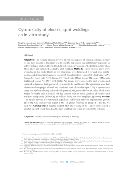
Braz J Oral Sci. January/March 2008 - Vol. 7 - Number 24
Cytotoxicity of intraoral orthodontic
elastics
Rogério Lacerda Dos Santos1
Matheus Melo Pithon1
Márlio Vinícius de Oliveira2
Gabriella da Silva Mendes3
Maria Teresa Villela Romanos4
Antônio Carlos de Oliveira Ruellas5
1
Student of the Master’s degree program in
Orthodontics, Federal University of Rio de
Janeiro-UFRJ, Brazil
2
Specialist in Orthodontics by the School of
Pharmacy and Dentistry of Alfenas, Efoa/
Ceufe, Brazil
3
Student of the Master’s degree program in
Microbiology, Federal University of Rio de
Janeiro-UFRJ, Brazil
4
Assistant Professor of Microbiology, Federal
University of Rio de Janeiro-UFRJ, Brazil
5
Invited Professor of the Specialization Course
in Orthodontics, School of Pharmacy and
Dentistry of Alfenas - Efoa/Ceufe and Professor
of Orthodontics, Federal University of Rio de
Janeiro-UFRJ Orthodontics
Abstract
Aim: The present study compared in vitro four types of latex intraoral
elastics (5/16 = 7.9 mm, mean load) regarding their possible cytotoxic
effects. Material and Methods: The sample was allocated to four
groups of 24 elastics: Group A (American Orthodontics), Group M
(Morelli), Group T (TP Orthodontics), and Group U (Uniden).
Cytotoxicity assays were performed using cell culture medium
containing epithelioid-type cells (Hep-2 line) derived from human
laryngeal carcinoma. Two methods for evaluating the cytotoxicity,
the agar overlay test and “dye-uptake” test, were employed at two
different moments (0 and 24 h). Data were compared by analysis of
variance (ANOVA) and Tukey’s test (p<0.05). Results: The results
showed statistically significant difference (p<0.05) between Groups
T and M, T and U, A and M, and A and U, for dye-uptake test at 0
and 24 h. No cell lysis was found in Groups T and A for the agar
overlay test, whereas 80% cell lysis was found in Groups M and U
after 24 h according to the Stanford’s response index (RI).
Conclusion: Both TP Orthodontics and American Orthodontics
elastics evoked less cell lysis than Morelli and Uniden elastics.
Key Words:
cytotoxicity; elastics; biocompatibility; orthodontics
Received for publication: November 11, 2007
Accepted: April 15, 2008
Correspondence to:
Rogério Lacerda dos Santos
Rua Uberaba 606, centro
Araújos- MG - Brazil
CEP 35603-000
E-mail: [email protected]
1520
Braz J Oral Sci. 7(24):1520-1525
Cytotoxicity of intraoral orthodontic elastics
Introduction
The biocompatibility of dental materials has been theme of
great speculation and uncertainty. There are, particularly in
Orthodontics, several materials keeping direct contact with
organic tissues for long periods of time. Recent studies have
investigated the biocompatibility of different types of
orthodontic materials1-2. Intraoral elastics are among these
materials. Their contact with oral mucosa may be prolonged
for months. Therefore, the possibility that elastomers release
toxic substances has been addressed, as such substances
could damage the cells1-2.
Latex consists of cis-1,4-polyisoprepen and in liquid state it
is conserved by adding a preserving agent (usually ammonia).
When liquid latex is manufactured several chemicals are added
so that the desired properties are achieved3.
As variations exist in the composition of latex elastics,
differences in their properties also occur. This may be one of
the reasons why manufacturers produce elastics of different
sizes, in order to compensate the variation of physical
properties 4 . Depending on how the elastics are stored,
alterations in their composition may occur 3, since latex is
highly sensitive to ozone and other free radical releasing
systems, such as sunlight or ultra-violet light, which trigger
the latex polymer chain. Under clinical conditions, orthodontic
elastics are replaced with new ones before achieving this
stage 3.
Prevulcanized latex consist of the pure natural latex having
the highest molecular weigh3 and stabilisers, such as zinc
oxide and chemically vulcanized products, then resulting in a
mixture which is heated until 70o C5. Although zinc is known
to be neurotoxic6, the amount released by orthodontic elastics
can be ingested as research studies show no evidence of
harm7.
Anti-ozone and anti-oxidant agents are also added to latex
during manufacturing of orthodontic elastics 3 . This
process has the advantage of producing latex with higher
mechanical properties, thus increasing its strength and
elasticity5,7. However, natural latex is not into the category
of materials known to be entirely inoffensive 8-9. Allergy
caused by latex proteins has been well documented 10 ,
including immediate hypersensitivity reactions11. Amongst the
allergic reactions caused by orthodontic elastics, swelling and
stomatitits, erythematous oral lesions, respiratory reactions,
and even anaphylactic shock, the most severe form of allergy1213
, can be cited. Latex allergy occurs in 3-17% of the cases14.
It is necessary, therefore, to test the biocompatibility of dental
materials by toxicity assays15. The use of cell culture medium
for testing the toxicity of dental products is a valid way of
understanding the biological behavior of such materials8. The
objective of the present in vitro study was to test the
hypothesis that there is no difference in the cytotoxicity
between elastics of different manufacturers.
Material and Methods
Four different types of intraoral elastics (5/16" = 7.9 mm, mean
load) made of natural latex were selected for studying their
cytotoxicity on two in vitro methods (Table 1). The samples
were allocated into four groups of 24 elastics each, according
to their manufacturers: Group A (American Orthodontics,
Sheboygan, Wisconsin, USA), Group M (Morelli, Sorocaba,
São Paulo, Brazil), Group T (TP Orthodontics, Lodi, California,
USA), and Group U (Uniden, Sorocaba, São Paulo, Brazil).
The elastics used in this study came from the same production
lot for each tested trademark. Dental copper amalgam
(Vigodent, Rio de Janeiro, Brazil) with standard size and weight
was used as positive control, whereas stainless steel wire
(American Orthodontics, Sheboygan, Wisconsin) was used
as negative control (Table 1).
Cell culture containing Hep-2 line cells (human laryngeal
carcinoma) was maintained in Eagles’ minimum essential
medium (Cultilab, Campinas, São Paulo, Brazil) by adding 0.03
mg/mL of glutamine (Sigma, St. Louis, Missouri, USA), 50
µg/ml of garamicine (Schering Plough, Kenilworth, New
Jersey, USA), 2.5 mg/ml of fungizone (Bristol-Myers-Squibb,
New York, USA), 0.25% sodium bicarbonate solution (Merck,
Darmstadt, Germany), 10 mM of HEPES (Sigma), and 10%
fetal calf serum (Cultilab, Campinas, São Paulo, Brazil) for
growth medium or no fetal calf serum for maintenance
medium only. Next, the cell culture medium was incubated at
37oC for 48 h.
Table 1 - Elastic and control groups used for the assays.
Groups
Trademark
Color
Ref erence number
T
TP Orthodontics
Natural
360-012
A
American Orthodontics
Natural
000-113
M
Morelli
Natural
60.01.205
U
Uniden
Natural
000-1204
Positive Control
Dental copper amalgam, Pratic NG 2, Vigodent
Negativ e Control
Stainless steel wire, American Orthodontics , 0.019”x 0.025”
1521
Braz J Oral Sci. 7(24):1520-1525
Cytotoxicity of intraoral orthodontic elastics
Table 2 - Response indices (RI) used for evaluating the degree of cytotoxicity of the tested materials
according to the parameters established by Stanford19
The elastics had their both sides previously sterilised with
ultraviolet light (Labconco, Kansas, Missouri, USA) during
30 min.
Two methods were used for determining the cytotoxicity of
orthodontic elastics: the first one, called “dye-uptake”16, is
based on neutral red dye incorporated into live cells; the
second one, called agar overlay, uses the agar diffusion
technique17-18. It was used in this experiment only two times
of evaluation 0 and 24 h, a time that, usually these elastics
are changed to each 24 h for the patient. Time zero represents
the contact and immediate removal of the elastic of cell
culture medium. The timing of 24 h represents the
maintenance of elastic in the cell culture medium for 24 h
after removal.
Dye-uptake
Volumes of 100 µL of Hep-2 cells were distributed into 96well microplates. After 48 h, the growth medium was replaced
with 100 µL of Eagles’ minimum essential medium (MEM)
obtained following incubation in the different types of
elastics at 0 and 24 h. Positive and control groups consisted
of culture medium put in contact with amalgam and stainless
steel wire, respectively.
After 24-h incubation, 100 µL of 0.01% neutral red dye (Sigma,
St. Louis, Missouri, USA) were added to the culture medium
in the 96-well microplates, which were incubated again for 3
h at 37oC so that the red dye could penetrate the live cells.
Following this period, 100 µL of 4% formaldehyde solution
(Vetec, Rio de Janeiro, Brazil) in PBS (130 mM of NaCl; 2 mM
of KCl; 6 mM of Na2HPO4 2H2O; 1 mM of K2HPO4 1 mM; pH
7.2) were added in order to promote cell attachment to the
plate. After 5 min, 100 µL of 1% acetic acid (Vetec, Rio de
Janeiro, Brazil) and 50% methanol (Vetec, Rio de Janeiro,
Brazil) were added in order to remove the dye.
After 20 min, a spectrophotometer (BioTek, Winooski,
Vermont, USA) at 492 nm wavelength ((l = 492 nm) was used
for reading the data.
Agar Overlay Technique
For this test, Hep-2 cells were grown in 10-cm-diameter Petri
1522
dishes of. After reaching a confluence with cell density of
approximately 106 cells/mL, the cell culture was replaced with
two-fold concentrated MEM mixture and 3% agar, and 3
samples of each elastic were carefully placed on the agar
layer. Next, the Petri dishes were incubated for 24 h at 37oC in
5% CO2 atmosphere. Thereafter, the cells were attached by
using 4% formaldehyde in PBS and stained with 1% crystal
violet dye (Vetec, Rio de Janeiro, Brazil) so that the lysis
halos could be measured by using a millimeter rule.
The results were recorded as response indices (RI) according
to parameters established by Stanford19, being represented
by two numbers separated by a slash in which the first number
means the halo size (diffusion of toxic substance) and the
second one means the amount of cell lysis (Table 2). Such
indices were calculated in relation to the halos measured
following use of crystal violet dye.
Data were compared by analysis of variance (ANOVA). Tukey
multiple-comparisons test was used for identifying
differences between the groups. Significance level was set
at p<0.05.
Results
Dye-uptake test
There were statistically significant differences between
groups T (TP Orthodontics) and M (Morelli), T (TP
Orthodontics) and U (Uniden), A (American Orthodontics)
and M (Morelli), A (American Orthodontics) and U (Uniden),
and M (Morelli) and U (Uniden) (p=0.00). No statistically
significant difference was found between Groups T (TP
Orthodontics) and A (American Orthodontics) (p=0.754) at 0
h for dye-uptake method (Table 3).
After immersing the elastics, the 24-h cytotoxicity assay
using dye-uptake method showed results similar to those of
0-h cytotoxicity and agar overlay assays.
There were statistically significant differences (p=0.00)
between groups T and M, T and U, A and M, and A and U.
On the other hand, Groups T and A as well as M and U had
no statistically significant differences between them (p=0.975
and p=0.972, respectively) after 24 h for the dye-uptake assay
(Table 3).
Braz J Oral Sci. 7(24):1520-1525
Cytotoxicity of intraoral orthodontic elastics
Table 3 - Dye-uptake technique: Descriptive statistics for optical density
N=24. Values followed by same letters are not significantly different (p>0.05) for the same time .SD: standard deviation
Agar Overlay Technique
The positive control had RI = 4/5 (Table 4), which means a
diffusion of toxic substance greater than 1.0 cm in length
and more than 80% of lysed cells, but not involving the
whole Petri dish. The negative control had RI = 0/0, which
means absence of cell lysis (Figures 1 and 2).
Morelli and Uniden elastics were cytotoxic, as their RI was
4/5 (Table 4). This means that the diffusion of toxic substance
was greater than 1.0 cm and 80% of the cells within the halo
had been lysed (Figures 5 and 6). On the other hand, TP
Orthodontics and American Orthodontics elastics showed
biocompatibility with respect to cytotoxicity as their RI was
0/0 (Figures 3 and 4).
Discussion
In the present study, dental copper amalgam and stainless
steel wire were used as positive and negative controls (Table
1), respectively, because they have proved to be adequate
for this type of assay18,20-21. Dental amalgam is potentially
cytotoxic due the presence of mercury, but there are also
other neurotoxic substances depending on their composition
and manufacturer6.
Toxic materials show zones corresponding to cell death22.
The positive control had RI = 4/5 (Table 4), which means a
diffusion of toxic substance greater than 1.0 cm in length
and more than 80% of lysed cells, but not involving the
whole Petri dish. The negative control had RI = 0/0, which
means absence of cell lysis (Figures 1 and 2). These results
were similar to those obtained by Moreira Pacheco18,21.
Sterilization is a prerequisite for cytotoxicity assay and it
can be done by autoclavation23. However, the elastics become
hardened after this procedure because of the heat17, resulting
in degradation and subsequent release of toxic substances.
In this study, ultraviolet light was used in the present study
for sterilizing both sides of the elastics24 during 30 min. All
elastics were found to have the same color and malleability
following UV light sterilization.
The results showed that Morelli and Uniden elastics were
cytotoxic, as their RI was 4/5 (Table 4). This means that the
diffusion of toxic substance was greater than 1.0 cm and
Table 4 - Cytotoxicity assay using the agar overlay test.
p
1523
Braz J Oral Sci. 7(24):1520-1525
Fig. 1 - Petri dishes with stainless steel wires
without halo formation of cytotoxicity. (negative
control).
Fig. 4 - Petri dishes with intraoral elastics
(group A) without halo of cytotoxicity or cell
lysis.
Cytotoxicity of intraoral orthodontic elastics
Fig. 2 - Petri dishes containing dental copper
amalgam with halo of cytotoxicity around it
(positive control).
Fig. 5 - Petri dishes with intraoral elastics (group Fig. 6 - Petri dishes with intraoral elastics
M) with halo of cytotoxicity of 18.5 mm and > (group U) with halo of cytotoxicity of 13.5
80% of cell lysis.
mm and 100% of cell lysis.
80% of the cells within the halo had been lysed (Figures 5
and 6). On the other hand, TP Orthodontics and American
Orthodontics elastics showed biocompatibility regarding
cytotoxicity as their RI was 0/0 (Figures 3 and 4). However,
variations occur in the composition of latex elastics4, which
might explain the difference in the results regarding both
trademarks.
By recognizing the importance of the biological
characteristics of dental materials, the American Dental
Association (ADA) have developed a series of tests in order
to develop a reliable method for assessing both toxicity and
irritation such materials may cause to oral tissues 22. The
cytotoxic potential can be assessed by means of in vitro
and in vivo (animals) experiments22, which represents one of
the ways of analyzing the safety and efficacy regarding
dental materials25.
Regarding in vitro experiments, three different methods can
be employed: evaluation of chromium release, Millipore
1524
Fig. 3 - Petri dishes with intraoral elastics
(group T) without halo of cytotoxicity or cell
lysis.
membrane filtration and agar overlay test19.
The dye-uptake technique 16 , in which neutral red dye is
incorporated by live cells, allowing that the initial cytotoxic
effect of dental materials on cells can be evaluated by a
spectrophotometer was used for reading the data through
optical density.
The percentage of viable cells was obtained by comparing
the mean optical density (OD) of control cells (no contact
with the materials) with that of cell cultures put in contact
with different elastics, resulting in 50% toxicity for the cell
cultures (CC50) (Table 3).
According to Schmalz 8 , the major risk factor for using
potentially cytotoxic intraoral elastics would be that
substances released by them could be ingested by the patient
over time, which might result in diseases caused by
accumulation of the toxic substance. It is known that latex is
not entirely biocompatible as it may cause allergic
reactions 3,14 and interact with food14,26 and medications27.
Braz J Oral Sci. 7(24):1520-1525
Nevertheless, the potential for hypersensitivity may not be
related to the potential for cytotoxicity. In other words, the
material may be allergenic but not cytotoxic, although the
contrary may not be true.
As dental latex elastics are widely used in clinical orthodontics,
the cytotoxic effects they may produce should be taken into
account, particularly intraoral elastics because of their
continuous and prolonged contact with the mucosa. Therefore,
materials proved biocompatible should be selected if such a
concern occurs. Previous studies on the toxicity of orthodontic
latex elastics have shown that they were toxic to gingival
fibroblasts 9. The cytotoxic effect was demonstrated after
exposing the elastics to the culture medium as the Morelli and
Uniden elastics were found to cause more cell death in
comparison to TP Orthodontics and American Orthodontics
elastics, thus suggesting that the former are less biocompatible
than the latter. However, as in vitro experiments cannot
reproduce the oral environment in all its aspects, those elastics
should not be considered clinically inert. Variations occur in
the composition of the latex elastics and this could explain the
difference of results between the trademarks in this study.
Although orthodontic elastics are cytotoxic under in vitro
conditions, this effect is not always reflected in the clinical
practice7,9. It is important for the orthodontist to know how to
manage patients with a latex allergy and how to deal with this
problem28. An alternative for patients with allergy to latex, is
to use latex-free elastics, which can be used in orthodontics
without jeopardizing the orthodontic treatment29.
In conclusion, TP Orthodontics and American Orthodontics
intraoral elastics were found to produce less cell lysis, whereas
Moreover, Morelli and Uniden intraoral elastics were found to
cause significantly more cell lysis, suggesting a less
biocompatibility of these elastics and greater possibility of
stimulation to the allergic reactions.
Cytotoxicity of intraoral orthodontic elastics
8.
9.
10.
11.
12.
13.
14.
15.
16.
17.
18.
19.
20.
21.
22.
23.
24.
References
1.
2.
3.
4.
5.
6.
7.
Kao CT, Ding SJ, He H, Chou MY, Huang TH. Cytotoxicity of
orthodontic wire corroded in fluoride solution in vitro. Angle
Orthod. 2007; 77: 349-54.
Vannet BMRAV, Hanssens JL. Cytotoxicity of two bonding
adhesives assessed by three-dimensional cell culture. Angle Orthod.
2007; 77: 716-22.
Weiss ME, Hirshman CA. Latex allergy. Can J Anaesth. 1992; 39:
528-32.
Bishara SE, Andreasen GF. A comparison of time related forces
between plastic alastiks and latex elastics. Angle Orthod. 1970;
40: 319-28.
Perrella FW, Gaspari AA. Natural rubber latex protein reduction
with an emphasis on enzyme treatment. Methods 2002; 27: 7786.
Lobner D, Asrari M. Neurotoxicity of dental amalgam is mediated
by zinc. J Dent Res. 2003; 82: 243-6.
Hanson M, Lobner D. In vitro neuronal cytotoxicity of latex and
nonlatex orthodontic elastics. Am J Orthod Dentofacial Orthop.
2004; 126: 65-70.
25.
26.
27.
28.
29.
Schmalz G. Use of cell cultures for toxicity testing of dental materials—
advantages and limitations. J Dent. 1994; 22 Suppl 2: S6-11.
Holmes J, Barker MK, Walley EK, Tuncay OC. Cytotoxicity of
orthodontic elastics. Am J Orthod Dentofacial Orthop. 1993;
104: 188-91.
Palosuo T, Alenius H, Turjanmaa K. Quantitation of latex allergens.
Methods 2002; 27: 52-8.
Wakelin SH, White IR. Natural rubber latex allergy. Clin Exp
Dermatol. 1999; 24: 245-8.
Everett FG, Hice TL. Contact stomatitis resulting from the use of
orthodontic rubber elastics: report of case. J Am Dent Assoc.
1974; 88: 1030-1.
Tomazic VJ, Withrow TJ, Fisher BR, Dillard SF. Latex-associated
allergies and anaphylactic reactions. Clin Immunol Immunopathol.
1992; 64: 89-97.
Turjanmaa K, Alenius H, Makinen-Kiljunen S, Reunala T, Palosuo
T. Natural rubber latex allergy. Allergy. 1996; 51: 593-602.
Babich H, Sinensky MC. Indirect cytotoxicity of dental materials:
a study with Transwell inserts and the neutral red uptake assay.
Altern Lab Anim. 2001; 29: 9-13.
Neyndorff HC, Bartel DL, Tufaro F, Levy JG. Development of a
model to demonstrate photosensitizer-mediated viral inactivation
in blood. Transfusion. 1990; 30: 485-90.
Wigg MD, Menezes LM, Quintão CCA, Moreira TC, Chevitarese
O. Elásticos extra-orais: avaliação da citotoxicidade. Ortod Gaúcha.
1997; 1: 151-7.
Moreira TC, Quintão CAC, Menezes LM, Wigg MD, Chevitarese
O. Elásticos plásticos: avaliação da citotoxicidade após
esterilização. Rev SBO. 1998; 3: 172-7.
Stanford JW. Recommended standard practices for biological
evaluation of dental materials. Int Dent J. 1980; 30: 140-88.
Ferreira SMS. Doenças virais e controle de infecção no ambiente
de trabalho odontológico. Rio de Janeiro: Conselho Regional de
Odontologia do Rio de Janeiro; 1994.
Pacheco MCT, Wigg MD, Chevitarese O, Elias CN. Biocompatibilidade
das soldagens ortodônticas. Rev. SBO 1995; 2: 233-8.
Phillips RWS. Materiais dentários. Rio de Janeiro: Guanabara
Koogan; 1993.
Guimarães JR. Biossegurança Odontológica. Dental Gaucho
Maquart & Cia; 1994.
Matta ENR, Calasans-Maia JA, Ruellas ACO, Wigg MD. Avaliação
da citotoxicidade In vitro de elásticos Ortodônticos com
tratamento de superfície. J Bras Ortodon Ortop Facial. 2004; 9:
587-93.
Moreira TC, Quintão CAC, Menezes LM, D. WM, Chevitarese
O. Avaliação da citotoxicidade de materiais ortodônticos pelo
teste de difusão em ágar. Manual prático. Rio de Janeiro:
Universidade Federal do Rio de Janeiro; 1997.
Carey AB, Cornish K, Schrank P, Ward B, Simon R. Crossreactivity of alternate plant sources of latex in subjects with
systemic IgE-mediated sensitivity to Hevea brasiliensis latex. Ann
Allergy Asthma Immunol. 1995; 74: 317-20.
Towse A, O’Brien M, Twarog FJ, Braimon J, Moses AC. Local
reaction secondary to insulin injection. A potential role for latex
antigens in insulin vials and syringes. Diabetes Care. 1995; 18:
1195-7.
Hain MA, Longman LP, Field EA, Harrison JE. Natural rubber latex
allergy: implications for the orthodontist. J Orthod. 2007; 34: 6-11.
Gandini P, Gennai R, Bertoncini C, Massironi S. Experimental
evaluation of latex-free orthodontic elastics behavior in
dynamics. Prog Orthod. 2007; 8: 88-99.
1525
Download