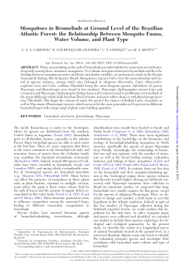
Journal of Vector Ecology 384 December 2014 Mosquito fauna of the Guapiaçu Ecological Reserve, Cachoeiras de Macacu, Rio de Janeiro, Brazil, collected under the influence of different color CDC light traps Júlia dos Santos Silva1,2, Márcia Souto Couri1, Alessandro Ponce de Leão Giupponi3, and Jeronimo Alencar4 Departamento de Entomologia, Museu Nacional, Universidade Federal do Rio de Janeiro, Rio de Janeiro, Rio de Janeiro, Brasil, [email protected] 2 Laboratório de Transmissores de Leishmanioses, Instituto Oswaldo Cruz/FIOCRUZ, Rio de Janeiro, Rio de Janeiro, Brasil 3 Laboratório de Referência Nacional em Vetores das Riquetsioses – LIRN, Instituto Oswaldo Cruz/FIOCRUZ, Rio de Janeiro, Rio de Janeiro, Brasil 4 Laboratório de Diptera, Instituto Oswaldo Cruz/FIOCRUZ, Rio de Janeiro, Rio de Janeiro, Brasil 1 Received 8 June 2014; Accepted 11 August 2014 ABSTRACT: The objective of the present study was to identify mosquito fauna and to evaluate whether different light bulb colors influence the attraction of light traps in the Guapiaçu Ecological Reserve. Samples were obtained monthly during the period of February, 2012 to January, 2013. Centers for Disease Control (CDC) light traps with incandescent light bulbs and LED (ultraviolet, blue, green, and red) bulbs were utilized. In total, 8,170 specimens were captured, including 59 species. The presence of Anopheles nimbus (Theobald 1902) and Orthopodomyia fascipes Coquillet 1906 were recorded for the first time in the state of Rio de Janeiro. The green LED trap attracted the highest number of specimens and presented the highest diversity and mosquito average. The blue and green LED traps attracted the highest number of species. However, the differences between lights were not significant. The most common species were Coquillettidia juxtamansonia (Chagas 1907), Culex declarator Dyar and Knab 1906, and Culex ribeirensis Forattini and Sallum 1985. Journal of Vector Ecology 39 (2): 384-394. 2014. Keyword Index: Mosquitoes, vectors, Atlantic Forest, attractiveness, LED. INTRODUCTION Environmental preservation areas, such as parks and forests reserves, contribute towards our knowledge with information about the biology, ecology, and biodiversity of insect vectors. The faunal study of culicids in natural environments is of considerable importance because of their role in pathogen transmission to humans and other vertebrates. Diversity studies of Culicidae in parks and forests reserves may clarify still unknown habits of these vectors (Hutchings et al. 2005) and facilitate the identification, monitoring, and controlling of mosquito populations. Currently, several types of automatic traps based on the attraction exerted by a light source and suction through ventilators have been developed to assist in the capture of small insects in field studies. Such traps are frequently used to capture culicids. The most well-known of these traps today is the CDC light trap described by Sudia and Chamberlain (1962). In a single insect species, different parts of the eyes are often equipped with receptors of different spectral sensitivity. In the case of attraction by artificial light, this depends on the sensibility of the receptors at different wavelengths. According to Briscoe and Chittka (2001), this sensitivity is between 350 and 600 nm for Culicidae, which can also be described as principally among the ultraviolet, green, and blue spectra. Also in the literature specific to culicids, the maximum wavelength was confirmed as a UVA band (Clements 1999). However, some species of mosquitoes and other dipterans of medical importance are attracted by artificial light and not all mosquito species respond equally to visual stimuli or to different wavelengths of light (Service 1993). According to Burkett and Butler (2005), there is a lack of information on the attractiveness of different light wavelengths in different mosquito species in areas where research on mosquito control is based on the number of specimens and species caught in light traps. With advances in lighting technology, light emitting diodes (LEDs) have been developed that can be selected to emit a specific color (Cohnstaedt et al. 2008). These colored LEDs, when used in CDC traps, have a high light intensity and require significantly less energy (around 0.125 mA/hr in comparison to 150 mA/hr of standard CM-47 lamps). This type of configuration has functioned particularly well for capturing disease vectors, but perfecting these techniques to increase the quantity of mosquitoes captured using these more precise light sources is greatly needed (Tchouassi et al. 2012). Furthermore, the use of LEDs provides a more energy efficient and accurate system for determining the spectral sensitivity of insects (Bentley et al. 2009). This study of mosquitoes in the Guapiaçu Ecological Reserve (REGUA) aimed to characterize the fauna from different areas in REGUA and to evaluate the influence of different light bulb colors of light traps in attracting these insects. Additionally, knowledge of the attractiveness of different types of lamps will add to data on the biology and Vol. 39, no. 2 Journal of Vector Ecology behavior of the different species in their natural habitat, which was previously unknown. Results presented here will assist in the targeting of monitoring programs, since we will be able to use colors more effectively for capturing mosquitoes and know the effectiveness of LEDs in comparison to the incandescent light traps. MATERIALS AND METHODS Study sites Captures were conducted in the Guapiaçu Ecological Reserve (REGUA), a private reserve in the remaining area of the Atlantic Forest, which also includes sections of floristic recovery similar to the original biocenotic structure. Its vegetation cover is divided into two altitudinal zones: dense lowland Ombrophilous Forest (up to 500 m in altitude) and dense montane Ombrophilous Forest (from 500 to 1500 m) (Veloso et al. 1991). The Guapiaçu Ecological Reserve currently consists of 3,760 ha, in addition to another 3,300 ha per an agreement with local landowners, totaling 7,000 ha. Intended to protect the Atlantic Forest in the Upper River Guapiaçu Basin, the reserve is located in the district of Guapiaçu, Cachoeiras de Macacu, Rio de Janeiro, approximately 60 km from the municipal of Rio de Janeiro (Figure 1). Samples were collected in areas of preserved forest and near reconstituted wetlands in the reserve. Six capture sites distributed between two distinct areas of 385 the reserve were established: four sites were located in areas of preserved forest and two sites were located near the wetlands that were reconstructed beginning in 2005. Geographical coordinates were obtained using GPS and verified in Google Earth software. Maps were prepared in Arcview10 and Google Earth and edited in Adobe Photoshop CS5 and CorelDraw X5. Locations are specified and indicated in Figures 1 and 2. Mosquito collection Samples were taken monthly during the period of February, 2012 to January, 2013 for three consecutive nights between 17:30 and 09:00. Five CDC light traps from BioQuip®, model 2836BQ, were used simultaneously and allocated at fixed points at a height of 2.0 m and were placed 40 m apart from one another per night. The following light bulb types were utilized in the traps: incandescent (4 watts); and UV (390 nm), blue (430 nm), green (570 nm) and red (660 nm) LEDs. The CO2 was not used in order to avoid interference with the influence of the colors. All captured mosquitoes were sacrificed by exposure to chloroform and kept in standard plastic pots, which were taken to the laboratory for triage and assembly of specimens. Taxonomic identification of culicids was conducted by direct observation of morphological characters via stereoscopic microscope and based on dichotomous keys developed by Kumm 1933, Lane 1953a,b, Pecor et al. 1992, Consoli and Lourenço-de-Oliveira 1994 and Forattini 2002. Figure 1. Distribution map of the Culicidae capture sites in the Guapiaçu Ecological Reserve (REGUA) highlighted by: A) Location of the municipality of Cachoeiras de Macacu in the state of Rio de Janeiro, Brazil, and in detail, the location of REGUA in the municipality of Cachoeiras de Macacu; B) The six capture sites in the REGUA (yellow circle) with the park headquarters highlighted as well (red square); C) Geographic coordinates of Culicidae capture sites in the REGUA. 386 Journal of Vector Ecology Data analysis Indices were calculated based on the total number of mosquitoes caught in all the traps in order to gain a better understanding of the mosquito fauna of REGUA. Additionally, indices were determined for each of the different light bulb colors with the aim to analyze their efficiencies separately. Analysis of variance (ANOVA) with significance level of 5% was used to analyze possible differences in the attractiveness of each of the five colors used in CDC traps among the species. To evaluate the most abundant species of mosquitoes, “Index of Species Abundance” (ISA) was utilized. This index was converted into a scale from zero to one for “Standardized Index of Species Abundance” (SISA), according to the definitions set forth by Roberts and Hsi (1979). In this index, the value of 1 corresponds to the most abundant species and was calculated using the formula: ISA = a + Rj / K; and SISA = c - ISA / c - 1, where: a: number of samplings in which the species was absent multiplied by c; c: for each sampling, the species must be distributed in positions which vary from 1 to n, with 1 being attributed to the most frequent species. The c comprises the largest value of n obtained considering all samplings, plus 1; Rj: the sum of the positions of each species; K: number of samplings. Species dominance was defined according to the categories established by Friebe (1983), using the definitions: eudominant > 10%, 5% < dominant < 10%, 2% < subdominant < 5%, 1% < recessive < 2% and rare < 1%. D% = (i / t) . 100, where “i” is the total of individuals of a particular species; and “t” is the total number of specimens collected. Calculation of Williams mean (Xw) (Haddow 1960) is based on the mosquito incidence values throughout the study period for each trap per month. This calculation is designed to show the trend of the natural species distribution without strong interference from extreme incidences related to particularities associated with each month studied. Calculation of Williams mean: Log (Xw + 1) = Σ log (ni + 1) where, N ni = number of captured specimens N = number of samples To evaluate and compare differences in mosquito community composition for each trap, Shannon-Wiener Diversity index (H’) and Shannon Equitativity (EH’) were employed (Shannon 1948). The Diversity index (H’) was chosen because it is appropriate for random samples of species in a community or subcommunity. Equitativity (EH’) refers to the distribution of individuals among species, being proportional to diversity and inversely proportional to dominance. The indices were calculated using the following formulas: H’ = ∑pi ln pi, where: pi is the proportion of species in relation to the total number of specimens found in samplings conducted and EH´ = H’ / Hmáx’, and Hmáx’ = lns, where H’ = Shannon-Wiener index; s = number of species December 2014 sampled. The Margalef diversity index (α) was also used (Margalef 1958) and is interesting as a comparison because it takes into account the number of species and the natural logarithm of the total number of individuals. This index was calculated by the formula: α = (S-1) / ln N, where S = number of species and N = number of individuals found. The similarity between the traps as well as the number of species was estimated by the Sørensen qualitative similarity index (SI), which is based on the presence or absence of species (Felfili et al. 1993). SI = 2c / a + b, where: a and b = species from a given area; c = species common to both areas. To calculate the quantitative similarity, the Morisita-Horn index (CMH) was used, which permits the establishment of the degree of similarity between the different traps based on community composition and species abundance (Volpato and Anjos 2001): CMH = 2 ∑ (ani . bni) / (da + db) . (aN . bN), where: da = ∑ ani2 / aN2, db = _ bni2 / bN2, ani and bni = number of individuals of each species in a particular trap, aN and bN = total number of individuals in a particular trap. RESULTS During the twelve-month study period, 8,170 specimens were captured, comprising two subfamilies, 14 genera, and 59 species of Culicidae. The subfamily Anophelinae was represented by one genus and 11 species and the subfamily Culicinae was represented by 13 genera and 48 species (Table 1). For the first time, the presence of Anopheles nimbus (Theobald 1902) was recorded in the state of Rio de Janeiro in the forested area close to reconstituted wetlands in REGUA. Its occurrence was recorded for the June, 2012 capture in the area at Site 4 and included one male specimen in the blue LED trap and one female specimen in the red LED trap. The second occurrence was recorded for the October, 2012 capture at Site 6, when one male specimen and two female specimens were collected in the blue LED trap and one male specimen was collected in the green LED trap (Figures 1 and 2) . We also recorded a new occurrence of the species Orthopodomyia fascipes Coquillett 1906, in the state of Rio de Janeiro in a forested preservation area. The occurrence of Or. fascipes was observed in the December, 2012 capture at Site 2 (Figure 2), with one female specimen collected in a UV LED trap. There were 36 captures conducted between February, 2012 and January, 2013. Although the ANOVA analysis did not indicate significant differences between the traps (F = 0.387; df = 4; p = 0.817), the green LED trap attracted the greatest number of specimens (N = 2,231), the highest Shannon Diversity (H´ = 2.31), the largest Equitativity value (EH´ = 0.55) and the highest mosquito average (Xw = 143.06). Traps with blue and green LEDs attracted the greatest number of species (S = 51 and S = 50, respectively) (Figures 3 and 4). As a comparison, we also used the Margalef Diversity index (α), which takes into account the number of species caught. This index showed the blue LED trap had the greatest diversity, followed by the green LED trap (α = 6.6 and α = 6.4, respectively). The CDC trap with red LED showed the lowest Shannon and Equitability indexes, the lowest number of 534 231 61 147 317 1351 Coquillettidia (Coquilletidia) juxtamansonia (Chagas 1907) Culex (Culex) declarator Dyar and Knab 1906 Culex (Culex) nigripalpus Theobald 1901 Culex (Melanoconion) ribeirensis Forattini and Sallum 1985 Others* Total 100.0% 23.5% 10.9% 4.5% 17.1% 39.5% 4.5% D% 87.29 20.27 7.24 1.07 9.72 33.84 2.04 Xw 1583 301 151 136 259 663 73 N 100.0% 19.0% 9.5% 8.6% 16.4% 41.9% 4.6% D% UV LED 105.18 18.50 8.62 2.22 11.36 36.94 2.91 Xw 1950 386 164 152 485 696 67 N 100.0% 19.8% 8.4% 7.8% 24.9% 35.7% 3.4% D% Blue LED 110.44 21.49 6.70 1.82 12.69 38.88 3.61 Xw 2231 577 177 105 298 909 165 N 100.0% 25.9% 7.9% 4.7% 13.4% 40.7% 7.4% D% Green LED 143.06 33.48 9.46 2.68 15.28 56.56 5.87 Xw 1055 213 114 51 110 481 86 N 100.0% 20.2% 10.8% 4.8% 10.4% 45.6% 8.2% D% Red LED 69.66 14.62 5.24 0.86 5.57 31.85 Xw 1.70 0.564 Cq. fasciolata Cq. fasciolata 0.807 Cx. ribeirensis Cx. ribeirensis 0.825 Cx. declarator Cx. declarator Species 0.991 Cq. juxtamansonia SISA UV LED Cq. juxtamansonia Species Incandescent light bulb Species Ur. calosomata 0.722 Cq. fasciolata 0.843 Cx. ribeirensis 0.898 Cx. declarator 0.948 Cq. juxtamansonia SISA Blue LED Species Ur. ditaenionata Cx. nigripalpus 0.519 Ma. titillans 0.776 Cq. fasciolata 0.784 Cx. ribeirensis 0.855 Cx. declarator 0.997 Cq. juxtamansonia SISA Green LED Species 0.535 0.557 0.580 0.765 Cx. (Melanoconion) Atratus group 0.829 Cx. ribeirensis 0.937 Cx. declarator 0.999 Cq. juxtamansonia SISA Red LED 0.547 0.708 0.789 0.992 SISA Table 2. Abundance index (SISA) of the most abundant species (SISA> 0.5) of Culicidae captured in each trap in the Guapiaçu Ecological Reserve (REGUA), Cachoeiras de Macacu, Rio de Janeiro, Brazil. *Others: Anopheles (Nyssorhynchus) albitarsis Lynch-Arribalzaga 1878, An. (Nys.) aquasalis Curry 1932, An. (Nys.) argyritarsis Robineau-Desvoidy 1827, An. (Kerteszia) cruzii Dyar and Knab 1908, An. (Anopheles) eiseni Coquillett 1902, An. (Nys.) evansae (Brethes 1926), An. (Ano.) intermedius (Peryassú 1908), An. (Stethomyia) nimbus (Theobald 1902), An. (Nys.) rangeli Gabaldon, Cova Garcia and Lopez 1940, An. (Nys.) strodei Root 1926, An. (Nys.) triannulatus (Neiva and Pinto 1922), Aedeomyia (Aedeomyia) squamipennis (Lynch Arribalzaga 1878), Aedes (Ochlerotatus) fluviatilis (Lutz 1904), Aedes hastatus/serratus/oligopistus, Ae. (Och.) rhyacophilus (Da Costa Lima 1933), Ae. (Och.) scapularis (Rondani 1848), Ae. (Och.) serratus (Theobald 1901), Ae. (Protomacleaya) terrens (Walker 1856), Coquillettidia (Rhynchotaenia) albicosta (Peryassu 1908), Cq. (Rhy.) chrysonotum (Peryassú 1922), Culex (Culex) abnormalis Lane 1936, Cx. (Cux.) usquatus Dyar 1918, Cx. (Mel.) Atratus group, Culex (Mel.) spp. Theobald 1903, Culex (Microculex) davisi Kumm 1933, Cx. (Mcx.) imitator Theobald 1903, Cx. (Mcx.) pleuristriatus Theobald 1903, Culex (Mcx.) spp. Theobald 1907, Culex ocellatus Theobald 1903, Haemagogus (Conopostegus) leucocelaenus (Dyar and Shannon 1924), Limatus durhamii Theobald 1901, Li. flavisetosus De Oliveira Castro 1935, Mansonia (Mansonia) titillans (Walker 1848), Orthopodomyia fascipes (Coquillett 1906), Psorophora (Janthinosoma) ferox (Von Humboldt 1819), Runchomyia (Runchomyia) frontosa (Theobald 1953), Ru. (Run.) reversa Lane and Cerqueira 1942, Sabethes (Sabethoides) chloropterus (Von Humboldt 1819), Sa. (Peytonulus) identicus Dyar and Knab 1907, Sabethes undosus/ fabricii, Uranotaenia (Uranotaenia) apicalis Theobald 1903, Ur. (Ura.) calosomata Dyar and Knab 1907, Ur. (Ura.) ditaenionata Prado 1931, Ur. (Ura.) geometrica Theobald 1901, Ur. (Ura.) lowii Theobald 1901, Ur. (Ura.) pallidoventer Theobald 1903, Ur. (Ura.) pulcherrima Lynch-Arribalzaga 1891, Wyeomyia aporonoma Dyar and Knab 1906 e Wy. (Spilonympha) mystes Dyar 1924. 61 N Incandescent light bulb Coquillettidia (Rhynchotaenia) fasciolata Lynch-Arribalzaga 1891 Species Table 1. Absolute values (N), dominance (D%) and Williams means (Xw) of mosquito species collected in the Guapiaçu Ecological Reserve, Cachoeiras de Macacu, Rio de Janeiro, Brazil, in the period from February, 2012 to January, 2013. Vol. 39, no. 2 Journal of Vector Ecology 387 388 Journal of Vector Ecology December 2014 Figure 2. Illustrations of each of the six Culicidae capture sites in the Guapiaçu Ecological Reserve (REGUA), Cachoeiras de Macacu, Rio de Janeiro, Brazil: 1) Site 1, Green Trail, Preserved forest; 2) Site 2, Green Trail, Preserved forest; 3) Site 3, Manoel Alexandre river, Preserved forest; 4) Site 4, Yellow Trail, wetland; 5) Site 5, Green Trail, Preserved forest; 6) Site 6, Brown Trail, wetland. Vol. 39, no. 2 Journal of Vector Ecology Figure 3. ShannonWiener Diversity (Hʹ), Shannon Equitativity (EHʹ) and Margalef (α) index values and species richness (S) for each of the five CDC traps in the Guapiaçu Ecological Reserve (REGUA), Cachoeiras de Macacu, Rio de Janeiro, Brazil. Figure 4. Absolute values and Williams means (Xw) for each of the five CDC traps in the Guapiaçu Ecological Reserve, Rio de Janeiro in the period from February, 2012 to January, 2013. 389 Journal of Vector Ecology 390 December 2014 specimens, and lowest diversity value, mainly resulting from presenting the lowest number of species (S = 35) (Figure 3). The dominance index indicated Culex ribeirensis Forattini and Sallum 1985 was eudominant in the incandescent and red LED traps, and Culex declarator Dyar and Knab 1906 and Coquillettidia juxtamansonia (Chagas 1907) were eudominants in all CDCs, with Cq. juxtamansonia being the most abundant in all traps (SISA Incandescent, UV, blue, green, and red = 0.991, 0.948, 0.997, 0.999 and 0.992) (Tables 1 and 2). Among these species, the largest number of specimens Cq. juxtamansonia and Cx. ribeirensis were attracted by the green LED (27.7% and 23.5%), and the largest number of Cx. declarator were attracted by the blue LED (35.1%) (Figure 5). Considering separately each of the areas studied, the eudominant species present in the preserved forest were Cx. declarator (48.8%) and Culex nigripalpus Theobald 1901 (23.0%), while those present in the wetlands were Cq. juxtamansonia (45.6%), Cx. declarator (12.6%), and Cx. ribeirensis (10.5%). While Cq. juxtamansonia represented the largest percentage of abundance in the green LED trap, this species was also observed to have the highest average number of specimens for this color as well (Xw = 56.56) (Figure 5). For the species Culex nigripalpus Theobald 1901, which also was among the most dominant species, the highest means were found for the green lamps (Xw = 2.68), followed by blue, UV, incandescent, and red lamps (Table 1). The ANOVA analysis did not indicate significant differences between the traps for the most dominant species (Cq. juxtamansonia: F = 1.107; df = 4; p = 0.363, Cx. declarator: F = 1.305; df = 4; p = 0.280, Cx. nigripalpus: F = 0.399; df = 4; p = 0.808, and Cx. ribeirensis: F = 0.262; df = 4; p = 0.901). The Sørensen qualitative similarity index as well as the Morisita-Horn quantitative similarity index indicated that all the traps have similarity in species composition (IS > 0.5), with the incandescent and red traps (IS = 0.84) and incandescent and UV traps (CMH = 0.992) being the most similar. DISCUSSION For the first time, the presence of An. nimbus and Or. fascipes was recorded in the state of Rio de Janeiro. In Brazil, An. nimbus presents restricted distribution to forested plains with high rainfall. Although it is hematophagous, the species most likely does not have epidemiological importance in the transmission of human malaria. However, in the Brazilian Amazon, the Venezuelan equine encephalomyelitis (VEE) virus was isolated in a group of mosquitoes of this species (Travassos-da-Rosa et al. 1997). The Uma virus has also been isolated from this species in French Guiana (Pajot 1980) and the Macaua, Pixuna, and Tacaiuma viruses appear to have cycles involving daytime active vectors such as An. nimbus (Dégallier et al. 1992). From observations carried out at the Evandro Chagas Institute, Ananindeua, Pará, Brazil, the arbovirus Tembe, isolated from specimens from BelérnBrasília Highway, is believed to have An. nimbus as its likely vector (Pinheiro 1980). According to the literature, eleven Brazilian states have recorded the occurrence of An. nimbus, Figure 5. Dominance (D%) and Williams means (Xw) of mosquito species considered eudominant (N> 10%) for each CDC trap in the Guapiaçu Ecological Reserve (REGUA), Cachoeiras de Macacu, Rio de Janeiro, Brazil. including Acre, Amapá, Amazonas, Bahia, Goiás, Maranhão, Mato Grosso, Minas Gerais, Pará, Rondônia, and Roraima (Deane et al. 1948, Guedes et al. 1953, Ferraroni and Hayes 1977, Camargo et al. 1993). As observed by Deane et al. (1947), An. nimbus is found in forested suburbs and surrounding areas located within the forest. Their larvae are found in the collections of clear, cold water, which is shaded, still, or running slowly and often with grass and algae on the shore. Galvão et al. (1942) also reported finding the larvae of An. nimbus in streams and backwaters and Hutchings et al. (2002) observed breeding sites of this species in puddles on the ground. Deane et al. (1948) also noted An. nimbus adults were always found in Vol. 39, no. 2 Journal of Vector Ecology small numbers, even in places where larvae were abundant. The peak feeding activity of females usually occurs just before dusk with the day still clear between 17:30 and 18:30, which is also when this species is captured. However, Camargo et al. (1993) recorded anthropophilic and synanthropic behavior for this species in Goiânia. Although immature forms have not yet been captured in REGUA, the behavior of the species was equivalent to that described by Deane et al. (1948), given the area is under the strong influence of a large wetland of about 12 ha, and small areas of streams and swamps can be found in the forested area. Traps were always placed half an hour before the beginning of the evening (17:30). Thus, it is possible that the species was drawn to the trap during this period. The occurrence of Or. fascipes has been recorded in the states of Amazonas, Bahia, Goiás, Maranhão, Minas Gerais, Pará, Piauí, and Rondônia (Zavortink 1968, Luz and Lourenço-de-Oliveira 1996). The epidemiological importance of this species has not yet been registered. The biology of both Or. fascipes and An. nimbus is not well known. New records for the state of Rio de Janeiro, along with recent records for the species and the epidemiological potential of one of them, have demonstrated the importance of understanding the biology of both species. This study in REGUA showed that traps with blue and green LEDs attracted the greatest number of species and specimens, and showed the highest diversity value. The CDC trap with red LED showed the lowest diversity values and number of specimens. Previous studies on wavelength prevalence indicated a preference by mosquitoes for the blue-green range (400-600 nm), a decrease in attraction with increasing wavelength (Ali et al. 1989), and captures with infrared being the least successful (Burkett et al. 1998). Ali et al. (1989) used lamps painted in six different colors (white, yellow, green, orange, blue, and red) and found the five predominant species, Psorophora columbiae (Dyar and Knab 1906), Psorophora ciliata (Fabricius 1794), Culex salinarius Coquillett 1904, Cx. nigripalpus and Culex erraticus (Dyar and Knab 1906), were most attracted to the color blue, followed by the green and red lights. Browne and Bennett (1981) tested some wavelengths to assess the preference of Coquillettidia pertubans (Walker, 1856) through the landing rate and found that shorter wavelength (400-600 nm or bluegreen) attracted significantly more mosquitoes than longer wavelengths. Hoel et al. (2007), while testing the sensitivity to different colors of sand flies, simultaneously captured 5,845 mosquitoes of three genera (Aedes, Culex Linnaeus 1758, and Anopheles Meigen 1818) together with sand flies in the following order of attractiveness: green > incandescent > blue > red. Furthermore, Bentley et al. (2009) also observed in their capture of mosquitoes in resting boxes with luminous attractors a greater number of specimens attracted by blue LEDs followed by green, red, and lastly infrared. Gjullin et al. (1973) dipped lamps in ceramic paint and determined that Aedes sierrensis Ludlow 1905, Culex quinquefasciatus Say 1823, and Culex tarsalis Coquillett 1896 were more attracted to red light than to green, blue, orange, and white light, but the spectrum frequencies were 391 not provided in this study. According to Breyev (1963), many insects are not sensitive to red spectrum, and this finding is likely due to the low numbers of insects captured with this color (Tchouassi et al. 2012). Hoel et al. (2007) found more sand flies attracted by red light than in comparison with the other colors, demonstrating that this group is more attracted by red light. And, very recently, using the same Bioquip® traps, Tchouassi et al. (2012) conducted captures of culicids in the Rift Valley Province, Kenya, and within five months of study, their results indicated that the highest number of specimens was attracted by the incandescent lamp compared with the LEDs (UV, blue, green, red, and a combination of blue-green-red). It is important to emphasize that CO2 lures were used in the traps. The species Cq. juxtamansonia was the most abundant in all CDC traps, and the species Cx. declarator, Cx. nigripalpus, and Cx. ribeirensis also showed great occurrence. Representatives of the tribe Mansoninii are known to have the biological characteristic of their immature forms fixing on the tissues of aquatic plants such as Eichornia sp. (Forattini 2002). Their proliferation is increased by the offering of breeding sites, and when they occur in large numbers, these species are numerous and aggressive. (Tubaki et al. 1999), overtaking other species. In this case, the species of the tribe Mansoninii, principally Cq. juxtamansonia, presented in large numbers due to the influence of the massive flood area, which favored their proliferation. Due to these large numbers, this species was found to be widely present in all traps. This species is a vector of Wuchereria bancrofti (Cobbold 1877) in Brazil. Culex nigripalpus has been reported to prefer the colors green, blue, and white in decreasing order according to Burkett et al. (1998) and blue-green, orange, blue, white, red, and yellow-green second according to Burkett and Butler (2005). In REGUA, the highest means were found for the green lamps, followed by blue, UV, incandescent, and red lamps, but the ANOVA did not indicate significant differences. Eudominant species occurring both in the preserved forest and wetlands demonstrated considerable epidemiological potential. A vector of several arboviruses (Moju, Bussuquara and Catú viruses) (Travassos-da-Rosa et al. 1994), Cx. declarator has been recognized as an important vector for the Saint Louis encephalitis virus (SLE) and may also act as a vector for Dirofilaria immitis (Leidy 1856) (Labarthe et al. 1998); Cx. nigripalpus has been incriminated as a vector for SLE, eastern equine encephalitis (EEE), and West Nile viruses (Forattini 2002); Cx. ribeirensis previously has been involved in the transmission of EEE virus (Calisher et al. 1983). The species that occurred less frequently also present notable epidemiological potential, in particular: Anopheles albitarsis Lynch Arribalzaga 1878, Anopheles aquasalis Curry 1932, Anopheles cruzii Dyar and Knab 1908, Aedes scapularis (Rondani 1848), Haemagogus leucocelaenus (Dyar and Shannon 1924), Psorophora ferox (Von Humboldt 1819), and Sabethes chloropterus (Von Humboldt 1819). Among the anophelines: An. albitarsis is a malaria vector, usually appearing as a secondary vector; An. aquasalis is an important coastal vector of malaria in several locations in Brazil and the Americas; and An. cruzii is considered the primary vector 392 Journal of Vector Ecology of human and simian malaria (Forattini 2002). Among the culicines, Ae. scapularis has been incriminated as a vector of Rocio virus (Forattini et al. 1995), Wuchereria bancrofti and Dirofilaria immitis (Labarthe et al. 1998). Hg. leucocelaenus is considered an important species in yellow fever virus transmission in the southeast of the country (Vasconcelos 2003), in addition to having other viruses isolated, such as Ilhéus, Maguari, and Tucunduba (Hervé et al. 1986). Ps. ferox has been found naturally infected with arboviruses causing encephalitis (VEE, SLE, and Rocio), in addition to Ilhéus, Mayaro, Melao, Oriboca, West Nile and Uma viruses (Turell et al. 2005). Sa. chloropterus is a potential vector of yellow fever virus and SLE and Ilheus viruses have been detected in specimens collected in nature (Hervé et al. 1986). When we evaluated the efficiency of traps by month, most specimens were observed to occur in the green LED trap during the months of February, April, June, and July, in the blue LED trap during the months of March, May, September, October, and December, and in the UV LED trap during the months of August, November, and January. The major difference between the traps was in the number of specimens attracted by the green LED in June 2012 in comparison with the other traps. Analyzing qualitatively, in the months of April, June, July, August, and December, the trap with the green LED demonstrated higher species richness. Tchouassi et al. (2012) suggests environmental changes, such as storms or changes in vegetation, can lead a reduction in the luminosity of the LEDs, and consequently, variations in the attraction of mosquitoes. In this way, conducting the present study over 12 months for three consecutive nights reduced this type of influence. The following species occurred specifically in one of the traps: Sabethes chloropterus (incandescent); Ae. terrens (Walker 1856), Limatus durhamii Theobald 1901, Or. fascipes and Wyeomyia mystes Dyar 1924 (UV); Anopheles evansae (Brethes 1926), Anopheles triannulatus (Neiva and Pinto 1922), Ps. ferox and Runchomyia reversa Lane and Cerqueira 1942 (blue); An. albitarsis, Anopheles argyritarsis RobineauDesvoidy 1827, Ae. hastatus/serratus/oligopistus, Culex davisi Kumm 1933, Sabethes identicus Dyar and Knab 1907 and Wyeomyia sp. Theobald 1901 (green); and Aedes rhyacophilus (Da Costa Lima 1933) and Haemagogus leucocelaenus (red). No color preference can be assumed from these results given the species occurred in reduced numbers. However, these findings are interesting since many of these species, such as Sa. chloropterus, Ae. terrens, Li. durhamii, Or. fascipes, Wy. mystes, Ps. ferox, Ru. reversa, Sa. identicus, and Hg. leucocelaenus, are diurnal and this is an uncharacteristic behavior (Forattini 2002). Similar behavior was observed by Alencar et al. (2012) for Hg. leucocelaenus, a potential vector of yellow fever in the area of the Simplício Power Plant, Minas Gerais, Brazil, where some specimens were collected in CDC traps. The data obtained over twelve months enabled us to observe some differences between attractiveness. Our results suggest wavelengths in the blue to green range would be a great option for capturing a wide range of mosquito species. Tchouassi et al. (2012) used a combination of blue-green-red LEDs in a CDC light trap. A trap that utilizes a combination December 2014 like this (including incandescent, blue and green) would most likely be the ideal for capturing mosquitoes. Knowledge of the attractiveness of different lamp types has contributed to previously unknown data on the biology of different species and will assist in the targeting of monitoring programs. The REGUA has favorable ecological characteristics for hosting interactions with pathogens, especially considering the introduction and maintenance of wild arboviruses. Interestingly, REGUA also sees high tourist traffic, as it is often frequented by birdwatchers. Thus, such interactions could be potentiated due to the high frequency and abundance of mosquito species recognized as having vectorial capacity. Acknowledgments We thank Nicholas and Rachel Locke as president and vice president, and Jorge Bizarro as research coordinator of REGUA, for the facilities granted for carrying out the studies. The authors also thank CAPES and FAPERJ (E26/102.833/2011 and 112.076/2012) for financial support. All research was performed in accordance with scientific license number 34911 provided by SISBIO/ IBAMA for the capture of culicids throughout the Brazilian national territory. The field studies did not involve endangered or protected species. REFERENCES CITED Alencar, J., V.S. Mello, N.M. Serra-Freire, J.S. Silva, F. Morone, and A.E. Guimarães. 2012. Evaluation of mosquito (Diptera: Culicidae) species richness using two sampling methods in the hydroelectric reservoir of Simplício, Minas Gerais, Brasil. Zool. Sci. 29: 218-222. Ali, A., J.K. Nayar, J.W. Knight, and B.H. Stanley. 1989. Attraction of Florida mosquitoes (Diptera: Culicidae) to artificial light in the field. Annu. Conf. Calif. Mosq.Vect. Contr. Assoc. 82–88. Bentley, M.T., P.E. Kaufman, D.L. Kline, and J.A. Hogsette. 2009. Response of adult mosquitoes to light-emitting diodes placed in resting boxes and in the field. J. Am. Mosq. Contr. Assoc. 25: 285-291. Breyev, K.A. 1963. The effect of various light sources on the numbers and species of blood-sucking mosquitoes (Diptera: Culicidae) collected in light traps. Entomol. Rev. 42: 155–168. Briscoe, A.D. and L. Chittka. 2001. The evolution of color vision in insects. Ann. Rev. Entomol. 46: 471-510. Browne, S.M. and F.G. Bennett. 1981. Response of mosquitoes (Diptera: Culicidae) to visual stimuli. J. Med. Entomol. 18: 505-521. Burkett, D.A. and J.F. Butler. 2005. Laboratory evaluation of colored light as an attractant for female Aedes aegypti, Aedes albopictus, Anopheles quadrimaculatus, and Culex nigripalpus. Fla. Entomol. 88: 383-389. Burkett, D.A., J.F. Butler, and D.L. Kline. 1998. Field evaluation of colored light-emitting diodes as attractants for woodland mosquitoes and other Diptera in north central Florida. J. Am. Mosq. Contr. Assoc. 14: 186–195. Calisher, C.H., T.L.M. Coimbra, O.S. Lopes, D.J. Muth, L.A. Vol. 39, no. 2 Journal of Vector Ecology Sacchetta, D.B. Francy, J.S. Lazuick, and C.B. Cropp. 1983. Identification of New Guama and Group C serogroup bunyaviruses and an ungrouped virus from southern Brazil. Am. J. Trop. Med. Hyg. 32: 424–431. Camargo, M.F., I.G. Silva, and E. Isac. 1993. Ocorrência de Anopheles (Stethomyia) nimbus (Theobald, 1903) (Diptera, Culicidae) no ambiente domiciliar, na área urbana de Goiânia, Goiás, Brasil. Rev. Pat. Trop. 22: 101. Clements, A.N. 1999. The Biology of Mosquitoes. Sensory Reception and Behaviour. vol. 2., Wallingford Caby, London, U.K. Cohnstaedt, L.W., J.I. Gillen, and L.E. Munstermann. 2008. Light-emitting diode technology improves insect trapping. J. Am. Mosq. Contr. Assoc. 24: 331–334. Consoli, R.A.G.B. and R. Lourenço-de-Oliveira. 1994. Principais mosquitos de importância sanitária no Brasil. Editora Fiocruz, Rio de Janeiro, Brazil. Deane, L.M., O.R. Causey, and M.P. Deane. 1948. Notas sobre a distribuição e a biologia dos anofelinos das regiões nordestina e amazônica do Brasil. Mem. Inst. Oswaldo Cruz 104: 11-17. Deane, M.P., O.R. Causey, and L.M. Deane. 1947. Chave ilustrada para a identificação de 32 espécies de anofelinos das regiões nordestina e amazônica do Brasil pelos caractéres da larva, com a descrição de duas larvas. Rev. Serv. Esp. Saúde públ. 1: 335 - 384. Dégallier N., A.P.A. Travassos-da-Rosa, J.M.C. Silva, S.G. Rodrigues, P.F.C. Vasconcelos, J.F.S. Travassos-da-Rosa, G.P. Silva, and R.P. Silva. 1992. As aves como hospedeiras de arbovírus na Amazônia brasileira. Bol. Mus. Para. Emilio Goeldi, sér. Zool 8: 69-111. Felfili, J.M., M.C. Silva-Júnior, A.V. Rezende, J.W.B. Machado, B.M.T. Walter, P.E.N. Silva, and J.D. Hay. 1993. Análise comparativa da florística e fitossociologia da vegetação arbórea do cerrado sensu stricto na Chapada Pratinha, DF-Brasil. Acta Bot. Bras. 6: 27-47. Ferraroni, J.J. and J. Hayes. 1977. Estudo sobre um surto de malária entre os índios Mayongong e Sanomã (Norte de Roraima). Acta Amazonica 7: 401-406 Forattini, O.P. 2002. Culicidologia Médica: identificação, Biologia, Epidemiologia, vol. 2. Editora da Universidade de São Paulo, São Paulo, Brazil. Forattini, O.P., I. Kakitani, E. Massad, and D. Mauricci. 1995. Studies on mosquitoes (Diptera: Culicidae) and anthropic environment: 9- Synanthropy and epidemiological vector role of Aedes scapularis in South-Eastern Brazil. Rev. Saúde Pública 29: 199-207. Friebe, B. 1983. Zur Biologie eines Buchenwaldbodens: 3. Die Kaferfauna 41: 45-80. Galvão, A.L.A., R.G. Damasceno, and A.P. Marques. 1942. Algumas observações sobre a biologia dos anofelinos de importância epidemiológica de Belém/Pará. Arq. Hig. Saúde Pública 12: 51-111. Gjullin, C.M., D.G. Brandl, and J.J. O’Grady. 1973. The effect of colored lights and other factors on the numbers of Culex pipiens quinquefasciatus, Cx. tarsalis and Aedes sierrensis entering light traps. Mosq. News 33: 67–71. Guedes, A.S., J.R. Freitas, and S.H. Xavier. 1953. Contribuição 393 ao conhecimento da distribuição geográfica dos anofelinos e algumas observações sobre a biologia do Anopheles darlingi Root, 1926, no Estado de Minas Gerais, Brasil. Rev. Bras. Malariol. Doenças Trop. 5: 157165. Haddow, A.J. 1960. Studies on the biting-habits and medical importance of eats African mosquitoes in the genus Aedes. I- Subgenera Aedimorphus, Bankisinella and Dunnius. Bull. Entomol. Res. 50: 759-779. Hervé, J.P., N. Dégallier, A.P.A. Travassos-da-Rosa, F.P. Pinheiro, and G.C. Sá-Filha. 1986. Arboviroses: Aspectos ecológicos. In: Instituto Evandro Chagas: 50 anos de contribuição às ciências biológicas e à medicina tropical. Fund. Serv. Saúde Pública, Belém, Brazil. Hoel, D.F., J.F. Butler, E.Y. Fawaz, N. Watany, S.S. El-Hossary, and J. Villinski. 2007. Response of phlebotominae sand flies to light-emiting diode-modified light traps in southern Egypt. J. Vector Ecol. 12: 302-308. Hutchings, R.S.G., M.A.M. Sallum, and R.L.M. Ferreira. 2002. Culicidae (Diptera: Culicomorpha) da Amazônia Ocidental Brasileira: Querari. Acta Amazônica 32: 109122. Hutchings, R.S.G., M.A.M. Sallum, R.L.M. Ferreira, and R.W. Hutchings. 2005. Mosquitoes of the Jaú National Park and their potential importance in Brazilian Amazonia. Med. Vet. Entomol. 19: 428-441. Kumm, H.W. 1933. Mosquitos breeding in bromeliads, at Bahia, Brazil. Bull. Entomol. Res. 24: 561-573. Labarthe N., M.L. Serrão, F.Y. Melo, S.J. Oliveira, and R. Lourenço-de Oliveira. 1998. Potencial vectors of Dirofilaria immitis (Leidy, 1856) in Itacoatiara, Oceanic Region of Niterói municipality, State of Rio de Janeiro, Brazil. Mem. Inst. Oswaldo Cruz 93: 425-432. Lane, J. 1953a. Neotropical Culicidae, vol. 1. Editora Universidade de São Paulo, São Paulo, Brazil. Lane, J. 1953b. Neotropical Culicidae, vol. 2. Editora Universidade de São Paulo, São Paulo, Brazil. Luz, S.L.B. and R. Lourenço-de-Oliveira. 1996. Forest Culicinae Mosquitoes in the Environs of Samuel Hydroeletric Plant, State of Rondônia, Brazil. Mem. Inst. Oswaldo Cruz 91: 427-432. Margalef, R. 1958. Information theory in ecology. Gen. Syst. 3: 36-71. Pajot, F.X. 1980. Enfermedades Transmitidas por insectos em la Guayana Francesa. Bol. of Sanit Panam. 88: 218-227. Pecor, J.E., L.M. Varuni, R.E. Harbach, and E.L. Peyton. 1992. Catalog and illustrated review of the subgenus Melanoconion of Culex (Diptera: Culicidae). Contrib. Am. Entomol. Inst. 27: 1-228. Pinheiro, F.P. 1980. Situação das Arboviroses na região Amazônica. Ver. Fund. SESP 25: 37-58. Roberts, D.R., and B.P. Hsi. 1979. An index of species abundance for use with mosquito surveillance data. Environm. Entomol. 8: 1007-1013. Service, M.W. 1993. Mosquito Ecology: Field Sampling Methods. Chapman and Hall, London, UK. Shannon, C.E. 1948. A mathematical theory of communication. Bell. Syst. Techn. J. 27: 379-423, 623-656. 394 Journal of Vector Ecology Sudia W.D. and R.W. Chamberlain. 1962. Battery operated light trap, an improved model. Mosq. News 22: 126-129. Tchouassi, D.P., R. Sang, C.L. Sole, A.D.S. Bastos, L.W. Cohnstaedt, and B. Torto. 2012. Trapping of Rift Valley Fever (RVF) vectors using Light Emitting Diode (LED) CDC traps in two arboviral disease hot spots in Kenya. Parasit.Vectors 5: 94-100. Travassos-da-Rosa, A.P.A., E.S. Travassos-da-Rosa, J.F.S. Travassos-da-Rosa, N. Dégallier, P.F.C. Vasconcelos, and S.G. Rodrigues. 1994. Os arbovírus no Brasil: generalidades, métodos e técnicas de estudo. Belém, Instituto Evandro Chagas. Travassos-da-Rosa, A.P.A., J.F.S. Travassos-da-Rosa, F.P. Pinheiro, and P.F.C. Vasconcelos. 1997. Doenças infecciosas e parasitárias: enfoque amazônico. Editora CEJUP, Belém, Brazil. Tubaki, R.M., S. Hashimoto, M.F. Domingos, and S. Berenstein. 1999. Abundance and frequency of culicids, emphasizing anophelines (Diptera, Culicidae), at Taquaruçu dam in the Paranapanema basin, Southern Brazil. Rev. Brasil. Entomol. 43: 173-184. December 2014 Turell, M.J., M.L. O’Guinn, J.W. Jones, M.R. Sardelis, D.J. Dohm, D.M. Watts, R. Fernandez, A. Travassos-da-Rosa, H. Guzman, R. Tesh, C.A. Ludwig, J.A. Mangiafico, J. Kondig, L.P. Wasieloski, J. Pecor, M. Zyzak, G. Schoeler, C.N. Mores, C. Calampa, J.S. Lee, and T.A. Klein. 2005. Isolation of viruses from mosquitoes (Diptera: Culicidae) collected in the Amazon Basin region of Peru. J. Med. Entomol. 42: 891-898. Vasconcelos, P.F.C., A.F. Sperb, H.A. Monteiro, M.A.N. Torres, M.R.S. Souza, H.B. Vasconcelos, L.B.L.F. Mardini, and S.G. Rodrigues. 2003. Isolations of yellow fever virus from Haemagogus leucocelaenus in Rio Grande do Sul State, Brazil. Trans. R. Soc. Trop. Med. Hyg. 97: 60-62. Veloso, H.P., A.L.R. Rangel-Filho, and J.C.A. Lima. 1991. Classificação da vegetação brasileira, adaptada a um sistema universal. IBGE, Departamento de Recursos Naturais e Estudos Ambientais, Rio de Janeiro, Brazil. Volpato, G.H. and L. Anjos. 2001. Análises das estratégias de forrageamento das aves que se alimentam no solo na Universidade Estadual de Londrina, Estado do Paraná. Ararajuba 9: 95-99. Zavortink, T.J. 1968. Mosquito studies (Diptera, Culicidae). VIII. A prodrome of the genus Orthopodomyia. Contrib. Am. Entomol. Inst. 3: 1-122. Copyright of Journal of Vector Ecology is the property of Society for Vector Ecology and its content may not be copied or emailed to multiple sites or posted to a listserv without the copyright holder's express written permission. However, users may print, download, or email articles for individual use.
Baixar