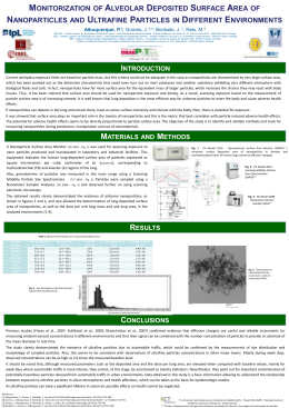
Monitorization of alveolar deposited surface area of nanoparticles and ultrafine particles in
different environments
1
2,3
3
4
Albuquerque, P. , Gomes , J.,Bordado , J., Reis, M.
1
ESTESL – Escola Superior de Tecnologia de Saúde de Lisboa – Instituto Politécnico de Lisboa, Av. D.
João II, Lote 4.69.01, 1990-096 Lisboa, Portugal
2
Área Departamental de Engenharia Química, ISEL – Instituto Superior de Engenharia de Lisboa –
Instituto Politécnico de Lisboa, R. Conselheiro Emídio Navarro, 1959-007 Lisboa, Portugal
3
IBB – Instituto de Biotecnologia e Bioengenharia / Instituto Superior Técnico – Universidade Técnica
4
de Lisboa, Av. Rovisco Pais, 1049-001 Lisboa, Portugal Instituto de Medicina Preventiva, Faculdade
de Medicina de Lisboa, Universidade de Lisboa, Av. Prof. Egas Moniz, 1649-028 Lisboa, Portugal
4
Instituto de Medicina Preventiva, Faculdade de Medicina de Lisboa, Universidade de Lisboa, Av. Prof.
Egas Moniz, 1649-028 Lisboa, Portugal
Email: [email protected]
Abstract
Nanotechnology is an important emerging industry with a projected annual market of around one trillion
dollars by 2015. It involves the control of atoms and molecules to create new materials with a variety of
useful functions. Although there are advantages on the utilization of these nano-scale materials,
questions related with its impact over the environment and human health must be addressed too, so
that potential risks can be limited at early stages of development. At this time, occupational health risks
associated with manufacturing and use of nanoparticles are not yet clearly understood. However,
workers may be exposed to nanoparticles through inhalation at levels that can greatly exceed ambient
concentrations.
Current workplace exposure limits are based on particle mass, but this criteria could not be adequate in
this case as nanoparticles are characterized by very large surface area, which has been pointed out as
the distinctive characteristic that could even turn out an inert substance into another substance
exhibiting very different interactions with biological fluids and cells. Therefore, it seems that, when
assessing human exposure based on the mass concentration of particles, which is widely adopted for
particles over 1 µm, would not work in this particular case. In fact, nanoparticles have far more surface
area for the equivalent mass of larger particles, which increases the chance they may react with body
tissues. Thus, it has been claimed that surface area should be used for nanoparticle exposure and
dosing. As a result, assessing exposure based on the measurement of particle surface area is of
increasing interest. It is well known that lung deposition is the most efficient way for airborne particles to
enter the body and cause adverse health effects. If nanoparticles can deposit in the lung and remain
there, have an active surface chemistry and interact with the body, then, there is potential for exposure.
It was showed that surface area plays an important role in the toxicity of nanoparticles and this is the
metric that best correlates with particle-induced adverse health effects. The potential for adverse health
effects seems to be directly proportional to particle surface area. The objective of the study is to identify
and validate methods and tools for measuring nanoparticles during production, manipulation and use of
nanomaterials.
With this aim, a Nanoparticle Surface Area Monitor, TSI3550, was used for assessing exposure to nano
particles produced and manipulated in laboratory and industrial facilities. This equipment indicates the
human lung-deposited surface area of particles expressed as square micrometers per cubic centimeter
2
3
of air (µm /cm ), corresponding to tracheobronchial (TB) and alveolar (A) regions of the lung. Also,
granulometry of particles was measured in the nano range using a Scanning Mobility Particle Size
Spectrometer, TSI3034. Particles were sampled using a Nanometer Sampler Analyser, TSI3089 and
observed further on using scanning electronic microscopy.
The obtained results clearly demonstrated the existence of airborne nanoparticles, as shown in figures 1
and 2, and also allowed the determination of lung-deposited surface area of nanoparticles, as well as
the dose per unit lung mass and unit lung area, in the analyzed environments [1-4].
References
[1] Albuquerque, P., Gomes, J., Bordado, J., Journal of Air & Waste Management Association, 62
(2012) 373-380.
[2] Gomes, J., Albuquerque, P., Miranda, R., Vieira, M., Journal of Toxicology and Environmental Health
– A, 75 (2012) 747-755.
[3] Gomes, J., Albuquerque, P., Miranda, R., Santos, T., Vieira, M., Inhalation Toxicology, 24 (2012)
774-781.
[4] Bordado, J., Gomes, J., Albuquerque, P., Journal of Air & Waste Management Association, 62
(2012) 1170-1180.
Figure 1 – TEM image of ultrafine particulates collected outdoor in Lisbon
Figure 2 – Size distribution of ultrafine particles collected outdoor in Lisbon
Download