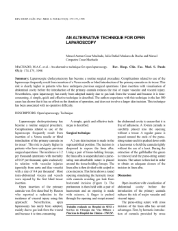
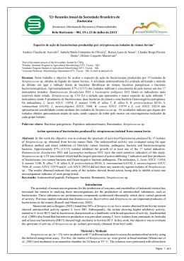
Acta Scientiae Veterinariae, 2014. 42: 1210.
RESEARCH ARTICLE
ISSN 1679-9216
Pub. 1210
Two-stage Rumenostomy in Buffaloes
Pedro Paulo Maia Teixeira1,2, Marco Augusto Machado Silva3 & Rinaldo Batista Viana4
ABSTRACT
Background: Rumenostomy may be performed for therapeutic and digestibility research purposes in bovines, small ruminants and camelids. Several studies requires romenostomy in buffaloes in order to sample ruminal content for laboratorial
assays. However, complications and outcome of rumenostomy was poorly studied in buffaloes. Therefore, the aim of the
current study was to describe a two-stage rumenostomy technique in buffaloes, focused on intra and post-operative period.
Materials, Methods & Results: Nine Murrah buffaloes were submitted to a 36-h and 12-h of food and water fastening. The
animals were given acepromazine and maintained in standing position. Flank local anesthesia was carried out. A circular
skin incision was carried out in the center of the left flank, followed by divulsion of the external and internal obliques and
transversus abdominus muscles, and incision of the peritoneum. Subsequently, a segment of the dorsal aspect of the rumen
was grasped and pulled through the flank incision. The rumen was attached to the peritoneum and skin incision margins
in four points (dorsal, ventral, cranial and caudal). Additional simple interrupted sutures attaching the rumen serosa to the
skin were applied subsequently. Four additional interrupted horizontal mattress sutures were applied equidistantly, taking
bites only in the skin and rumen serosa. Following 12 h, the second stage was carried out. The buffaloes were prepared
and restrained as performed for the first stage. A circular flap was excised from the exteriorized rumen and the silicone
romenostomy cannula was placed. Clinical parameters, postoperative recovery, weight and behavioral pain scale were
assessed. Positioning and anesthesia regimen were adequate for the achievement of the procedure. However, two animals
fell in the restraint chute during the first surgical stage. Mild ischemia of the exteriorized rumen segment was observed on
the second surgical stage, which resulted in less hemorrhage and enhanced cannula positioning. Complete cicatrization
and permanent adhesion of the rumen to the skin were achieved. No ruminal leakage to the abdominal cavity occurred.
No signs of pain were reported. There were few cases of laxity of the romenostomy opening leading to drop of cannula,
myiasis on the margin of the stoma site and few cases of mild ruminal content leakage on the long-term assessment.
Discussion: Restraint in standing position was considered adequate, although lateral recumbence constitutes another
option. However, higher risk of contamination and technical difficulties in placing the cannulas are expected if lateral
recumbence is considered. In other trials using acepromazine, no accidental recumbence occurred. Xylazine was also
indicated for chemical restraint of buffaloes. It is known that flexible cannulas provide better anatomic adjustment and
adaptation as well as being effective for sampling ruminal content, as seen in the current study. Ruminal leakage is one
of the most frequent complications of romenostomy, which may affect animal’s welfare. The animals in the current study
presented no variations on the body score, even though on those presenting cannula loosening or ruminal content leakage.
Moreover, no significant changes of the ruminal content parameters were noticed. Myiasis was also reported following
ruminal surgical interventions, which were mainly attributed to extensive breeding. Loss of the cannula, subcutaneous
emphysema and suture dehiscence are common complications of romenostomy. Nonetheless, none of those complications
were found on the current study. Thus, romenostomy was feasible and efficient for sampling and performing assays of the
ruminal content in buffaloes.
Keywords: rumen cannulation, Bubalus bubalis, ruminal stoma, surgery.
Received: 22 March 2014
1
Accepted: 18 September 2014
2
Published: 28 September 2014
Division of Veterinary, State University Midwest, Guarapuava, PR, Brazil. Division of Veterinary Obstetrics, DPVMAP, College of Agricultural Sciences
and Veterinary Medicine, São Paulo State University “Julio de Mesquita Filho”, Jaboticabal Campus, (FCAV/UNESP), Jaboticabal, São Paulo, Brazil.
3
College of Agronomy and Veterinary, Passo Fundo University (UPF), Passo Fundo, RS, Brazil. 4Institute of Animal Health and Production, Federal
Rural University of Amazonia (ISPA/UFRA), Belém, PA Brazil. CORRESPONDENCE: P.P.M. Teixeira [[email protected] - Tel.: +55 (42)
3629-8217]. Division of Veterinary, State University Midwest (UNICENTRO), CEDETEG Campus, Guarapuava, Rua Simeão Camargo Varela de Sá
n. 03. Vila Carli. CEP 85040-080 Guarapuava, PR, Brazil.
1
P.P.M. Teixeira, M.A.M. Silva & R.B. Viana. 2014. Two-stage Rumenostomy in Buffaloes.
Acta Scientiae Veterinariae. 42: 1210.
INTRODUCTION
8 cm ventrally to the transverse process of the lumbar
vertebrae, at the midpoint between the coxae tuber
and the 13th rib (Figure 1A and 1B). The external and
internal obliques and the transverse abdominal muscles
were dissected and retraced according on the direction
of their fibbers, following by incision of the peritoneum
using Mayo straight round-tip scissors.
A part of the rumen was grasped using two
Glock non-traumatic forceps and pulled out through
the laparotomy site (Figure 1C). The exteriorized ruminal surface was attached to the peritoneum and skin
using manufactured autoclaved polyamide (0.60 mm
of diameter) in a simple continuous pattern, which
started as four interrupted sutures applied dorsal, ventral cranial and caudally, taking 1 cm of skin margin.
The ruminal serosa was progressively approximated
to the peritoneal surface and skin margin as the
suture was applied. Four reinforcement interrupted
horizontal mattress sutures were placed 1 cm from
the skin margins, taking bite only on the ruminal USP
was used. Additional interrupted horizontal mattress
sutures were placed for better apposition of the skin
to the rumen serosa as necessary (Figure 1C, 1D e
1E). All sutures were performed using a “s” shaped
triangular needle.
The animals were given flunixin meglumine3
(1.1 mg/kg, IM, SID, for 3 days), a single dose of an
association of penicilins4 (30.000 UI/kg of benzathine
penicillin, IM) and wound care included repellent
topical ointment over the ruminal surface (Figures
1F, 2A e 2B).
The second surgical stage was carried out
following 12 days post-op. The same patient preparation and restraint used in the first stage was adopted.
A circular incision was performed accomplishing all
layers of the rumen, followed by insertion of the ruminal cannula (Figure 2D). A four inches in diammeter
silinicone cannula for bovines5 was used (Figure 2E
and F). Wound care was continued using the same
protocol as used for the previous stage was adopted.
Ointment was applied to the skin and ruminal surface,
surrounding the cannula.
Clinical parameters, wound cicatrization and
complications were assessed between surgeries ando
n the post-operative period of the second stage if
present. Weight was measured for 30 days by the first
surgical stage, as well as pain behavior. The animals
were observed for six months following rumenostomy.
Romenostomy is used for therapeutical purposes in cases of reccurrent timpanism and ruminal
disorders. However, the main aspect of cannulation
of the gastrointestinal tract is to provide access to the
digestive chambers for assaying in situ digestibility
and nutrition on breeding animals, especially concerning nutrition in ruminants [6, 7]. Several surgical
techniques were reported, mainly in bovines, small
ruminants, alpacas and llamas [2, 3, 8, 10].
Although several studies regarding nutrition
of buffaloes were carried out, which were basically
conduced following cannulation of the rumen for
laboratorial assays, [4, 5], there are no studies focusing
on the technical aspect and postoperative outcome of
rumenostomy in the bubaline specie. Moreover, buffaloes present several morphophysiologic and behavioral
different characteristics from the bovine specie, which
may affect differently their response to surgical trauma.
Based on those facts, the aim of the current
study was to describe and assess a two-stage rumenostomy technique in buffaloes, as well as to evaluate
intra and long-term postoperative complications and
behavioral changes.
MATERIALS AND METHODS
Nine Murrah breed neutered buffaloes, mean
age of 24 ± 6 months old, weighting 320 ± 25 kg, were
assessed. The animals were confined in Brachiaria
humidicula and natural grass paddocks on a holm
area, receiving ad libitum water and mineral salt. The
animals were selected following general clinical examination, parasitologic assays and complete hemogram.
Rumenostmoy was carried out in two stages.
On the first stage, 36-h and 12-h food and water fasting was performed, respectively. The buffaloes were
tranquilized using acepromazine1, (0.10 mg/kg, intramuscularly) and restrained in standing position in
restraint chutes. Wide clipping and asseptic prepare
were performed on the left paralumbar area. Local
anesthesia was carried out using 2% lidocaína chloride
without vasoconstrictor2, employing the “inverted L”
infiltration technique, associated to incision line subcutaneous and intramuscular block.
Subsequently, the incision line was delimited using the cannula cap as reference. A circular
skin incision was performed on the center of the left
paralumbar fossa and the skin was excised, beginning
2
P.P.M. Teixeira, M.A.M. Silva & R.B. Viana. 2014. Two-stage Rumenostomy in Buffaloes.
Acta Scientiae Veterinariae. 42: 1210.
Figure 1. Intra-operative images of the first of two stages of the two-stage rumenostomy in buffaloes. Demarking the incision line with the
cannula cap (A), cicular skin dieresis on the left paralumbar space (B). Access to the rumen through the skin defect and flank musclese; grasping the left dorsal portion of the rumen, beginning of the rumenopexy (C). Progression of the attachment of the rumen to the peritoneum (D).
End of the suture showing the exposed ruminal surface (E). Early post-operative image of the first surgical stage, showing the patient’s posture
and use of repelent intment on the exposed ruminal surface (F).
Figure 2. Images of the evolution of the surgical wound following the accomplishment of the first stage and end of the two-step rumenostomy in
buffaloes. Surgical wound following 24 h of rumenopexy (A); following four days of surgery (B); right before the second surgical stage (C). Exeresis
of the exposed part of the rumen; mild hemorrhage is seen (D); following cannula placement (E); the patient in the early post-operatiove period (F).
RESULTS
Although some technical difficulty was found
for applying sutures to the flank abdominal muscles
due to their thickness on the bubaline specie, no major
complication occurred during suture.
The exposed part of the rumen became ischemic
in all patients, which enhanced cannula positioning and
The sedation, anesthesia and restraint used in
the current study was fairly adequate for the accomplishment of the procedure. However, two animals
(22%) fell in the restraint chute during surgery on the
first surgical stage.
3
P.P.M. Teixeira, M.A.M. Silva & R.B. Viana. 2014. Two-stage Rumenostomy in Buffaloes.
Acta Scientiae Veterinariae. 42: 1210.
Besides affecting the hygienic aspect, ruminal leakage impacts the animal’s welfare [11]. Even though
tere were few cases of leakage of ruminal fluid and
dropping of the cannula in the current study, the
animals were kept in good body score during all the
study, revealing no major impact over the ruminal
physiology.
Myiasis was also found in other trial [10]. In
the current study, myiasis occurred in one animal,
which was attributed to extensive managing.
The two-stage rumenostomy technique provided both cannula adjustment and wound cicatrization.
Loss of cannulas, subcutaneous emphysema, wound
dehiscence and surgical site infections are the most
common post-surgical complications of rumenostomy
in ruminants [8]. Nevertheless, none of such complications occurred in the current study regarding the
bubaline specie.
prevented hemorrhage. The silicone-based cannula was
also important for proper adjustment to the stoma.
Complete cicatrization and adhesions among
the rumen and the abdominal wall were observed.
There was no leakage of the ruminal fluid to the abdominal cavity and no cases of peritonitis occurred.
Except for the spontaneous recumbence in two
animals, no early post-surgical complications occurred.
The long-term follow-up revealed few cases of dropping of the cannula and small amounts of leakage of
ruminal content and myiasis in an animal (11%). The
animals were able to be used for assays on nutrition
and no other complications were noted.
DISCUSSION
Although restraint in standing position was
fairly adequate in the current study, lateral recumbence
was also stated as a successful restraint for surgical
approach. However some risk of contamination and
difficulty in placing the cannula is advised [6].
Acepromazine was cited for tranquilization
and penile exposure in another trial, however with no
cases of recumbence in the restraint chute [1]. Lowdose xylazine should be considered if recumbence is
not desired [7].
No technical difficulty in passing the sutures
through the flank muscles was reported before in species subjected to rumenostomy [2,3,9,11], as seen in
the buffaloes on the current study. However, no major
complications occurred during such surgical maneuver.
The flexibility of the silicone-based cannula
used in the current trial was important for placing
the apparatus thgough the stoma. The use of flexible
cannulas provides better anatomical adjustment and is
efficient for retrieval of ruminal content with minimal
risk of local necrosis and peritonitis [11].
Ruminal content leakage is reported as
one of the major complications of rumenostomy.
CONCLUSION
The two-stage rumenostomy was feasible
in buffaoes and provided minimal impact over the
animal’s welfare, wile being adeequate for the accomplishment of assays on nutrition on the bubaline specie.
SOURCES AND MANUFACTURERS
1
Acepran 1%®, Vetnil. SP, Brazil.
2
Lidovet®, Brasvet. RJ, Brazil.
3
Desflan®, Ourofino. SP, Brazil.
4
Pencivet® Plus PPU, MSD Saúde Animal. SP, Brazil.
5
Cânula para rúmen 4” - silicone, KEHL® SP, Brazil.
Ethical approval. The current study was approved by the Comittee for Ethics and Animal Use in Research conduced of the
“Universidade Federal Rural da Amazônia”.
Declaration of interest. The authors report no conflicts of
interest. The authors alone are responsible for the content and
writing of the paper.
REFERENCES
1 Bittencourt R.H.F.P.M., Silva M.C., Viana R.B., Vale W.G., Araújo C.V. & Moreira V.M.T.S.M. 2005. L´uso del
maleato di acepromazina per l´esteriorizzazion del pene dei bufali (Bubalus bubalis). Bubalus Bubalis. 4: 75-82.
2 Cabrera C.R., Lopez C.A., Maiztegui V.J. & Marin G.M.P. 1996. Fistulacion y canulacion permanente del compartimento 1 (rumen) en alpacas. Avances em Ciencias Veterinarias. 11(2): 108-111.
3 Cattelan J.W., Janola J.C., Valadão C.A.A. & Macoris D.G. 1990. Rumenostomia canulada em dois tempos operatórios em bovinos e ovinos. Ars Veterinárial. 6(2): 112-119.
4 Delgado D.C., Rosabal Y. & Cairo J. 2005. In situ ruminal degradability of Pennisetum purpureum Cuba CT-115 in
commercial river buffaloes and Zebu. Cuba Jornal Agricultury Science. 39(2): 181-185.
4
P.P.M. Teixeira, M.A.M. Silva & R.B. Viana. 2014. Two-stage Rumenostomy in Buffaloes.
Acta Scientiae Veterinariae. 42: 1210.
5 Franzolin R., Rosales F.P. & Soares W.V.B. 2010. Effects of dietary energy and nitrogen supplements on rumen
fermentation and protozoa population in buffalo and zebu cattle. Revista Brasileira de Zootecnia. 39(3): 549-555.
6 Laflin S.L. & Gnad D.P. 2008. Rumen Cannulation: Procedure and Use of a Cannulated Bovine. Veterinary Clinics
Food Animal Practice. 24(2): 335-340.
7 Minervino A.H.H., Barrêto Júnior R.A., Rodrigues F.A.M.L., Ferreira, R.N.F., Saut J.P.E., Queiroz G.F., Dos
Reis L.F. & Ortolani E.L. 2009. Biópsia hepática por laparotomia paracostal em bovinos e búfalos. Ciência Rural.
39(3): 798-802.
8 Muzzi L.A., Muzzi R.A.L. & Gabellini E.L.A. 2009. Técnica de fistulação e canulação do rúmen em bovinos e ovinos.
Ciências Agrotecnica. 33(Edição Especial): 2059-2064.
9 Sarma K.K., Baishya M.P. & Sharma M. 2010. Rumenostomy in Iake (Propeophagus grunniens) for Experimental
Research. Intas Polivet. 11(2): 404-045.
10 Sanni B.D., Olainipekun E.O., Sackey A.K., Fadason S.T. & Gyang E.O. 2002. Post-surgical complications from
students` large animal surgical exercise. Nigerian Veterinary Journal. 23(2): 40-45.
11 Stedile R., Beck C.A.C., Nóbrega F.S., Silva Filho A.P.F., Ferreira M.P., Alievi M.M., Schiochet F., Bordin A.I.,
Gonzalez P.C. & Lampert M. 2008. Rumenostomia com colocação de cânula flexível em ovinos. Acta Scientiae
Veterinariae. 36(1): 35-38.
www.ufrgs.br/actavet
5
1210
Download