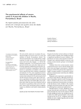
in
linical and Laboratorial
Research in Dentistry
Restorative Dentistry
The effects of ionizing radiation on the development
of human caries lesions in vitro
• Deborah Pereira Lee Undergraduate Student, School of Dentistry, University of São Paulo, São Paulo, SP, Brazil • Luciana
Cardoso Espejo-Trung Department of Operative Dentistry, School of Dentistry, University of São Paulo, São Paulo, SP,
Brazil • Maria Regina Lorenzetti Simionato Department of Microbiology, Institute of Biomedical Sciences, University of São
Paulo, São Paulo, SP, Brazil • Fabio de Abreu Alves Department of Stomatology, School of Dentistry, University of São Paulo,
São Paulo, SP, Brazil • Moacyr Domingos Novelli Department of Stomatology, School of Dentistry, University of São Paulo,
São Paulo, SP, Brazil • Maria Aparecida Alves de Cerqueira Luz Department of Operative Dentistry, School of Dentistry,
University of São Paulo, São Paulo, SP, Brazil
abstract
|
descrIPtors
|
resuMo
|
descrItores
|
Radiotherapy is associated with several undesired side effects, such as rampant radiation caries. The aim
of this study was to evaluate the effect of ionizing radiation on the development of carious lesions using a
bacterial system in vitro. Fifteen sound human molars were selected and sectioned into buccal (A, control)
and lingual (B, irradiated) dental fragments, which were considered dependent. Group B was submitted
to radiotherapy according to the protocol for head and neck oncological treatment. The two groups were
exposed to a cariogenic challenge using a bacterial system with S. mutans for 10, 20 and 30 days (n = 5).
The variabels depth, extension and area for lesions formed at the enamel-dentin junction were measured
by software coupled with light microscopy. Optical coherence tomography (OCT) was used to visualize
the morphological characteristics of the lesions. Only the 20-day period of culture immersion for caries
development resulted in significantly better lesion comparisons, by light microscopy. Of the three lesion
dimensions analyzed, lesion depth (lD) differed statistically between groups A and B (p = 0.013). Analysis
using OCT allowed the visualization of carious lesions without showing the carious layers. Within the limitations of this study, we can conclude that radiation treatment of sound teeth before a cariogenic challenge
in vitro causes deeper carious lesions than in those teeth not subjected to radiation treatment.
Dental Caries; Head and Neck Neoplasms; Radiotherapy; Streptococcus mutans.
Efeito da radiação ionizante no desenvolvimento de lesões de cárie em dentes humanos in vitro • A radioterapia é associada
a diversos efeitos colaterais, entre eles a cárie de radiação. O objetivo desse estudo foi avaliar o efeito da radiação ionizante no desenvolvimento de lesões cariosas utilizando um sistema bacteriano in vitro. Foram selecionados quinze terceiros molares humanos inclusos
que foram seccionados em fragmentos vestibular (A, controle) e lingual (B, irradiado), e foram mantidos pareados até o término do estudo. O grupo B foi submetido à radioterapia de acordo com protocolo utilizado no tratamento radioterápico de cabeça e pescoço. Ambos os grupos foram expostos a um desafio cariogênico utilizando um sistema bacteriano com S. mutans por 10, 20 e 30 dias (n = 5). As
variáveis de profundidade, extensão e área das lesões formadas no limite amelo-dentinário foram medidas por um software acoplado
ao microscópio de luz. A Tomografia de coerência óptica (TCO) foi utilizada para visualizar as características morfológicas das lesões.
Os resultados da microscopia de luz mostraram que, no período de 20 dias de desafio bacteriano, ocorreu um resultado significante,
comparando a profundidade das lesões formadas entre os grupos A e B (p = 0.013). A análise de TCO não permitiu visualizar as camadas de cárie das lesões. Podemos concluir, dentro das limitações do estudo, que o tratamento radioterápico pode levar à formação de
lesões de cárie mais profundas do que aquelas que se desenvolvem em dentes sem exposição à radiação ionizante.
Cárie Dentária; Neoplasias de Cabeça e Pescoço; Radioterapia; Streptococcus mutans.
corresPondIng author
|
• Maria Aparecida Alves de Cerqueira Luz Department of Operative Dentistry, School
of Dentistry, University of São Paulo. • Av. Prof. Lineu Prestes, 2227 São Paulo, SP,
Brazil • 05508-000 E-mail: [email protected]
• Received Jan 07, 2013 • Accepted Jun 24, 2013
46
● Clin Lab Res Den 2014; 20 (1): 46-53
Lee DP • Espejo-Trung LC • Simionato MRL • Alves FA • Novelli MD • Luz MAAC •
Introduction
toms because of loss of pulpal sensitivity. These le-
Cancer is one of the major health problems in
sions are observed in anterior and posterior teeth,
the world. In Brazil, the National Cancer Institute
which may develop four weeks after completion of
estimates that there were 14,170 new cases in 2012,
radiotherapy or 3–6 months after the beginning of
with 9,990 men and 4,180 women being diagnosed.
treatment. 4,8,9 The multifactorial disease termed ra-
In 2009, 6,510 deaths occurred due to oral cancer.1
diation caries is not well-defined by the literature, 4,7
Radiotherapy is often used in palliative or cura-
nor by its histological features. Several authors sug-
tive treatment of malignancies, since this method is
gest that radiation caries occurs due to pH changes
capable of destroying tumor cells using ionizing ra-
caused by a decrease in oral salivary flow because
diation beams. The total radiation dose to be used is
of salivary gland damage from treatment of head
usually split into equal daily doses in order to guar-
and neck cancer. 4,5,6,2,9,10 Others, such as Poyton11
antee tissue tolerance; this way, the biological effect
and Anneroth et al.,12 suggest that these lesions
reaches the largest number of neoplastic cells.
are caused by changes in the crystalline structure
Many patients with head and neck cancer are
of hard tissues as a consequence of direct radiation
exposed to high doses of radiotherapy in extensive
to the teeth. Franzel and Gerlach13 concluded that
fields of radiation that include the oral cavity, max-
enamel and dentin are strongly affected by high
illa, mandible and salivary glands. Radiotherapy
and low energy irradiation, and, consequently, that
preserves the tissue structure but causes adverse
the mechanical properties of the enamel are dam-
reactions that appear in the oral cavity.
2,3
The se-
aged by cancer radiation treatment.
verity of these reactions depends on the volume
Considering the small number of studies about
and radiation location, total dose, fractionation,
this topic and the disagreement in the literature
age, clinical conditions of the patient and associ-
about the role of radiotherapy in radiation caries,
ated treatments. These reactions can occur in the
the aim of this study was to evaluate the hypothesis
acute phase (during or within the first weeks of
that radiotherapy is able to increase the caries risk
treatment) or in the chronic phase (months or years
of human teeth subjected to a cariogenic challenge,
after the radiotherapy), and include the following
using a bacterial method in vitro.
conditions:
Objective
• mucositis,
• candidiasis,
• taste alteration,
• osteoradionecrosis,
• soft tissue necrosis,
• dry mouth and
• radiation caries.4,5,6,2,7
The aim of this study was to evaluate, in vitro,
the effect of ionizing radiation on the development
of carious lesions using a bacterial system.
Materials and Methods
Fifteen extracted, non-erupted human third
molars without previous lesions or visible enamel
Rampant caries from radiation is a common
and important complication
4,3
defects were stored in distilled water at 4°C. Ap-
of treatment and
proval by the Ethics Committee, School of Dentist-
usually develops at the cervical level of the tooth,
ry, University of São Paulo was obtained. After the
starting at the labial surface and moving sequen-
apical 2/3 of the roots had been removed, the teeth
tially to the lingual surface in a chronic process.
were sectioned with double-faced diamond discs
The change usually occurs without painful symp-
(Discoflex, KG Sorensen, Cotia, SP, Brazil) into two
Clin Lab Res Den 2014; 20 (1): 46-53 ● 47
The effects of ionizing radiation on the development of human caries lesions in vitro
fragments:
• buccal (A, control) and
• lingual (B, irradiated).
300 ml of TSB with 5% sucrose and 3 ml of the
inoculum broth, and were maintained in this bacterial system and transferred to a fresh tube every
24 h. During the incubation period, tests were per-
Group B was submitted to radiotherapy accord-
formed to check for contaminants. After 10, 20 and
ing to the protocol for head and neck oncological
30 days, the groups were removed from each bacte-
treatment. The dental fragments from groups A and
rial system and cleaned.
B were paired, and all of them were cleaned with an
aqueous slurry of pumice and anionic detergent.
Optical coherence tomography (OCT; OCP930SR, Thorlabs, Newton, New Jersey, USA) was
The fragments from Group B were irradiated
used to visualize the morphological characteris-
according to the following protocol: 2 Gy per day
tics of the lesions.16,17,18 In this study, OCT was used
for 35 days, for a total of 70 Gy, by a linear accel-
in order to detect alterations of the tissues optical
erator device (2100C 6×; Clinac iX, Varian Medi-
properties and for qualitative analysis of the cari-
cal Systems, Palo Alto, CA, USA). The sample was
ous lesions at the enamel-dentin junction.
randomly divided into 3 groups (n = 5), with each
After OCT analysis, each specimen was embed-
one being subjected to a cariogenic challenge for a
ded in epoxy resin (Arazym, Redlease, São Paulo,
specific period of time:
SP, Brazil) and sectioned in a buccolingual direc-
• Group 1, 10 days;
• Group 2, 20 days;
• Group 3, 30 days.
tion, resulting in sections with 300 µm thickness,
exposing the enamel-dentin junction. Three of
these sections were randomly chosen from the central area of the lesions and were ground by hand to
During the cariogenic challenge there were
a thickness of 80–120 µm using sandpaper for light
specimen losses in Group 1 and Group 2. Conse-
microscopy analysis. Each section was examined
quently, an n = 4 was considered for all the groups,
under a light microscope (Citoval 2 with CarlZeiss
randomly ruling out one specimen of Group 3.
lens, Laboral 4, Zeiss, USA) coupled with software
Steel wires were attached to each dental frag-
(DIRACOM 3 Imagelab 2000, Bio Rad Canton,
ment, and the surfaces of the fragment were paint-
MA, USA) in order to take the following measure-
ed with acid-resistant nail varnish (Risque, Niasi
ments (variables):
Ind. de Cosméticos, Taboão da Serra, SP, Brazil),
• lesion extension (LE), from the external lesion
except on a 3 × 5 mm window at the enamel-dentin
margin to the opposite margin at the enamel-
junction. The specimen/wire sets were sterilized
dentin junction;
with gamma irradiation (25 kgy), using a Gama-
• lesion depth (LD), from the enamel-dentin junc-
cel 220 device (Atomic Energy of Canada, Chalk
tion to the deepest point of the lesion in dentin;
River, Ontario, Canada), at the Institute of Energy
• lesion area (LA), calculated using the software’s
and Nuclear Research. The development of carious
“calculated area” tool, based on the selected
lesions was induced in vitro with a bacterial sys-
area of the lesion.
tem following the method used by Gama-Teixeira
et al.14 and Espejo et al.15 The microorganism used
The data were subjected to statistical analysis
was S. mutans ATCC 25.175. The specimens of the
using Student’s t-test for pairwise comparisons of
same group were immersed in a tube containing
each period of cariogenic challenge (p < 0.05).
48 ● Clin Lab Res Den 2014; 20 (1): 46-53
Lee DP • Espejo-Trung LC • Simionato MRL • Alves FA • Novelli MD • Luz MAAC •
|
Figure 1 OCT Images. Twenty
days of cariogenic challenge. A: A
thin, whitish stripe corresponding to
less scattered light representing less
demineralized enamel can be seen on
the non-irradiated specimen. B: A wide
stripe corresponding to more scattered
light representing more demineralized
enamel can be seen on the
irradiated specimen.
|
Figure 2 Light microscopy
analysis (60×). A: Non-irradiated
specimen; B: Irradiated specimen.
The red line indicates the region and
direction of depth measurement;
E = enamel; D = dentin.
A
B
A
Results
B
iogenic challenge on enamel and/or dentin and of
The images taken with OCT are based on the op-
the enamel-dentin junction (Figure 1A), correspond-
tical properties of the sample and they are made us-
ing to the transverse area delineated by a homoge-
ing false color mapping, where the white color corre-
neous whitish stripe that runs continuously on the
sponds to a high spreading coefficient equivalent to
healthy enamel. The healthy dentin exhibited a simi-
demineralized areas and the black color corresponds
lar whitish stripe that was less homogeneous than
to a low spreading coefficient. In this study, the im-
that of the enamel, but had a clearer surface bound-
age taken with OCT allowed observation of the de-
ary. Enamel and dentin displayed distinct spreading
mineralization depth of the area exposed to the car-
characteristics in the OCT images. The tissue struc-
Clin Lab Res Den 2014; 20 (1): 46-53 ● 49
The effects of ionizing radiation on the development of human caries lesions in vitro
Table 1 | Descriptive analysis of the
Control
Treatment
data and comparison between
groups (n = 4) considering
the factors analysed.
Mean
10 days
Depth (µm)
20 days
Area (µm2)
a
1.08
b
1.66
a
0.07
0.75
Mean
1.58
0.37
2.26
c
0.81
a
0.22
1.68
0.35
2.16
Total
1.48
0.52
2.00
11.49
d
3.84
SD
a
30 days
10 days
Extension (µm)
Irradiated
SD
0.57
9.78
d
3.57
d
2.07
20 days
d
15.91
4.29
11.38
30 days
18.40d
2.94
15.99d
3.45
Total
15.27
4.51
12.38
3.93
10 days
14.61
e
2.94
18.12
e
20 days
26.11e
7.36
12.30
27.23e
9.33
30 days
35.85e
15.31
41.17e
8.48
Total
25.52
13.78
28.84
12.49
Different letters in the same row indicate statistically significant differences analyzed using the paired t test
for each factor (α < 0.05).
tures with carious lesions showed a whitish stripe
affirmed that radiotherapy indirectly causes caries
deeper than did the healthy teeth (Figure 1B).
by compromising the salivary glands tissue, causing
Through the light microscopy analysis (Figure
hyposalivation, but without directly damaging the
2A and 2B), it was possible to make comparisons
dental tissue. Conversely, several authors12,11 affirm
between the carious lesions of both groups studied,
that these lesions occur owing to modifications of
irradiated and non-irradiated teeth. The data are
the crystalline structure of hard tissues as a conse-
presented in Table 1. The comparison between the
quence of the direct action of radiation to the teeth.
periods of time of cariogenic challenge showed that
Others5 claim that both hyposalivation and direct
only after 20 days of the bacterial cariogenic chal-
radiation make the teeth more susceptible to decal-
lenge was there a significant difference between the
cification and affect the development of radiation
lesions formed in both groups studied. Compar-
caries. We agree with Springer et al.7 and Aguiar
ing the depth of the lesions of the irradiated and
et al.4 that both generic caries and radiation caries
non-irradiated teeth after 20 days of cariogenic
should be treated as a multifactorial disease, i. e.,
challenge, the control group (1.66 ± 0.75) showed
in addition to the changes in the dental tissues, the
a smaller lesion depth than the irradiated group
salivation decrease might also favor lesion develop-
(2.26 ± 0.81) with p = 0.013. Comparing the exten-
ment, in addition to other modifying factors, such as
sion and the area of the lesions, it was not possi-
food diet, social conditions, education, and others.2
ble to find statistically significant differences with
In this in vitro study, a comparison of the size of
p = 0.22 and p = 0.87, respectively, for each of the
the carious lesions in the control group and in the
cited variables.
irradiated group was made. After subjecting both
tooth groups to the same bacterial cariogenic chal-
Discussion
lenge, the irradiated tooth lesions were found to be
This study did not consider the saliva factor vari6,9,19,3
able because several researchers
50 have already
● Clin Lab Res Den 2014; 20 (1): 46-53
deeper than those on non-irradiated teeth. However,
the literature does not contain conclusive consider-
Lee DP • Espejo-Trung LC • Simionato MRL • Alves FA • Novelli MD • Luz MAAC •
ations regarding the effect of radiation alone on den-
optical coherence tomography (OCT) was not con-
tal tissues, except for histological changes demon-
clusive because it was difficult to observe the dif-
strated by chromatography analysis on pulp tissue
ferences among the carious lesions, probably be-
7
according to Springer et al. These findings may of-
cause the images of the lesions appeared somewhat
fer an additional explanation for the great incidence
tenuous and, for this reason, were not clear enough
of carious lesions in irradiated patients, mainly in
to make a comparison. Due to these difficulties of
the cervical region of the teeth at the enamel-dentin
interpretation, common light microscopy analysis
junction, since pain sensitivity may be modified due
associated with the measurements of the carious
to histological changes of pulp tissue.
lesions was added to this study in order to obtain
The irradiation protocol adopted in this study
was calculated by a physicist operating the equip-
quantitative data that enabled a more objective
comparison.
ment in such a way that all teeth received the same
We considered that this result pointed indi-
radiation dose. The dental fragments received 2 Gy
rectly to a tissue alteration because the saliva factor
of radiation per day, and, over 35 days, the frag-
and other modifying factors, such as socioeconom-
ments received a total of 70 Gy, which represents
ic and cultural factors, are not present in this case.2
the maximum radiation that a patient receives dur-
The result is in accordance with that of Springer et
ing radiotherapy treatment of the head and neck.
al.7 that verified that a total dose of 31.5 Gy of ra-
Because OCT images did not have good contour
diation would be sufficient to damage the collagen
definition, it was impossible to estimate their di-
fibers present in dentin, enamel and pulp tissues of
mensions clearly; therefore, we could merely affirm
human third molars using chromatographic analy-
or deny the presence of caries by using this mode
sis. The extension of the lesion was similar for all
of lesion analysis. Comparing the periods of time
groups because it depends directly on the contact of
of the bacterial cariogenic challenge—10, 20 and
the bacterial biofilm present on the exposed enam-
30 days—using light microscopy associated with
el and dentin area, which had the same dimensions
a measurement of the lesions, we observed differ-
in all specimens. Although the depth of the lesions
ences in carious lesions only after the intermediate
was statistically higher in irradiated teeth than
period of time (20 days). It was observed that the
in non-irradiated teeth in the 20-day cariogenic
lesions that developed over 10 days of cariogenic
challenge group, the area of the lesions did not dif-
challenge were too small, and the lesions that de-
fer significantly between the groups. This differ-
veloped over 30 days were too large, for compari-
ence can be explained by the small sample size of
sons. This usually occurs using the bacterial chal-
each group and by the high standard deviation of
lenge, since the bacterial strains may have different
the lesion extension measurements, since the cal-
pathogenicity, and the teeth may have different
culation of area used extension versus depth. The
susceptibility. These were the reasons why we used
depth measurements, in contrast, exhibited a low
different periods of challenge in this study. After
standard deviation; for this reason they showed a
20 days of cariogenic challenge, it was possible to
clear difference between non-irradiated and irra-
observe differences among the depths of the le-
diated teeth. The tooth crown location chosen for
sions, which were deeper in irradiated teeth than in
development of the lesions was the region of the
non-irradiated ones. Therefore, the results after 20
enamel-dentin junction because previous studies
days were used in the discussion.
showed a great clinical incidence of carious lesions
The analysis method initially adopted using
in this dental area in irradiated head and neck pa-
Clin Lab Res Den 2014; 20 (1): 46-53 ● 51
The effects of ionizing radiation on the development of human caries lesions in vitro
tients.9 The collagen fibers, more present in dentin
Conclusion
than in enamel, could have suffered alterations
Within the limitations of this in vitro study, it
due to the radiation-enabled progression of the le-
can be concluded that radiation treatment of sound
sion in the direction of the pulp, since it has been
teeth may cause deeper carious lesions than in
19
non-irradiated teeth subjected to the same cario-
shown that collagen fibers present in dental pulp
undergo histological modification after radiation
treatment, which may change pain sensitivity and
favor a higher incidence and larger development of
genic challenge.
Acknowledgments
caries lesions in patients. This interpretation leads
The authors of this study are thankful to FUN-
indirectly to the conclusion that the ionizing radia-
DECTO; to Prof. Anderson Zanardi de Freitas from
tion used for oncological head and neck treatment
the Nuclear and Energy Research Institute (IPEN-
can induce tissue alterations in enamel and/or den-
CNEN/SP), São Paulo, SP, Brazil; and to the Tech-
tin that facilitate the development of dental caries;
nological Center for Radiation, A. C. Camargo Hos-
however, more studies are needed to analyze irra-
pital.
diated tissues.
References
1. Brazil. Ministry of Health. National Institute of Cancer
7. Springer IN, Niehoff P, Warnke PH, Böcek G, Kovács G, Suhr
José Alencar Gomes da Silva. Estimate/2012 - Cancer Inci-
M, et al. Radiation caries--radiogenic destruction of dental
dence in Brazil [Internet]. Rio de Janeiro: Inca; 2011 [cited
collagen. Oral Oncol. 2005 Aug;41(7):723-8.
2013 May 23]. Available from: URL:http://www.saude.sp.gov.
8. Jham BC, Reis PM, Miranda EL, Lopes RC, Carvalho AL,
br/resources/ses/perfil/gestor/homepage/estimativas-de-
Scheper MA, et al. Oral health status of 207 head and neck
incidencia-de-cancer-2012/estimativas_incidencia_can-
cancer patients before, during and after radiotherapy. Clin
cer_2012.pdf. Oral Investig. 2008 Mar;12(1):19-24.
2. Moore S, Burke MC, Fenlon MR, Banerjee A. The role of
9. Silva AR, Alves FA, Antunes A, Goes MF, Lopes MA. Patterns
the general dental practitioner in managing the oral care
of demineralization and dentin reactions in radiation-related
of head and neck oncology patients. Dent Update. 2012
Dec;39(10):694-6,698-700,702.
caries. Caries Res. 2009;43(1):43-9.
10. Silva AR, Alves FA, Berger SB, Giannini M, Goes MF, Lopes
3. Thariat J, Ramus L, Darcourt V, Marcy PY, Guevara N, Odin
MA. Radiation-related caries and early restoration failure in
G, et al. Compliance with fluoride custom trays in irradiated
head and neck cancer patients. A polarized light microscopy
head and neck cancer patients. Support Care Cancer. 2012
and scanning electron microscopy study. Support Care Can-
Aug;20(8):1811-4.
cer. 2010 Jan;18(1):83-7.
4. Aguiar GP, Jham BC, Magalhães CS, Sensi LG, Freire AR.
A review of the biological and clinical aspects of radiation
caries. J Contemp Dent Pract. 2009 Jul 1;10(4):83-9.
5. Jham BC, da Silva Freire AR. Oral complications of radiotherapy in the head and neck. Braz J Otorhinolaryngol.2006
Sep-Oct;72(5):704-8.
6. Meurman JH, Grönroos L. Oral and dental health care of oral
cancer patients: hyposalivation, caries and infections. Oral
Oncol. 2010 Jun;46(6):464-7.
11. Poyton HG. The effects of radiation on teeth. Oral Surg Oral
Med Oral Pathol. 1968 Nov;26(5):639-46.
12. Anneroth G, Holm E, Karlsson G. The effect of radiation on
teeth. A clinical, histologic and microradiographic study. Int
J Oral Surg. 1985 Jun;14(3):269-74.
13. Fränzel W, Gerlach R. The irradiation action on human dental
tissue by X-rays and electrons -- a nanoindenter study. Z Med
Phys. 2009;19(1):5-10.
14. Gama-Teixeira A, Simionato MR, Elian SN, Sobral MA,
Luz MA.Streptococcus mutans-induced secondary car-
52 ● Clin Lab Res Den 2014; 20 (1): 46-53
Lee DP • Espejo-Trung LC • Simionato MRL • Alves FA • Novelli MD • Luz MAAC •
ies adjacent to glass ionomer cement, composite resin and
17.Lee C, Darling CL, Fried D. Polarization-sensitive opti-
amalgam restorations in vitro. Braz Oral Res.2007 Oct-
cal coherence tomographic imaging of artificial deminer-
Dec;21(4):368-74.
alization on exposed surfaces of tooth roots. Dent Mater.
15. Espejo LC, Simionato MR, Barroso LP, Netto NG, Luz MA.
2009 Jun;25(6):721-8.
Evaluation of three different adhesive systems using a bacte-
18. Maia AM, Fonsêca DD, Kyotoku BB, Gomes AS. Characteriza-
rial method to develop secondary caries in vitro. Am J Dent.
tion of enamel in primary teeth by optical coherence tomog-
2010 Apr;23(2):93-7.
raphy for assessment of dental caries. Int J Paediatr Dent.
16. Azevedo CS, Trung LC, Simionato MR, Freitas AZ, Matos AB.
Evaluation of caries-affected dentin with optical coherence
tomography. Braz Oral Res. 2011 Sep-Oct;25(5):407-13.
2010 Mar;20(2):158-64.
19. Specht L. Oral complications in the head and neck radiation
patient. Introduction and scope of the problem. Support Care
Cancer. 2002 Jan;10(1):36–9.
Clin Lab Res Den 2014; 20 (1): 46-53 ● 53
Baixar