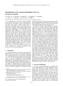
ISSN 1981-5484
Acta Veterinaria Brasilica, v.7, n.1 p.73-75, 2013
FACIAL HEMANGIOSSARCOMA IN A BLUE-FRONTED
AMAZON PARROT (Amazona aestiva)
[Hemangiossarcoma facial em papagaio verdadeiro (Amazona aestiva)]
Guilherme Augusto Marietto-Gonçalves1*, Fabrizio Grandi2,3
1
Avian Pathology Laboratory, Sao Paulo State University, Brazil;
Laboratory of Investigative and Comparative Pathology, Sao Paulo State University, Botucatu-SP, Brazil;
3 Department of Pathology, Medical School, Sao Paulo State University, Botucatu-SP, Brazil;
2
ABSTRACT - Here we describe the first case report of an hemangiosarcoma in a male Blue-fronted
amazon parrot (Amazona aestiva). The mass was localized near the inferior portion of the right orbit. A
fine needle aspiration biopsy (FNAB) was performed and a presuntive diagnosis of
hemangioma/hemangiosarcoma was established. Further histopathological evaluation revealed a
mesenchymal neoplasm composed basically by well formed vascular channels lined by pleomorphic
endothelial cells. Thus, the final diagnosis was a well-differentiated cutaneous hemangiosarcoma.
Keywords: Hemangiossarcoma, birds, Amazon parrot, Pathology.
RESUMO – Aqui se descreve o primeiro caso de hemangiossarcoma em um macho de Papagaio
verdadeiro (Amazona aestiva). A massa estava localizada próxima a porção inferior da órbita direita. Uma
aspiração
utilizando
agulha
fina
foi
realizada
e
o
diagnóstico
presuntivo
de
hemangioma/hemangiossarcoma foi estabelecido. Em seguida, a avaliação histopatológica revelou uma
neoplasia mesenquimal composta, basicamente, por canais vasculares bem formados alinhados com
células endoteliais pleiomórficas. Desse modo, o diagnóstico final foi de hemangiossarcoma cutâneo bem
diferenciado.
Palavras-Chave: Hemangiossarcoma, pássaros, papagaio verdadeiro, patologia.
*
Corresponding author: [email protected].
73
Acta Veterinaria Brasilica, v.x, n.x, p.x-x, 2013
Hematoxilin and Eosin (HE) in order to achieve
a definitive diagnosis. Histologically, the mass
were restricted to dermis, highly cellular and
had an expansible growth. The neoplastic cells
were plump to fusiform and aligned on collagen
trabeculae that formed an anastomosing
meshwork of blood-filled channels of variable
size. The cells were moderately pleomorphic
with and eosinophilic discrete cytoplasm, oval
nuclei, and distinct nucleoli (Figure 1). The
vascular spaces were not completely enclosed
by endothelial cells, and blind-end trabeculae
could be observed. Some vessels were filled by
adherent thrombi. Rare mitotic figures could be
seen. Based on those findings, the final
diagnosis was well differentiated cutaneous
hemangiosarcoma. Finally, the animal died two
weeks after the procedure due to surgical
complications such uncontrollable nasal
bleeding and difficulty feeding.
INTRODUTION
Hemangiosarcoma also known as angiosarcoma
or malignant hemangioendothelioma is a
malignant neoplasm. Is characterized by
pleomorphic endothelial cells aligned on
multiple collagen trabeculae that forms bloodfilled vascular spaces of variable sizes (Maxie,
2007). It is locally invasive and multicentric
tumor that tends to present with inflammation
and necrosis. It may occur primarily in the skin
or as a result of metastasis from a visceral
primary source, and is commonly found in adult
and old animals (Jones et al., 1997; Reavill,
2001; Maxie, 2007). Here we describe the first
case report of an hemangiosarcoma in a bluefronted amazon parrot (Amazona aestiva).
CLINICAL REPORT
An 15-year-old, 320g captive male Amazona
aestiva was presented for examination after a
progressive facial enlargment localized near the
inferior portion of the right orbit. On physical
evaluation, a mass presumably extending into
subcutaneous, measuring 1,5 x 0,8 cm, adhered
to deep tissues, firm, warm, reddish, and nonulcerated was noted in aforementioned region.
Clinically, the animal was anorexic and
depressed.
Samples
for
cytopathologic
examination were obtained by fine needle
aspiration biopsy (FNAB) from 2 different areas
using a fine needle (22G1 ¼´´) and a 10 ml
syringe. Previously to cytological examination,
the animal was sedated with an subcutaneous
administration of ketamine HCl (25 mg/Kg
IM,). The collected material was spread on
histological slides, air-dried, methanol fixed,
and stained with Giemsa. The smears were
highly cellular and composed predominately by
erythrocytes, heterophils and lesser amounts of
monocytes. A presuntive diagnosis of
hemangioma or hemangiosarcoma was stated.
After routine anesthetic procedures (atropine
sulfate 0,02 mg/kg IM. ketamine HCl 20mg/kg
and midazolam 4 mg/kg IM) and local
antisepsis, the animal was submitted to a
excisional biopsy in order to remove the mass.
The lesion was not completely removed during
surgery due to the deep adherence to the
underlying tissues which in turn could
compromise postoperative recovery.
Figure 1. The mass present cells were plump to fusiform
and aligned on collagen trabeculae that formed an
anastomosing meshwork of blood-filled channels of variable
size. The cells were moderately pleomorphic with and
eosinophilic discrete cytoplasm, oval nuclei, and distinct
nucleoli (HE, 40x).
DISCUSSION
In psittacines birds the neoplasm has been
described in Melopsittacus undulates (Freeman
et al., 1999), Pionus spp., Agapornis spp.,
Psittacus erithacus (Reavill, 2004), Primolius
auricollis
(Reavill,
2001),
Nymphycus
hollandicus (Sledge et al., 2006), Amazona
ochrocephala oratrix (Hanley, 2005), A.
farinosa (Rossi, 1998), A. amazonica (Mickley
et al., 2009), and also already described in A.
aestiva (Godoy et al., 2009).
In avian, the most common affected anatomic
areas are the skin of peripheral beak region and
cloaca, neck, dorsum, hind and forelimbs
(Campbell, 1986; Reavill, 2001; Schmidt et al.,
The tumor was fixed in 10% neutral-buffered
formalin, trimmed, routinely processed and
embedded in paraffin. Routine histological
sections 4μm in thickness were stained with
74
Acta Veterinaria Brasilica, v.x, n.x, p.x-x, 2013
2003). Some internal organs such liver, bones,
and ovary can also be affected (Rossi, 1998;
Hanley, 2005; Mickley et al., 2009). Surgical
excision of cutaneous tumors in domestic birds
is usually curative with a good prognosis if no
metastases are present (Bauck, 1997; Maxie,
2007). Grossly, the tumor mass can present as
eryhtematous areas with black spots due to the
inflammatory process associated with multiple
necrotic foci which can lead to follicle loss
(Campbell, 1986; London & Vail, 1996).
Clinically ill birds can present lethargy,
anorexia, weight loss, edema, dyspnea, petechial
hemorrhage, and anemia. The last two signs are
secondary to trauma of the neoplastic mass
(Latimer, 1994).
(Eds). Tratado de Animais Selvagens. São Paulo, Roca,
p.247-248.
Definite diagnosis based on cytology can be
very difficult to impossible since the smears
contain only blood and rarely pleomorphic
neoplastic endothelial cells (Campbell, 1986).
Despite this, a presuntive diagnosis of
hemangiosarcoma can be readily made based on
FNAB. However, histopathological analysis is
considered the definitive diagnostic method
allowing differentiation between vascular
neoplasms, hematomas, filariosis, cutaneous
cysts and Poxvirus infection in birds.
London, C.A., Vail, D.M. 1996. Tumor biology. In:
Whithrow, S.J., Macewen, E.G., (Eds). Small Animal
Clinical Oncology. Philadelphia, WB Saunders Company,
p.16-31.
Godoy, S.N., Alves, V.A.F., Kanamura, C.T., Matushima,
E.R. 2009. Principais processos neoplásicos encontrados em
psitacídeos mantidos em cativeiro. Pesq. Vet. Bras. 29(6):
445-451.
Hanley, C.S., Wilson, G.H., Latimer, K.S., Frank, P.,
Hernandez-Divers,
S.J.
2005.
Interclavicular
hemangiosarcoma in a Double yellow-headed amazon parrot
(Amazona ochrocephala oratrix). J. Avian Med. Surg.
19(2):130-137.
Jones, T.C., Hunt, R.D., King, N.W. 1997. Veterinary
Pathology. 6th ed. Baltimore, Willians and Wilkins, 1392p.
Latimer, K.S. 1994. Oncology. In: Ritchie, B.W., Harrison,
G.J., Harrison, L.R. (Eds). Avian Medicine: Principles and
Applications. LakeWorth, Wingers Publishing, p.640-672.
Maxie, M.G. 2007. Jubb, Kennedy and Palmers: Pathology
of Domestic Animal, 5th ed. Philadelphia, Saunders Elsevier.
Mickley, K., Buote, M., Kiupel, M., Graham, J., Orcutt, C.
2009. Ovarian hemangiosarcoma in an Orange winged
amazon parrot (Amazona amazonica). J. Avian Med. Surg.
23(1):29-35.
Reavill, D.R. 2001. Pet bird oncology. In: Proceedings of
the Association of Avian Veterinarians conference. Avian
Specialty Advanced Program: Orlando, FL; 2011:29-43.
REFERENCES
Reavill, D.R. 2004. Tumors of pet birds. Vet. Clin. North.
Am. Exot. Anim. Pract. 7(3):537-560.
Bauck, L. 1997. Avian Dermatology. In: Altman RB, Clubb,
S.L., Dorrestein, G.M., Quesenberry, K. (Eds). Avian
Medicine and Surgery. Philadelphia, WB Saunders
Company, p.557-568.
Rossi G. 1998. A poorly-differentiated hepatic
haemangiosarcoma in a Amazon farinosa parrot. Avian
Pathol. 27(4):427-430.
Campbell, T.W. 1986. Neoplasia. In: Harrison, G.J.,
Harrison, L.R. (Eds). Clinical Avian Medicine and Surgery.
Philadelphia,WB Saunders Company, p.500-508.
Schmidt, R.T., Reavill, D.R., Phalen, D.N. 2003. Pathology
of Pet and Aviary Birds. Ames, Iowa State Press, 234p.
Freeman, K.P., Hahn, K.A., Adams, W.H. 1999. Radiation
therapy for hemangiosarcoma in a budgerigar. J. Avian Med.
Surg. 13(1):40-44.
Sledge, D.G., Radi, Z.A., Miller, D.L., Lynn, B.S. 2006.
Subcutaneous Haemangiosarcoma in a Cockatiel
(Nymphicus hollandicus). J. Vet. Med. A Physiol. Pathol.
Clin.
Med.
53(6):293-295.
Godoy, S.N. 2006. Psittaciformes (arara, papagaio,
periquito). In: Cubas, Z.S., Silva, J.C.R., Catão-Dias, J.L.
75
Download