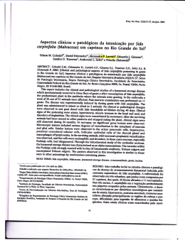
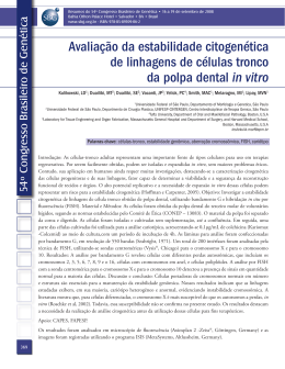
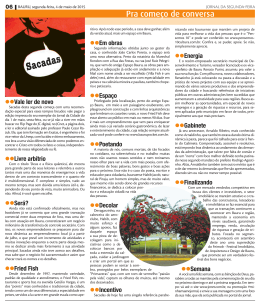
Maria da Conceição Constantino Fernandes
C ONTAMINAÇÃO
DA
B ARRINHA
DE
POR METAIS PESADOS :
E SMORIZ /L AGOA
E FEITOS
E HISTOLÓGICOS EM
L IZA
DE
P ARAMOS
BIOQUÍMICOS
SALIENS
Universidade de Trás-os-Montes e Alto Douro
Vila Real 2007
UNIVERSIDADE DE TRÁS-OS-MONTES E ALTO-DOURO
Tese de Doutoramento em Ciências do Ambiente
Contaminação da Barrinha de Esmoriz/Lagoa de Paramos por
metais pesados: efeitos bioquímicos e histológicos em Liza saliens
Tese original submetida à Universidade
de Trás-os-Montes e Alto Douro para a
obtenção do grau de Doutor em Ciências do
Ambiente, de acordo com o estipulado no
Decreto-Lei 216/92, de 13 de Outubro.
De: Maria da Conceição Constantino Fernandes
Orientadores: Professora Doutora Maria Antónia Santos Mendes Salgado,
Instituto Ciências Biomédicas Abel Salazar, Universidade do Porto
Professor Doutor António Augusto Fontaínhas Fernandes, Universidade de
Trás-os-Montes e Alto Douro
Vila Real, 2007
Ao David e ao Simão por
todo o amor e paciência
AGRADECIMENTOS
A realização desta Tese de Doutoramento contou com o apoio, incentivo e
colaboração de um conjunto de pessoas, pelo que a todos manifesto o meu
agradecimento. Para além disso, gostaria de destacar aqueles aos quais quero
deixar expresso o meu profundo reconhecimento.
À Professora Doutora Maria Antónia Santos Mendes Salgado e ao Professor
Doutor António Augusto Fontaínhas Fernandes que de forma incondicional e
empenhada, aliado à forma carinhosa e à inesgotável paciência, sempre me
apoiaram e orientaram. Ao longo destes anos foram profícuas as discussões e as
sugestões, levando ao culminar desta tese e ao estreitamento da amizade. A vossa
confiança, ajuda e dedicação foi muito importante para mim, pelo que aqui
testemunho o meu sentido agradecimento.
À Escola Superior Agrária de Bragança, pela oportunidade e as facilidades
concedidas.
Ao Peralta e ao Henrique Ferrão da brigada de Bombeiros de Esmoriz e à
minha colega Catarina Teixeira, pela valiosa contribuição no trabalho de campo,
imprescindível para a realização deste trabalho.
À D. Rosa Vara do departamento de Agro-Indústrias da ESAB, pela forma
sempre carinhosa e disponível, por toda a ajuda e profunda amizade.
Ao Engenheiro David Cabral da ESAB, pela ajuda na determinação dos metais
nos tecidos e pelas extensas conversas.
Ao Professor Doutor António Afonso do ICBAS, por toda a disponibilidade e
amizade, pelas críticas, sugestões e ajuda sempre pronta e tão importante para a
realização deste trabalho.
À minha colega Ana Ferreira e ao Professor Doutor Mike Weber da Estação
Litoral da Aguda, pela utilização do laboratório e ajuda na classificação dos
espécimes, para além da amizade sempre presente.
Ao Professor Doutor Eduardo Rocha do ICBAS, por todas as sugestões e a
inestimável ajuda na identificação das lesões histológicas do fígado.
À D. Helena Galano do laboratório de Histologia do ICBAS, pela ajuda técnica
na preparação do fígado para microscopia óptica.
i
Aos elementos do departamento de Química do ICBAS, ao coordenador Professor
Doutor Anii Kijjoa pela facilidade de acesso ao laboratório e pelo incentivo, aos
meus colegas Adelina Costa e Luís Vieira, pela amizade e apoio e à Sónia pela boa
disposição, o meu reconhecimento.
À Amélia Cortez e à Júlia Bessa, do ICBAS, quero agradecer profundamente a
total amizade, ajuda e o incentivo que sempre me prestaram ao longo destes anos.
Vou ter saudades!
À Rosalina, do departamento de Engenharia Biológica e Ambiental da
UTAD, pelos empréstimos de material, à Donzília Costa e à Ana Maria Fraga do
laboratório de histologia, pela colaboração técnica na preparação do material
para microscopia óptica, aos elementos do departamento de Química, ao
coordenador Professor Doutor Luís Carvalho, ao Sr. Duarte e ao Carlos Costa pela
colaboração no doseamento dos metais nos tecidos, a todos agradeço o valioso
auxílio, a inteira disponibilidade e simpatia que sempre me concederam.
À minha colega Mariza Monteiro da UTAD, pelo conhecimento partilhado e
ajuda na identificação das lesões da brânquia.
Ao meu colega Francisco Ferreira da UTAD, pela enorme simpatia e
disponibilidade, pela ajuda prestada e pela amizade criada.
À Professora Doutora Maria Armanda Reis Henriques, pela sua total
disponibilidade, sugestões e facilidades na utilização do laboratório do CIIMAR.
À minha colega Marta Ferreira do CIIMAR,
pelos
ensinamentos
e
colaboração na execução dos estudos bioquímicos das enzimas do stress
oxidativo.
À minha grande amiga Gisela Macedo por todo o apoio e companheirismo,
pelas inúmeras conversas e pela boa disposição.
Por
último,
fica
o
meu
profundo
reconhecimento
à
minha
família,
especialmente aos meus Pais, Irmãos, Cunhadas e aos meus Filhos, a quem em
particular é difícil expressar por palavras tudo quanto lhes devo. Por todo o apoio
que me concederam o meu incondicional amor e o meu sincero obrigada.
ii
RESUMO
O presente trabalho centrou os seus objectivos na análise de alguns
parâmetros químicos, bioquímicos, hematológicos e histológicos em tainha, Liza
saliens, como medida do impacto da exposição crónica a metais pesados na
Barrinha de Esmoriz/Lagoa de Paramos. Neste laguna costeira a contaminação por
metais pesados é uma preocupação porque são poluentes que apresentam ao
mesmo tempo persistência, capacidade de bioacumulação e toxicidade.
A análise das concentrações totais de metais pesados (Cr, Cu, Pb e Zn), na
água e nos sedimentos da Barrinha de Esmoriz/Lagoa de Paramos, mostrou que a
ordem relativa de contaminação foi “Zn > Cu ~ Pb > Cr”. Nos sedimentos as
concentrações de metais foram muito superiores às da água e a média por estrato
de profundidade não mostrou diferenças entre as estações amostradas, permitindo
cálcular a média de concentração de exposição para os peixes de 241 mg Zn·Kg-1,
83 mg Cu·Kg-1, 87 mg Pb Kg-1 e 47 mg Cr·Kg-1, sugerindo um padrão de deposição
semelhante ao longo do tempo.
A quantificação dos metais, Cr, Cu, Pb e Zn, no fígado, na brânquia e no
músculo de Liza saliens revelou existirem diferenças na bioacumulação de metais,
entre diferentes tecidos. Os resultados mostram que o Cu e o Zn são os metais
mais preocupantes neste ecossistema, atendendo aos valores de bioacumulação.
A relação dos factores de bioacumulação (BFAs) em relação aos sedimentos,
do Cu na brânquia e no fígado com a idade dos peixes, revelou que o tempo de
exposição afecta a capacidade homeostática de regulação deste metal, levando
progressivamente à bioacumulação do Cu. Apesar da elevada concentração de Cu
no meio, a concentração encontrada na brânquia é inferior à encontrada no fígado,
sugerindo que as tainhas conseguem transferir o Cu da brânquia para o fígado que
mostrou ser o principal órgão envolvido no metabolismo do Cu. Assim, os níveis de
Cu acumulados pelo fígado de L. saliens mostram ser um bom indicador da
contaminação deste metal na laguna.
A relação dos BFAs do Zn com a idade dos peixes, mostrou que o fígado
parece regular a concentração do metal ao longo do tempo; na brânquia, embora
ocorra alteração da regulação do metal, o aumento é repartido com o tecido
muscular. Assim, a regulação do Zn parece envolver os três tecidos.
A brânquia, devido ao permanente contacto com os poluentes e à sua função
que propicia rápida entrada de poluentes para a corrente sanguínea, mostrou estar
histologicamente muito afectada. O destacamento do epitélio foi a alteração
histológica mais grave pelo seu índice de extensão e severidade, seguida da
iii
hiperplasia devido ao seu índice de extensão. Comparando com o fígado, bem como
com tainhas recolhidas no mar, as brânquias de L. saliens aparecem com muitas
lesões, mostrando uma prevalência maior do número de lesões e de cada lesão.
Além disso, as alterações observadas foram comuns às que ocorrem devido à
presença de Cu e Zn, referidas na bibliografia.
No fígado, a presença de extensas zonas de parênquima heterogéneo parece
depender da maior acumulação de Cu e Zn no tecido. Apesar disso, as funções
metabólicas
do
nomeadamente
fígado
os
aparentemente
mecanismos
não
envolvidos
se
na
encontram
comprometidas,
destoxificação,
visto
que
a
prevalência do número de lesões e das diferentes lesões é moderada e o tecido não
se encontra invadido por lesões pré neoplásicas, nem neoplásicas.
Vários indicadores bioquímicos e morfológicos sugerem uma situação geral de
stress nas tainhas da laguna, como o aumento da glicemia e da proteinemia e o
aumento do factor de condição que podem ser indicativos de efeitos tóxicos. O
aumento do peso relativo do fígado pode estar relacionado com as lesões
encontradas e com modificações metabólicas, incluindo a acumulação de gordura.
Os valores de maior actividade da aspartato aminotransferase plasmática nos
peixes da laguna também podem resultar de alterações no metabolismo proteico,
eventualmente impostas pela exposição aos metais.
Apesar
disso,
os
peixes
mostraram
possuir
alguns
mecanismos
compensatórios na regulação iónica e no metabolismo da biotransformação, para
fazer face à bioacumulação dos metais. Na brânquia, os mecanismos envolvem
aumento do K+ plasmático como consequência do destacamento do epitélio e o
aumento do fósforo na compensação do equilíbrio osmótico e ácido-base. O
aumento da actividade da superóxido dismutase na brânquia é indicador de uma
resposta compensatória, face ao aumento do stress oxidativo que ocorre devido à
acumulação de metais neste órgão. No fígado, estes mecanismos incluem a indução
da actividade da catalase como resposta ao stress oxidativo devido à acumulação
de Cu no tecido.
Os resultados de natureza química, bioquímica, hematológica e histológica,
obtidos no presente estudo indicam que as condições ambientais na Barrinha de
Esmoriz/Lagoa de Paramos são frágeis. A exposição crónica das tainhas a
concentrações moderadas de Cu e Zn, afecta a resposta reguladora destes metais e
envolve alterações metabólicas e estruturais, quer adaptativas com vista à
desintoxicação, quer tóxicas face à acumulação de metais.
iv
Não obstante as medidas de revitalização da Barrinha de Esmoriz/Lagoa de
Paramos
a
contaminação
em
metais
deve
continuar
a
nomeadamente avaliando a recuperação da população piscícola.
v
ser
monitorizada,
ABSTRACT
This
work
focusced
on
the
analysis
of
some
chemical,
biochemical,
haematological and histological parameters of mullet, Liza saliens, to assess the
impact of chronical exposure to heavy metals in Esmoriz-Paramos coastal lagoon.
In this lagoon heavy metals contamination is a concern since they are persistent,
toxic and prone to bioaccumulate.
The first approach to this study was investigated heavy metal concentrations
(Cr, Cu, Pb and Zn) in the water and sediments of the lagoon. These analyses
showed the following relative order of contaminationin “Zn > Cu ~ Pb > Cr” in both
matrices, although metal concentrations in the sediments were higher than in
water. Metals sediments were determined in different sites and along depth, and
significant differences were found between sites but not in depth, suggesting a
similar pattern of sediment deposition over time, and allowing to calculate mean
concentrations of fish exposure: 241 mg Zn·Kg-1, 83 mg Cu·Kg-1, 87 mg Pb Kg-1
and 47 mg Cr·Kg-1.
The same metals were also quantified in the living fish in order to evaluate
their bioaccumulation. The results showed that Cu accumulated mostly in liver and
Zn in gill, leading to conclude that these are the metals of concern in this lagoon.
BAFs of Cu in tissues were calculated relative to the sediments and a positive
relation between Cu-BAF in liver and gill with fish age, revealed that time of
exposure affects homeostatic regulation of copper. Despite the presence of high Cu
concentration in the surroundings, Cu concentration in gill was much lower than in
liver, suggesting that mullets are able to transfer Cu from gills to liver, the mais
organ of Cu metabolism. Therefore the levels os Cu in liver seem to be a good
indicator of Cu contamination in this lagoon.
The relation between Zn-BAF in liver and gill with fish age revealed that liver
keeps Zn under regulation. On the contrary, Zn increased in gill with time of
exposure, but this increase was shared with the muscle, where it remained
constant. Then, Zn metabolism involves the three tissues.
Gill histological alterations revealed extensive and severe epithelial lifting and
extensive hyperplasia, and higher prevalence of lesions than mullets collected in
the sea. In addition, gill damage observed was similar to that has been described in
fish exposed to metals.
In liver, histological changes were less pronounced than in gill, but extensive
areas of heterogeneous parenchyma were observed, wich depend on Cu and Zn
accumulation. Since the prevalence of lesions was mild and neither pre-neoplastic,
vi
nor neoplastic alterations were observed, metabolic functions, such as metal
detoxification seemed to be unaffected.
The measurement of several biochemical and morphological indicators suggest
a general stress condition in the lagoon fish, such as hyperglycemia and
proteinemia and increased condition factor. The higher aspartate aminotransferases
can suggeste an alteration in protein metabolism. The HSI was also increased in L.
saliens which may be related with liver lesions and metabolic modifications, such as
lipid accumulation.
Despite these observations, fish showed some compensatory mechanisms. In
gill, this include a direct relation of epithelial lifting with an increase of K+ in plasma
and an increase of phosphorus concentration in an osmotic and acid-base
compensatory mechanism. The increase of superoxide dismutase activity in gill and
catalase in liver was seen as a response to oxidative stress caused by metals
accumulated in these organs.
In conclusion, the chemical, biochemical, hematological and histological results
of
this
work
indicate
that
Liza
saliens
chronical
exposure
to
moderate
concentrations of Cu and Zn, affect the regulatoring mechanisms and envolves
structural and metabolic changes of adaptive nature to promote detoxification and
even toxic.
At present, several actions have been implemented in Esmoriz/Paramos
lagoon to improve water quality, nevertheless chemical monitorization and fish
recovery assessmente is recommended.
vii
ÍNDICE DE FIGURAS
Fig. 1.1 - Diminuição da profundidade média da laguna, com evidente
redução da área inundada.
18
Fig. 1.2 - Exemplo de situação frequente na laguna, por forte presença de
detritos e matéria em suspensão na água.
18
Fig. 2.1 - Location of study area and sampling map.
38
Fig 2.2 - Relationships between Cu-liver and length in fish from the
Esmoriz-Paramos lagoon.
43
Fig. 3.1 – Map showing the Esmoriz/Paramos lagoon sampling locations.
Water inflows: noth, Ribeira de Paramos (Pa); south, Vala de Maceda (M).
Channel (P).
Fig. 3.2 – Mean tissue concentrations of Cu and Zn in Liza saliens from
Esmoriz/Paramos lagoon.
Fig. 3.3 – Relationships between fish age and BAFs: (A) Cu-BAFs in gill,
(B) Cu-BAFs in liver, and (C) Zn-BAFs in gill.
Fig. 4.1 – A, Gill filament of mullet collected from the sea; Gill
histopathology of mullet (Liza saliens) from the Esmoriz-Paramos lagoon:
B, High severity of filamentar epithelium hyperplasia that induced
completely lamellar fusion (F). High severity vasodilation (V) in the
lamellar vascular axis and necrosis with rupture of covering filamentar
epithelium (arrow); C, Aneurism with high grade of severity that extends
through the entire lamellar vascular axis and lifting of lamellar epithelium
(arrow); D, High severity degree of lifting of filamentar and lamellar
epithelium. In filament, the lifting might be due to degeneration of
epithelial cells and edema. The lifting observed in lamellae is probably due
to the high levels of edema. (Magnification, 200x).
Fig. 5.1 - Gill histopathology of Liza saliens from the Esmoriz-Paramos
lagoon. A, High severity of lamellar epithelium hyperplasia with fusion of
adjacent lamellae (200x); B, Aneurism, with high grade of severity that
extends through the entire lamellar vascular axis and lifting of lamellar
epithelium (200x); C, Filamentar epithelium hyperplasia with focal
lamellar fusion (200x); D, Gill filament of fish collected from the sea with
no histopathological changes (200x).
Fig. 6.1 - Relationships between SOD and GST activities (a), SOD and fish
age (b), CAT and gill copper content (c), and CAT and gill alterations (d)
in Liza saliens from Esmoriz-Paramos lagoon.
Fig. 6.2 - Relationships between hepatic CAT and Cu-liver (a), lipid
peroxidation and hepatic GST activity (b) lipid peroxidation and Cu-liver
(c) and liver protein and fish length (d), in Liza saliens, from EsmorizParamos lagoon.
Fig. 7.1 - Histological sections of L. saliens liver showing: (A) extensive
heterogeneous parenchyma with large spectrum of vacuolization (Mv) and
poorly vacuolated (Pv) tissue 40x; higher magnification of heterogeneous
parenchyma with hyaline tissue showing different spectrum of vacuolated
hepatocytes (Mv) visible as white unstained areas and poorly vacuolated
(Pv) visible as moderate to strong basophilia 100x; (C) large area of lytic
necrosis (Ne) 100x; (D) nuclei hypertrofia (Nu) and simultaneous
nucleolus (Nc) 100x; (E) focal hepatocyte death (arrow) 250x
viii
54
57
58
71
83
95
96
111
ÍNDICE DE TABELAS
Table 2.1 – Certified metal concentrations (mg.Kg
reference material, results of analyses (N=10),
obtained].
-1
dry weight) in
and [recoveries
Table 2.2 – Mean concentrations of metals in the Esmoriz-Paramos
lagoon water: dissolved and bound to particulate matter fractions.
Table 2.3 – Metal concentrations in sediments from the Esmoriz-Paramos
lagoon: minimum (min), maximum (max) and mean ± sd (mg.kg -1
d.w.).
40
40
41
Table 2.4 - Metals distribution in muscle and liver of the L. saliens from
lagoon (mg.Kg-1 d.w. ± standard deviation). N=35
42
Table 2.5 – Mean values and standard deviations of length (L), weight
(W), condition factor (K) and hepato-somatic index (HSI) of mullet from
the lagoon and sea.
42
Table 3.1 - Certified metal concentrations in reference material and
results of analyses (N=10), in mg.Kg -1 dry weight (mean ± sd).
55
Table 3.2 - Sediments metal concentrations (mg metal·Kg-1 d. w, mean
± (sd), N=2) from 11 stations along depth.
56
Table 3.3 - Metal concentration of sediment exposure and the
bioaccumulation factors in fish tissues (mean ± sd).
57
Table 4.1 - Prevalence of the number and kind of lesions.
70
Table 4.2 - Prevalence (%) of the severity and extension scores for each
lesion.
70
Table 4.3 – Relationship between histological assessment values for each
gill lesion, fish age and metal levels, and relationship between metal
levels and fish age.
72
Table 5.1 - Plasma electrolytes concentrations in fish collected from the
lagoon and from the sea. Mean (sd) and [range].
82
Table 6.1 - Lesions prevalences in gill and histopathological assessment
in Liza saliens from Esmoriz-Paramos lagoon (N= 13).
Table 6.2 - Gill and liver copper and zinc contents in Liza saliens from
Esmoriz-Paramos lagoon (N= 13).
Table 6.3 - Enzymatic activities, liver lipid peroxidation and liver protein
levels from lagoon fish (N = 13). [CAT liver and gill activities from sea
fish N=7].
Table 7.1 – Sample prevalence of the number and categories of hepatic
alterations in Liza saliens from Esmoriz-Paramos lagoon
Table 7.2 – Plasma parameters comparison between mullets sampled in
lagoon and in sea, within the same size class (range 25-47 cm). Mean
(sd)
ix
93
94
94
109
110
ÍNDICE GERAL
Agradecimentos
i
Resumo
iii
Abstract
vi
Índice de figuras
viii
Índice de tabelas
ix
Índice geral
x
1. Introdução geral
1.1. Metais
1.1.1. Considerações iniciais
1.1.2. Biomarcadores
1.1.3. Bioacumulação e toxicidade
1.1.3.1. Factores abióticos
1.1.3.2. Factores bióticos
1.1.4. Parâmetros de condição
1.1.5. Histopatologia
1.1.6. Parâmetros do stress osmótico
1.1.7. Parâmetros do stress oxidativo
1.1.8. Parâmetros hematológicos
2
2
3
4
5
6
9
9
11
11
13
1.2. Barrinha de Esmoriz/Lagoa de Paramos
1.2.1. Caracterização geral
1.2.2. Caracterização ecológica
1.2.3. Fontes de poluição
1.2.4. Rede hidrográfica
1.2.5. Estado da Barrinha
14
14
15
16
16
17
1.3. Biologia da tainha
1.3.1 Sistemática e distribuição geográfica
1.3.2. Características ecológicas
19
19
19
1.4. Objectivos e metodologia geral
21
1.5. Referências
22
2. Heavy metals in water, sediment and tissues of Liza saliens from EsmorizParamos lagoon, Portugal
2.1. Abstract
35
2.2. Introduction
36
2.3. Material and Methods
37
2.5. Discussion
43
2.6. References
46
3. Bioaccumulation of heavy metals in Liza saliens from the Esmoriz-Paramos costal
lagoon, Portugal
3.1. Abstract
52
3.2. Introduction
52
x
3.3. Material and Methods
53
3.4. Results
55
3.5. Discussion
59
3.6. Conclusions
61
3.7. References
61
4. Histopathological gills changes in wild leaping grey mullet (Liza saliens) from the
Esmoriz-Paramos coastal lagoon, Portugal
4.1 Abstract
66
4.2. Introduction
66
4.3. Material and Methods
68
4.4. Results
69
4.5. Discussion
72
4.6. References
74
5. Changes in plasma electrolytes and gill histopathology in wil Liza saliens from the
Esmoriz-Paramos costal lagoon, Portugal
5.1. Intoduction
79
5.2. Material and Methods
80
5.3. Results and Discussion
81
5.4. References
85
6. Oxidative stress response in gill and liver of Liza saliens, from the EsmorizParamos coastal lagoon, Portugal
6.1. Abstract
89
6.2. Introduction
89
6.3. Material and Methods
91
6.4. Results
93
6.5. Discussion
97
6.6. References
99
7. Liver histopathological alterations and plasma transaminase activities in wild
mullet Liza saliens from Esmoriz-Paramos lagoon, Portugal
7.1. Abstract
105
7.2. Introduction
106
7.3. Materials and methods
107
7.4. Results
108
7.5. Discussion
112
7.6. References
114
8. Discussão geral
119
8.1. Referências
130
xi
C A P Í T U L O
1
INTRODUÇÃO GERAL
-1-
1. INTRODUÇÃO GERAL
1.1. Metais
1.1.1. Considerações iniciais
A variedade de substâncias xenobióticas que usualmente são referidas como
poluentes e que podem afectar os seres vivos é muito vasta, merecendo especial
destaque os metais pesados. As concentrações de metais pesados nos solos e
sedimentos não poluídos são geralmente baixas, dependendo essencialmente da
sua pedogénese. No entanto, estudos sobre a distribuição de metais pesados em
ecossistemas indicam que muitas áreas próximas de complexos urbanos e
industriais, incluindo as de extracção de minério, apresentam concentrações
elevadas destes elementos. Este aumento de metais ocorre devido a descargas de
efluentes, à deposição em aterros e à emissão atmosférica, com a consequente
deposição, contribuindo desta forma para o impacto negativo no ambiente. O uso
de agro-químicos, designadamente de fertilizantes e pesticidas, contendo várias
combinações de metais pesados, sendo alguns aplicados de forma intensiva,
também contribui para a entrada antropogénica deste tipo de poluentes (Alloway,
1993; Novotny, 1995).
O efeito particularmente negativo dos metais pesados resulta da elevada
persistência destes compostos no ambiente e da susceptibilidade de acumulação
nos seres vivos, podendo atingir níveis tóxicos (Alloway, 1993). O ecossistema
aquático é o que apresenta maior risco de poluição uma vez que as substâncias
químicas, entre as quais os metais, podem ser eventualmente depositadas.
Fenómenos de lixiviação, via efluentes ou via erosão, conduzem inevitavelmente ao
transporte e à acumulação dos metais no meio aquático (Chapman et al., 2003),
com a consequente distribuição nas fases que o integram, nomeadamente na água,
nos sólidos em suspensão, nos sedimentos e biota. Os organismos aquáticos
encontram-se progressivamente mais expostos a elevadas concentrações de metais
e,
consequentemente,
numerosos
estudos
referem
um
aumento
da
sua
concentração em peixes (Hornung e Ramelow, 1987; Romeo et al., 1999).
Contrariamente a outros poluentes, os metais embora sejam passíveis de uma
certa regulação pelo organismo, não são irreversivelmente transformados ou
metabolizados (Drexler et al., 2003). O aumento da sua presença no meio
aquático, bem como a sua acção potencialmente tóxica, conduz a que os metais
como poluentes sejam encarados com preocupação. É conhecido, a nível de
-2-
exposição aguda, que a presença de metais causa alterações morfológicas e
bioquímicas nos peixes, podendo mesmo conduzir à morte. A título de exemplo,
refira-se que o mecanismo da toxicidade aguda do Cu nos peixes é bem conhecido
e envolve a acção directa em órgãos alvo (Handy, 2003). Contudo, a análise da
informação actual sobre toxicidade crónica de metais nos peixes, permite concluir
que os estudos que têm sido efectuados sobre os efeitos de vários anos de
exposição ainda são escassos.
1.1.2. Biomarcadores
A monitorização de todos os contaminantes que representam uma potencial
ameaça para o ambiente é difícil, quer sejam de natureza antropogénica, quer
naturais. No entanto, a necessidade de avaliar o impacto da poluição na qualidade
do ambiente, conduziu à análise de respostas bioquímicas que reflectem a
potencialidade dos xenobióticos em prejudicar determinados processos fisiológicos
(McCarthy e Shugart, 1990). Neste sentido, surgiram os marcadores de efeito
biológico de contaminantes nos organismos, designados índices de stress no
passado e que na actualidade são conhecidos como biomarcadores em diversos
estudos de ecotoxicologia (McCarthy e Shugart, 1990; Livingstone, 1993; Timbrell,
1996).
Embora
o
conceito
de
biomarcador
tenha
evoluído,
actualmente
são
considerados como sendo alterações traduzidas por uma resposta biológica, desde
os níveis molecular, celular e fisiológico até comportamental, as quais podem estar
relacionadas com a exposição de produtos químicos ou radiações libertadas no
ambiente (NRC, 1989; Adams, 1990; Depledge et al., 1993; Peakall, 1994).
Dado que as alterações a nível molecular, bioquímico e fisiológico ocorrem
numa fase precoce da exposição aos contaminantes, quando comparadas com as
registadas ao nível da população ou comunidade os biomarcadores apresentam a
possibilidade de funcionarem também como indicadores precoces de exposição (Au,
2004). O termo biomarcador foi definido pela “National Academy of Sciences” dos
USA como sendo “uma variação induzida por xenobióticos em componentes
celulares ou bioquímicos ou processos, estruturas, ou funções que são medidos
num sistema biológico ou amostra” (Lam e Wu, 2003).
O objectivo do uso dos biomarcadores é o de relacionar a presença de
substâncias tóxicas no meio com os efeitos no organismo, sendo que geralmente
estes efeitos podem depender da toxicidade da substância, concentração e do grau
de exposição. Apesar do uso de biomarcadores ser amplamente reconhecido,
-3-
existem algumas limitações à sua aplicabilidade que abarcam a complexidade e
custo da metodologia, bem como os vários graus de especificidade. Muitos
biomarcadores, talvez a maioria, são pouco específicos e traduzem uma resposta
de stress do organismo devido à presença do contaminante (Lam e Wu, 2003). A
aplicação de diferentes biomarcadores permitirá assim uma interpretação mais
abrangente da resposta biológica do indivíduo, face à presença do xenobiótico. Por
outro lado, a exposição crónica a contaminantes pode implicar alterações no
indivíduo, com vista a desenvolver mecanismos de adaptação/compensatórios a
distintos níveis e, neste caso, a resposta dos biomarcadores pode ser variável.
O fígado, considerando a sua diversidade funcional, possui um papel
fisiológico de importância vital. Para além do seu papel na digestão, o fígado é um
local de acumulação de lípidos, glicogénio e de vitaminas A e D, e encontra-se
envolvido na secreção dos pigmentos biliares, bem como noutras actividades
metabólicas. Por outro lado, é o principal órgão envolvido na acumulação,
biotransformação e excreção de poluentes nos peixes (Hinton e Laurén, 1990;
Triebskorn et al., 1997).
As
brânquias
são
órgãos
vitais
responsáveis
pelas
trocas
gasosas,
osmoregulação e equilíbrio ácido-base, encontram-se envolvidas na excreção de
compostos azotados e também desempenham um papel activo na excreção de
poluentes (Hughes, 1984; Wood e Soivio, 1991; Evans et al., 1999). Dada a sua
localização, podem estar continuamente expostas aos poluentes no meio aquático
e, se por um lado são a primeira barreira entre o meio exterior e o meio interno,
são também o local inicial onde eventualmente podem ser exercidos efeitos tóxicos
(Playe et al., 1992; McDonald e Wood, 1993). Neste sentido a aplicação de
biomarcadores de contaminação no fígado e na brânquia têm vindo a assumir uma
importância crescente em estudos de ecotoxicologia de peixes (Hinton e Laurén,
1990).
1.1.3. Bioacumulação e toxicidade
Os estudos que envolvem bioacumulação de metais num sistema biológico
potencialmente indicam a biodisponibilidade e a toxicidade destes contaminantes. A
concentração de metais nos tecidos dum organismo, embora seja um indicador da
exposição ao contaminante, não apresenta necessariamente uma relação directa
com a concentração no meio, dado que a biodisponibilidade dos metais depende de
diversos factores abióticos e bióticos. Por outro lado, embora a bioacumulação
represente o balanço entre a entrada do contaminante e a sua depuração pelo
organismo, envolve vários processos como a absorção, metabolismo, redistribuição,
-4-
compartimentação em tecidos específicos e excreção que vão influenciar a
acumulação e a toxicidade.
Em termos gerais, quanto maior a bioacumulação de um poluente, maior é o
risco de eventual toxicidade, embora nos peixes a acumulação de metais e a sua
eventual toxicidade seja um processo complexo que depende de diversos factores
que se referem de seguida.
1.1.3.1. Factores abióticos
A biodisponibilidade e a bioacumulação de metais no meio aquático podem ser
controladas por diferentes processos. A biodisponibilidade tem sido definida de
várias formas, embora a mais elucidativa considere a concentração de metais que é
absorvida pelo organismo (Plette et al., 1999).
As principais vias de entrada de metais nos peixes são a ingestão e a
absorção, em particular através das brânquias, atendendo a que pela derme o
contributo é menor (Drexler et al., 2003). Os metais distribuem-se no meio
aquático entre as fases aquosa e sólida e a especiação química pode condicionar a
biodisponibilidade dos metais em ambas as fases. Alguns processos como a
solubilidade, complexação, precipitação e adsorção que por sua vez dependem das
propriedades do metal e das condições físico-químicas do meio, podem afectar a
biodisponibilidade dos metais (Novotny, 1995; Plette et al., 1999; Chapman et al.,
2003. Assim, num sistema natural a biodisponibilidade dos metais não é constante,
pois vai depender da concentração total, bem como da dinâmica das condições do
próprio sistema.
As
condições
físico-químicas
do
meio
que
podem
influenciar
a
biodisponibilidade dos metais, incluem a concentração de oxigénio dissolvido, pH,
temperatura, dureza da água, concentração de iões e a presença de agentes
capazes de formar complexos com os metais, como é o caso da matéria orgânica
(EPA, 1999; Plette et al., 1999; Chapman et al., 2003; Rajotte et al., 2003). De um
modo geral, o ião metálico livre é a forma mais biodisponível (Drexler et al., 2003)
e as condições que favorecem a complexação, adsorção e precipitação, tendem a
diminuir a disponibilidade dos metais na fracção solúvel (Novotny, 1995). Contudo,
o papel da dieta como veículo de entrada e de transferência de contaminantes pode
assumir um papel relevante. Com o aumento da dureza da água, os metais tendem
a precipitar (EPA, 1999) podendo a sua biodisponibilidade aumentar quando a via
de entrada predominante é a ingestão de partículas.
-5-
1.1.3.2. Factores bióticos
Os factores biológicos também podem influenciar a bioacumulação dos metais,
entre os quais se destacam a espécie, comportamento, fisiologia, morfologia, idade,
tecido alvo e a capacidade reguladora do indivíduo.
O mecanismo de acumulação dos metais no peixe depende do tipo de dieta e
do metabolismo, incluindo a assimilação, regulação e desintoxicação (Manon Vaal et
al. 1997; Clearwater, 2002). O comportamento alimentar pode levar a um aumento
da exposição ao contaminante, atendendo a que a ingestão de partículas em
suspensão na água ou no sedimento pode representar uma fonte importante nos
organismos detritívoros (Peplow e Edmonds, 2005). Alguns estudos demonstraram
que diferentes espécies de peixes da mesma área geográfica, apresentavam
diferentes concentrações de metais, consoante o tipo de dieta (Kalay et al., 1999;
Canli e Atli, 2003; Marcovecchio, 2004).
Alguns metais, como o Cu e o Zn, desempenham funções essenciais em
concentrações reduzidas. O Zn actua como componente metálico de diversas
enzimas, sendo um cofactor regulador, funcional e estrutural. Alguns exemplos
incluem a fosfatase alcalina, superóxido dismutase, e as ADN e ARN polimerases
(Eisler, 1993; Kiekens, 1993; EPA, 2005). O Zn também é importante para o
desenvolvimento normal das gónadas dos peixes (Kime, 1999; Kotze et al., 1999).
O Cu é um elemento essencial para o metabolismo celular, pois é constituinte de
algumas metaloenzimas, nomeadamente do complexo citocromo-oxidase, e assume
um papel importante na síntese de hemoglobina (AEP, 1996).
A bioacumulação dos metais essenciais, como o Cu e Zn, pode ser regulada
pelo peixe, num intervalo de concentrações (Kiekens, 1993; Drexler et al., 2003;
Rajotte et al., 2003). Todavia, estes metais podem apresentar efeitos tóxicos
quando o equilíbrio homeostático é afectado e as concentrações nos tecidos são
aumentadas (Eastwood e Couture, 2002). Pelo contrário, a bioacumulação dos
metais não essenciais, como são os casos do Pb e Cr, reflecte a biodisponibilidade
do meio, dado que não são activamente regulados (Chattopadhyay et al., 2002).
O tamanho do peixe é outro dos factores que pode também influenciar a
bioacumulação de metais (Al-Yousuf et al., 2000; Canli e Atli, 2003). Em termos
genéricos, os peixes mais jovens acumulam maior quantidade de metais, porque
apresentam uma maior taxa de metabolismo e apresentam uma superfície corporal
maior em relação ao volume, comparando com os mais velhos (Borga et al., 2004).
A acumulação dos metais essenciais, como é o caso do Cu, também depende da
fase de crescimento. Em situação de exposição crónica os peixes adultos
-6-
apresentam maior capacidade para regular a concentração do Cu, em comparação
com os juvenis da mesma espécie (Handy, 2003).
Os níveis de metais que podem ser acumulados em cada tecido também são
variáveis, com as funções fisiológicas do órgão. O músculo não é um tecido activo
ao nível da acumulação e da regulação de metais quando comparado com o fígado
e
a
brânquia
(Romeo
et
al.,
1999).
Em
consequência,
alguns
estudos
demonstraram que existe uma correlação entre a exposição aos metais e a sua
concentração na brânquia e fígado de peixes (Al-Yousuf et al., 2000; Olsvik et al.,
2001; Chattopadhyay et al., 2002; Sekhar et al., 2003; Karadede et al., 2004).
A sazonalidade influencia a bioacumulação de metais através de alterações
fisiológicas
e
comportamentais
no
peixe,
bem
como
de
alterações
na
biodisponibilidade dos metais, atendendo a que esta responde às alterações do
meio. A reprodução, intimamente ligada a este aspecto, pode conduzir à
mobilização de Zn, o qual é necessário para a maturação das gónadas, em
particular, nas fêmeas (Kotze et al., 1999) e em alterações de comportamento,
como migrações e hábitos alimentares.
Existem outros factores que afectam a bioacumulação dos metais, entre os
quais se destacam os níveis e a duração de exposição. Vários estudos mostraram
um aumento da bioacumulação como resposta a gradientes de concentração, em
particular durante a exposição aguda. No entanto, durante a exposição crónica a
relação da bioacumulação com a concentração de exposição pode ser diferente, em
especial, para os metais essenciais, pois o organismo tem tempo para a regulação
(Olsvik et al., 2001; Rajotte et al., 2003; Kraemer et al., 2005), designadamente
na exposição crónica, a excreção do Cu pelo fígado é estimulada (Handy, 2003).
Do exposto, resulta que a bioacumulação de metais nos peixes nem sempre
reflecte a concentração de exposição biodisponível, uma vez que as relações entre
as concentrações nos tecidos e no meio podem variar com o metal (essenciais ou
não), espécie (caso do comportamento alimentar) e tecido (fígado versus músculo).
Vários exemplos mostram que a toxicidade do contaminante está relacionada
com a elevada concentração encontrada nos tecidos. De um modo geral, quanto
maior a bioacumulação de metais, maior é o risco de toxicidade. No entanto não
existe uma relação directa, pois a toxicidade depende também de diversos factores,
nomeadamente do tipo de organismo, fase de crescimento, características do metal
e do tempo de exposição, entre outros.
A sensibilidade à presença de metais depende da espécie e pode ser
manifestada de diferente modo. Estudos laboratoriais com truta, salmão, tainha e
-7-
peixe-gato, mostraram diferentes efeitos negativos no seu crescimento e/ou
sobrevivência quando expostos a concentrações de Cu na dieta (Clearwater, 2002).
A toxicidade aguda dos metais pode ser marcadamente influenciada pela
idade (Eisler, 1993). Em geral, os peixes mais jovens são mais susceptíveis à
toxicidade dos metais do que os adultos, dado que acumulam uma maior
quantidade, e porque em fases mais precoces, os órgãos e o metabolismo, não se
encontram completamente desenvolvidos.
Alguns estudos revelam que ocorre aclimatação dos peixes, envolvendo
processos fisiológicos que se traduzem num aumento da tolerância ao metal (Hollis,
et al., 1999; Stubblefield et al., 1999; Alsop e Wood, 2000; Chapman et al., 2003;
Drexler at al., 2003). As metalotioneínas são proteínas de baixo peso molecular que
regulam os níveis de metais essenciais na célula, designadamente de Cu e Zn. A
indução da síntese de metalotioneínas é um mecanismo comum, que possibilita aos
peixes resistir ao aumento de metais (Olsvik et al., 2001; Chapman, et al., 2003),
permitindo a sequestração de metais, em particular do Cd, além dos referidos
(AETE, 1999; Drexler et al., 2003; Kraemer et al., 2005). O aumento da excreção
no peixe através de diferentes órgãos, intestino (fezes), rim (urina), fígado (bílis),
brânquias e pele, e a redução da absorção, são também mecanismos envolvidos na
regulação e toxicidade dos metais (Bury et al., 2002; Clearwater, 2002; Grosell et
al., 2002; Chapman et al., 2003).
Adicionalmente, a exposição crónica dos peixes aos metais pode conduzir ao
desenvolvimento de outros mecanismos de adaptação que permitam minimizar os
efeitos tóxicos. Alterações da estratégia reprodutiva ou da locomoção são exemplos
adaptativos com vista a poupar energia (Handy, 2003).
A toxicidade dos metais pode ser afectada pelos parâmetros físico-químicos da
água. O aumento da temperatura resulta na diminuição dos níveis de oxigénio na
água, aumento do metabolismo e traduzem-se no aumento da toxicidade dos
metais (Rajotte et al., 2003). Por exemplo, temperaturas elevadas e baixo teor de
oxigénio dissolvido tendem a aumentar a toxicidade do Zn (EPA, 1999; Eisler,
1993).
O efeito tóxico também depende de outros factores, como a presença de
outros compostos na água. A toxicidade do Cu nos peixes de água doce é reduzida
com a presença de cálcio na água (Grosell et al., 2003), sendo a toxicidade do Zn
diminuída com a presença de cálcio e magnésio (EPA, 1999). A interacção do Zn
com outros metais existentes no meio também pode afectar a sua toxicidade. Em
geral, a exposição a misturas de Zn e Cu em vários organismos aquáticos, incluindo
os peixes, resulta em toxicidade para além do efeito aditivo (Eisler, 1993).
-8-
1.1.4. Parâmetros de condição
A determinação de alguns índices relacionados com o crescimento e a
reprodução em peixes é útil na avaliação crónica da toxicidade dos metais. As
alterações no tamanho do fígado devido a stress ambiental revestem-se de um
particular
interesse.
Atendendo
às
funções
metabólicas
que
este
órgão
desempenha, o índice hepatossomático (IHS) que relaciona o peso do fígado com o
peso total do peixe, pode ser utilizado para avaliar indirectamente o tamanho deste
órgão (Dethloff e Schmitt, 2000). Apesar de vários estudos relacionarem este índice
com a exposição a contaminantes, a sua relação com os metais não é consensual.
Na realidade, alguns peixes expostos a metais não mostraram variação nos valores
de IHS (Martin e Black, 1996; Eastwood e Couture, 2002), enquanto outros
mostraram uma diminuição (Norris et al., 2000).
O factor de condição de Fulton (K) relaciona o peso total do peixe com seu
comprimento, sendo um indicador das reservas energéticas a longo tempo. Embora
se trate de um parâmetro que não é específico, pode fornecer informação sobre o
efeito de contaminantes crónicos e vários estudos relacionam este indicador com a
exposição aos metais. Em geral, a presença crónica de metais conduz a uma
diminuição dos valores de K (AETE, 1999; Laflamme et al., 2000; Norris et al,
2000; Eastwood e Couture, 2002; Rajotte et al., 2003).
Embora K e IHS possam variar com o ciclo reprodutivo e a presença de
diversos contaminantes, a sua utilização em estudos de campo, pode constituir
uma evidência de alterações ocorridas nos peixes em consequência da exposição a
metais.
1.1.5. Histopatologia
As alterações que ocorrem nos órgãos alvo, como o fígado e a brânquia,
revestem-se
de
particular
interesse,
dadas
as
funções
fisiológicas
que
desempenham. A presença de lesões internas no fígado e na brânquia dos peixes
tem sido usada como biomarcador da exposição aguda ou crónica a contaminantes,
nomeadamente de metais (Hinton e Laurén, 1990; Munday e Nowak, 1997;
Tricklebank, 2001; Au, 2004).
A etologia das alterações encontradas no fígado é conhecida com base no
estudo dos mamíferos, cujo conhecimento é mais vasto. No entanto, o interesse
por esta área de estudo nos peixes tem aumentado nos últimos tempos,
representando os peixes um modelo para avaliar a saúde dos ecossistemas
aquáticos e em toxicologia patológica (Law, 2003).
-9-
Entre as lesões que têm sido encontradas em estudos deste tipo, incluem-se as
neoplasias malignas, como o carcinoma hepatocelular, e lesões toxicopáticas
iniciais, como as não neoplásicas, designadamente o polimorfismo celular e nuclear
(Mikaelian et al., 2002; Stentiford et al., 2003). O desenvolvimento de lesões préneoplásicas, apontadas como estádios iniciais da formação de neoplasias hepáticas,
bem como de lesões neoplásicas, resulta de uma resposta a longo termo. Trata-se
de uma resposta que é característica de exposição crónica a contaminantes e, deste
modo, utilizadas como biomarcador histopatológico (Köhler et al., 2002; Stentiford
et al., 2003; Au, 2004).
Embora as lesões histopatológicas no fígado de peixes não sejam, em geral,
específicas de um determinado composto tóxico, alguns estudos relacionam a sua
presença e incidência com o tempo de exposição e a concentração de poluentes,
nomeadamente dos metais, tanto em laboratório (Schwaiger et al., 1997; Arellano
et al., 1999; Paris-Palacios et al., 2000; Varanka et al., 2001; Olojo et al., 2005;
Van Dyk et al., 2007) como em estudos de campo (Hinton et al., 1992; Teh et al.,
1997; Wilhelm Filho et al., 2001; Mikaelian et al., 2002; Köhler et al., 2002;
Stentiford et al., 2003).
O efeito dos metais na histologia da brânquia também tem sido estudado em
diversas espécies de peixes. As brânquias, pela sua localização e ampla superfície,
são
sensíveis
à
presença
de
vários
contaminantes,
sofrendo
alterações
histopatológicas do epitélio, como por exemplo destacamento, hipertrofia, necrose,
fusão de lamelas primárias e secundárias e edema dos capilares. Estas alterações
branquiais ocorrem como resposta à exposição de poluentes e embora não sejam
específicas de um determinado tóxico (Au, 2004), diferentes estudos em laboratório
mostraram existir relação entre a manifestação de determinadas lesões e a
presença de metais (Skidmore e Tovell, 1972; Schwaiger et al., 1997; Arellano et
al. 1999; De Boeck et al. 2001; Mazon et al., 2002; Martinez, et al., 2004;
Monteiro et al. 2005; Olojo et al., 2005).
Em síntese, diversos trabalhos relacionam a presença de metais com a
presença de alterações histomorfológicas no fígado e na brânquia de peixes,
considerando a histopatologia como um biomarcador de exposição.
No caso dos estudos de campo, apesar de diversos factores poderem
condicionar as alterações histopatológicas, a análise da sua prevalência, severidade
e extensão e a comparação com resultados obtidos em condições experimentais,
poderão
constituir
indicadores
de
exposição
nomeadamente metais.
- 10 -
crónica
a
contaminantes,
1.1.6. Parâmetros do stress osmótico
A presença de metais no meio aquático, além de poder induzir alterações
estruturais na brânquia, também pode afectar a capacidade respiratória e a
regulação, quer iónica quer osmótica dos peixes (Wendelaar e Lock, 1992; Martinez
et al. 2004). Nos peixes de água doce em geral, concentrações moderadas de
metais influenciam a regulação iónica, enquanto concentrações elevadas podem
alterar a capacidade respiratória, devido a alterações estruturais, podendo mesmo
conduzir à morte (Playle, 1998).
A diminuição de sódio no plasma tem sido amplamente usada como um
indicador de stress ambiental, devido à inibição da actividade da Na+/K+-ATPase
branquial (Mazon et al. 2002). Diversos estudos demonstraram existir uma
diminuição do teor plasmático em sódio, como resposta à toxicidade aguda causada
por metais, em peixes de água doce (Mazon et al., 2002; Martinez et al. 2004;
Taylor et al., 2004; Monteiro et al., 2005). De igual modo, as concentrações
plasmáticas de cloro, potássio e cálcio também têm sido utilizadas como
indicadores da função osmótica. A título de exemplo, refira-se que o Cu pode
induzir a inibição da captação activa de iões, através da inibição da Na+/K+-ATPase
(Li et al., 1998) e aumentar a permeabilidade do epitélio, conduzindo a um
acréscimo do fluxo iónico (Lauren e McDonald, 1985).
As potenciais variações da capacidade de osmoregulação, traduzem-se numa
diminuição do nível destes electrólitos (Mazon et al. 2002; Zsigmond et al. 2002). A
acção tóxica do Zn pode traduzir-se na alteração da regulação do cálcio, podendo
conduzir a uma hipocalcemia (Alsop e Wood, 2000).
Os metais com um potencial efeito negativo na regulação iónica podem ser
agrupados nas seguintes categorias: os que afectam iões Na+/Cl-, como é o caso do
Cu, e os que influenciam o Ca2+, designadamente o Zn, de acordo com a afinidade
para os substratos (Alsop e Wood, 2000; Chapman et al., 2003). Assim, podem
ocorrer efeitos antagonistas, aditivos ou sinérgicos quando o peixe é exposto a
diversos metais. Por outro lado, embora os mecanismos de resposta da capacidade
osmoreguladora da brânquia, à toxicidade aguda induzida por metais, sejam
identificáveis, o processo pode ser diferente na exposição crónica e merece ser
estudado com maior detalhe.
1.1.7. Parâmetros do stress oxidativo
A presença de metais nos organismos vivos pode também originar a formação
de espécies reactivas de oxigénio (EROs), também conhecidas por oxiradicais (Ercal
- 11 -
et al., 2001; Durmaz et al., 2006; Lesser, 2006). As EROs podem ter influência ao
nível da actividade de enzimas, da peroxidação lipídica, do ADN e, em casos
extremos, conduzir à morte celular (Livingstone, 2001). As EROs podem ser
desintoxicadas por acção de enzimas que participam nas fases I e II do mecanismo
de biotransformação e por enzimas do stress oxidativo, nomeadamente a
superóxido dismutase (SOD) e a catalase (CAT).
A SOD representa um grupo de metaloenzimas envolvidas na função
antioxidante que catalisam a conversão do anião O2– · em H2O2 , enquanto a CAT
cataliza a reacção do peróxido de oxigénio em água e oxigénio (Livingstone, 2001;
Lushchak et al.,
2001; Ozmen et al, 2004; Lesser, 2006). A glutationa S-
transferase (GST) é uma enzima multifuncional da fase II que se encontra
envolvida na conjugação dos xenobióticos com a glutationa, tendo como resultado
um aumento da solubilidade dos xenobióticos, conduzindo a uma rápida excreção
(Livingstone, 2003).
A alteração da actividade destas enzimas ocorre, particularmente no fígado,
tendo sido usada na monitorização da exposição a contaminantes, constituindo um
biomarcador de contaminação em numerosos estudos (Livingstone, 2001; Orbea et
al, 2002; Livingstone, 2003; Regoli et al, 2004; Sen e Kirikbakan, 2004; Ferreira et
al., 2005; Li et al., 2005). Os danos oxidativos, como o caso da peroxidação
lipídica, também têm sido utilizados como potenciais biomarcadores em estudos de
toxicologia ambiental (Sayeed et al., 2003; Bláha et al., 2004; Ozmen et al, 2004;
Almroth et al., 2005; Durmaz et al., 2006).
Diversos estudos realizados em laboratório com peixes indicam que as
actividades da SOD e da CAT são geralmente induzidas pelo stress oxidativo,
devido à presença de metais (Paris-Palacios et al., 2000; Sanchez et al., 2005) ou
outros poluentes (Palace et al., 1996; Figueiredo-Fernandes et al., 2006).
Resultados similares têm sido obtidos em estudos de campo (Porte et al., 2002;
Deviller et al., 2005; Ferreira, et al., 2005). Contudo, estes resultados não são
consensuais. Na realidade, outros estudos realizados em peixes revelaram uma
diminuição da actividade da SOD na presença de poluentes, tanto em laboratório
(Peixoto et al., 2006) como em zonas poluídas (Wilhelm Filho et al., 2001).
De igual modo, os resultados sobre os efeitos dos poluentes na actividade da
GST não são conclusivos, revelando existir indução em determinadas condições
(Sen e Kirikbakan, 2004; Camargo e Martinez, 2006), inibição da actividade
enzimática (Martinez-Lara et al., 1996; Wilhelm Filho et al., 2001), enquanto outros
sugerem não existir alteração (Porte et al., 2002; Sanchez et al., 2005).
- 12 -
Em síntese, embora o stress oxidativo possa levar ao estímulo da actividade destas
enzimas, esta resposta não é consensual, pelo que merece ser estudada.
1.1.8. Parâmetros hematológicos
Os indicadores hematológicos não são específicos na sua resposta a
compostos químicos indutores de stress. No entanto, podem fornecer informação
relevante em estudos de avaliação ambiental, uma vez que constituem uma
indicação da fisiologia geral e do estado de saúde dos organismos (Wester et al.,
1994). Nomeadamente, o aumento da actividade das aminotransferases, AST e
ALT, no plasma é usado no diagnóstico de lesões causadas por poluentes, em
diversos tecidos do peixe (Bucher e Hofer, 1990; De la Torre, et al., 2000; Yang e
Chen, 2003). Na realidade, a actividade de enzimas plasmáticas possibilita uma
avaliação do estado interno do peixe (Lusková et al., 2002), tendo vindo a ser
aplicada na monitorização do efeito de diversos contaminantes (Oluah, 1999;
Bernet et al. 2001; Begun, 2004).
As aminotransferases dos peixes, à semelhança de outros vertebrados, são
enzimas intracelulares que estão, genericamente, envolvidas no catabolismo dos
aminoácidos. Este grupo de enzimas cataliza a transferência de grupos amino
durante a conversão de aminoácidos a alfa-cetoácidos.
A aspartato aminotransferase (AST), localizada nas células do fígado entre
outros órgãos, catalisa a transaminação de aspartato e alfa-cetoglutarato em
oxaloacetato e glutamato (Mosby, 1995; Ekrem, 2004).
Por seu lado, a alanina aminotransferase (ALT) tem como função catalizar a
transaminação da alanina e alfa-cetoglutarato em piruvato e glutamato. Esta
enzima
encontra-se
em
diversos
tecidos,
em
particular
no
fígado,
sendo
considerada hepato-específica, visto que na degeneração ou necrose hepática se
observa um aumento significativo da sua actividade no plasma (Mosby, 1995).
A fosfatase alcalina (ALP) é um grupo de enzimas envolvido na separação dos
grupos fosfato terminais dos estéres orgânicos, presente no intestino, nos rins e
principalmente nos hepatócitos (Bernet et al., 2001).
A presença de metais pesados pode influenciar a actividade destas enzimas. A
indução da actividade da AST e ALT, pela presença de Zn, Cu e Cd em condições
controladas, foi demonstrada para Clarias albopunctatus, Cyprinus carpio e
Carassius auratus gibelio (Oluah, 1999; Varanka et al., 2001; Zikic´ et al., 2001),
bem como em condições de exposição crónica a metais pesados na Perca
flavescens (Levesque et al., 2002). A presença de outros poluentes também pode
- 13 -
influenciar a actividade destas enzimas. Peixes recolhidos em zonas com níveis de
poluição diversos mostraram um aumento da actividade plasmática da ALP
(Zsigmond et al., 2002) e da AST (Beyer, 1996; Van der Oost et al., 1998).
No entanto, a presença de poluentes nem sempre induz a actividade das
transaminases e da ALP no plasma. Foi registado o aumento na actividade
enzimática da AST e ALT em truta (Salmo trutta) sob exposição aguda a efluentes
domésticos, enquanto que em condições de exposição crónica, a actividade destas
enzimas voltavam aos valores considerados normais para a espécie (Bucher e
Hofer, 1990). Bernet et al. (2001) observaram, por outro lado, uma diminuição da
actividade da ALP em trutas (Salmo trutta) expostas a efluentes contaminados.
Além disso, não foram encontradas diferenças na actividade da AST e ALT em
enguias
(Anguilla
anguilla)
recolhidas
em
locais
com
níveis
de
poluição
consideráveis, em comparação com peixes de um local de referência (Van der Oost
et al., 1996).
Do exposto, pode-se sintetizar que a resposta aos poluentes varia com a
espécie de peixe e com o tipo de poluente. Apesar disso, a actividade das enzimas
plasmáticas poderá vir a ser considerado um biomarcador promissor para a
presença de metais.
1.2. Barrinha de Esmoriz/Lagoa de Paramos
1.2.1. Caracterização geral
A Barrinha de Esmoriz/Lagoa de Paramos é uma laguna costeira situada no
litoral Norte de Portugal, entre os concelhos de Ovar e Espinho que ocupa uma área
de cerca de 396 ha. Esta laguna apresenta uma forma grosseiramente triangular,
com cerca de 1500 m de comprimento, na direcção Norte-Sul, e cerca de 700 m de
largura, na direcção Este-Oeste. Encontra-se afastada do mar cerca de 400 m,
separada por dunas e pela faixa de areia. Na zona envolvente da Barrinha existem
áreas de pinhal (Sul), bosques ripícolas/húmidos (Este e Sul), praia e dunas
(Oeste),
campos
agrícolas
(Norte,
Leste
e
Sul),
planície
com
vegetação
rasteira/arbustiva (Norte) e construções (Norte, Este e Sul).
A frente litoral da Barrinha apresenta uma configuração suave, com uma
inclinação da ordem dos 3%, da parte emersa da praia, e de 1.5%, nos fundos
adjacentes à linha de costa (Almeida, 1998). A acumulação de água nesta laguna
resulta essencialmente dos volumes drenados por duas ribeiras principais, Ribeira
- 14 -
de Paramos e Vala de Maceda. De um modo geral, trata-se de uma laguna com
baixa profundidade, apresentando os seus fundos cobertos por sedimentos móveis.
Até ao início deste estudo (2004), a ligação da laguna com o mar era
efectuada ocasionalmente por um canal de largura variável com cerca de 600 m de
comprimento, de orientação Este-Oeste, com tendência a fechar por acção do
transporte sólido litoral. A comunicação com o mar verificava-se na altura das
marés-vivas, quando ocorria precipitação muito elevada ou, de forma artificial,
através de dragagens. Quando a comunicação com o mar estava interrompida e a
Barrinha estava cheia, a área do plano de água chegava aos 56 ha, correspondendo
a um volume aproximado de 230.000 m3 de água (SIMRIA, 2002). Durante o Verão
a Barrinha funcionava como um sistema isolado, sem comunicação com o mar,
sendo o nível da água mantido pelos caudais dos seus afluentes, a cotas entre os
1.5-2.0 m, podendo pontualmente chegar aos 2.5 m (Almeida, 1998).
1.2.2. Caracterização ecológica
De forma geral, as lagunas costeiras constituem biótopos importantes para
um número considerável de espécies animais e vegetais que podem ser
classificadas como endémicas, raras ou ameaçadas. A Barrinha de Esmoriz/Lagoa
de Paramos é a zona húmida mais significativa no litoral Norte, entre a Ria de
Aveiro e o Estuário do Rio Minho (ICN, 2006).
Esta laguna apresentou onze habitats classificados como protegidos, incluídos
no Anexo I da Directiva Habitats (Directiva 92/43/CEE, transposta pelo DL Nº
226/1997) que vão desde dunas fixas com vegetação herbácea, a florestas aluviais
residuais. Este local foi considerado estratégico de estadia e/ou passagem para um
grande número de aves e espécies dependentes deste ecossistema, que constam
do anexo A1 do DL Nº 140/1999, referente a espécies de interesse comunitário,
cuja conservação requer a designação de zonas de protecção especial. Esta laguna
foi classificada como prioritária na Directiva Habitat e foi incluída na Lista Nacional
de Sítios (Resolução do Conselho de Ministros Nº 76/2000).
Esta laguna costeira já foi o suporte de uma grande diversidade de espécies
piscícolas, que pelas suas exigências em termos de qualidade ambiental foram
desaparecendo, permanecendo as mais resistentes e de menor valor económico,
como a tainha.
Actualmente, a Barrinha de Esmoriz/Lagoa de Paramos encontra-se integrada
na Reserva Ecológica Nacional, classificada como biótopo CORINE (no âmbito da
- 15 -
Comissão da União Europeia) e integrada na rede Natura 2000 (SIMRIA, 2002; ICN,
2006).
1.2.3. Fontes de poluição
A Barrinha tem sido o destino final de inúmeras descargas de efluentes. As
fontes de poluição na bacia de drenagem para este sistema lagunar foram até ao
início deste trabalho sobretudo descargas não controladas de águas residuais
domésticas e industriais e situações difusas referentes às actividades agrícola e
agropecuária (SIMRIA, 2002; AMRIA, 2006).
A área da Bacia Hidrográfica que drena para a Barrinha de Esmoriz/Lagoa de
Paramos apresenta uma forte ocupação humana e industrial, englobando parte dos
concelhos de Espinho, Santa Maria da Feira e de Ovar. Por outro lado, as zonas
agrícolas que constituem áreas bem representativas, localizam-se principalmente
em zonas adjacentes às linhas de água. Embora nesta região as parcelas de terreno
agrícola, em geral, não excedam os 5 ha são exploradas intensivamente,
recorrendo aos fertilizantes e pesticidas (Almeida, 1998).
A zona Este da envolvente limítrofe da Barrinha também apresenta uma área
habitacional, com diferentes actividades, como é o caso das oficinas de reparação
de automóveis com efluentes residuais directos para a laguna.
Em
termos
de
ocupação
industrial,
destacam-se
diversas
unidades
pertencentes aos sectores de cortiça, curtumes, calçado, madeiras, papel, têxtil
(cordoaria e tapeçarias), reparação automóvel, metalurgia e metalomecânica,
indústria de produtos químicos (abrasivos, detergentes, tintas, vernizes) e
indústrias agropecuárias, que se concentram predominantemente no Concelho de
Santa Maria da Feira, no núcleo industrial da freguesia de Paramos e na zona
industrial de Esmoriz (Almeida, 1998; SIMRIA, 2002). Os efluentes destas
indústrias eram escoados pelas redes de drenagem de águas residuais urbanas,
quando existentes e apresentavam carga poluente muito diversa, sendo frequente
a presença de metais pesados (SIMRIA, 2002).
1.2.4. Rede hidrográfica
A Bacia Hidrográfica que drena para a Barrinha de Esmoriz/Lagoa de Paramos
tem aproximadamente 78 Km2 de área e 44 Km de perímetro (Almeida, 1998;
SIMRIA, 2002). A rede hidrográfica é constituída por duas linhas de água principais,
com direcção dominante Este-Oeste, a Ribeira de Paramos que desagua a Norte da
laguna e a Vala de Maceda, que desagua a Sul da Barrinha. Estas linhas de água
- 16 -
apresentavam problemas de qualidade associados quer a descargas de efluentes
domésticos e industriais, quer ao lançamento de resíduos diversos (SIMRIA, 2002;
AMRIA, 2006).
Nas últimas décadas, tem-se verificado uma marcada diminuição, em largura
e
profundidade,
do
canal
de
comunicação
com
o
mar
e
um
acentuado
assoreamento da Barrinha.
A diminuição do canal deveu-se principalmente ao arrastamento pelo vento de
material das dunas e da praia e a consequente entrada na embocadura da
Barrinha. A deposição continuada de sedimentos contaminados transportados pelas
linhas de água, para além de contribuir para a diminuição da qualidade da água e
sedimentos, tem sido o aspecto mais determinante para o assoreamento da
Barrinha. O volume de transporte sólido drenado para a Barrinha de Esmoriz/Lagoa
de Paramos estimou-se em 14 000 m3 por ano, contribuindo a Vala de Maceda
maioritariamente, com cerca de 52%, e a Ribeira de Paramos com cerca de 39%
(Almeida, 1998). Esta situação foi também agravada pela deficiente renovação de
massas de água, em consequência das alterações no sistema natural de ligação
desta laguna ao mar. Outras causas apontadas para o assoreamento da Barrinha
foram o despejo de entulhos e são a tomada de terrenos adjacentes e o
consequente aterro.
1.2.5. Estado da Barrinha
No início deste estudo, a Barrinha de Esmoriz/Lagoa de Paramos encontravase
num
estado
avançado
de
degradação,
devido
a
diversos
factores.
O
assoreamento progressivo que levou à redução da área submersa (Figura 1.1), a
taxa reduzida de renovação da água e os problemas de qualidade associados à
ocupação humana, agrícola e industrial da Bacia Hidrográfica, bem como da zona
envolvente, teve como consequência a diminuição da qualidade da água da
Barrinha e dos seus efluentes, contribuindo para a alteração profunda que se
verificou neste ecossistema.
- 17 -
Fig. 1.1 - Diminuição da profundidade média da laguna, com
evidente redução da área inundada.
A vegetação da zona envolvente encontrava-se danificada e era visível a deposição
de lixo variável, entulho e outros. A água da Barrinha na maioria das vezes
transportava diversos detritos e apresentava um forte cheiro nauseante (Figura
1.2).
Fig. 1.2 - Exemplo de situação frequente na laguna, por forte
presença de detritos e matéria em suspensão na água.
A ictiofauna encontrada era muito pouco diversificada, sendo dominada pela
presença da tainha, concordante com a diminuição da qualidade da sua água e
sedimentos.
Na actualidade, o estado desta laguna é diferente, denotando esforços para a sua
revitalização. Para além de concluída a obra de gestão que controla a abertura da
- 18 -
Barrinha ao mar, foi efectuado o alargamento do Sistema Multimunicipal à
Barrinha, rede que engloba colectores, ETARs e exutores submarinos (SIMRIA,
2006). Assim a qualidade da água poderá sofrer uma importante melhoria e a
recuperação da laguna poderá ser uma realidade.
1.3. Biologia da tainha
1.3.1 Sistemática e distribuição geográfica
As tainhas pertencem à classe dos Actinopterygii, peixes caracterizados por
apresentarem barbatanas suportadas por raios, esqueleto interno tipicamente
calcificado e aberturas branquiais protegidas por um opérculo ósseo. Representam
o grupo dominante dos vertebrados, apresentando milhares de espécies em todos
os ambientes aquáticos (Wikipédia, 2006; ITIS, 2006)
A superordem Acanthopterygii, à qual pertencem estes peixes, representa um
grupo monofilético que se caracteriza por várias estruturas e especializações
funcionais, como o aparelho mandibular faríngeo e o mecanismo mandibular oral,
englobando mais de metade de todas as famílias de peixes. A tainha é o nome
vulgar de vários peixes da família Mugilidae que actualmente constituem o único
membro da ordem dos Mugiliformes, da qual fazem parte várias dezenas de
espécies e 17 géneros, entre os quais os géneros Liza e Mugi. Este último engloba a
maior parte das espécies (Thomson, 1980; CSBD, 2006; Wikipédia, 2006).
Os mugilídeos encontram-se distribuídos por todo mundo, preferindo águas
costeiras
temperadas
ou
tropicais.
De
uma
forma
geral,
distribuem-se
preferencialmente junto à costa, na desembocadura de rios ou nos seus cursos
baixos. A tainha é nativa da costa Ibérica e de abundância comum (Drake e Arias,
1991; Blasco et al., 1998). Actualmente, Liza saliens é a única espécie que abunda
na Barrinha e, por este motivo, foi usada como modelo no presente estudo.
1.3.2. Características ecológicas
As tainhas apresentam uma elevada tolerância a uma vasta amplitude de
parâmetros de qualidade da água, adaptando-se a condições aquáticas muito
adversas. Com efeito, as tainhas são encontradas em habitats que outros géneros
de peixes não toleram, sendo também muito resistentes à presença de substâncias
tóxicas naturais ou de origem antropogénica.
- 19 -
São peixes eurialinos, estendendo-se o seu habitat desde a água salgada, salobra,
existindo até algumas espécies que vivem em água doce (Minos et al., 1995). A
Liza saliens tolera níveis de salinidade muito baixos, sendo usualmente encontrada
em águas salobras. Segundo Koutrakis (2004) a ocorrência de diferentes
mugilídeos em estuários e rios é influenciada pelos níveis de salinidade e no caso
de L. saliens a maior percentagem de capturas, de acordo com este autor, ocorreu
em zonas de baixa salinidade (0-3‰).
Em relação à temperatura, tratam-se de peixes euritérmicos, preferindo no
entanto águas quentes (Minos et al., 1995). No caso de L. saliens, tolera variações
de 5 a 27 ºC (CSBD, 2006).
As tainhas são peixes gregários, que se encontram principalmente junto à
costa, mantendo-se nas camadas de água superficiais. A L. saliens prefere águas
de profundidade acima dos 5-700 m (CSBD, 2006), entrando em lagoas e estuários
de rios para se alimentarem. Os juvenis aparecem nos estuários cerca de 1 a 1,5
meses após a desova, dependendo da distância da zona de reprodução em relação
à costa, visto que os ovos e os alevins são transportados por correntes marítimas
(Koutrakis, 2004).
A maturação das gónadas e a reprodução tem lugar exclusivamente no mar
(Fehri-Bedoui et al., 2002), nas zonas pelágicas, obrigando por isso a migrações
sazonais. Geralmente, a maturação sexual ocorre a partir dos 3 anos, e é frequente
que a maturação dos machos ocorra mais cedo do que a das fêmeas. Os machos de
L. saliens atingem a maturidade sexual a partir dos 3 anos e as fêmeas após os 4
anos (CSBD, 2006). Algumas espécies de tainha, como Mugil cephalus têm tido
assinalável êxito em cultivo. No entanto, a desova espontânea destas fêmeas não
ocorre, visto não alcançarem a maturação sexual completa, sendo em alternativa
induzida por hormonas, como a gonadotropina. Em contrapartida, os machos
apresentam maturação sexual espontânea na estação normal de reprodução
(Landeta, 1983).
A época da desova para cada espécie depende da latitude devido ao
fotoperíodo e à temperatura (Landeta, 1983). No mar Cáspio e dependendo da
temperatura, a reprodução de L. saliens ocorre de Junho a Setembro (CSBD,
2006). Os diferentes períodos de reprodução entre as espécies originam sucessivos
picos de abundância de indivíduos ao longo do tempo nos sistemas estuarinos, com
vantagens do ponto de vista alimentar. Em geral, coexistem não mais de 2 a 3
espécies de mugilídeos, conseguindo aproveitar os recursos alimentares disponíveis
(Koutrakis, 2004).
- 20 -
As tainhas são omnívoras, alimentando-se de fitoplancton, zooplacton, organismos
bentónicos e detritos. São peixes com uma grande versatilidade do ponto de vista
alimentar, sendo frequente a sua presença junto a efluentesnão tratados, como o
que se pode usualmente observar no rio Douro e na Barrinha/Lagoa de Esmoriz.
Em termos económicos, a tainha é um peixe comestível pouco utilizado em
Portugal e utilizado, particularmente, no Egipto, Espanha, França, Grécia, Israel,
Itália, Irão e Tunísia, entre outros países, onde as capturas comerciais ascendem a
várias toneladas (Fehri-Bedoui et al., 2002; CSBD, 2006).
1.4. Objectivos e metodologia geral
Os mecanismos envolvidos na toxicidade crónica de metais no meio aquático
não se encontram completamente esclarecidos, já que a maioria da informação
advém por um lado de um número restrito de espécies e, por outro, de estudos
com pouca duração, quer no campo, quer no laboratório. No último caso, tem a
agravante de não ser considerada a variabilidade inerente aos sistemas naturais.
O presente trabalho representa um contributo para o conhecimento dos
mecanismos da acção sub-letal dos metais, particularmente, em peixes.
Devido
à
presença
abundante
de
tainha
(Liza
saliens)
na
Barrinha
de
Esmoriz/Lagoa de Paramos e pelas características apresentadas por esta, em
particular, a contaminação por metais pesados e o isolamento da laguna, este
mugilídeo foi usado para estudar o impacto da exposição crónica a metais.
No início deste trabalho, esta laguna caracterizava-se pela interrupção do
canal de comunicação com o mar, sendo esta muito ocasional, o que dificultava a
migração dos peixes. Em consequência, os peixes capturados apresentam fortes
possibilidades de aí terem permanecido toda a sua vida, representando por isso o
modelo ideal para estudos de exposição crónica. O peixe é considerado um bom
modelo em estudos de toxicidade, visto que se encontra em contacto directo com
os poluentes e, adicionalmente, muitos dos mecanismos bioquímicos envolvidos nos
processos de toxicidade são comuns aos do Homem.
Como grupo controlo, utilizou-se um grupo de mugilídeos de alto mar, que
foram capturados em simultâneo com os da laguna. Não obstante não serem peixes
sedentários, supõem-se estarem sujeitos a menor concentração de metais ou de
outros poluentes.
A avaliação da contaminação causada pelos metais pesados neste ecossistema
envolveu, numa primeira fase deste estudo, a sua quantificação. Assim, no capítulo
2 são descritas as metodologias e os resultados da determinação de metais nos
- 21 -
sedimentos e na água, e estabeleceu-se a sua relação com a concentração
encontrada em alguns tecidos de Liza saliens.
No capítulo 3 os factores de bioacumulação dos metais nos diferentes tecidos
de L. saliens, nomeadamente brânquias, fígado e músculo, foram determinados, em
função da contaminação nos sedimentos. A relação da bioacumulação dos metais
nos tecidos com o tempo de exposição foi ainda estudada.
Os
efeitos
potencialmente
tóxicos
da
exposição
aos
metais
foram
investigados, recorrendo a vários biomarcadores histopatológicos e bioquímicos,
nos órgãos de entrada, como as brânquias e nos órgãos metabólicos, como o
fígado, em L. saliens.
Assim, no capítulo 4 foram pesquisadas as alterações na histologia das células
branquiais
de
L.
saliens
e
avaliadas
as
suas
prevalências.
As
alterações
histopatológicas da brânquia, caracterizadas por grau de extensão e grau de
severidade, foram relacionadas com a concentração de metais na brânquia. Foi
ainda calculado um factor de impacto de cada lesão que permitiu avaliar o estado
geral da população de peixes da laguna.
As alterações nas funções de regulação desempenhadas pela brânquia foram
investigadas no capítulo 5. Os níveis de iões plasmáticos que interferem no
metabolismo de regulação osmótica e iónica foram estudados e relacionados com
as lesões e os metais encontrados na brânquia.
No capítulo 6 fez-se o estudo da actividade das enzimas envolvidas no stress
oxidativo. A alteração da actividade destas enzimas foi pesquisada na brânquia e no
fígado e os danos oxidativos foram avaliados no fígado.
As alterações na histologia do tecido hepático e a sua relação com os metais
encontrados
neste
órgão,
bem
como
a
actividade
das
aminotransferases
plasmáticas foram investigadas no capítulo 7.
Finalmente no capítulo 8 é apresentada uma síntese das principais conclusões
dos efeitos da contaminação crónica de metais em Liza saliens, residente na
Barrinha de Esmoriz/Lagoa de Paramos.
1.5. Referências
Adams, S.M., 1990. Status and use of biological indicators for evaluating the
effects of stress on fish. American Fisheries Society Symposium, 8: 1-8.
- 22 -
AEP (Alberta Environmental Protection). 1996. Alberta water quality guideline for
the protection of freshwater aquatic life: Copper. Standards and Guidelines
Branch. Environmental Assessment Division. Environmental Regulatory Service.
AETE Aquatic Effects Technology Evaluation (AETE) Programe, 1999. Technical
evaluation of fish methods in environmental monitoring for the mining industry
in Canada. Environment Consultants North Vancouver, BC, pp.12-22
Alloway, B.J., 1993. The origins of heavy metals in soils. In: Heavy Metals in Soils.
Ed B.J. Alloway. Blackie Academic & Professional, pp. 29-39
Almeida, C.S.D., 1998. Estudo de pesticidas na Barrinha de Esmoriz/Lagoa de
Paramos. Desenvolvimento do método de extracção de triazinas em fase sólida
e identificação por cromatografia líquida de alta eficiência com detecção por
Diodo Array. MSc Thesis, Instituto de Ciências Biomédicas de Abel Salazar,
Universidade do Porto, pp. 4-22
Almroth, B.C., Sturve J., Berglund A. e Förlin, L., 2005. Oxidative damage in
eelpout (Zoarces viviparous), measured as protein carbonyls and TBARS, as
biomarkers. Aquat. Toxicol., 73: 171-180
Alsop, D.H. e Wood, C.M., 2000. Kinetic analysis of zinc accumulation in the gills of
juvenile rainbow trout: effects of zinc acclimation and implications for biotic
ligand modeling. Environ. Toxicol. Chem., 19: 1911-1918.
Al-Yousuf, MH, El-Shahawi, MS e Al-Ghais SM., 2000. Trace metals in liver, skin
and muscle of Lethrinus lentjan fish species in relation to body lenght and sex.
Sci. Total Environ., 256:87-94.
AMRIA, Plano Municipal da Água, 2006. http://www.amria.pt
Arellano, J.M., Storch, V. e Sarasquete C., 1999. Histological changes and copper
accumulation in liver and gills of the senegales sole, Solea senegalensis.
Ecotoxicol. Environ. Saf., 44: 62-72.
Au, D.W.T., 2004. The application of histo-cytopathological biomarkers in marine
pollution monitoring: a review. Mar. Pollut. Bull., 48:817-834.
Begum, G., 2004. Carbofuran insecticide induced biochemical alterations in liver
and muscle tissues of the fish Clarias batrachus (Linn) and recovery response.
Aquat. Toxicol., 66:83-92.
Bernet, D., Schmidt, H., Wahli, T. e Burkhardt-Holm P., 2001. Effluent from a
sewage treatment works causes changes in serum chemistry of brown trout
(Salmo trutta L.). Ecotox. Environ. Saf., 48:140-147.
Beyer, J., 1996. Fish biomarkers in marine pollution monitoring: evaluation and
validation in laboratory and field studies. Academic Thesis, University of Bergen,
Norway.
- 23 -
Bláha, L., Kopp, R., Simková, K. e Mares, J., 2004. Oxidative stress biomarkers are
modulated in silver carp (Hypophthalmichthys molitrix Val.) exposed to
microcystin-producing cyanobacterial water bloom. Acta Veterinary BRNO,
73:477-482.
Blasco, J., Rubio, J.A., Forja, J. Gómez-Parra, A. e Establier, R., 1998. Heavy
metals in some fishes of the Mugilidae Family from salt-ponds of Cádiz Bay, SW
Spain. Ecotox. Environ. Restor., 1(2): 71-77.
Borgå, K., Fisk, A.T., Hoekstra, P.F. e Muir D.C.G., 2004. Biological and chemical
factors of importance in the bioaccumulation and trophic transfer of persistent
organochlorine contaminants in artic marine food webs. Env. Toxicol. Chem.,
23(10):2367-2385.
Bucher, F. e Hofer, R. ,1990. Effects of domestic wastewater on serum enzyme
activities of brown trout (Salmo trutta). Comp. Biochem. Physiol., 97C,: 381385.
Bucher, F. e Hofer, R., 1990. Effects of domestic wastewater on serum enzyme
activities of brown trout (Salmo trutta). Comp. Biochem. Physiol., 97: 381385.
Bury, N.R., Balasaria, S., Qui, A., Glover, C., Walker, P. e Hogstrand, C., 2002. Gill
metal handling as determinant of metal induced osmoregulatory disruption in
freshwater fish. Comp. Biochem. Physiol. Part A 132: S1-S12
Camargo,
M.M.P.
e
Martinez,
C.B.R.,
2006.
Biochemical
and
physiological
biomarkers in Prochilodus lineatus submitted to in situ tests in an urban stream
in southern Brazil. Environ. Toxicol. Pharmacol., 21(1): 61-69.
Canli, M. e Atli G., 2003. The relationships between heavy metal (Cd, Cr, Cu, Fe,
Pb, Zn) levels and the size of six Mediterranean fish species. Environ. Pollut.,
121:129-136.
Chapman, P.M., Wang, F., Janssen, C.R. e Kamunde, R.R.C.N., 2003. Conducting
ecological risk assessments of inorganic metals and metalloids: current status.
Human Ecological Risk Assessm., 9(4):641-697.
Chattopadhyay, B., Chatterjee, A. e Mukhopadhya, S.K., 2002. Bioaccumulation of
metals in the East Calcutta wetland ecosystem. Aquatic Ecosyst. Health Manag.,
5:191-203.
Clearwater, S. 2002. Metals in the aquatic food web: bioavailability and toxicity to
fish” Fact Sheet on Environmental Risk Assessment. Intern. Council on Mining
and Metals, 6:1-7.
CSBD,
Caspian
Sea
Biodiversity
http://www.caspianenvironment.org
- 24 -
Database,
2006.
De Boeck, G, Vlaeminck, A, Balm, PH, Lock, RA, De Wachter, B e Blust R., 2001.
Morphological and metabolic changes in common carp, Cyprinus carpio, during
short-term
copper
exposure:
interactions
between
Cu2+
plasma
cortisol
elevation. Environ Toxicol. Chem., 20: 374-381.
De la Torre, F.R., Salibián, A. e Ferrari, L., 2000. Biomarkers assessment in juvenile
Cyprinus carpio exposed to waterborne cadmium. Environ. Pollut., 109: 277282.
Depledge, M.H., Amaral-Mendes,J.J., Daniel, B., Halbrook, R.S., Kloepper-Sams, P.,
Moore, M.N e Peakall, D.B., 1993. The conceptual basis of the biomarker
approach. In: Peakall, D. e Shugart , L.R. (Eds), Biomarkers. Springer, Berlin,
Heidelberg, pp. 15-29.
Derexler, J., Fisher, N., Henningsen, G., Lanno, R., McGeer, J. e Sappington, K.,
2003. Issue paper on the bioavailability and bioaccumulation of metals. U.S.
EPA Risk Assessment Forum.
Dethloff, G.M. e Schmitt, C.J., 2000. Condition factor and organo-somatic índices.
In Biomonitoring of Environmental Status and Trends (BEST) Program: selected
methods for monitoring chemical contaminants and their effects in aquatic
ecosystems. Ed Schmitt C.J.and Dethloff G.M. U.S. Geological Survey, Biological
Resources Division, Columbia, (MO): Information and Technology Report
USGS/BRD-2000-0005.
Deviller, G., Palluel, O., Aliaume, C., Asanthi, H., Sanchez, W., Franco Nava, M.A.,
Blancheton, J-P. e Casellas, C., 2005. Impact assessment of various rearing
systems on fish health using multibiomarker response and metal accumulation.
Ecotox. Env. Saf., 61: 89-97.
Drake, P. e Arias, A.M.,1991. Composition and seasonal fluctuations of the
ichthyoplankton community in a shallow tidal channel of Cadiz Bay (S.W. Spain).
J. Fish Biolog., 39(2): 245-263
Durmaz, H., Sevgile, Y. e Üner, N., 2006. Tissue-specific antioxidative and
neurotoxic responses to diazinon in Oreochromis niloticus. Pestic. Biochem.
Physiol., 84: 215-226.
Eastwood, S. e Couture, P., 2002. Seasonal variations in condition and liver metal
concentrations of yellow perch (Perca flavescens) from a metal-contaminated
environment. Aquat. Toxicol., 58: 43-56.
Eisler, R., 1993. Zinc hazards to fish, wildlife, and invertebrates: a synoptic review.
U.S. Department of the Interior Fish and Wildlife Service. Patuxent Wildlife
Research Center. Laurel Maryland 20708.
- 25 -
Ekrem, A.Ç., 2004. Blood chemistry (electrolytes, lipoproteins and enzymes) values
of black scorpion fish (Scorpaena porcus L., 1758) in the Dardanelles, Turkey. J.
Biological Scienc., 4(6):716-719.
EPA, 2005. Toxicological review of zinc and compounds.U.S. EPA, Washington D.C.
EPA,1999. Biological assessment of the Idaho water quality standards for numeric
water quality criteria for toxic pollutants. U.S. EPA, Region 10, Seattle Wa.
Ercal, N., Gurer-Orhan, H. e Aykin-Burns, N., 2001. Toxic metals and oxidative
stress Part I: Mechanisms involved in metal induced oxidative damage. Curr.
Topics Medicinal Chem., 1: 529-539.
Evans, D.H., Piermarini, P.M. e Potts, W.T.W., 1999. Ionic Transport in the fish gill
epithelium. J. Experim. Zool., 283:641-652.
Fehri-Bedoui, R., Gharbi, H. e El Abed, A., 2002. Période de reproduction et
maturité sexuelle de Liza aurata (poisson, Mugilidae) dês cotes Este et Sud
Tunisiennes. Bull. Inst. Nat. Scien. Tech. Mer de Salammbô., 29:11-15
Ferreira, M., Moradas-Ferreira, P. e Reis-Henriques, M.A.,2005. Oxidative stress
biomarkers in two resident species, mullet (Mugil cephalus) and flounder
(Platichthys flesus), from a polluted site in River Douro Estuary, Portugal. Aquat.
Toxicol., 71:39-48.
Figueiredo-Fernandes, A., Fontaínhas-Fernandes, A., Peixoto, F., Rocha, E. e ReisHenriques, M.A., 2006. Effect of paraquat on oxidative stress enzymes in tilapia
Oreochromis niloticus at two levels of temperature. Pestic. Biochem. Physiol.,
85:97-103.
Grosel, M., McDonald, M.D., Walsh, P.J. e Wood, C.M., 2002. Mechanism of
osmoregulatory disturbance in marine teleost fish during copper exposure.
Comp. Biochem. Physiol. Part A 132: S1-S12
Grosell, M., Wood, C.M. e Walsh, P.J., 2003. Copper homeostasis and toxicity in the
elasmobranch Raja erinacea and the teleost Myoxocephalus octodecemspinosus
during exposure to elevated water-borne copper. Comp. Biochem. Physiol., 135:
179-190.
Handy, R.D., 2003. Chronic effects of copper exposure versus endocrine toxicity: two
sides of the same toxicological process? Comp. Biochem. Physiol., 135: 25-38
Hinton, D.E. e Laurén, D.J., 1990. Integrative histopathological approaches to
detecting effects of environmental stressors on fishes. Americ. Fisheir. Soc.. 8:
51-66.
Hinton, D.E., Baumann, P.C., Gardner, G.R., Hawkins, W.E., Hendricks, J.D.,
Murchelno,
Biomarkers:
R.A.
e
Okihiro,
Biochemical,
M.S.,
1992.
Physiological
- 26 -
Histopathologic
and
biomarkers.
Histological
Markers
In
of
Anthropogenic Stress. Ed RJ Huggett, RA Kimerle, PM Mehrle, HL Bergamna.
Boca Raton: Lewis Publishers, pp. 155-209.
Hollis, L., James, C., MacGeer, D., McDonald, G. e Wood, C. M., 1999. Cadmium
accumulation, gill Cd binding, acclimation, and physiological effects during long
term sublethal Cd exposure in rainbow trout. Aquat. Toxicol., 46: 101-119
Hornung, H. e Ramelow, G.J., 1987. Distribution of Cd, Cr, Cu and Zn in eastern
Mediterranean fishes. Mar. Pollut. Bull., 18:45-49.
Hughes, C.M., 1984. General anatomy of the gills. In Fish Physiology. Ed Hoar RDJ,
Marshal WS. New York: Academic Press. vol. XA, pp. 1-72.
ICN, Instituto de Conservação da Natureza, 2006. http://www.icn.pt
ITIS, Integrated Taxonomic Information System, 2006. http://www.cbif.gc.ca
Kalay, M., Ay, O. e Canli, M., 1999. Heavy metal concentrations in fish tissues from
the Northeast Mediterreanean Sea. Bull. Environ. Contam. Toxicol., 63: 673681.
Karadede, H., Oymak, S.A. e Ünlü, E., 2004. Heavy metals in mullet, Liza abu, and
catfish, Silurus triostegus, from the Atatürk Dam lake (Euphrates), Turkey.
Environ. Intern., (30): 183-188.
Kiekens, L., 1993. Zinc. In Heavy Metals in Soils. Ed B.J. Alloway. Blackie Academic
& Professional, Pp. 261-277
Kime, D.E., 1999. A strategy for assessing the effects of xenobiotics on fish
reproduction. Sci. Total Environ., 225: 3-11
Köhler, A., Wahl, E. e Söffker, K., 2002. Functional and morphological changes of
lysosomes as prognostic biomarkers of toxic liver injury in marine flatfish
(Platichthys flesus (L.)). Env. Toxicol. Chem., 21(11): 2434-2444.
Kotze, P., Preez, H.H. e Van Vuren, J.H.J., 1999. Bioaccumulation of copper and
zinc in Oreochromis mossambicus and Clarias gariepinus, from the Olifants
river, Mpumalanga, South Africa. Water SA, 25 (1): 99-110.
Koutrakis, E.T., 2004. Temporal occurrence and size distribution of grey mullet
juveniles (Pisces, Mugilidae) in the estuarine systems of the Stymonikos Gulf
(Greece). J. Appl. Ichthyol., 20: 76-78.
Kraemer, L.D., Campbell, P.G.C. e Hare, L., 2005. Dynamics of Cd, Cu and Zn
accumulation in organs and sub-cellular fractions in field transplanted juvenile
yellow perch (Perca flavescens). Environ. Poll., 138: 324-337.
Laflamme, J.-S., Couillard, Y., Campbell, P.G.C. e Hontela, A., 2000. Interrenal
metallothionein and cortisol secretion in relation to Cd, Cu and Zn exposure in
yellow perch, Perca flavescens, from Abitibi lakes. Can. J. Fish. Aquat. Sci.,
57:1692-1700.
- 27 -
Lam, P.K.S. e Wu, R.S.S., 2003. Use of biomarkers in environmental monitoring.
STAP Workshop on The use of bioindicators, biomarkers and analytical methods
for the analysis of POPs in the developing countries.
Landeta, I. P., 1983. Cultivo integral de múgil. In Acuicultura marina animal. Ed.
Coll Morales. Mundi-Prensa Madrid, pp. 231-235.
Lauren, D.J. e McDonald, D.G., 1985. Effects of copper on branchial ionoregulation
in the rainbow trout, Salmo gairdneri Richardson: modulation by water hardness
and pH. J. Comp. Physiol., 155: 635-644.
Law, J.M., 2003. Issues related to the use of fish models in toxicologic pathology:
session introduction. Toxicol. Pathol., 31: 49-52.
Lesser, M.P., 2006. Oxidative stress in marine environments: Biochemistry and
physiological ecology. Annual Review Physiol., 68: 253-278.
Levesque, H.M., Moon, T.W., Campbell, P.G.C. e Hontela, A., 2002. Seasonal
variation in carbohydrate and lipid metabolis of yellow perch (Perca flavescens)
chronically exposed to metals in the field. Aquat. Toxicol., 60:257-267.
Li, J. Quabius, S.E., Wendelaar Bonga, S., Flick, G. e Lock, R.A.C., 1998. Effects of
water-borne
copper
on
branchial
cells
and
Na+/K+-ATPase
activities
in
Mozambique tilapia (Oreochromis mossambicus). Aquat. Toxicol., 43:1-11.
Li, X.Y., Chung, I.K., Kim, J.I. e Lee, J.A., 2005. Oral exposure to Microcystis
increases activity-augmented antioxidant
enzymes in the liver
of
loach
(Misgurnus mizolepis) and has no effect on lipid peroxidation. Comp. Biochem.
Physiol., 141: 292-296.
Livingstone, D.R., 1993. Biotechnology and pollution monitoring: use of molecular
biomarkers in the aquatic environment. J. Chem. Tech. Biotech., 57: 195-211.
Livingstone, D.R., 2001. Contaminant reactive oxygen species production and
oxidative damage in aquatic organisms. Mar. Pollut. Bull., 42: 656-666.
Livingstone, D.R., 2003. Oxidative stress in aquatic organisms in relation to
pollution and aquaculture. Revue Médicine Véterinary, 154 (6): 427-430.
Lushchak, V., Lushchak, L.P., Mota, A.A. e& Hermes-Lima, M., 2001. Oxidative
stress and antioxidant defences in goldfish Carassius auratus during anoxia and
reoxygenation. American J. Physiol. Regulat. Integrat.Comp.. Physiol., 280:
100-107.
Lusková, V., Svoboda, M. e Kolárová, J., 2002. The effect of diazinon on blood
plasma biochemistry in carp (Cyprinus carpio L.). Acta Vet. BRNO, 71:117-123.
Manon, Vaal, Wal, J.T., van der Hermens, J. e Hoekstra, J., 1997. Pattern analysis
of the variation in the sensitivity of aquatic species to toxicants. Chemosphere,
35 (6): 1291-1309.
- 28 -
Marcovecchio, J.E., 2004. The use of Micropogonias furnieri and Mugil liza as
bioindicators of heavy metals pollution in La Plata river estuary, Argentina. Sci.
Total Environ., 323: 219-226.
Martin, Jr. L.K. e Black, M.C., 1996. Biomarker assessment of the effects of
petroleum refinery contamination on channel catfish. Ecotoxicol. Environ. Saf.,
33: 81-87.
Martinez, C.B.R., Nagae, M.Y., Zaia, C.T.B.V. e Zaia, D.A.M., 2004. Acute
morphological and physiological effects of lead in the neotropical fish Prochilodus
lineatus. Braz. J. Biol., 64(4):797-807.
Martinez-Lara, E., Toribio, F., López-Barea, J. e Bárcena, J.A., 1996. Glutathione-Stransferase isoenzyme patterns in the gilthead seabream (Sparus aurata)
exposed to environmental contaminants. Comp. Biochem. Physiol., 113: 215220.
Mazon,
A.F.,
Monteiro,
E.A.S,
Pinheiro,
G.H.D.
e
Fernandes,
M.N.
,2002.
Hematological and physiological changes induced by short-term exposure to
copper in the freshwater fish, Prochilodus scrofa. Brazilian J. Biol. ,62(4A): 621631.
McCarthy, J.F. e Shugart, L.R., 1990. Biological markers of environmental
contamination. In: McCarthy, J.F. e Shugart, L.R. (Eds), Biomarkers of
Environmental Contamination. Lewis Publishers, Boca Raton, pp. 3-16.
McDonald, D.G. e Wood, C.M., 1993. Branchial mechanisms of acclimation to
metals in freshwater fish. In: Fish ecophysiology Ed J. Rankin. London Chapman
& Hall, pp. 297-321
Mikaelian, I., De Lafontaine, Y., Harshbarger, C., Lee, L.L.J. e Martineau, D., 2002.
Health of lake whitefish (Coregonus clupeaformis) with elevated tissue levels of
environmental contaminants. Env. Toxicol. Chem., 21(3): 532-541.
Minos, G., Katselis, G., Kaspiris, P. e Ondrias, I., 1995. Comparison of the change
in morphological pattern during the growth in length of the grey mullets Liza
ramada and Liza saliens from Western Greece. Fisheries Res., 23:143-155
Monteiro, S.M., Mancera, J.M., Fontaínhas-Fernandes, A. e Sousa, M., 2005. Copper
induced alterations of biochemical parameters in the gill and plasma of
Oreochromis niloticus. Comp. Biochem. Physiol., 141:375-383.
Mosby, 1995. (Diccionario Mosby de medicina y ciencias de la salud). Ed MosbyDoyma Libros, S.A., pp. 38, 114, 503, 1083
Munday, B.L. e Nowak, B.F., 1997. Histopathology as a tool in ecotoxicology: adva
pitfalls. In Ecotoxiciology: Responses, Biomarkers and Risk Assessment. Ed J
Zelikoff, JM Shepers. Paris
- 29 -
Norris, D.O., Camp, J.M., Maldonado, T.A. e Woodling, J.D., 2000. Some aspects of
hepatic function in feral brown trout, Salmo trutta, living in metal contaminated
water. Comp. Biochem. Physiol., 127: 71-78
Novotny, V., 1995. Diffuse sources of pollution by toxic metals and impact on
receiving waters. In Heavy metals. Ed Solomons & Forstner. Springer, pp. 3352.
NRC, 1989. Biological Markers in Reproductive Toxicology. National Academy Press,
Washington DC, USA.
Olojo, E.A.A., Olurin, K.B., Mbaka, G. e Oluwemimo, A.D., 2005. Histopathology of
the gill and liver tissues of the African catfish Clarias gariepinus exposed to lead.
African J. Biotechnol., 4(1):117-122.
Olsvik, P.A., Gundersen, P., Andersen, R.A. e Zachariassen, K.E., 2001. Metal
accumulation and metallothionein in brown trout, Salmo trutta, from two
Norwegian rivers differently contaminated with Cd, Cu and Zn. Comp. Biochem.
Physiol., 128: 189-201.
Oluah, N.S., 1999. Plasma aspartate aminotrabsferase activity in the catfish Clarias
albopunctatus exposed to sublethal zinc and mercury. Bull. Environ. Contam.
Toxicol., 63: 343-349.
Orbea, A., Ortiz-Zarragoitia, M., Solé, M., Porte, C. e Cajaraville, M.P., 2002.
Antioxidant enzymes and peroxisome proliferation in relation to contaminant
body burdens of PAHs and PCBs in bivalve molluscs, crabs and fish from the
Urdaibai and Plentzia estuaries (Bay of Biscay). Aquat. Toxicol., 58: 75-98.
Ozmen, I., Bayir, A., Cengiz, M., Sirkecioglu, A.N. e Atamanalp, M., 2004. Effects of
water reuse system on antioxidant enzymes of rainbow trout (Oncorhynchus
mykiss W., 1792). Veterin. Medicine Czech, 49(10): 373-378.
Palace. V.P., Dick. T.A., Brown. S.B., Baron. C.L. e Klaverkamp, J.F., 1996.
Oxidative stress in Lake sturgeon (Acipenser fulvescens) orally exposed to
2,3,7,8-tetrachlori-dibenzofuran. Aquat. Toxicol., 35: 79-92.
Paris-Palacios, S., Biagianti-Risbourg, S. e Vernet, G., 2000. Biochemical and
(ultra)structural
hepatic
perturbations
of
Brachydanio
rerio
(Teleostei,
Cyprinidae) exposed to two sublethal concentrations of copper sulphate. Aquat.
Toxicol., 50: 109-124.
Peakall, D.W., 1994. Biomarkers: the way forward in environmental assessment.
Toxicol. Ecotoxicol. News, 1: 55-60.
Peixoto, F., Simões, D., Santos, D. e Fontaínhas-Fernandes, A., 2006. Toxicological
effects of oxyfluorfen on oxidative stress enzymes in tilapia Oreochromis
niloticus. Pestic. Biochem. Physiol., 85: 91-96.
- 30 -
Peplow, D. e Edmonds, R., 2005. The effects of mine waste contamination at
multiple levels of biological organization. Ecological Engineer., 24:101-119.
Playle, R.C. 1998. Modelling metal interactions at fish gills. Sci. Total Environ., 219:
147-163.
Playle, RC, Gensemer, RW e Dixon, DG., 1992. Copper accumulation on gills of
fathead minnows: influence of water hardness, complexation and pH of the gill
micro-environment. Environ. Toxicol. Chem., 11: 381-391.
Plette. A.C.C., Nederlof. M.M., Temminghoff. E.J.M. e Van Riemsdijk, W.H., 1999.
Bioavailability of heavy metals in terrestrial and aquatic systems: a quantitative
approach. Environ. Toxicol. Chem., 18(9):1882-1890.
Porte, C., Escarpín, E., Garcia de la Parra, L.M., Biosca, X. e Albaigés, J., 2002.
Assessment of coastal pollution by combined deermination of chemical and
biochemical markers in Mullus barbatus. Mar. Ecol. Progress Series, 235: 205216.
Rajotte, J., Pyle, G. e Couture, P., 2003. Indicators of chronic metal stress in wild
yellow
perch
from
metal-contaminated
environments.
In:
Conference
Presentations, Mining and Environment, 28th Annual Meeting.
Regoli, F., Frenzilli, G., Bochetti, R., Annarumma, F., Scarcelli, V., Fattorini, D. e
Nigro, N., 2004. Time-course variations of oxyradical metabolism, DNA integrity
and lysossomal stability in mussels, Mytilus galloprovincialis, during a filed
translocation experiment. Aquat. Toxicol., 68: 167-178.
Romeo, M., Siau, Y., Sidoumou, Z. e Gnassia-Barelli, M., 1999. Heavy metal
distribution in different fish species from the Mauritânia coast. Sci. Total
Environ., 232:169-175.
Sanchez, W., Palluel, O., Meunier, L., Coquery, M., Porcher, J-M. e Aït-Aïssa, S.,
2005. Copper-induced oxidative stress in three-spined stickleback: relationship
with hepatic metal levels. Environ. Toxicol. Pharmacol., 19: 177-183.
Sayeed, I., Parvez, S., Pandey, S., Bin-Hafeez, B., Haque, R. e Raisuddin, S., 2003.
Oxidative stress biomarkers of exposure to deltamethrin in freshwater fish,
Channa punctatus Bloch. Ecotox. Environ.l Saf., 56: 295-301.
Schwaiger, J., Wanke, R., Adam, S., Pawert, M., Honnen, W. e Triebskorn, R.,
1997. The use of histopathological indicators to evaluate contaminant-related
stress in fish. J. Aquatic Ecosys. Stress Recov., 6:75-86.
Sekhar, K.C., Chary, N.S., Kamala, C.T., Raj, D.S.S. e Rao, A.S., 2003.
Fractionation studies and bioaccumulation of sediment-bound heavy metals in
Kolleru lake by edible fish. Environ. Internat., 29:1001-1008.
- 31 -
Sen, A. e Kirikbakan, A., 2004. Biochemical characterization and distribution of
glutathione
S-transferase
in
leaping
mullet
(Liza
saliens).
Biochemistry
(Moscow), 69(9): 993-1000.
SIMRIA (Saneamento Integrado dos Municípios da Ria). 2002. Avaliação da
Contaminação da Barrinha de Esmoriz. Portugal.
SIMRIA (Saneamento Integrado dos Municípios da Ria). 2006. http://www.simria.pt
Skidmore. J.F. e Tovell, P.W.A., 1972. Toxic effects of zinc sulphate on the gills of
rainbow trout. Water Res., 6: 217-230.
Stentiford, G.D., Longshaw, M., Lyons, B.P., Jones, G., Green, M. e Feist, S.W.,
2003. Histopathological biomarkers in estuarine fish species for the assessment
of biological effects of contaminants. Mar. Env. Res., 55:137-159.
Stubblefield, W.A., Steadman, B.L., La Point, T.W. e Bergman, H.L., 1999.
Acclimation induced changes in the toxicity of zinc and cadmium to rainbow
trout. Environ. Toxicol. Chem.,18(12): 2875-2881.
Taylor, L.N., McFarlane, W.J., Pyle, G.G., Couture, P. e McDonald, D.G., 2004. Use
of performance indicators in evaluating chronic metal exposure in wild yellow
perch (Perca flavenscens). Aquat. Toxicol., 67: 371-385
Teh, S.J., Adams, S.M. e Hinton, D.E., 1997. Histopathologic biomarkers in feral
freshwater fish populations exposed to different types of contaminant stress.
Aquat. Toxicol., 37: 51-70.
Thomson, J.M., 1980. Family Mugilidae. Grey mullets. In Freshwater fishes of
south-eastern Australia. Ed McDowall R.M. Sydney: Reed, pp. 162-166.
Timbrell, J.A., 1996. Editorial. Biomarkers, 1: 1-2.
Tricklebank, K.A., 2001. Histological alterations in fish from Sydney reefs: possible
biomarkers for environmental effects ? Biomarkers, 6: 26-32.
Triebskorn, R., Köhler, H.R., Honnen, W., Schramm, M. e Adams, S.M., 1997.
Induction of heat shock proteins, changes in liver ultrastructure, and alterations
of fish behaviour: are these biomarkers related and are they useful to reflect the
state of pollution in the field? J. Aquat, Ecos. Stress Recov., 6:57-73.
Van der Oost, R., Goksøyr, A., Celander, M., Heida, H. e Vermeulen, N.P.E., 1996.
Biomonitoring of aquatic pollution with feral eel (Anguilla anguillla). IIBiomarkers: pollution-induced biochemical responses. Aquat. Toxicol., 36:189222.
Van der Oost, R., Lopes, S.C.C., Komen, H., Satumalay, K., van den Bos, R., Heida,
H. e Vermeulen, N.P.E., 1998. Assessment of environmental quality and inland
water pollution using biomarker responses in caged carp (Cyprinus carpio); use
of a bioactivation:detoxication ratio as biotransformation index (BTI). Mar.
Environ. Pollut., 46: 315-319.
- 32 -
Van Dyk, J.C., Pieterse, G.M. and van Vuren, J.H.J., 2007. Histological changes in
the liver of Oreochromis mossambicus (Cichlidae) after exposure to cadmium
and zinc. Ectoxicol. Environ. Saf. (online available).
Varanka, Z., Rojik, I., Varanka, I., Nemcsók, J. e Ábrahám, M., 2001. Biochemical
and morphological changes in carp (Cyprinus carpio L.) liver following exposure
to copper sulfate and tannic acid. Comp. Biochem. Physiol., 128:467-478.
Wendelaar Bonga, S.E. e Lock, R.A.C., 1992. Toxicants and osmoregulation in fish.
Netherl. J. Zool., 42: 478-493.
Wester, P.W., Vethaak, D. e van Muiswinkel, W.B., 1994. Fish as biomarkers in
immunotoxicology. Toxicology, 86: 213-232.
Wikipédia, 2006. http://pt.wikipedia.org
Wilhelm Filho, D., Torres, M.A., Tribess, T.B., Pedrosa, R.C. e Soares, C.H.L., 2001.
Influence of season and pollution on the antioxidant defences of the cichlid fish
acará (Geophagus brasiliensis). Brazil. J. Med. Biological Res., 34:719-726.
Wood, C.M. e Soivio, A., 1991. Environmental effects on gill function: An
introduction. Physiol. Zool., 64 (1): 1-3.
Yang, J-L. e Chen, H-C., 2003. Serum metabolic enzyme activities and hepatocyte
ultrastructure of common carp after gallium exposure. Zool. Stud., 42(3): 455461.
Žikic, R.V. Štajn, A. Š., Pavlovic, S.Z., Ognjanovic, B.I. e Saicic, Z.S., 2001.
Actívities of superoxide dismutase and catalase in erythrocytes and plasma
transaminases of goldfish (Carassius auratus gibelio Bloch.) exposed to
cadmium. Physiol. Res., 50:105-111.
Zsigmond, J., Valtonen, E.T., Jeney, G. e Jokinen, E.I., 2002. Effects of pulp and
paper mill effluent (BKME) on physiological parameters of roach (Rutilus rutilus)
infected by the digenean Rhipidocotyle fennica. Folia Parasitologica, 49: 103108.
- 33 -
C A P Í T U L O
2
HEAVY METALS IN WATER, SEDIMENT AND TISSUES OF LIZA SALIENS FROM
ESMORIZ-PARAMOS LAGOON, PORTUGAL
Co-authors: A.Fontaínhas-Fernandes, D. Cabral and M.A.Salgado
[Accepted by ENVIRONMENTAL MONITORING AND ASSESSMENT]
- 34 -
2. HEAVY
METALS IN WATER, SEDIMENT AND TISSUES OF
LIZA
SALIENS FROM
ESMORIZ-
PARAMOS LAGOON, PORTUGAL
2.1. Abstract
Esmoriz-Paramos lagoon is an ecosystem of great ecological importance that is
located on the northwest coast of Portugal and has been degraded as a result of
industrial and anthropogenic activities. To assess metal contamination in the lagoon
its distribution among the different components of this habitat was evaluated.
Concentrations of heavy metals (Cr, Cu, Pb and Zn) were measured in water,
sediment and in tissues, liver and muscle of the dominant fish in the lagoon, the
mullet, Liza saliens.
Metals in water were predominantly bound to particulate matter and their
concentrations occasionally exceeded the limits of chronic reference values. Metal
concentrations in sediments varied among sampled sites and their relative order of
concentrations was Zn > Cu ~ Pb> Cr, the same pattern observed for metals in
water. Metals in fish tissues showed higher concentrations in liver (262 mg Cu·Kg-1
and 89 mg Zn·Kg-1) than in muscle (< 3 mg Cu·Kg-1 and 26 mg Zn·Kg-1), while Pb
and Cr were not detected. These results suggest that Cu and Zn are the metals of
major
concern
in
this
lagoon.
Mullet
detritivorous
feeding
habits,
metal
bioaccumulation patterns and the high metal concentrations in the sediments
relative to the water suggest that sediments can be the most important source of
contamination in this ecosystem.
The positive relationship between Cu in liver and fish length found demonstrates
that time of exposure is a crucial factor in bioaccumulation. Higher condition indices
(K and HSI) in mullets from the lagoon compared to mullets from the sea, may
suggest alterations in metabolism of the lagoon population.
In this lagoon, metals chronic exposure can contribute to considerable fish
stress.
Keywords: Esmoriz-Paramos - heavy metals - sediment - water - fish
- 35 -
2.2. Introduction
The pollution of the aquatic environment with heavy metals has become a
worldwide problem during recent years, because they are indestructible and most
of them have toxic effects on organisms (MacFarlane & Burchet, 2000). Among
environmental pollutants, metals are of particular concern, due to their potential
toxic effect and ability to bioaccumulate in aquatic ecosystems (Miller, Sweet,
Adams, Omann, Passino-Reader, & Meter, 2002; Censi, Spoto, Saiano, Sprovieri,
Mazzola, Nardone, Geronimo, Punturo, & Ottonello, 2006). The final fate of metals
is typically the sediments via sedimentation, adsorption to particulate matter,
precipitation and/or co-precipitation (Chapman, Wang, Janssen, & Kamunde,
2003).
In aquatic ecosystems heavy metals are accumulated in sediments, where may
reach concentrations several orders of magnitude greater than in the overlying
water (Bryan & Langston, 1992). Sediment-associated metals pose a direct risk to
detritus and deposit-feeding organisms, and may also represent long-term sources
of contamination to higher trophic levels (Eimers, Evans, & Welbourn, 2001; MarínGuirao, Cesar, Marín, Lloret & Vita, 2005). Due to the ecological importance and the
persistence
of
pollutants
in
the
aquatic
ecosystems,
sediments
are
more
appropriate to be monitored in environmental evaluations and understand their
potential toxic impacts (Von Gunten, Sturm, & Moser; Kwon & Lee, 1997; Kwon &
Lee, 2001; Ghrefat, & Yusuf, 2006; Cesar, Choueri, Riba, Morales-Caselles, Pereira,
Santos, Abessa, & DelValls, 2007).
Trace metals can be accumulated by fish, both through the food chain and
water. The potential metal effects on fish can be assessed examining their
accumulation in target tissues (Rajotte, Pyle, & Couture, 2003; Mendil & Uluözlö,
2007). Heavy metals like copper and zinc are essential for fish metabolism, while
others such as lead have no function in biological systems. However, all may exert
harmful effects on fish, depending, among others, upon concentration.
Several studies have demonstrated that high concentrations of metals can affect
wild fish species (Romeo, Siau, Sidoumou, & Gnassia-Barelli, 1999). The use of
condition factor (K) and hepato-somatic index (HSI) can provide insight into longerterm responses of fish to stressors (Dethloff and Schmitt, 2000). Condition factor is
a commonly measured variable that can be used as a general index of fish health.
Although it may not be specific, several studies have related it with metal
contamination (Laflamme, Couillard, Campbell, & Hontela, 2000; Eastwood &
- 36 -
Couture, 2002). The hepatic-somatic index reflects the status of liver and its
variation may be associated to contaminant exposure (Dethloff & Schmitt, 2000).
Coastal lagoons are considered a priority habitat, according to the European
Community Habitats Directive. The Esmoriz-Paramos lagoon is located on the
Northwest coast of Portugal, it is included in the National Ecological Reserve and is
classified as CORINE biotope. The quality of this ecosystem has been degraded due
to agriculture and industrial activities, including cork, leader, wood, textile, paper,
painting and metallurgical industries (SIMRIA, 2002). Over the past decades,
population in this region has increased and it is estimated to grow from the actual
60 000 to 237 000 inhabitants in 2030 (Almeida, 1998; SIMRIA, 2002). However,
despite these many inputs, the amounts and spatial distribution of metals in water,
sediments and biota have been poorly documented.
The dominant fish in the lagoon is Liza saliens, whose family has been
considered sentinel organisms for pollution impact research (Chen & Chen, 1999;
Marcovecchio, 2004; Ferreira, Moradas-Ferreira, & Reis-Henriques, 2005). Mullets
feeding behaviour, filter and detritus-mud feeder, exposed them to sediments and
water contamination (Minos, Katselis, Kaspiris, & Ondrias, 1995).
The aim of this study was to assess metal contamination in this ecosystem, by
measuring Cr, Cu, Pb and Zn in water, sediments and in tissues of fish Liza saliens
from the Esmoriz-Paramos lagoon. Condition indices (K and HSI) of mullet from the
lagoon were compared with mullet caught at the sea.
2.3. Material and Methods
Study area
The Esmoriz-Paramos lagoon, 1500 m in length (N-S), 700 m wide (W-E), 2.5 m
maximum depth and a catchment area of 78 Km2, is located on the Northwest coast
of Portugal. It receives two main water inflows (Figure 2.1): one from the North
(Paramos) and another from the South (Maceda), and occasional effluents from
local housing and small industry. The lagoon communication with the sea is
established through a non-permanent channel with small dimensions, particularly in
the shoreline. Major inputs of contaminants into the lagoon are mostly industrial or
municipal,
mainly
untreated
sewage
from
its
tributaries
contaminated soils and surfaces (Almeida, 1998; SIMRIA, 2002).
- 37 -
and
runoff
from
Sample collection
Surface water samples were collected, in three sites: channel (P), Paramos (Pa)
and
Maceda
(M),
between
November
2003
and
September
2004,
using
polyethylene bottles (Figure 2.1), according to standard procedures (Forrest,
2000). Heavy metals were analysed in the dissolved and bound to particulate
matter fractions.
Sediment cores (30 cm depth) were also collected, between February and March
2003, using polymethyl methacrylate corers (7 cm diameter x 60 cm height),
according to general standard procedures. The sampling sites were chosen to cover
the lagoon area, including the main tributaries (E1 and E5) and occasional water
inflows (E6 and E9). Triplicate cores were taken from each site and vertically
fractioned in 2 cm sections, from 0-10 cm, and 4 cm sections, from 10-30 cm. The
triplicate fractions of same depth were pooled, homogenized and stored at -20ºC
until metal analyses. Appropriate decontamination procedures were employed
throughout.
Fig 2.1 - Location of study area and sampling map.
Organic matter and grain size analyses were done for all sites, in 3 core sections, 010, 10-20 and 20-30 cm. Organic matter was determined by gravimetric analysis
using 5 g of dried sediment incinerated at 375 ºC for 16 h. Grain size determination
was carried out in a Retsch GmbH, AS 200 sieve, using 100 g of dried sediments
and 2, 1, 0.5, 0.25, 0.125, 0.063 and < 0.063 mm sieves at 1.6 mm vibration
height for 10 minutes. Results are expressed as percentage.
- 38 -
Fish, from the lagoon and from the sea, 14 Km northwards from the lagoon, were
captured by gill net in April 2004. Fish handling was in accordance with institutional
guidelines for animal welfare. They were anaesthetised and their total length and
weight recorded to calculate condition factor (K = body weight (g)/(fish length
(cm))3 x 100). Livers were removed and weighed to obtain hepato-somatic index
(HSI = liver weight (g)/ body weight (g) x 100).
The liver and dorsal muscle of fish from the lagoon were dissected and stored in
plastic bags at -20 ºC, until metals analysis.
Analytical methods
Total metal concentrations were measured using a Philips PU9100X flame
atomic absorption spectrophotometer (FAAS). Sediment samples were oven-dried,
homogenized and digested according to HMSO (1986) and the results expressed in
mg·Kg-1 dry weight.
Water samples were filtered through 0.45 µm membrane filters (Schleicher &
Schuell), using a volume of 0.25-1 L The dissolved metal fraction was quantified by
direct analysis of the filtered water, after acidification with HNO3 (0.2%).
Quantification of metals bound to particulate matter was done digesting filters as
described for the sediments. The results are expressed in mg·L-1.
Liver and muscle tissues were freeze-dried and digested according to Ferreira,
Cortesão, Castro, & Vale (1990) and metal concentrations are expressed in mg·kg-1
dry weight. The analyses were done in duplicates and the chemicals used were of
analytical reagent grade. Blank assays were done with Milli-Q50 water and blank
filters. Certified reference materials (National Research Council of Canada) were as
follows: PACS-2 for sediment, DOLT-3 for liver, and DORM-2 for muscle (Table
2.1).
Data analyses
Statistical
analyses
were
carried
out
using
SPSS
statistical
program.
Kolmogorov-Smirnov was used to test normal variable distribution and two-way
ANOVA was used to compare differences in sediment metal concentrations along
depth and among sites. Multiple comparisons between sites were made using
Tamhane test, since homogeneity of variance could not be assumed. Differences in
metal concentrations in water were tested with Mann-Whitney. Differences between
tissue metal concentrations were compared with t test, as well as differences
between condition indices of the two fish populations. Relationships between
sediment parameters (metal concentrations, organic matter and grain size) were
evaluated with Spearman´s correlations, while Pearson´s correlations were applied
- 39 -
to the relationships between tissue metals content and fish length and weight. The
significance was set at 0.05.
Table 2.1 – Certified metal concentrations (mg.Kg -1 dry weight) in reference material,
results of analyses (N=10), and [recoveries obtained].
REFERENCE
Cr
λ (357.9 nm)
Cu
λ (324.8 nm)
Pb
λ (217nm)
Zn
λ (213.9 nm)
CERTIFIED VALUE
90.7 ± 4.6
310 ± 12
183 ± 8
364 ± 23
ANALYZED VALUES
49.48 ± 4.21
[51-60%]
306 ± 4
[97-101%]
179 ± 10
[93-104%]
362 ± 18
[95-106%]
CERTIFIED VALUE
-
31.20 ± 1.00
0.319 ± 0.045
86.60 ± 2.40
29.13 ± 0.59
[93-96%]
a
86.41 ± 1.62
[97-101%]
2.34 ± 0.16
0.065 ± 0.007
25.60 ± 2.30
a
27.34 ± 1.84
[95-112%]
DORM-2 DOLT-3 PACS-2
material
ANALYZED VALUES
CERTIFIED VALUE
34.7 ± 5.5
ANALYZED VALUES
(1)
(1)
19.9 ± 1.1
[54-61%]
2.45 ± 0.19
[93-109%]
(1) N=6; a: below the quantification limit; Values were rounded of the same decimal than the certified value
2.4. Results
Table 2.2 shows metal concentrations in water, as dissolved and bound to
particulate matter. Metals in water were predominantly bound to particulate matter.
Table 2.2 – Mean concentrations of metals in the Esmoriz-Paramos lagoon water:
dissolved and bound to particulate matter fractions.
Sampling
Months
sites
M
P
Pa
Metal concentration (mg.L-1)
Zn
Cu
Pb
Bound
Dissolved
Bound
Dissolved
Bound
Dissolved
Nov
Jan
Mar
Set
n.d.
n.d.
0.006
< 0.063
0.267
n.d.
n.d.
< 0.063
0.006
0.006
0.003
< 0.03
n.d.
n.d.
n.d.
< 0.03
n.d.
n.d.
0.010
< 0.038
n.d.
n.d.
n.d.
< 0.038
Nov
Jan
Mar
0.112
0.019
0.012
0.699
n.d.
n.d.
0.015
0.006
0.005
n.m.
n.d.
n.d.
n.d.
n.d.
0.011
n.d.
n.d.
n.d.
Set
n.m.
n.m..
n.m.
n.m.
n.m.
n.m.
Nov
Jan
Mar
Set
0.134
n.d.
0.034
< 0.063
0.519
n.d.
n.d.
< 0.063
0.031
0.014
0.025
< 0.03
n.d.
n.d.
n.d.
< 0.03
0.024
n.d.
0.026
< 0.038
n.d.
n.d.
n.d.
< 0.038
Mean ± sd
0.264 ± 0.341
0.012 ± 0.010
0.018 ± 0.008
n.m.: not measured; n.d.: not detected, detection limits for dissolved fraction: 0.038 mg Zn.L-1, 0.005 mg
Cu.L-1, 0.004 mg Pb.L-1 and 0.012 mg Cr.L-1. Detection limits for bound fraction as in sediments.
- 40 -
However, Zn occurred occasionally in both fractions and Cr was not detected. Zn
was significantly higher than Cu. No significant differences in water metal
concentrations among sites sampled were found.
Metal concentrations in sediments are presented in Table 2.3. The ranges of
mean concentrations were as follows: 15-545 mg Zn·kg-1, 6-232 mg Cu·kg-1, 2-255
mg Cr·kg-1, and 7-299 mg Pb·kg-1. The distribution of heavy metals in sediments
varied significantly among sampling sites. The lowest mean metals concentrations
were measured at E1, except for Cr that was lowest in E2. The highest metal
concentrations
in
sediments
were
found in
E8
and
E9,
and
the
highest
concentrations of Pb were found in E6.
Results also revealed that organic matter varied significantly among site,
showing higher percentages at E3, E6, E7, E8 and E9, but not with depth. A
positive correlation between organic matter and metal concentrations in sediments
was found for Cu, Pb and Zn within 0-20 cm and for Cr within 20-30 cm depth.
Table 2.3 – Metal concentrations in sediments from the Esmoriz-Paramos lagoon:
minimum (min), maximum (max) and mean ± sd (mg.kg -1 d.w.).
Cr
Cu
sites
min-max
mean
min-max
E1
26-57
41 ± 9
14-41
E2
2-47
24 ± 16
13-118
E3
25-156
73 ± 38
62-154
E4
n.d.-72
37 ± 22
E5
25-58
42 ± 9
E6
29-86
E7
21-52
Pb
mean
Zn
min-max
mean
min-max
mean
22 ± 8
15-67
32 ± 16
44-134
80 ± 26
74 ± 37
<7-66
40 ± 19
50-427
221 ± 129
107 ± 28
35-102
75 ± 20
163-485
339 ± 104
<6-108
48 ± 39
<7-114
59 ± 41
33-240
129 ± 79
42-102
65 ± 21
43-99
70 ± 16
141-298
222 ± 51
48 ± 17
55-163
117 ± 41
95-299
153 ± 70
96-384
265 ± 108
31 ± 11
77-139
106 ± 22
90-144
115 ± 18
196-486
337 ± 92
E8
21-63
32 ± 14
85-184
119 ± 34
75-184
130 ± 38
247-544
374 ± 100
E9
37-255
103 ± 75
125-232
170 ± 37
71-150
117 ± 24
331-545
457 ± 73
E10
n.d.-59
41 ± 19
<6-126
47 ± 48
18-278
126 ± 105
15-216
82 ± 75
E11
19-56
40 ± 11
15-64
40 ± 15
17-54
40 ± 12
73-188
148 ± 35
n.d. not detected detection limits: 0.048 mg Zn.Kg-1, 0.013 mg Cu.Kg-1, 0.019 mg Pb.Kg-1 and 0.037 mg
Cr.Kg-1.
Sediment grain size analysis was characterised mostly by coarse sand (>0.25 mm;
63%), followed by fine sand (0.25-0.063 mm; 19%) and fine particles including
clay plus silt (<0.063 mm; 18%). No significant relationship between metals and
grain size was found, although a large percentage of fine particles in E8, E3 and E7
sites (30%, 32% and 34%, respectively) were observed.
- 41 -
Averaging metal concentrations of all sites, the depth profile did not show any
significant differences. The calculation of mean metal concentrations was then 241
mg Zn·Kg-1, 83 mg Cu·Kg-1 (Fernandes, Fontaínhas-Fernandes, Peixoto, & Salgado,
2006), 87 mg Pb ·Kg-1 and 47 mg Cr·Kg-1. The resultant order of metal
concentrations in sediments was Zn > Cu ~ Pb> Cr.
Metal content in fish tissues are shown in table 2.4. Cu and Zn were significantly
higher in liver, ranging 51-547 mg Cu·kg-1 and 26-190 mg Zn ·kg-1, than in muscle,
where Zn ranged 13-64 mg·kg-1 and Cu was always below 2.64 mg·Kg-1
(Fernandes, Fontaínhas-Fernandes, Peixoto, & Salgado, 2007) and Pb and Cr, were
not detected in none of the tissues.
Table 2.4 - Metals distribution in muscle and liver of the Liza saliens
from lagoon (mg.Kg-1 d.w. ± standard deviation). N=35
Tissues
Cr
Cu
Pb
Zn
Liver
«a
262.08 ± 140.71
«a
88.64 ± 32.00
Muscle
«a
< 2.64
«a
25.71 ± 10.08
a: below the analytical detection limits (the same as in table 2.3).
Table 2.5 compares condition indices between mullets caught at Esmoriz-Paramos
lagoon and at sea, within the same size class. Condition factor (K) was significantly
higher in fish from the lagoon than in fish from the sea (range 0.72-1.10 g.cm-3 and
0.31-0.99 g.cm-3, respectively). Fish from the lagoon also showed higher HSI than
fish from the sea (range 1.76-4.38 % and 0.36-2.21 %, respectively).
Table 2.5 – Mean values and standard deviations of
length (L), weight (W), condition factor (K) and hepatosomatic index (HSI) of mullet from the lagoon and sea.
Lagoon
Sea
N = 30
N = 32
28.63 ± 2.21
28.05 ± 3.20
230.08 ± 62.05
173.43 ± 81.79
K (g.cm )
0.95 ± 0.08 **
0.75 ± 0.14
HSI (%)
2.56 ± 0.65 **
1.26 ± 0.41
L (cm)
W (g)
-3
**p<0.01
- 42 -
A positive relationship between Cu-liver content and lagoon fish length was also
found (Figure 2.2).
(a) Pearson´s r = 0.37; p= 0.03
600
300
0
20
30
lenght (cm)
40
50
60
Fig 2.2 - Relationships between Cu-liver and length in fish from
the Esmoriz-Paramos lagoon.
2.5. Discussion
The present study showed that metal concentrations in the water were
predominantly bound to particulate matter. However, an exception was registered
for Zn in November, when the dissolved fraction reached 0.7 mg·L-1. Zn was the
metal showing the highest total concentration in water, while Cr was not detected
at all. Generally, mean metals concentrations in the water were within or
occasionally exceeded the limit of chronic reference values, suggested by USEPA
(1999): 0.11 mg Zn·L-1, 0.012 mg Cu·L-1 and 0.0032 mg Pb·L-1.
Metal concentrations in sediments varied among sites. The highest metal
concentrations were found in sites with reduced water inflow, near occasional
effluents of domestic and small industry origin. The low water exchange associated
to high organic matter composition in these areas, probably favours metal
deposition. It is well known that metals (Cu, Zn and Pb) have high affinity to humic
substances present in organic matter (Sekhar, Chary, Kamala, Raj, & Rao, 2003).
The present study shows a positive relationship between organic matter and the
metal content in sediments.
On contrary, the mean metal concentrations of all sites did not vary along
depth, which reflects a similar pattern of deposition over time. Adsorption of heavy
- 43 -
metal ions onto organic particulate debris has been demonstrated (Chattopadhyay,
Chatterjee, & Mukhopadhyay, 2002). Metal accumulation in sediments results
mainly from this deposition and, to a lesser extent, from diffusion of dissolved
metals in the water (Chapman, Wang, Janssen, & Kamunde, 2003). Total
suspended solids (TSS) load varied with particles input, water chemistry and flow
regime. The high TSS measured, 76 mg.L-1; n= 3x12 (Fernandes; personal
communication) and the slow water flow into the lagoon, enhances particle settling
and metal accumulation in sediments. The relative order of concentrations in the
sediments was Zn > Cu ~ Pb> Cr, similar to that in water.
Trace metal contamination in sediment can affect the water quality and the
bioaccumulation of metals in aquatic organisms, resulting in potential long-term
implications on human health and ecosystem. This study revealed that that L.
saliens displayed high concentrations of Cu and Zn in liver, and Zn in muscle than
related species from other polluted ecosystems (Blasco, Rubio, Forja, Gómez-Parra,
& Establier, 1998; Yilmaz, 2003; Joyeux, Filho, & Jesus, 2004; Karadede, Oymak, &
Ünlü, 2004). As reported by Mansour & Sidky (2002, 2003), feeding behaviour
make Mugil sp. more vulnerable to sediment-pollution, than other fish species, such
as Solea sp. and Tilapia sp. Mullet detritivorous feeding habits and the high
concentrations of metals in lagoon sediments, relatively to the water, suggest that
sediments can be the most important source of contamination in this ecosystem.
The relationship between metals concentrations in tissues and sediments found
in the present study reveals that direct up-take was not the unique process
involved in bioaccumulation. This was particularly evidenced for Pb and Cr since
they were not detected in tissues, regardless sediment high concentrations. The
bioaccumulation of metals depends on the total metal content of exposure,
chemical
composition
and
several
environmental
and
biological
conditions.
Speciation of metals is expected to influence metal bioavailability and thereby metal
content in biota (Plette, Nederlof, Temminghoff, & Van Riemsdijk, 1999; Sekhar,
Chary, Kamala, Raj, & Rao, 2003). On the other hand, metal biological
characteristics, such as bioactivity, play an important role in the homeostatic
regulation.
Environmental concentrations of non-essential toxic metals, such as Pb and Cr,
can determine their accumulation (Chattopadhyay, Chatterjee, & Mukhopadhyay,
2002).
However, the physico-chemical conditions of the lagoon must have
regulated Cr and Pb low bioavailability. On the contrary the accumulation of
bioactive metals, such as Cu and Zn, are under homeostatic control and increased
metal content in fish tissues may be an indicator of increased metal availability,
- 44 -
overload of homeostatic control processes and therefore potential metal stress
(Eastwood & Couture, 2002). The high content of Cu-liver in L. saliens may be
attributed to their bioavailability and loss of liver homeostatic capacity. The hepatic
Zn reflects its bioavailability, and it is apparently under regulation, as previously
demonstrated (Fernandes, Fontaínhas-Fernandes, Peixoto, & Salgado, 2007). The
referred previous study also revealed that time of Cu-exposure is other crucial
factor involved in liver bioaccumulation in this ecosystem (Fernandes, FontaínhasFernandes, Peixoto, & Salgado, 2007). We found a similar relationship between Culiver content and fish length.
Alterations in fish condition indices (K and HSI) are sometimes indicative of
toxicant effects (Mayer, Versteeg, McKee, Folmar, Graney, McCume, & Rattner,
1992). Condition indices were higher in fish collected in the lagoon than in fish
caught in the sea. Several studies have shown lower K in the presence of metal
contamination (Laflamme, Couillard, Campbell, & Hontela, 2000; Eastwood &
Couture, 2002; Rajotte, Pyle, & Couture, 2003). Previous studies suggested that K
was not significantly different in fish exposed to pollutants compared to the controls
(Van der Oost, Lopes, Komen, Satumalay, van den Bos, Heida, & Vermeulen, 1998;
Gadagbui & Goksoyr, 1996), while others, reported that an increase of K is
sometimes
indicative
of
toxicant
effects
(Figueiredo-Fernandes,
Fontaínhas-
Fernandes, Peixoto, Rocha, & Reis-Henriques, 2006).
In the present study we found an increase of HSI that can be due to metabolic
modifications
induced
by
chronic
exposure
to
heavy
metals.
Stephensen,
Svavarsson, Sturve, Ericon, Adolfson-Erici, & Förlin (2000) consider that the higher
HSI found in the sculpin (Myoxocephalus scorpius) caught in polluted areas can be
indicative of increased activity of xenobiotic biotransformation enzymes. Results
from different studies with fish exposed to different pollutants, like PCB, OCP and
PCDD also showed a significant increase of HSI (Newsted & Giesy, 1993; Arnold,
Pluta, & Braunbeck, 1995; Gadagbui & Goksoyr, 1996). In contrast, some studies
revealed that HSI of fish exposed to metals are not significantly different from the
controls (Martin & Black, 1996; Eastwood & Couture, 2002). Norris, Camp,
Maldonado, & Woodling (2000) found significant sex and site differences in HSI,
with large livers observed in fish living at the uncontaminated site.
In conclusion, metal contamination in water and sediments from EsmorizParamos lagoon followed the order Zn > Cu ~ Pb> Cr. Metals in the water were
within, or occasionally exceeded, reference values, whereas in the sediments they
were high and probably constituted the main source of bioaccumulation in fish
tissues. Concentrations of metals found in fish liver were high and followed an
- 45 -
inverse
order
relative
to
water
and
sediments,
Cu
>
Zn,
showing
that
bioaccumulation of metals in mullets was driven by biotic as well as abiotic factors.
The results of this study supplied valuable information on the metals levels in
water, sediment, and Liza saliens from the Esmoriz-Paramos lagoon, and also show
that this lagoon is an area of environmental concern.
Acknowledgements
This study was partially supported by Foundation for Science and Technology (FCT) through
the research project POCTI/QUI/15089/1999 and by the Center of Studies for Technological,
Environmental and Life Sciences (CETAV) from University of Trás-os-Montes and Alto Douro,
Portugal. The authors would like to thank Esmoriz fire brigade for their support in fish
sampling.
2.6. References
Almeida C.S.D. (1998). Estudo de pesticidas na Barrinha de Esmoriz-Lagoa de
Paramos. Desenvolvimento do método de extracção de triazinas em fase sólida
e identificação por cromatografia líquida de alta eficiência com detecção por
Diodo Array. MSc Thesis, ICBAS, Oporto University.
Arnold, H., Pluta, H.J., & Braunbeck, T. (1995). Simultaneous exposure of fish to
endosulfan and disulfoton in vivo: ultrastructural, stereological and biochemical
reactions in hepatocytes of male rainbow trout (Oncorhynchus mykiss). Aquatic
Toxicology, 33, 17-43.
Blasco, J., Rubio, J.A., Forja, J., Gómez-Parra, A., & Establier, R. (1998). Heavy
metals in some fishes of the Mugilidae family from salt-ponds of Cadiz bay SW
Spain. Ecotoxicology Environmental Research, (2), 71-78.
Bryan, G.H., & Langston, W.J. (1992). Bioavailability, accumulation and effects of
heavy metals in sediments with special reference to United Kingdom estuaries:
a review. Environmental Pollution Bulletin, 48, 405-408.
Censi, P., Spoto, S.E:, Saiano, F., Sprovieri, M., Mazzola, S., Nardone, G.,
Geronimo, S.I., Punturo, R. & Ottonello, D. (2006). Heavy metals in coastal
water systems. A case study from the northwestern Gulf of Thailand.
Chemosphere, 64, 1167-1176.
Cesar, A., Choueri, R.B., Riba, I., Morales-Caselles, C., Pereira, C.D.S., Santos,
A.R., Abessa, D.M.S. & DelValls, T.A. (2007). Comparative sediment quality
assessment in different littoral ecosystems from Spain (Gulf of Cadiz) and Brazil
(Santos and São Vicente estuarine system). Enviromental International (in
press).
- 46 -
Chapman, P.M., Wang, F., Janssen, C.R., & Kamunde, R.C.N. (2003). Conducting
ecological risk assessments of inorganic metals and metalloids: current status.
Human Ecological Risk Assessment, 9(4), 641-697.
Chattopadhyay, B., Chatterjee, A., & Mukhopadhyay, S.K. (2002). Bioaccumulation
of metals in the East Calcutta wetland ecosystem. Aquatic Ecosystem Health
Management, 5, 191-203.
Chen, M-H., & Chen, C-Y. (1999). Bioaccumulation of sediment-bound heavy
metals in grey mullet, Liza macrolepis. Marine Pollution Bulletin, (39), 239-244.
Dethloff G.M., & Schmitt C.J. (2000). Condition factor and organo-somatic índices.
U.S. Geological Survey, Information and Technology Report USGS/BRD-20000005.
Eastwood, S. & Couture, P. (2002). Seasonal variations in condition and liver metal
concentrations of yellow perch (Perca flavescens) from a metal-contaminated
environment. Aquatic Toxicology, 58, 43-56.
Eimers, R.D., Evans, R.D., & Welbourn, P.M. (2001). Cadmium accumulation in the
freshwater isopod Asellus racovitzai: the relative importance of solute and
particulate sources at trace concentrations. Environmental Pollution, 111, 247253.
Fernandes, C., Fontaínhas-Fernandes, A., Peixoto, F., & Salgado, M.A. (2007).
Bioaccumulation of heavy metals in Liza saliens from the Esmoriz-Paramos
coastal lagoon, Portugal. Ecotoxicology and Environmental Safety, 66, 426-431
Ferreira, A.M., Cortesão, C., Castro, O., & Vale, C. (1990). Accumulation of metals
and organochlorines in tissues of the oyster Crassostrea angulata from the Sado
estuary. Science of the Total Environment, 97/98, 627-639.
Ferreira, M., Moradas-Ferreira, P., & Reis-Henriques, M.A. (2005). Oxidative stress
biomarkers in two resident species, mullet (Mugil cephalus) and flounder
(Platichthys flesus), from a polluted site in River Douro Estuary, Portugal.
Aquatic Toxicology, 71, 39-48.
Figueiredo-Fernandes, A., Fontaínhas-Fernandes, A., Peixoto, F., Rocha, E., & ReisHenriques, M.A. (2006). Effects of gender and temperature on oxidative stress
enzymes in Nile tilapia Oreochromis niloticus exposed to paraquat. Pesticide
Biochemistry and Physiology, 85: 97-103.
Forrest B.J. (2000). Collecting water-quality samples for dissolved metals-in-water.
Compiled by Forrest B.J. USEPA, Region 6.
Gadagbui, B.K.M., & Goksøyr, A. (1996). CYP1A and other biomarker responses to
effluents from a textile mill in the Volta river (Ghana) using caged tilapia
(Oreochromis niloticus) and sediment-exposed mudfish (Clarias anguillaris).
Biomarkers, 1, 252-261.
- 47 -
Ghrefat, H. & Yusuf, N. (2006). Assessing Mn, Fe, Cu, Zn, and C. Cd pollution in
bottom sediments of Wadi Al-Arab Dam, Jordan. Chemosphere, 65, 2114-2121.
HMSO (1986). Methods for the determination of metals in soils, sediments and
sewage sludge and plants by hydrochloric-nitric acid digestion, with a note on
the determination of the insoluble metal contents. Her Majesty’s Stattionery
Ofice, London.
Joyeux, J-C., Filho, E.A.C., & Jesus, H.C. (2004). Trace metal contamination in
estuarine fishes from Vitória Bay, ES, Brazil. Brazilian Archives of Biology and
Technology, 47(5), 765-774.
Karadede, H., Oymak, S.A., & Ünlü, E. (2004). Heavy metals in mullet, Liza abu,
and catfish, Silurus triostegus, from the Atatürk Dam lake (Euphrates), Turkey.
Environment International, (30), 183-188.
Kwon, Y-T., & Lee, C-W. (2001). Sediment metal speciation for the ecological risk
assessment. Analyical Science, 17, 1015-1017.
Laflamme, J.-S., Couillard, Y., Campbell, P.G.C. & Hontela, A. (2000). Interrenal
metallothionein and cortisol secretion in relation to Cd, Cu and Zn exposure in
yellow perch, Perca flavescens, from Abitibi lakes. Canadian Journal of Fish.
Aquatic Sciences, 57, 1692-1700.
MacFarlane, G.B., & Burchettt, M.D. (2000). Cellular distribution of Cu, Pb, and Zn
in the Grey Mangrove Avicemnia marina (Forsk.). Vierh Aquatic Botanic, 68, 4559.
Mansour SA, & Sidky M.M. (2002). Ecotoxicological studies. 3. Heavy metals
contaminating
water
and
fish
from
Fayoum
Governorate,
Egypt.
Food
Chemistry, 78, 15-22.
Mansour,
S.A.,
&
Sidky,
M.M.
(2003).
Ecotoxicological
studies.
The
first
comparative study between Lake Qarun and Wadi El-Rayan wetland (Egypt)
with respect to contamination of their major components. Food Chemistry, 82,
181-189.
Marcovecchio, J.E. (2004). The use of Micropogonias furnieri and Mugil liza as
bioindicators of heavy metals pollution in La Plata river estuary, Argentina.
Science of the Total Environment, (323), 219-226.
Mariín-Guirao, L., Cesar, A., Marín, A., Lloret, J. & Vita, R. (2005). Establishing the
ecological status of soft-bottom mining-impacted coastal waterbodies in the
scope of the Water Framework Directive. Marine Pollution Bulletin, 50, 374-387.
Martin, L.K. & Black, M.C. (1996). Biomarker assessment of the effects of
petroleum
refinery
contamination
on
Environmental Safety, 33, 81-87.
- 48 -
channel
catfish.
Ecotoxicology
and
Mayer, F.L., Versteeg, D.G., McKee, M.J., Folmar, L.C., Graney, R.L., McCume,
D.C., & Rattner, B.A. (1992). Metabolic products as biomarkers. In: Hugget RJ,
Kimerly
RA,
Mehrle
PM,
Bergman
HL
(eds),
Biomarkers:
biochemical,
physiological and histological markers of anthropogenic stress (pp 5-86).
Chelsea: Lewis Publishers.
Miller, G.G., Sweet, L.I., Adams, J.V., Omann, G.M., Passino-Reader, D. & Meter,
P.G. (2002).In vitro toxicity and interactions of environmental contaminants
(Arochlor 125 and mercury) and immunomodulatory agents (lipopolysaccharidae
and cortisol) on thymocytes from lake trout (Salvelinus namaycus). Fish and
Shellfish Immunology, 13, 11-26.
Minos, G., Katselis, G., Kaspiris, P. & Ondrias, I. (1995). Comparison of the change
in morphological pattern during the growth in length of the grey mullets Liza
ramada and Liza saliens from Western Greece. Fisheries Research, 23, 143-155.
Mendil, D. & Uluözlö, Ö.D. (2007). Determination of trace metal levels in sediment
and five fish species from lakes in Tokat, Turkey. Food Chemistry, 101, 739745.
Newsted, J.L. & Giesy, J.P. (1993). Effect of 2,3,7,8-tetrachlorodibenzo-pdioxin(TCDD) on the epidermical growth receptor in hepatic plasma membranes
of rainbow trout (Oncorhynchus mykiss). Toxicology Applied Pharmacology, 119,
41-51.
Norris, D.O., Camp, J.M., Maldonado, T.A., & Woodling, J.D. (2000). Some aspects
of hepatic function in feral brown trout, Salmo trutta, living in metal
contaminated water. Comparative Biochemistry and Physiology, 127, 71-78.
Plette, A.C.C., Nederlof, M.M., Temminghoff, E.J.M., & Van Riemsdijk, W.H. (1999).
Bioavailability of heavy metals in terrestrial and aquatic systems: a quantitative
approach. Environmental Toxicological Chemistry, 18(9), 1882-1890.
Rajotte, J., Pyle, G., & Couture, P. (2003). Indicators of chronic metal stress in wild
yellow perch from metal-contaminated environments. Conference Presentations,
Mining and Environment, 28th Annual Meeting.
Romeo, M., Siau, Y., Sidoumou, Z., & Gnassia-Barelli, M. (1999). Heavy metal
distribution in different fish species from the Mauritânia coast. Science of the
Total Environment, 232, 169-175.
Sekhar, K.C., Chary, N.S., Kamala, C.T., Raj, D.S.S., & Rao, A.S. (2003).
Fractionation studies and bioaccumulation of sediment-bound heavy metals in
Kolleru Lake by edible fish. Environment International, 29, 1001-1008.
SIMRIA (2002). Avaliação da Contaminação da Barrinha de Esmoriz. International
Report, Saneamento integrado dos municípios da Ria, Aveiro, Portugal.
- 49 -
Stephensen, E., Svavarsson, J., Sturve, J., Ericon, E., Adolfson-Erici, M., & Förlin,
E. (2000). Biochemical indicators of pollution exposure in shorthorn sculpin
(Myoxocephalus scorpius), caught in four harbours on the sothwest coast of
Iceland. Aquatic Toxicology, 48, 431-442.
USEPA (1999). Biological Assessment of the Idaho Water Quality Standards for
numeric Water Quality Criteria for Toxic Pollutants. Washington, D.C.
Van der Oost, R., Lopes, S.C.C., Komen, H., Satumalay, K., van den Bos, R., Heida,
H., & Vermeulen, N.P.E. (1998). Assessment of environmental quality and inland
water pollution using biomarker responses in caged carp (Cyprinius carpio): use
of a bioactivation:detoxication ratio as biotransformation index (BTI). Marine
Environmental Pollution, 46, 315-319.
Von Gunten, H.R., Sturm, M. & Moser, R.N. (1997). 200-Year recorded of metals in
lake sediments and natural background concentrations. Environmental Science
and Technology, 31(8), 2193-2197.
Yilmaz, A.B. (2003). Levels of heavy metals (Fe, Cu, Ni, Cr, Pb and Zn) in tissue of
Mugil cephalus and Trachurus mediterraneus from Iskenderun Bay, Turkey.
Environmental Research, (92), 277-281.
- 50 -
C A P Í T U L O
3
BIOACCUMULATION OF HEAVY METALS IN LIZA SALIENS FROM THE ESMORIZPARAMOS COASTAL LAGOON, PORTUGAL
Co-authors: A. Fontaínhas-Fernandes, F. Peixoto and M.A. Salgado
[Published by ECOTOXICOLOGY AND ENVIRONMENTAL SAFETY]
- 51 -
3. BIOACCUMULATION
COSTAL LAGOON,
OF HEAVY METALS IN
LIZA
SALIENS FROM THE
ESMORIZ-PARAMOS
PORTUGAL
3.1. Abstract
Heavy metal (Cu and Zn) concentrations in liver, gill and muscle of leaping
grey mullet Liza saliens, from the Portuguese Esmoriz-Paramos coastal lagoon were
measured
to
evaluate
their
bioaccumulation
as
a
function
of
sediment
contamination. The highest metal concentrations were observed in the liver (254
mg Cu.kg-1) and gill (114 mg Zn.kg-1). Bioaccumulation factors (BAFs) were found
to follow the order: Cu-liver > Cu-gill > Cu-muscle and Zn- gill > Zn-liver > Znmuscle. The highest BAFs were observed in the organs mainly implicated in metal
metabolism and a significant positive relationship was found between BAFs and fish
age. These results suggest the loss of homeostatic capacity of L. saliens under
chronic metal exposure leading to bioaccumulation. Furthermore, Cu-liver and Zngill accumulation can be good environmental indicators of metal stress in L. saliens.
Keywords: Bioaccumulation - chronic exposure - heavy metals - Liza saliens
3.2. Introduction
Metals can be taken up by fish from water, food, sediments and suspended
particulate material (Hardersen and Wratten, 1998). However, the presence of a
given metal at high concentrations in water or sediments does not involve direct
toxicological risk to fish, especially in the absence of significant bioaccumulation. It
is known that bioaccumulation is to a large extent mediated by abiotic and biotic
factors that influence metal uptake (Rajotte et al., 2003).
Due to the deleterious effects of metals on aquatic ecosystems, it is necessary
to monitor their bioaccumulation in key species, because this will give an indication
of the temporal and spatial extent of the process, as well as an assessment of the
potential impact on organism health (Kotze et al., 1999).
The Esmoriz-Paramos coastal lagoon represents an ecosystem of great
physical as well as ecological significance. However, as a result of industrial,
agricultural and anthropogenic
progressively
degrading
over
(Saneamento
Integrado
dos
activities,
the
past
Municípios
- 52 -
this
aquatic
decades
da
Ria),
ecosystem
(Almeida,
2002).
has
1998;
The
heavy
been
SIMRIA
metal
contamination has been an important factor in the decline of water and sediments
quality and may adversely affect fish health.
In the present work the leaping grey mullet, Liza saliens, was studied because
it is the most dominant species in the lagoon. It is a filter feeder and also feeds on
detritus mud, and is therefore exposed to contaminated sediment. In turn, metals
bound to the sediments might pose a threat to aquatic biota either through leaching
into the aqueous phase or by direct contact with the organisms (D’Itri, 1990).
The aim of the present study was to assess metal concentrations of Cu and Zn in
sediments and their bioaccumulation in various fish tissues, such as muscle, liver
and gill. In order to evaluate the effect of chronic fish exposure to metals, the
relationships
between
bioaccumulation
factors
(BAFs)
and
fish
age
were
determined.
3.3. Material and Methods
The Esmoriz-Paramos Lagoon is located on the Northwest coast of Portugal
and receives two main water inflows (Fig. 3.1). The communication of the lagoon
with the sea is established through a non-permanent channel, and the system
receives mostly untreated effluents via its tributaries. The particle deposition rate
has been relatively high during the last 25 years and the wet area of the lagoon has
decreased (Almeida, 1998; SIMRIA, 2002): the annual solid transport from land,
estimated to be as much as 14,000 m3 (Almeida, 1998), caused a ca. 20%
reduction of the immersed area between 1970 and 1995 (SIMRIA, 2002).
Sampling stations were set within the wet area of the lagoon (Fig. 3.1). Eleven
sediment cores (30 cm long) were collected in February-March 2003, whereas 34 L.
saliens specimens were captured during April 2004, in the post-spawning period in
accordance
with
institutional
guidelines
for
animal
welfare.
Fishes
were
anesthetized and liver, gill and dorsal muscle samples were taken and stored at -20
ºC until analyses. The age was determined by reading the annual ring structure of
scales.
Sediments were extruded and sectioned in 2-4 cm thick slices, which were
dried for 24 h at 105 ºC and digested with hydrochloric-nitric acid mixture
according to HMSO (1986). Analysis of sediments included organic matter
determination and granulometry for each station and at two depths (0-10 cm and
10-20 cm). Organic matter determination was done by gravimetric analysis after
incineration of 5 g of dried sediment at 375ºC for 16 h. Grain size determination
was done using 100 g of sieved dried sediments (2, 1, 0.5, 0.25, 0.125, 0.063 and
- 53 -
< 0.063 mm) at 1.6 mm vibration height for 10 minutes (Retsch GmbH, AS 200).
Results from duplicate samples are expressed in percentages.
Fig. 3.1 – Map showing the Esmoriz/Paramos lagoon
sampling locations. Water inflows: noth, Ribeira de Paramos
(Pa); south, Vala de Maceda (M). Channel (P).
Fish tissues were digested based on the method of Ferreira et al. (1990). Metal
concentrations in sediments, liver and muscle were measured by flame atomic
absorption spectrometry (Philips PU9100X). The gill soft tissue was also aciddigested by nitric acid in a ratio of W (g): V (ml) = 1:20 and analysed in a graphite
furnace atomic absorption spectrometer (UNICAMP 939 AA - GF90).
Certified reference materials, i.e PACS-2 (sediment from National Research
Council of Canada-NRC), DOLT-3 (dogfish liver from NRC) and DORM-2 (dogfish
muscle from NRC) were analysed to check analytical accuracy and precision. Few
discrepancies were observed between the metal concentrations of analysed and
certified reference materials (Table 3.1). The standard recoveries obtained were:
97-101% and 95-106%, respectively, for Cu and Zn in PACS-2; 93-96% and 97101%, respectively for Cu and Zn in DOLT-3; and 93-109%, and 95-112%,
respectively for Cu and Zn in DORM-2. The precision of the methods for the
sediment and tissue analysis, for both metals, varied between 1-5% and 2-8%,
respectively.
All reagents were analytical grade and blank determinations were done using
the same procedure with Milli-Q50 water. Results were expressed in mg·Kg-1 dry
weight.
- 54 -
Table 3.1 - Certified metal concentrations in reference material and
results of analyses (N=10), in mg.Kg -1 dry weight (mean ± sd).
REFERENCE
PACS-2
Cu
Zn
CERTIFIED VALUE
310 ± 12
364 ± 23
ANALYZED VALUE
306 ± 4
362 ± 18
CERTIFIED VALUE
31.20 ± 1.00
86.60 ± 2.40
ANALYZED VALUE
29.13 ± 0.59
86.41 ± 1.62
CERTIFIED VALUE
2.34 ± 0.16
25.60 ± 2.30
DOLT-3
DORM-2
ANALYZED VALUE
2.45 ± 0.19
a
27.34 ± 1.84
Note: Values were rounded to the same decimal place as certified value.
a
N=6
Bioaccumulation factors (BAFs) were calculated according to the formula BAF =
metal concentration (mg·Kg-1 dry wt.) in tissue/metal concentration (mg·Kg-1 dry
wt.) in sediment x 100%, according to Barron (1995).
Statistical analyses were carried out using the SPSS statistical package
program. The Kolmogorov-Smirnov method was used to test normal variable
distribution and two-way ANOVA was used to compare differences in metal
concentrations in the sediments along depth and stations. Multiple comparisons
between stations were made using the Tamhane test, since homogeneity of
variance could not be assumed. Metal tissues concentrations were also compared
by one-way ANOVA. BAFs in the different tissues were tested by Pearson rank
correlation and linear regression analysis was applied to BAFs and fish age. Partial
correlations analyses were used to determine associations between Zn-BAFs.
Nonparametric correlations were applied for organic matter and grain size. The
significance level (α) was set at 0.05.
3.4. Results
Total metal concentrations in the sediments along depth are shown in Table
3.2. No significant differences were found along depth, although they varied among
stations, ranging from 4-232 mg Cu·Kg-1 and 15-545 mg Zn·Kg-1. Stations E6, E8
and E9 showed significantly higher levels of copper whereas E3, E7, E8, and E9
were contaminated by zinc.
- 55 -
Table 3.2 - Sediments metal concentrations (mg metal·Kg-1 d. w, mean ± (sd),
N=2) from 11 stations along depth.
Depth
(cm)
E1
E2
E3
E4
E5
E6
E7
41
(0.9)
30
(0.4)
24
(0.4)
25
(0.4)
20
(1.3)
18
(0.4)
18
(0.0)
16
(0.4)
14
(0.9)
15
(1.3)
33
(0.9)
13
(1.8)
28
(3.3)
104
(9.8)
69
(4.9)
118
(0.0)
104
(6.5)
79
(9.8)
81
(9.8)
107
(6.5)
80
(1.6)
62
(1.6)
86
(1.6)
112
(3.1)
93
(1.6)
104
(1.6)
136
(0.0)
154
(0.0)
125
(3.1)
121
(3.1)
83
(2.1)
108
(3.1)
91
(4.1)
81
(5.2)
41
(4.1)
37
(4.1)
16
(0.0)
9
(0.9)
86
(2.1)
45
(2.1)
50
(3.7)
77
(3.7)
82
(0.5)
102
(0.5)
55
(3.7)
70
(3.7)
45
(2.1)
42
(1.1)
130
(2.1)
127
(2.1)
148
(10.6)
152
(4.2)
157
(2.1)
104
(4.2)
67
(2.1)
68
(0.0)
55
(6.4)
163
(2.1)
77
(1.5)
87
(1.5)
113
(1.5)
79
(1.5)
93
(0.0)
105
(6.0)
124
(0.0)
139
(3.0)
132
(1.5)
115
(1.5)
22
74
107
48
65
117
106
0-2
134
(2.5)
91
(0.2)
282
(19.6)
200
(0.7)
283
(8.2)
2-4
97
3.0)
50
(4.0)
163
(8.7)
187
(2,0)
141
(6.8)
4-6
84
(1.6)
61
(1.5)
264
(5.1)
240
(11.5)
191
(4.8)
6-8
95
(3.2)
427
(1.0)
279
(0.0)
208
(4.7)
250
(9.5)
364
(5.2)
264
(9.4)
116
(3.4)
227
(0.7)
384
(9.5)
E8
E9
E10
E11
126
(2.8)
98
(2.8)
102
(8.3)
68
(0.9)
40
(2.8)
21
(0.0)
31
(0.5)
35
(6.4)
51
(0.0)
64
(1.4)
29
(2.3)
40
(0.0)
60
(0.9)
44
(0.5)
29
(1.4)
15
(0.9)
Mean
Cu
0-2
2-4
4-6
6-8
8-10
10-14
14-18
18-22
22-26
26-30
Mean
<6
<6
93
125
(0.0)
(9.3)
85
137
(1.5)
(3.7)
105
161
(0.0)
(0.0)
135
154
(0.0)
(1.9)
86
157
(3.0)
(1.9)
101
230
(0.0)
(1.9)
112
232
(6.0)
(7.4)
184
182
(0.0)
(3.7)
166
182
(4.5) (14.9)
123
139
(10.5) (3.7)
<4
<4
<4
<4
82
75
87
96
79
89
84
86
76
77
119
170
47
40
280
(3.3)
404
(13.6)
216
(0.0)
126
(4.3)
231
246
(13.2)
384
(6.1)
100
(3.9)
122
(13.5)
187
302
(0.8)
501
(7.6)
174
(10.7)
179
(12.2)
247
381
(5.8)
485
(1.5)
151
(9.0)
183
(7.9)
280
67
(4.3)
145
(4.6)
245
Zn
8-10
74
328
(1.6) (18.9)
330
196
(11.2) (14.0)
330
(4.3)
232
(5.8)
361
356
(24.2) (13.2)
255
(9.9)
298
320
473
(25.5) (17.3) (11.3)
10-14
84
196
423
(5.3) (10.9) (12.3)
157
(1.4)
298
(0.7)
164
(7.8)
359
(0.8)
318
(7.4)
545
(1.5)
32
(0.3)
161
(9.5)
249
14-18
72
323
(0.0) (19.9)
485
(5.8)
64
(7.4)
213
(2.0)
96
(0.9)
412
(5.8)
391
(14.0)
331
(8.3)
21
(2.6)
188
(4.0)
236
18-22
60
(3.9)
387
(4.4)
51
(3.0)
259
(7.5)
109
(1.7)
486
(1.6)
516
535
(14.0) (12.9)
18
(1.6)
153
(0.7)
250
22-26
53
237
(6.4) (24.9)
379
(2.2)
33
(3.7)
176
(18.3)
515
(17.4)
15
(1.3)
146
(25.3)
251
26-30
44
(6.6)
318
(9.9)
467
(5.1)
36
(3.8)
177
(9.5)
393
(6.1)
22
(0.3)
73
(0.6)
235
Mean
80
221
339
129
222
457
82
148
175
(5.0)
241
426
(20.0) (25.5)
544
(9.9)
270
349
439
(13.8) (24.7) (26.3)
265
337
374
Based on both lagoon area and solid transport rate, the sediment deposition was
estimated to be at most 2 cm per year. Since fish mobility is random around all the
stations and no significant differences were found between mean concentrations of
each metal along depth (Table 3.2), the average sediment concentration was taken
into account for metal exposure: 241 mg Zn·Kg-1 and 83 mg Cu·Kg-1.
- 56 -
Concentrations of Cu and Zn in the liver, gill and muscle were significantly different
(P<0.05) from each other (Fig. 3.2). Liver contained Cu levels significantly higher
(P<0.001) than the gill and muscle, 30- and 96-fold, respectively.
400
(mg/kg d.w.)
Cu
Zn
200
0
liver
gill
muscle
Fig. 3.2 – Mean tissue concentrations of Cu and Zn in
Liza saliens from Esmoriz/Paramos lagoon.
The mean concentration of Cu in the liver was 253.68 mg·Kg-1 with a range of 51544 mg·Kg-1, while in the muscle it was always below 2.64 mg·Kg-1. The level of Zn
was significantly higher in the gill than in liver and muscle tissues (P<0.001). The
mean concentration of Zn in the gill was 114.41 mg·Kg-1, 4.4- and 1.3-fold the
content in the muscle and liver, respectively.
Table 3.3 shows the comparison of BAFs. Similarly to the concentration values
of Fig. 3.2, the variability is high, and much more accentuated for Cu. Actually, the
highest and lowest BAFs are a characteristic of copper: 305% and < 3% in the liver
and muscle, respectively. Zinc displayed the highest BAF (47%) in the gill, whereas
the lowest value (11%) was found in the muscle. Thus, the BAFs for these metals
were found to follow the order: Cu-liver > Cu-gill > Cu-muscle and Zn-gill > Znliver > Zn-muscle.
Table 3.3 - Metal concentration of sediment
bioaccumulation factors in fish tissues (mean ± sd).
Sediment exposure
concentration (mg.Kg-1 d.w.)
BAF (%)
Pooled livers
Pooled muscles
Pooled gills
exposure
and
the
Zn
Cu
241 ± 145
83 ± 52
36.37 ± 13.26
304.98 ± 160.66
10.72 ± 4.23
< 3.17
47.45 ± 10.27
10.34 ± 3.36
Fish age varied between 6 and 12 years. A clear dependence upon age was found
for Cu-BAF in liver and gill, as well as for Zn-BAF in the gill (Fig 3.3).
- 57 -
20
y = 1.16x + 0.18
2
BAF (%)
R = 0.411
10
0
0
5
age (years)
10
15
(A)
700
BAF (%)
y = 48.72x - 120.60
2
R = 0.314
350
0
0
5
age (years)
10
15
10
15
(B)
100
BAF (%)
y = 3.13x + 20.09
R2 = 0.318
50
0
(C)
0
5
age (years)
Fig. 3.3 – Relationships between fish age and
BAFs: (A) Cu-BAFs in gill, (B) Cu-BAFs in liver, and
(C) Zn-BAFs in gill.
- 58 -
3.5. Discussion
The present study shows that metals concentrations in the sediments tend to
vary among stations, and some stations (E3, E6, E7, E8, and E9) displayed
particularly high levels. This variation may be due to the differences in the sources
of
metal
pollution
and
physical-chemical
conditions
favouring
sediment
contamination. The correlations between the percentage of organic matter and Cu
and Zn in the sediments showed that they are directly related. Spearman´s test
gave particularly good correlations for Cu, at depths 0-10 and 10-22 cm (r = 0.873,
p< 0.01; r = 0.933, p < 0.01) and for Zn at the same depths (r = 0.673, p =
0.023; r = 0.767; p = 0.016). Grain size analyses, in the 0-10 and 10-20 cm of
depth, showed a wide range of grain sizes, from sand to clay, yet no evidence of
significant relationships with metal content was found.
The 30 cm of sediment sampled include the entire range of sediments that the
oldest fish found in the lagoon (12 years) has been exposed to. Rather surprisingly,
no significant differences were found between mean concentrations of metals along
depth, despite the variations mentioned above. Therefore, average concentrations
of 241 mg Zn·Kg-1 and 83 mg Cu·Kg-1, assuming random fish mobility around
stations, can be used to assess the fish-sediment accumulation factors.
Bioaccumulation is species-dependent and therefore feeding habits and life
style can be strongly related to the sediment exposure (Chen and Chen, 1999). On
the other hand, bioavailability of metals can be influenced by inorganic and organic
factors that control metal speciation and thereby bioaccumulation (Sekhar et al.,
2003). The uptake of sediment-associated contaminants by fish may occur by
respiratory and dietary routes, whereas the dermal route is usually a minimal
contributor of exposure, due to the often effective barrier provided by the external
epithelium. Our data show that mean concentrations of metals in the gill, liver and
muscle are very variable. Previous studies also indicated that different fish species
from the same area contained different metal levels in their tissues (Kalay et al.,
1999; Canli and Atli, 2003; Marcovecchio, 2004). The metal accumulation in
different fish organs depends on their physiological role, behaviour and feeding
habits, as well as regulatory ability, as reported by Chattopadhyay et al. (2002) and
Clearwater (2002). Other factors, such as sex and size, may also influence metal
bioaccumulation (Al-Yousuf et al., 2000; Canli and Atli, 2003). The concentrations
of essential metals, such as Cu and Zn in organisms, tend to be highly regulated
compared to nonessential. Fish can use different strategies of metal homeostasis to
achieve a steady-state balance. The mechanisms of reducing metal accumulation
and toxicity include uptake inhibition, increased elimination and detoxification, and
- 59 -
storage. This study showed that the highest BAF for Cu was in liver and for Zn was
in the gill. The lowest BAFs, for both metals, were found in muscle. Similar findings
were obtained in the muscle of Liza macrolepis and Oreochromis mossambicus
(Chen and Chen, 1999; Kotze et al., 1999).
Gill is the first organ to be exposed to resuspended sediment particles, so they
can be significant sites of interaction with metal ions. On the other hand, the liver
has a key role in basic metabolism (Moon et al.‚ 1985) and is the major site of
accumulation, biotransformation and excretion of contaminants in fish (Triebskorn
et al., 1994, 1997). It is well known that large amount of metallothionein induction,
caused by contamination, occurs in liver tissues of fish (Olsvik et al., 2001). In
contrast, the muscle tissues are not considered an active site for metal
accumulation (Romeo et al., 1999).
Liver and gill are known to be target organs for Cu in fish (Arellano et al.,
1999). Results of the present study showed a positive correlation between the two
BAFs (Cu-liver and Cu-gill) and the highest BAF, found in liver, revealed that this
organ is involved in the metabolism of copper. On other hand, the Cu-BAF in liver
increased with age, showing that time of exposure affects Cu bioaccumulation in L.
saliens. Prolonged exposure may result in impairment of the normal detoxification
response for copper, leading to liver bioaccumulation.
Although an increase in Cu-BAF in gill with age was observed, the ratio
between Cu-BAFs in liver and gill (31- fold) shows that gill is not the primary organ
involved in copper metabolism. As mentioned, gill can uptake heavy metals at
higher levels due to their distinct role in metal exposure. They are involved in Zn
regulation, either reducing influx or increasing efflux rates, reaching a steady state
(Kraemer et al., 2005). The highest Zn-BAF found in gill, and the metal increase
over time can indicate loss of homeostatic capacity by this organ.
The role of liver in zinc metabolism should also be considered, but zinc is
apparently well regulated in this organ, since no increase of BAF with age was
found. No relationships were found between Zn-BAFs in muscle and age or Zn-BAFs
in gill and muscle. However, an increase of Zn-BAF in muscle and gill depending on
fish age (r = 0.365; p = 0.036) was evident. This suggests that the metabolism of
zinc is related with muscle tissue. The relationship found between chronic exposure
and metal bioaccumulation in aquatic biota, shows that usually Zn content is well
regulated (Marcovechio and Moreno, 1993; McGeer et al., 2003). However, the
lowest BAF found in muscle does not exclude Zn toxic effects in fish. Marcovecchio
(2004) recorded mean values of zinc in muscle of 48.8 mg·Kg-1 (wet weight) in
Mugil liza, and Yilmaz (2003) found 51.13 mg·Kg-1 (wet weight) in Mugil cephalus,
- 60 -
both from contaminated sites. The present study found values twice as high (99.38
mg Zn·Kg-1) that clearly indicate a metal-contaminated environment, eventually
leading to toxic effects.
3.6. Conclusions
The relatively high content of metals found in liver and gill tissues may be due
to the metal concentrations in this ecosystem and time of exposure, which is a
function of fish age. The significant positive relationship observed between fish age
and bioaccumulation of copper in the metabolic organs (liver and gill) suggests a
loss of homeostatic ability. Zn metabolism seems to be dependent on the three
tissues. Zinc is homeostatically maintained within a range of concentrations in liver,
whereas its increase in the gill seems to be partitioned, within a range of
concentrations, with the muscle. In summary, the results of this study reveal that
Cu-liver and Zn-gill accumulation in this species can be used as environmental
indicators of metal stress.
Acknowledgments
This study was partially supported by Foundation for Science and Technology (FCT) through
the research project POCTI/QUI/15089/1999 and by the Center of Studies for Tecnhological,
Environmental and Life Sciences (CETAV) from University of Tras-os-Montes and Alto Douro,
Portugal. The authors would like to thank Dr. António Afonso for help on fish age
determination, and Dr. Mike Weber and Dr Ana Ferreira for fish identification. We also want
to thank Esmoriz fire brigade for their support in sediment and fish sampling.
3.7. References
Almeida, C.S.D., 1998. Estudo de pesticidas na Barrinha de Esmoriz/Lagoa de
Paramos. Desenvolvimento do método de extracção de triazinas em fase sólida
e identificação por cromatografia líquida de alta eficiência com detecção por
Diodo Array. MSc Thesis, Instituto de Ciências Biomédicas de Abel Salazar,
Universidade do Porto.
Al-Yousuf, M.H., El-Shahawi, M.S., Al-Ghais, S.M., 2000. Trace metals in liver, skin
and muscle of Lethrinus lentjan fish species in relation to body lenght and sex.
Sci. Total Environ. 256, 87-94.
Arellano, J.M., Storch, V., Sarasquete, C., 1999. Histological changes and copper
accumulation in liver and gills of the senegales sole, Solea senegalensis.
Ecotoxicol. Environ. Saf. 44, 62-72.
- 61 -
Barron, M.G., 1995. Bioaccumulation and concentration in aquatic organisms. In:
Hoffman, D.J., Rattner, B.A., Burton, G.A. Jr Cairns, J. (Eds.), Handbook of
Ecotoxicology. Lewis Publishers, Boca Raton, pp. 652-666.
Canli, M., Atli, G., 2003. The relationships between heavy metal (Cd, Cr, Cu, Fe, Pb,
Zn) levels and the size of six Mediterranean fish species. Environ. Pollut. 121,
129-136.
Chattopadhyay, B., Chatterjee, A., Mukhopadhyay, S.K., 2002. Bioaccumulation of
metals in the East Calcutta wetland ecosystem. Aquatic Ecosyst. Health Manag.
5, 191-203.
Chen, M-H., Chen, C-Y., 1999. Bioaccumulation of sediment-bound heavy metals in
grey mullet, Liza macrolepis. Mar. Pollut. Bull. 39, 239-244.
Clearwater, S., 2002. Metals in the aquatic food web: bioavailability and toxicity to
fish” Fact Sheet on Environmental Risk Assessment. Intern. Council on Mining
and Metals 6, 1-7.
D’Itri, F.M., 1990. The biomethylation and cycling of selected metals and metalloids
in aquatic sediment. In: Baudo, R. Giesy, J. Muntau, H. (Eds.), Sediments:
Chemistry and Toxicity of in-pace Pollutants.
Lewis Publishers, Ann Arbor,
pp.163-214.
Ferreira, A.M., Cortesão, C., Castro, O., Vale, C., 1990. Accumulation of metals and
organochlorines in tissues of the oyster Crassostrea angulata from the Sado
estuary. Sci. Total Environ. 97/98, 627-639.
Hardersen S., Wratten, S.D., 1998. The effects of carbaryl exposure of the
penultimate larval instars of Xathocnemis zealandica on emergence and
fluctuating asymmetry. Ecotoxicol. 7, 297-304.
HMSO, 1986. Methods for the determination of metals in soils, sediments and
sewage sludge and plants by hydrochloric-nitric acid digestion, with a note on
the determination of the insoluble metal contents. Her Majesty’s Stattionery
Ofice, London.
Kalay, M., Ay, O., Canli, M., 1999. Heavy metal concentrations in fish tissues from
the Northeast Mediterreanean Sea. Bull. Environ. Contam. Toxicol. 63, 673-681.
Kotze, P., Preez, H.H., van Vuren, J.H.J., 1999. Bioaccumulation of copper and zinc
in Oreochromis mossambicus and Clarias gariepinus, from the Olifants river,
Mpumalanga, South Africa. Water Sa 25, 99-110.
Kraemer L.D., Campbell P.G.C. and Hare L. 2005 Dynamics of Cd, Cu and Zn
accumulation in organs and sub-cellular fractions in field transplanted juvenile
yellow perch (Perca flavescens). Environmental Pollution. 138, 324-337.
- 62 -
Marcovecchio, J.E., 2004. The use of Micropogonias furnieri and Mugil liza as
bioindicators of heavy metals pollution in La Plata river estuary, Argentina. Sci.
Total Environ. 323, 219-226.
Marcovecchio, J.E., Moreno, V.J., 1993. Cadmium, zinc and total mercury levels in
the tissues of several fish species from La Palta river Estuary, Argentina.
Environ. Monitoriz. Assessment 25, 119-130.
McGeer, J.C., Brix, K.V., Skeaff, J.M., DeForest, D.K., Brigham, S.I., Adams, W.J.,
Green, A., 2003. Inverse relationship between bioconcentration factor and
exposure concentration for metals: implications for hazard assessment of metals
in the aquatic environment. Environ. Toxicol. Chem. 22, 1017-1037.
Moon, T.W., Walsh, P.J., Mommsen T.P., 1985. Fish hepatocytes: a model
metabolic system. Can. J. Fish Aquatic Sci. 42, 1772-1782.
Olsvik, P.A., Gundersen, P., Andersen, R.A., Zachariassen, K.E., 2001. Metal
accumulation and metallothionein in brown trout, Salmo trutta, from two
Norwegian rivers differently contaminated with Cd, Cu and Zn. Comp. Biochem.
Physiol. Part C. 128, 189-201.
Rajotte, J., Pyle G., Couture P., 2003. Indicators of chronic metal stress in wild
yellow perch from metal-contaminated environments. Conference presentations,
mining and environment. 28th Annual Meeting.
Romeo, M., Siau, Y., Sidoumou, Z., Gnassia-Barelli, M., 1999. Heavy metal
distribution in different fish species from the Mauritânia coast. Sci. Total
Environ. 232, 169-175.
Sekhar, K.C., Chary, N.S., Kamala, C.T., Raj, D.S.S., Rao, A.S., 2003. Fractionation
studies and bioaccumulation of sediment-bound heavy metals in Kolleru lake by
edible fish. Environ. Intern. 29, 1001-1008.
SIMRIA (Saneamento Integrado dos Municípios da Ria), 2002. Avaliação da
Contaminação da Barrinha de Esmoriz. An International Report, Aveiro,
Portugal.
Triebskorn, R., Köhler, H.R., Flemming, J., Braunbeck, T., Negele, R.D., Rahmann,
H., 1994. Evaluation of bis(tri-n-butyltin)oxide (TBTO) neurotoxicity in rainbow
trout (Oncorhynchus mykiss). I. Behaviour, weight increase, and tin contents.
Aquat. Toxicol. 30, 189-197.
Triebskorn, R., Köhler, H.R., Honnen, W., Schramm, M., Adams, S.M., 1997.
Induction of heat shock proteins, changes in liver ultra-structure, and alterations
of fish behaviour: are these biomarkers related and are they useful to reflect the
state of pollution in the field? J. Aquatic Ecossyst. Stress Recov. 6, 57-73.
- 63 -
Yilmaz, A.B., 2003. Levels of heavy metals (Fe, Cu, Ni, Cr, Pb and Zn) in tissue of
Mugil cephalus and Trachurus mediterraneus from Iskenderun Bay, Turkey.
Environ. Res. 92, 277-281.
- 64 -
C A P Í T U L O
4
HISTOPATHOLOGICAL GILLS CHANGES IN WILD LEAPING GREY MULLET
(LIZA SALIENS) FROM THE ESMORIZ-PARAMOS COASTAL LAGOON, PORTUGAL
Co-authors: A. Fontaínhas-Fernandes, S.M. Monteiro and M.A. Salgado
[Accepted by ENVIRONMENTAL TOXICOLOGY]
- 65 -
4. HISTOPATHOLOGICAL
FROM THE
GILLS CHANGES IN WILD LEAPING GREY MULLET
(LIZA
SALIENS)
ESMORIZ-PARAMOS COASTAL LAGOON, PORTUGAL
4.1 Abstract
The histopathological changes are among the most recognized responses to
environmental stressors, namely heavy metals. Liza saliens were sampled, in the
Portuguese Esmoriz-Paramos coastal lagoon to assess their gill histopathological
response to Cu and Zn contaminated sediments. A lesion prevalence index, severity
and extension scores of each lesion, as well as an assessment value (severity x
extension), were determined to evaluate the effect of environmental heavy metal
exposure.
The main histopathological changes observed were aneurisms, hyperplasia,
lifting and vasodilation. A high prevalence of lesions (65 to 85%) was found, in
addition a high number of simultaneous lesions. Vasodilation and hyperplasia were
the lesions that showed higher prevalence indexes. The lifting, followed by
hyperplasia, were the lesions with highest assessment value. The prevalence of the
number of lesions found in L. saliens collected from Esmoriz-Paramos lagoon was
high when compared with mullet caught in the sea. A previous work showed that
fish collected in the lagoon showed elevated Cu and Zn levels in their gills, in a
positive correlation with age. However, no positive correlation was observed
between assessment value and gill metal levels.
Keywords: Histopathological changes – gill – metals - Liza saliens
4.2. Introduction
The increase of domestic, industrial and agricultural activities has resulted in
an increasing number of freshwater systems being impacted by the contaminants
present in wastewater release. The heavy metal contamination of the aquatic
ecosystems
is
a
severe
problem
because
these
pollutants
persist
in
the
environment and are potentially harmful to most organisms.
Heavy metals, like copper and zinc, are essential for the normal metabolism of
fish. However they are present at high concentrations in freshwaters, generally as a
result of industrial pollution (Alsop and Wood, 2000; Arellano et al., 1999). High
levels of heavy metals in the aquatic environment induce fish gill structure damage,
- 66 -
affecting physiological functions such as respiratory gas exchange, acid-base
balance, water and ion exchange (Wendelaar Bonga and Lock, 1992).
Histopathological studies, in laboratory and in field experiments, have proved
to be a sensitive tool to detect direct toxic effects of chemical compounds within
target organs of fish (Schwaiger et al., 1997; Au, 2004). The gills of freshwater fish
are the largest fraction of the total body surface area, which is in direct contact with
the water (Hughes, 1984). The complexity and constant contact with the
surrounding water make the gill the first target to waterborne pollutants (Perry and
Laurent, 1993). In fact, pollutants enter the organism through the gills and exert
their primary toxic effects on the branchial epithelium (Playe et al., 1992). Thus,
changes in fish gills are among the most commonly recognized responses to
environmental stressors (Mallat, 1985; Au, 2004) and are an indicative of physical
and chemical stress. Although they are not diagnostic of particular chemicals or
mode of action, the prevalence, severity and extension of the lesions might be an
accurate indicator of the toxicant levels.
The Esmoriz-Paramos is a coastal lagoon on the Northwest coast of Portugal
that plays a crucial role on the ecology of the ecosystem. However, as a result of
industrial, agricultural and other anthropogenic activities, this aquatic ecosystem
has been degraded over the last decades. The heavy metal contamination has been
an important factor to the decline of water and sediment quality and may adversely
affect fish health (Fernandes et al., 2007).
In the present work the leaping grey mullet Liza saliens was studied because
it is the dominant species in the Esmoriz-Paramos lagoon. It is a filter feeder and
also a detritus-mud feeder, and is therefore exposed to contaminated sediments. In
fact, the sediment compartment is the final receptor of the insoluble or weakly
water soluble pollutants acting both as a deposit and as a source of pollutants
(Fracácio et al., 2003).
Data from a previous study showed that the metal concentrations in the
sediments collected in different stations of the Esmoriz-Paramos lagoon did not
varied significantly with depth and its mean metal concentrations were 234 mg
Zn·kg-1 d.w. and 84 mg Cu·kg-1 d.w. (Fernandes et al., 2007). The range of metal
concentrations in the water was 0.006-0.811 ppm for Zn and 0.003-0.031 ppm for
Cu and was high when compared with reference guidelines (Fernandes et al., 2007
accepted).
The purpose of the present study was to evaluate the gill histopathological
changes in Liza saliens caught in the Portuguese Esmoriz-Paramos coastal lagoon,
- 67 -
and to determine the prevalence of the number of lesions and the prevalence of the
severity and extension of each lesion.
4.3. Material and Methods
Study Area and Fish Sampling
The Esmoriz-Paramos coastal lagoon is located on the Northwest coast of
Portugal and is about 1500 m long (N-S), 700 m wide (W-E) and presents a
maximum depth of 2.5 m. It receives mostly untreated effluents via its tributaries:
one from the north, Ribeira de Paramos and other from the south, Vala de Maceda.
The lagoon communication with the sea is established occasionally when its channel
extends through the sand dunes to the ocean. The particle deposition rate has been
relatively high during the last 20 years and the wet area of the lagoon has
decreased (SIMRIA, 2002).
Liza saliens from their natural populations were captured during April 2004 in
Esmoriz-Paramos lagoon, using a gill net. Mullet from the sea, 14 Km northwards
from the lagoon, were also caught during April 2004. Fish was anesthetized and gill
samples were randomly taken for histopathological examination and metal
analyses. Fish age was determined by reading the annual ring structure of scales
removed from opercular region (Muir and Den Haas, 2003).
For metals quantification the gill soft tissue was lyophilized, and digested
overnight with nitric acid (supra pure grade) at 60 ºC. The digested samples were
analysed in a graphite furnace atomic absorption spectrometer (UNICAMP 939 AAGF90). Blank determinations were done using the same procedure with Milli-Q50
water. The results were expressed in mg·kg-1 dry weight. The analytical accuracy
and precision was checked using certified reference materials, i.e DOLT-3 and
DORM-2 (National Research Council of Canada).
Light Microscopy
For light microscopy, the gill tissue previously fixed in buffered formalin (10%)
fluid for 48 h, was dehydrated in graded ethanol concentrations, and embedded in
paraffin wax. Sagital sections (5 µm of thickness) were stained with hematoxylineosin (HE). Changes observed in gill tissue were analyzed in a light microscope
Nykon and photographed using an Olympus digital camera (Camedia 5050).
Analysis of the Gill Lesions
A score system was used to rank the severity and extension of the gill lesions.
The severity of the lesions was scored, according to Schwaiger et al. (1997), as
- 68 -
follows: 0 = no pathological alterations, 1 = focal mild pathological alterations, 2 =
moderate pathological alterations, 3 = severe pathological alterations. The
extension of lamellae affected was scored as follows: 0 = 0%, 1 ≤ 10%, 2 = 11 to
49%, 3 = 50 to 69%, 4 ≥ 70%.
For each fish and lesion, the product severity x extension was calculated to
establish an assessment value per gill filament varying through 1-12. Assessment
values ≥ 6 were assumed to be of high impact. To reduce bias, the codes for
individuals were broken only after the final histopathological examination. The
number of lesions, the scores of severity and extension, and the large assessment
values (6-12) were recorded and their prevalence index calculated. The prevalence
was registered as the percentage of fishes that evidenced the lesion.
Statistical Analysis
The relationships between the assessment value for each fish lesion and gill
metal levels, and fish age were tested using Pearson’s correlation test, as well as
the relationship between gill metal levels and fish age. The level of significance was
set at p< 0.05.
4.4. Results
Fish age varied between 6 and 13 years old. A mucous layer was frequently
observed on the gills surface. Gill epithelium was typical of other teleost fish.
Filamentar epithelium was constituted by several cell types, namely chloride cells,
pavement and mucous cells. The filament is vertically intersected by the lamella
vascular axis, which in its apical region is covered by pavement cells. The lamellar
vascular axis is supported by endothelial modified cells, the pillar cells.
Gill epithelium of exposed fish showed different histopathological effects. These
main lesions were lamellar capillary aneurisms, epithelial hyperplasia, occasionally
resulting in lamellar fusion, epithelial lifting and vasodilation (Fig 4.1). These
lesions were observed at varying degrees of extension and severity.
Table 4.1 shows the prevalence of the number of gill lesions and the
prevalence of each lesion. All the fishes presented at least one histological
alteration in the gill. However, the prevalence of 3 or 4 different lesions (76%) was
found to be higher than the prevalence of 1 or 2 lesions (24%). All the lesions
showed high prevalence, being the vasodilation the one with the highest value
recorded, whereas aneurism showed the lowest. The prevalence of the number of
- 69 -
lesions found in L. saliens collected from Esmoriz-Paramos lagoon was high when
compared with mullet caught in the sea.
Table 4.1 - Prevalence of the number and kind of lesions.
Parameters
Prevalence (%)
Reference
(N=10)
Esmoriz-Paramos
lagoon (N=34)
1–2
71
24
3–4
29
76
0
65
Hyperplasia
14
82
Lifting
43
74
Vasodilation
53
85
Number
of lesions
Aneurism
Lesion
The number of lesions was estimated as the number of different gill lesions present
simultaneously in each fish; The prevalences indicates the percentage of fish
presenting 1-2, 3-4 number of gill lesions, or a specific gill lesion (aneurism,
hyperplasia, lifting or vasodilation).
The prevalence relative to severity and extension scores for each lesion and the
large assessment value are presented in Table 4.2. There is a predominance of
lesions with low severity and extension (score 1), except lifting which showed 32
and 24% of the fishes affected, respectively, in a very extensive and severe way.
Reference
(N=10)
Lagoon
(N=34)
Lesions
Table 4.2 - Prevalence (%) of the severity and extension scores for each lesion.
Extension
3
2
score
4
Vasodilation
0
0
Aneurism
0
Hyperplasia
Severity
2
3
Assessment value
6 to 12
1
1
14
86
57
43
0
2
0
0
0
0
0
0
0
0
0
43
57
100
0
0
1
Lifting
0
0
57
43
71
29
0
2
Vasodilation
0
7
24
69
48
38
14
10
Aneurism
0
0
14
86
50
36
14
5
Hyperplasia
14
14
25
47
50
43
7
21
Lifting
32
12
20
36
44
32
24
40
Assessment value was obtained by multiplying the severity and extension from each lesion. High impact is
assumed to be assessment value ≥ 6.
- 70 -
F
A
B
C
D
V
V
Fig. 4.1 – A, Gill filament of mullet collected from the sea; Gill histopathology of mullet (Liza
saliens) from the Esmoriz-Paramos lagoon: B, High severity of filamentar epithelium
hyperplasia that induced completely lamellar fusion (F). High severity vasodilation (V) in the
lamellar vascular axis and necrosis with rupture of covering filamentar epithelium (arrow); C,
Aneurism with high grade of severity that extends through the entire lamellar vascular axis
and lifting of lamellar epithelium (arrow); D, High severity degree of lifting of filamentar and
lamellar epithelium. In filament, the lifting might be due to degeneration of epithelial cells and
edema. The lifting observed in lamellae is probably due to the high levels of edema.
(Magnification, 200x).
- 71 -
The analysis of metal concentrations in the gills showed that the zinc levels were
significantly higher than copper. The minimum and maximum concentrations were
69 to 191 mg Zn·kg-1 and 3 to 16 mg Cu·kg-1, respectively. Table 4.3 shows the
relationships between the histological assessment values for each gill lesion, fish
age and metals levels and the relationships between gill metal levels and fish age.
The concentrations of Cu and Zn in gills were positively correlated with fish age
(p<0.01). Lifting was the single lesion showing significant negative correlations with
both metals and with fish age.
Table 4.3 – Relationship between histological assessment values for each gill
lesion, fish age and metal levels, and relationship between metal levels and fish
age.
Age
Cu-gill
Zn-gill
AVA
-0.064
0,016
0.116
AVH
0.250
0.334
0.259
AVL
-0.395 *
-0.517 **
-0.374 *
AVV
-0.155
-0.087
0.033
Age
0.577 **
0.450 **
AVA - assessment value of aneurism; AVH - assessment value of hyperplasia; AVL - assessment
value of lifting; AVV - assessment value of vasodilation; fish age was determined by reading the
annual ring structure of scales; p < 0.05; ** p < 0.01;
4.5. Discussion
Histological alterations in marine organisms have been identified as useful
biomarkers of environmental contamination (Hinton and Laurén, 1990, Hinton et
al., 1992; Munday and Nowak, 1997; Tricklebank, 2001). The histopathological
changes observed in Liza saliens, collected from Esmoriz-Paramos lagoon, were
previously identified in other teleosts exposed to copper or zinc (Arellano et al.,
1999; Chen and Lin, 2001; De Boeck et al. 2001). However, these alterations are
not specifically induced by these metals. In fact, other pollutants may cause the
observed changes, such as endosulfan (Nowak, 1992), arsenic (Hwang and Tsai,
1993), nickel (Pane et al., 2004) or cadmium (Reid and McDonald, 1988).
The results of the present study reveal that all the analysed fish from EsmorizParamos lagoon showed some degree of gill injury and high percentage of fishes
had more than two types of lesions simultaneously. The prevalence of the number
of lesions found in L. saliens collected from lagoon was high when compared with
fish caught in the sea, which is considered an unimpacted area.
However, considering the extension and the severity, not all the lesions had high
impact. The severity and extension of aneurism and vasodilation was generally low,
resulting in a few number of fishes with high impact assessment. On the other
hand, fish presenting lesions with high impact assessment were the ones with more
- 72 -
extense lesions. In fact, about 44% of the fish showing lifting and 28% presenting
hyperplasia had a severe extension. As a result, lifting, fallowed by hyperplasia,
were the lesions with the highest number of fish showing elevated assessment
value.
Lifting constitutes a typical defence mechanism that increases the diffusion
distance between blood and the waterborne pollutants, and was probably induced
by the incidence of severe edema (Arellano at al., 1999; Pane et al., 2004;
Schwaiger et al., 2004). Hyperplasia of filamentar epithelium, in turns, reduces the
superficial area exposed to the pollutants. Previous experimental studies have
demonstrated that lifting and hyperplasia of the epithelium are frequent lesions
observed in gill epithelium of fish exposed to Cu (Arellano et al., 1999). The
exposure of rainbow trout (O. mykiss) to Zn can also lead to epithelial lifting and
lamellar aneurism (Skidmore and Tovell, 1972). However, epithelial lifting and
hyperplasia of undifferentiated epithelial cells are non-specific alterations that can
be
caused
by
other
factors,
namely
ammonia
or
protozoan
ectoparasites
(Schwaiger et al., 1997; Martinez et al., 2004).
The aneurisms are gill lesions that may result from extended vasodilation with
rupture of pillar cells, and are less often associated with heavy metals (Mallatt,
1985). However, experimental studies reveal that exposure to Cd (Thophon et al.
2003), Pb (Martinez et al., 2004) and Cu (Mazon et al., 2002) induced this lesion in
the gill epithelium. On the other hand, field studies also referred the presence of
aneurisms (van den Heuvel et al. 2000; Handy et al., 2002).
It is generally accepted that heavy metal uptake occurs mainly from water,
food and sediment. The leaping grey mullet lives in close contact to the sediments
and its feeding strategy make them particularly vulnerable to sediment associated
pollution that may accumulate in the target organs such as liver, kidney and gills.
Gills are metabolic active tissues that accumulate heavy metals at higher levels as
has been observed in experimental and field studies (Allen, 1995; Karadede and
Unlu, 2000; Yilmaz, 2003). The age, size and feeding habitats of fish, besides their
retention
time
in
polluted
waters,
affect
the
heavy
metal
accumulation
(Schumacher et al., 1992; Al-Yousuf et al., 2000; Canli and Atli, 2003).
Corroborating these results, the present study showed a positive relationship
between Cu and Zn concentrations in gills and fish age. However the present study
revealed no significant relationship between Cu and Zn gill content and the different
kind of lesions, except for fifting that showed a negative correlation.
In conclusion, this study supply new insights about histopathological gill changes
that are indicative of the poor environmental conditions of the studied are, mainly
- 73 -
showed by the epithelial lifting and hyperplasia assessment values and by a high
number of simultaneous lesions and a high prevalence for each lesion. However, it
is important to determine additional information about other contaminants present
in the Esmoriz-Paramos lagoon in order to explain the observed lesions.
Acknowledgements
The financial support for this project from FCT through the research project
POCTI/QUI/15089/1999 is gratefully acknowledged. The authors would like to thank Esmoriz
fire brigade for their support in fish sampling, Professor Eduardo Rocha from ICBAS by the
advices and photos, and Donzília Costa and Ana Fraga for technical help.
4.6. References
Allen P. 1995. Chronic accumulation of cadmium in the edible tissues of
Oreochromis aureus: modification by mercury and lead. Arch Environ Contam
Toxicol 29: 8-14.
Alsop DH Wood CM. 2000. Kinetic analysis of zinc accumulation in the gills of
juvenile rainbow trout: effects of zinc acclimation and implications for biotic
ligand modeling. Environ Toxicol Chem 19: 1911-1918.
Al-Yousuf MH, El-Shahawi MS, Al-Ghais SM. 2000. Trace metals in liver, skin and
muscle of Lenthrinus lentjan fish species in relation to body length and sex. Sci
Total Environ 256: 87-94.
Arellano JM, Storch V, Sarasquete C. 1999. Histological changes and copper
accumulation in liver and gills of the senegales sole, Solea senegalensis.
Ecotoxicol Environ Saf 44: 62-72.
Au D.W.T. 2004. The application of histo-cytopathological biomarkers in marine
pollution monitoring: a review. Mar Pollut Bull 48: 817-834.
Canli M, Atli G. 2003. The relationships between heavy metal (Cd, Cr, Cu, Fe, Pb,
Zn) levels and the size of six Mediterranean fish species. Environ Pollut 121:
129-136.
Chen JC, Lin, CH. 2001. Toxicity of copper sulfate for survival, growth, molting and
feeding of heveniles of the higher shrimp, Penaeus monodon. Aquaculture 192:
55-65.
De Boeck G, Vlaeminck A, Balm PH, Lock RA, De Wachter B, Blust R. 2001.
Morphological and metabolic changes in common carp, Cyprinus carpio, during
short-term
copper
exposure:
interactions
elevation. Environ Toxicol Chem 20: 374-381.
- 74 -
between
Cu2+
plasma
cortisol
Fernandes, C., Fontaínhas-Fernandes, A., Cabral, D. and Salgado, M.A. 2007.
Heavy metals in water, sediment and tissues of Liza saliens from EsmorizParamos, Portugal. Environmental Monitoring and Assessment. (accepted)
Fernandes
C,
Fontaínhas-Fernandes
A,
Peixoto
F,
Salgado
M.A.
2007
Bioaccumulation of heavy metals in Liza saliens from the Esmoriz-Paramos
coastal lagoon, Portugal. Ecotoxicol Environ Safety 66:426-431.
Fracácio R, Verani NF, Espíndola ELG, Rocha O, Rigolin-Sá O, Andrade CA. 2003.
Alterations on growth and gill morphology of Danio rerio (Pisces, Ciprinidae)
exposed to the toxic sediments. Braz Arch Biol Technol 45: 685-695.
Handy, R.D., Runnalls, T. and Russell, P.M. 2002. Histopathological biomarkers in
three spined sticklebacks, Gasterosteus aculeatus, from several rivers in
southern England that meet the freshwater fisheries directive. Ecotoxicology 11:
467-479.
Hinton DE, Baumann PC, Gardner GR, Hawkins WE, Hendricks JD, Murchelno RA,
Okihiro MS. 1992. Histopathologic biomarkers. In: Biomarkers: Biochemical,
Physiological and Histological Markers of Anthropogenic Stress, RJ Huggett, RA
Kimerle, PM Mehrle, HL Bergamna, editors, Boca Raton: Lewis Publishers, pp.
155-209.
Hinton, DE, Laurén DJ. 1990. Integrative histopathological approaches to detecting
effects of environmental stressors on fishes. American Fisheires Society 8: 5166.
Hughes C.M. 1984. General anatomy of the gills. In: Hoar RDJ, Marshal WS,
editors. Fish Physiology. New York: Academic Press. vol. XA, p 1-72.
Hwang PP, Tsai YN. 1993. Effects of arsenic on osmoregulation in the tilapia
Oreochromis mossambicus reared in seawater. Mar Biol 117: 551-558.
Karadede H, Unlu E. 2000. Concentrations of some heavy metals in water,
sediment and fish epecies from the Ataturk Dam Lake (Turkey). Chemosphere
41: 1371-1376.
Mallat J. 1985. Fish gill structural changes induced by toxicants and other irritants:
a statistical review. Can J Fish Aquat Sci 42: 630-648.
Martinez CBR, Nagae MY, Zaia CTBV, Zaia DAM. 2004. Acute morphological and
physiological effects of lead in the neotropical fish Prochilodus lineatus. Braz J
Biol 64(4): 797-807.
Mazon AF, Monteiro EAS, Pinheiro GHD, Fernandes MN. 2002. Hematological and
physiological changes induced by short-term exposure to copper in the
freshwater fish, Prochilodus scrofa. Brazil J Biol 62(4A): 621-631.
- 75 -
Muir A, Den Haas TC. 2003. Manual of standart methods for the chippewas of
nawash calcified structure analysis facility. Chippewas of NawashFirst Nations,
Fish Assessment Programm.
Munday BL, Nowak BF. 1997. Histopathology as a tool in ecotoxicology: adva
pitfalls. In Ecotoxiciology: Responses, Biomarkers and Risk Assessment, J
Zelikoff, JM Shepers, editors, Paris, pp. 213-322.
Nowak BF, Deavin JG, Munday BL. 1992. Scanning electron microscopy in aquatic
toxicology. J Computer Assited Microsc 4: 241-246.
Pane EF, Haque A, Wood CM. 2004. Mechanistic analysis of acute, Ni-induced
respiratory toxicity in the rainbow trout (Oncorhynchus mykiss): an exclusively
branchial phenomenon. Aquat Toxicol 69: 11-24.
Perry SF, Laurent, P. 1993. Environmental effects on fish gill structure and function.
In: Rankim JC, Jensen FB, editors. Fish Ecophysiology. London: Chapman &
Hall, p. 231-264.
Playle RC, Gensemer RW, Dixon DG. 1992. Copper accumulation on gills of fathead
minnows: influence of water hardness, complexation and pH of the gill microenvironment. Environ Toicol Chem 11: 381-391.
Reid SD, McDonald DG. 1988. Effects of cadmium, copper and low pH on ion fluxes
in the rainbow trout, Salmo gairdneri. Can J Fish Aquat Sci 45: 244-253.
Schuhmacher M, Domingo JL, Corbella J, Bosque MA. 1992. Heavy metals in maribe
species from the Trerragona Coasta, Spain. J Environ Sci Health 27 (7): 19391948.
Schwaiger J, Ferling H, Mallow U, Wintermayr H, Negele RD. 2004. Toxic effects of
the non-steroidal anti-imflammatory drug diclofenac. Part I: histopathological
alterations and bioaccumulation in rainbow trout. Aquat Toxicol 68: 141-150.
Schwaiger J, Wanke R, Adam S, Pawert M, Honnen W, Triebskorn R. 1997. The use
of histopathological indicators to evaluate contaminant-related stress in fish. J
Aquatic Ecos Stress Recov 6: 75-86.
SIMRIA (Saneamento Integrado dos Municípios da Ria), 2002. Avaliação da
Contaminação da Barrinha de Esmoriz. An International Report, Aveiro,
Portugal.
Skidmore JF, Tovell PWA. 1972. Toxic effects of zinc sulphate on the gills of rainbow
trout. Water Research 6: 217-230.
Teh SJ, Adams SM, Hinton DE. 1997. Histopathologic biomarkers in feral freshwater
fish populations exposed to different types of contaminant stress. Aquat Toxicol
37: 51-70.
- 76 -
Thophon, S., Kruatrachue, M., Upatham, E.S., Pokethitiyook, P., Sahaphong, S. and
Jaritkhuan 2003. Histopathological alterations of white seabass, Lates calcarifer,
in acute and subchronic cadmium exposure. Environm Pollution 121: 307-320.
Tricklebank KA. 2001. Histological alterations in fish from Sydney reefs: possible
biomarkers for environmental effects ? Biomarkers 6: 26-32.
Van den Heuvel, M.R., Power, M., Richards, J., MacKinnon, M. and Dixon, D.G.
2000. Disease and gill lesions in yellow perch (Perca flavescens) exposed to oil
sands mining-associated water. Ecotoxicol Environ Saf 46: 334-341.
Wendelaar Bonga SE, Lock RAC. 1992. Toxicants and osmoregulation in fish.
Netherl J Zool 42: 478-493.
Yilmaz AB. 2003. Levels of heavy metals (Fe, Cu, Ni, Cr, Pb, and Zn) in tissue of
Mugil cephalus and Trachurus mediterraneus from Iskenderun Bay, Turkey.
Environ Research 92: 277-281.
- 77 -
C A P Í T U L O
5
CHANGES IN PLASMA ELECTROLYTES AND GILL HISTOPATHOLOGY
IN WILD
LIZA SALIENS FROM THE ESMORIZ-PARAMOS COSTAL
LAGOON,
PORTUGAL
Co-authors: A. Fontaínhas-Fernandes, S.M. Monteiro and M.A. Salgado
[Accepted by BULLETIN OF ENVIRONMENTAL CONTAMINATION AND TOXICOLOGY]
- 78 -
5. CHANGES
FROM THE
IN PLASMA ELECTROLYTES AND GILL HISTOPATHOLOGY IN WIL
LIZA
SALIENS
ESMORIZ-PARAMOS COSTAL LAGOON, PORTUGAL
5.1. Intoduction
The Esmoriz-Paramos is a lagoon of great ecological significance located on
the Northwest coast of Portugal. The quality of water and sediment within this
ecosystem has been gradually degraded due to the discharges of mostly untreated
industrial waste and domestic sewage. Contaminants include heavy metals that can
be taken up by fish from water, food, sediments, and suspended particulate
material. Fish inhabiting polluted water bodies tend to accumulate many chemicals
in high concentrations, even when the environmental contamination levels are low
(Colombo et al. 1995). The leaping grey mullet (Liza saliens) when in contact with
xenobiotics in the water and, when feeding, in the sediments is one of a few
dominant species living in this environment.
Previous studies have analyzed heavy metal concentrations in the water and
sediments of this lagoon, and evaluated their bioaccumulation in L. saliens. The
seasonal range of metal concentrations in surface water was 0.003-0.031 mg Cu·L1
, 0.006-0.811 mg Zn·L-1 and 0.01-0.026 mgPb·L-1, all mainly found in particulate
matter. The main metals found in sediments were Cu, Zn and Pb, respectively 83,
241 and 87 mg·Kg-1d.w. The Cu and Zn concentrations in L. saliens liver were 262
and 89 mg·Kg-1d.w and below the detection limit for Pb (0.073 ppm), whereas
these concentrations in the gill were 9 mg Cu·Kg-1 d.w.; 114 mg Zn·Kg-1 d.w. and
0.6 mg Pb·Kg-1 d.w. (Fernandes et al. 2007). Also in this study, were noted
significant age-related increases of metal concentration in tissue for Cu in liver and
gill and for Zn only in gill.
The gill is particularly sensitive to physical and chemical changes in the
aquatic environment and it is the main target organ in fish for toxic waterborne
heavy metals (McDonald and Wood 1993). Effects of metals on ionoregulatory gill
functions have been well demonstrated, including regulation of plasma electrolytes
(Mazon et al. 2002; Grosell et al. 2003; Martinez et al. 2004). The histological
effects of metals on fish gill have also been studied in several fish species (Arellano
et al. 1999; De Boeck et al. 2001). The mechanisms of acute Cu toxicity include the
osmoregulatory disturbances involving Na+, Cl- and K+ uptake by the gill (Mazon et
al. 2002; Grosell et al. 2003). Although the mechanisms of heavy metals toxicity
are well known in acute exposure, the process may differ in chronic exposure
(Handy 2003).
- 79 -
The objectives of this study were to evaluate the plasma electrolyte concentrations
of wild Liza saliens from the Esmoriz-Paramos lagoon and to assess the
osmoregulatory responses and branchial histopathological changes related to
chronic heavy metal exposure in this habitat.
5.2. Material and Methods
Mugilidae specimens were captured from the Esmoriz-Paramos lagoon and
from the sea, in April 2004 in post-spawning period. The water quality parameters
of the lagoon were monitored monthly during 2003-2004: temperature 10-23 ºC,
pH 6.9 -7.7 and salinity 0.17-2.60 ppt. Ten fish from the lagoon, with total length
of 27-45 cm, and 15 fish from the sea, with total length of 22-47 cm, were
randomly collected and quickly euthanized in the field by a sharp blow to the head.
Blood was drawn from the caudal vessels with heparinized syringes. The second
right gill arch was randomly collected for histopathology according to the usual
methodology (Pane et al. 2004) and immediately fixed in buffered formalin (10%)
for 48 h. The remaining gill tissue and liver samples were frozen at -20 ºC until
metal analyses. Fish age (7-12 years old) was determined by reading the annual
ring structure of scales.
Plasma was obtained by centrifugation (5 min, 10.000 g, 4ºC) and electrolyte
measurements were carried out for Na+ (mEq Na/L), Cl- (mEq Cl/L), K+ (mEq K/L),
Ca2+ (mg Ca/L) and inorganic phosphorus (mg P/dL), using an automatic dry
chemistry system analyzer (Clinical Diagnostics, VITROS 950). Na+, Cl- and K+ were
measured by direct ion-selective electrodes using SRM 919a and 956 from National
Institute for Standards and Technology. According to National Committee for
Clinical Laboratory Standards, colorimetric methods were employed to measure
inorganic phosphorus and calcium by the ammonium molybdate reaction and
arsenazo (III) dye formation, respectively.
Fish tissues were acid-digested based on the method of Ferreira et al. (1990).
Metal
concentrations
in
liver
were
measured
by
flame
atomic
absorption
spectrometry (Philips PU9100X). The gill metal concentrations were analyzed in a
graphite furnace atomic absorption spectrometer (UNICAMP 939 AA - GF90).
Certified reference materials, i.e DOLT-3 (dogfish liver from NRC) were analyzed to
check analytical accuracy and precision. Results were expressed in mg·Kg-1 dry
weight (Fernandes et al. 2007).
The gill tissue was prepared for analysis for light microscopy, being
dehydrated in graded ethanol concentrations, and embedded in paraffin wax.
- 80 -
Sagital sections (5 µm of thickness) were prepared and stained with hematoxylineosin (HE). Changes observed in gill tissue were analyzed using a Nykon E 200
microscope at magnification of 200x and photographed using an Olympus digital
camera (Camedia 5050).
To evaluate gill histopathological changes, 5 filaments and at least 100
lamellae per filament for each fish were evaluated using a scoring system to rank
severity and extent. Severity was scored as follows: 0 = no pathological alterations,
1 = focal mild pathological alterations, 2 = moderate pathological alterations, 3 =
severe pathological alterations (Schwaiger et al. 1997). A mean severity value for
each histopathological change was calculated per filament to normalize the results.
The extent of histopathological change was quantified, counting the number of
lamellae showing alterations in each filament. The extent of lamellae affected was
scored as follows: 0 = 0%, 1 ≤ 10%, 2 = 11 to 49%, 3 = 50 to 69%, 4 ≥ 70%.
The mean extent value of the 5 filaments was then calculated for each
histopathological change. Previous histopathological results, using a larger sample
of fish from the lagoon, have shown high prevalence of lesions with high extent
(Fernandes et al. 2007 accepted).
The relationships between plasma electrolyte concentrations, metal levels, and
histopathological evaluation were tested with nonparametric correlations (Kendall´s
r - p), and differences between metal concentrations in tissues were tested with
Mann-Whitney. A 5% significance level was applied throughout. Data are presented
as mean values (standard deviation).
5.3. Results and Discussion
Table 5.1 shows the average plasma ion levels in the two fish populations.
Plasma concentrations of P and K+, in fish from the lagoon were significantly higher
when compared with plasma from sea (p<0.01 and p=0.035, respectively).
Gill epithelium of fish from the lagoon showed histopathological changes. The
main lesions were epithelial hyperplasia, occasionally resulting in lamellar fusion,
epithelial lifting, vasodilation and lamellar capillary aneurisms (Fig. 5.1). At a
minimum, all the fish analyzed showed hyperplasia of filamentar and lamellar
epithelium and vasodilation of the vascular axis. Lifting of lamellar epithelium and
vascular aneurisms were found in 50% and 80%, respectively of the analyzed fish.
- 81 -
Table 5.1 - Plasma electrolytes concentrations in fish collected
from the lagoon and from the sea. Mean (sd) and [range]
Parameters
Ca (mg/L)
Cl (mEq/L)
K (mEq/L)
Na (mEq/L)
P (mg/dL)
Lagoon fish
(N=10)
7.06 (0.98)
[5.30-8.20]
129.66 (7.55)
[119-148]
5.31 (1.41)
[2.90-6.90]
152.28 (9.11)
[136-168]
15.88 (2.08)
[12.30-18.50]
Sea fish
(N=15)
6.47 (0.60)
[5.50-7.40]
134.23 (6.94)
[122-146]
4.26 (1.15)
[2.30-5.90]
161.27 (12.29)
[143-188]
12.05 (3.39)
[5.80-17.50]
These gill changes were observed at varying degrees of extent and severity. For
fish exhibiting aneurism, hyperplasia and vasodilation severity ranged from 1-3,
whereas severity ranged from 1-2 for epithelial lifting. The mean severity of
hyperplasia was 2 and for the rest of the changes was 1. The extent of hyperplasia
and lifting ranged from 1-4 with a mean of 2. The extent of vasodilation, ranged
from 1-2, and extent was 1 for aneurism; both of these lesions had an extent mean
of 1.
L. saliens from the lagoon exhibited highest Cu content in liver, ranging from
53 to 464 mg·kg-1 d.w. (mean: 283 mg·kg-1 d.w.), than in gill, where ranging from
7 to 16 mg·kg-1 d.w. (mean: 10 mg·kg-1 d.w.). Zn concentration was higher in gill
136 mg·kg-1 d.w. (range 107-191) than in the liver where 109 mg·kg-1 d.w. (range
63-190) were obtained.
The plasma electrolyte levels showed no significant correlation with Cu-gill or
Zn in tissues. However, K+ was positively correlated with liver Cu content
(Kendall´s r = 0.556, p = 0.037), and also with gill lifting (Kendall ´s r = 1, p =
0.042, for both severity and extent).
Phosphorus levels showed a positive correlation with severity of hyperplasia
(Kendall´s r = 0.568, p = 0.041). Cu-liver content was positively correlated with
fish length (Kendall ´s r = 0.556, p = 0.025).
The gill Cu and Zn levels of L. saliens found in this work are similar to those
observed in laboratory exposures that produced several histopathological changes,
such as lifting and hyperplasia of the epithelium (Skidmore and Tovell 1972;
Arellano et al. 1999).
- 82 -
A
B
C
D
Fig. 5.1 - Gill histopathology of Liza saliens from the Esmoriz-Paramos lagoon. A,
High severity of lamellar epithelium hyperplasia with fusion of adjacent lamellae
(200x); B, Aneurism, with high grade of severity that extends through the entire
lamellar vascular axis and lifting of lamellar epithelium (200x); C, Filamentar
epithelium hyperplasia with focal lamellar fusion (200x); D, Gill filament of fish
collected from the sea with no histopathological changes (200x).
Branchial Zn and Cu accumulations result from the chronic metal exposure (Taylor
et al. 2004). In fact, the highest Zn concentration was found in gill and an increase
kept within an interval was, over time, observed (Fernandes et al. 2007). The mean
gill to liver-Cu ratio was 1:28, and the range of hepatic Cu content found in this
study (53-464 mg·kg-1) suggests an above-normal accumulation (Paris-Palacios et
al. 2000). Furthermore, the increased Cu levels noted with increased fish length,
indicates some loss of liver homeostatic capacity, as also noted in previous studies
(Fernandes et al. 2007).
Besides metal accumulation and associated gill histopathological changes, the
stress caused by chronic metal exposure, can also affect the plasma K+ levels in L.
saliens. In this system, Cu-liver was a better environmental indicator of Cu stress,
rather than Cu-gill (Fernandes et al., 2007). Additionally, plasma K+ levels in fish
- 83 -
from the lagoon were higher than this electrolyte measured in fish from the sea
exposed to low concentrations of contaminants. Thus, the positive relationship
between Cu-liver and K+ concentrations can indirectly indicate that chronic
exposure affects this plasma electrolyte. Plasma K+ levels may increase in fish from
the lagoon due to osmotic adjustment, when compensating for a decline of other
serum components, or resulting from disruption of K+ regulatory ability (MarcaldoAllen et al. 2004). Plasma K+ alterations noted in this study may also have been
caused by the disruption of cell membrane integrity due to gill lesions. In fact, a
positive relationship was established between epithelial lifting (severity and extent)
and K+ levels. A comparison of plasma K+ levels between fish exhibiting epithelial
lifting (6.40 ± 0.36 mEq/L) and fish without this gill change (4.44 ± 1.31 mEq/L)
further reinforces this concept. Other investigations have also described an increase
of K+ levels with Cu acute toxicity (Mazon et al. 2002).
The mechanisms of acute Cu toxicity also include osmoregulatory disturbance
of Na+ uptake by the gill (Grosell et al. 2003; Taylor et al. 2004), related to
inhibition of branchial Na+/K+-ATPase (Mazon et al. 2002). In contrast, the present
results showed that plasma Na+ levels in fish from the lagoon were not affected. No
relationship between Na+ levels and gill changes or metals were established, nor
differences between Na+ levels from the two populations indicate any affects on the
Na+/K+-ATPase function.
Plasma Ca2+ and Cl- concentrations are parameters used to characterize the
general osmoregulatory condition of the fish. Both of these plasma electrolytes
generally decrease in cyprinids following exposure to different stressors (Jeney et
al. 2002; Mazon et al. 2002). No trends in Ca2+ or Cl- electrolytes in fish from the
lagoon were found. The same electrolytes had similar levels in the two populations,
which suggest that L. saliens was not sensitive enough or even adaptation has
taken place. Tsuzuki et al. (2001) reported that moderate salinities (3-5 ppt) can
reduce the stress response and ionic unbalances, because less osmotic work is
needed to maintain stable ion levels. Indeed, in this lagoon the annual salinity
range was 0.17-2.6 ppt, which may have contributed to a mild ionic stress
response.
The increase in phosphorus concentration found in L. saliens, from the lagoon
when compared with fish from the sea, could be a compensatory mechanism to
maintain the number of osmotically active particles in the plasma (Mercaldo-Allen
et al. 2004) and/or, as an important buffer involved in acid-base balance. In
addition, the relationship between phosphorus levels and severity of hyperplasia
- 84 -
may be explained by decreased cell integrity resulting in phosphorus release from
the cell.
Our results show potential evidence of anthropogenic effects on blood
chemistry parameters of L. saliens collected from Esmoriz-Paramos lagoon. The
chronic heavy metal toxicity may involve a series of adjustments or adaptations
that contribute to the leaping grey mullets long-term survival. The effect of chronic
Cu and Zn exposure in adult L. saliens in this lagoon included high gill and liver
metal content, histological damage to specific target organs (e.g the gills) and
indirectly
to
ionoregulatory
disturbances.
The
increase
of
plasma
K+
and
phosphorus could be a consequence of changes in gill permeability and cell
integrity, associated with the gill histopathological alterations. In conclusion, this
study provides information about the nature of chronic adverse and adaptive effects
on aquatic biota undergoing exposure to metals in Esmoriz-Paramos lagoon.
5.4. References
Arellano JM, Storch V, Sarasquete C (1999) Histological changes and copper
accumulation in liver and gills of the senegales sole, Solea senegalensis.
Ecotoxicol Environ Saf 44: 62-72
Colombo JC, Bilos C, Campanaro M, Presa MJR, Catoggio JA (1995) Bioaccumulation
of polychlorinated biphenyl and chlorinated pesticides by the Asiatic clam
Corbicula fluminae. Its use as sensitive organism in the Rio de la Plata Estuary.
Argentina Environ Sci Technol 29: 914-927
De Boeck G, Vlaeminck A, Balm PH, Lock RA, De Wachter B, Blust R (2001)
Morphological and metabolic changes in common carp, Cyprinus carpio, during
short-term copper exposure: interactions between Cu2+ and plasma cortisol
elevation. Environ Toxicol Chem 20: 374-381
Fernandes
C,
Fontaínhas-Fernandes
A,
Monteiro
SM,
Salgado
MA
(2007)
Histopathological changes in gills of wild Liza saliens from the Esmoriz-Paramos
coastal lagoon, Portugal. Environmental Toxicology. (accepted)
Fernandes
C,
Fontaínhas-Fernandes
A,
Peixoto
F,
Salgado
MA
(2007)
Bioaccumulation of heavy metals in Liza saliens from the Esmoriz-Paramos
coastal lagoon, Portugal. Ecotoxicol Environ Safety 66: 426-431
Ferreira AM, Cortesão C, Castro O, Vale C (1990) Accumulation of metals and
organochlorines in tissues of the oyster Crassostrea angulata from the Sado
estuary. Sci Total Environ 97/98: 627-639
- 85 -
Grosell M, Wood CM, Walsh PJ (2003) Copper homeostasis and toxicity in the
elasmobranch Raja erinacea and the teleost Myoxocephalus octodecemspinosus
during exposure to elevated water-borne copper. Comp Biochem Physiol Part C
135: 179-190
Handy RD (2003) Chronic effects of copper exposure versus endocrine toxicity: two
sides of the same toxicological process? Comp Biochem Physiol Part A 135: 2538
Jeney Z, Valtonen ET, Jeney G, Jokinen EI (2002) Effect of pulp and paper mill
effluent (BKME) on physiological parameters of roach (Rutilus rutilus) infected
by the digenean Rhipidocotyle fennica. Folia Parasitol 49: 103-108
Martinez CBR, Nagae MY, Zaia CTBV, Zaia DAM (2004) Acute morphological and
physiological effects of lead in the neotropical fish Prochilodus lineatus. Brazilian
J Biol 64: 797-807
Mazon AF, Monteiro EAS, Pinheiro GHD, Fernandes MN (2002) Hematological and
physiological changes induced by short-term exposure to copper in the
freshwater fish, Prochilodus scrofa. Brazilian J Biol 62: 621-631
McDonald DG, Wood CM (1993) Branchial mechanisms of acclimation to metals in
freshwater fish. In: Rankin J (ed) Fish Ecophysiolgy. London, Chapman & Hall
Mercaldo-Allen R, Dawson MA, Kuropat CA, Kapareiko D (2004) Variability in blood
chemistry of yellowtail flounder Limanda ferruginea with regard to sex, season
and geographic location. National Oceanic and Atmospheric Administration,
Technical Memorandum, NMFS-NE-180
Pane EF, Haque A, Wood CM (2004) Mechanistic analysis of acute, Ni-induced
respiratory toxicity in the rainbow trout (Oncorhynchus mykiss): an exclusively
branchial phenomenon. Aquat Toxicol 69: 11-24
Paris-Palacios
S,
(ultra)structural
Biagianti-Risbourg
hepatic
S,
Vernet
perturbations
of
G
(2000)
Brachydanio
Biochemical
rerio
and
(Teleostei,
Cyprinidae) exposed to two sublethal concentrations of copper sulphate. Aquat
Toxicol 50: 109-124
Schwaiger J, Wanke R, Adam S, Pawert M, Honnen W, Triebskorn R (1997) The use
of histopathological indicators to evaluate contaminant-related stress in fish. J
Aquatic Ecosystem Stress Recov 6: 75-86
Skidmore J. and Tovell PWA (1972) Toxic effects of zinc sulphate on the gills of
rainbow trout. Water Res 6: 217-230
Taylor LN, McFarlane WJ, Pyle GG, Couture P, McDonald DG (2004) Use of
performance indicators in evaluating chronic metal exposure in wild yellow perch
(Perca flavenscens). Aquat Toxicol 67: 371-385
- 86 -
Tsuzuki MY, Ogawa K, Strüssmann CA, Maita M, Takashima F (2001) Physiological
responses during stress and subsequent recovery at different salinities in adult
pejerrey Odontesthes bonariensis. Aquaculture 200: 349-362
- 87 -
C A P Í T U L O
6
OXIDATIVE STRESS RESPONSE IN GILL AND LIVER OF LIZA SALIENS, FROM THE
ESMORIZ-PARAMOS COASTAL LAGOON, PORTUGAL
Co-authors: A. Fontaínhas-Fernandes, M. Ferreira and M.A. Salgado
[Submitted to ARCHIVES ENVIRONMENTAL CONTAMINATION AND TOXICOLOGY]
- 88 -
6. OXIDATIVE
STRESS RESPONSE IN GILL AND LIVER OF
LIZA
SALIENS, FROM THE
ESMORIZ-PARAMOS COASTAL LAGOON, PORTUGAL
6.1. Abstract
Tissue-specific responses against the oxidative stress and lipid peroxidation
were analyzed in wild adult mullet (Liza saliens) caught in the portuguese costal
lagoon Esmoriz-Paramos. Parameters measured were catalase (CAT), superoxide
dismutase (SOD), and glutathione-S-transferase (GST) activities in liver and gill
tissues, and lipid peroxidation. The enzyme activities were related with gill
histopathological
alterations,
as
well
as
with
heavy
metals
(Cu
and
Zn)
concentrations in these tissues.
Gill epithelium of L. saliens showed histological alterations, such as epithelial
hyperplasia that results in lamellar fusion, epithelial lifting, vasodilatation and
lamellar aneurisms, which prevalences range 62 to 92%. The high Cu content was
found in liver (379 mg·kg-1), while the high Zn content was observed in gill (119
mg·kg-1).
SOD and CAT activities showed differences between gill and liver. The high
activities found were SOD in gill (10.1 U/mg prot.) and CAT in liver (39.2
mmol/min/mg prot.). In gill, CAT activity was negatively related with both Cu levels
and gill lifting, while a positive relationship was found between SOD activity and
fish age. A positive relationship was found, between hepatic CAT activity and Culiver that suggests a high metabolic level is related with Cu oxidative stress. The
decrease of gill CAT activity can be due to osmotic stress caused by damaged gill
epithelium. CAT activity in liver can be an appropriate biomarker of oxidative stress
in Esmoriz-Paramos lagoon.
Keywords: Oxidative stress - antioxidant enzyme – liver – gil - Liza saliens - heavy
metals
6.2. Introduction
The effects of pollutants in fish can be properly evaluated if bioaccumulation is
complemented with other biomarkers. Contaminants usually appear in the
environment as very complex mixtures that can cause interactive effects, thus
biomarkers offer an integrated measurement of these effects (Orbea et al. 2002;
Ferreira et al. 2005). Among pollutants that could be accumulated in fish, heavy
- 89 -
metals are of great interest because they could trigger oxidative stress in fish
(Bláha et al. 2004; Deviller et al. 2005), by reactive oxygen species (ROS)
generation (Durmaz et al. 2006; Lesser 2006).
Several studies revealed that exposure to contaminants in aquatic ecosystems
can enhance intracellular formation of ROS, which are able to origin oxidative
damage to biological systems (Livingstone 2003; Ferreira et al. 2005). ROS can be
detoxified by an enzymatic defence system, that includes superoxide dismutase
(SOD), catalase (CAT), and glutathione peroxidase (Halliwell and Gutteridge,
1989). SOD is the enzyme that catalyzes the dismutation of the superoxide anion
to O2 and H2O2 and CAT reacts with H2O2 to form water and molecular oxygen
(Livingstone 2001; Lushchak et al. 2001; Ozmen et al. 2004). Glutathione-Stransferase form a family of multifunctional phase II biotransformation enzymes,
present in cytosol of most cells, that catalyzes the conjugation of glutathione to a
variety of compounds (Livingstone 2003).
Recent data indicate that changes in the levels of antioxidant enzyme
activities can be used as contaminantion biomarkers in different aquatic organisms
(Orbea et al. 2002; Livingstone 2003; Regoli et al. 2004). Lipid peroxidation is one
of the main manifestations of oxidative damage induced by various compounds,
including metals (Ercal et al. 2001; Livingstone 2003; Lesser 2006), and it has also
been used as biomarker (Sayeed et al. 2003; Bláha et al. 2004; Ozmen et al. 2004;
Almroth et al. 2005; Durmaz et al. 2006).
The Esmoriz-Paramos is a coastal lagoon on the Northwest coast of Portugal,
which receives untreated industrial and domestic sewage that promotes a decline of
water and sediments quality and a decrease of biodiversity. Heavy metals have
been an important source of contamination in this ecosystem that can be up-taken
by fish from water, sediments, and suspended particulate material (Hardersen and
Wratten 1998).
The vertebrate model most used in ecotoxicological studies is fish, which
allows the evaluation of antioxidant responses in tissues and the hepatic oxidative
damage caused by metal contamination (Orbea et al. 2002). The liver is the main
detoxification organ (Olsvik et al. 2001). The gill is the osmoregulatory surface
tissue and it is the primary site of uptake of waterborne pollutants (McDonald and
Wood, 1993). The leaping grey mullet (Liza saliens) is the dominant specie in the
lagoon that contacting with water and sediments pollutants, through its detritus
feeding behaviour (Fernandes et al. 2007). Previous studies have shown Cu and Zn
bioaccumulation in liver and in gill of L. saliens from the lagoon, and that the metal
- 90 -
level increases for Cu-liver, Cu-gill and for Zn-gill with fish age (Fernandes et al.
2007).
The aim of the present study is to determine the antioxidant enzymes activities in
liver and gill in L. saliens from the Esmoriz-Paramos coastal lagoon. Lipid
peroxidation was also measured in liver to assess oxidative damage and fish age
was determined to assess time of exposure. Additionally, the enzyme activities
were related with gill alterations and heavy metals (Cu and Zn) content.
6.3. Material and Methods
Study Area
The Esmoriz-Paramos lagoon, 1500 m in length (N-S), 700 m wide (W-E), 2.5
m maximum depth and a catchment area of 78 Km2, is located on the Northwest
coast of Portugal (Almeida 1998). It receives two main water inflows: one from the
north and another from the south, and occasional effluents from local housing and
small industry. The lagoon communication with the sea is established through a
non-permanent channel with small dimensions, particularly in the shoreline. Major
inputs of contaminants into the lagoon are mostly industrial or municipal, untreated
sewage from its tributaries and runoff from contaminated soils and surfaces
(SIMRIA 2002).
Fish Sampling
Liza saliens from their natural populations were collected during April 2004 in
Esmoriz-Paramos lagoon, using a gill net. Mullet from the sea, 14 Km northwards
from the lagoon were also caught during April 2004 and CAT liver and gill activities
were measured and compared with equivalent activities from lagoon mullets. Fish
was anaesthetised and gill and liver samples were frozen in liquid nitrogen and
stored at -80 ºC, until biochemical assays. Gill samples were also randomly taken
for histopathological examination and gill and liver samples for metal analyses. The
fish age was determined by reading the annual ring structure of scales removed
from the opercular region (Muir and Den Haas 2003).
Biochemical Analysis
Livers were homogenised in ice-cold sodium phosphate buffer 50 mM,
Na2EDTA 0.1 mM, pH 7.8. Gills were homogenised in ice-cold imidazol buffer 50
mM, sucrose 150 mM, 10 mM Na2EDTA, pH 7.3 (homogenizer T 1500 Ystral GmH).
Mitochondrial fractions were obtained after centrifugation at 15 000 X g for 20 min.
at 4ºC.
- 91 -
SOD (EC 1.15.1.1) activity was determined by an indirect method involving the
inhibition of cytochrome c reduction at 550 nm (Mc Cord and Fridovich 1969). The
concentration of the reactives was: buffer 50 mM, pH 7.8, hypoxanthine 50 µM,
xanthine oxidase 1.98 mU/mL and cytochrome c 10 µM (Ferreira et al. 2005).
Enzyme activity was expressed in units per mg of protein (U/mg prot.), where 1
unit corresponds to 50% inhibition of the xantine oxidase reaction.
GST (EC. 2.5.1.18) activity was determined according to Habig et al. (1974)
adapted to microplate by Frasco and Guilhermino (2002). Concentrations of the
reactives were: glutathione (GSH) 10 mM in buffer 0.1 M, pH 6.5, and 1-chloro-2,4dinitrobenzene (CDNB) 60 mM in ethanol prepared just before the assay. The
reaction mixture was in a proportion of 4.95 mL (buffer): 0.9 mL (GSH solution):
0.15 mL (CDNB solution) (Ferreira et al. 2005). GST activity was measured every
20 s at 340 nm, during the first 5 min., and calculated using the period of linear
change in absorbance. Enzyme activity was expressed in nanomoles per minute per
mg of protein (nmol/min/mg prot.).
CAT (EC 1.11.1.6) activity was determined by measuring the consumption of
H2O2 monitored spectrophotometrically at 240 nm, according to Aebi (1974). The
reaction volume was 1 mL and contained 67.5 mM potassium phosphate buffer, pH
7.5, and 12.5 mM H2O2 (Ferreira et al. 2005). The CAT activity was expressed in
millimoles of decomposed hydrogen peroxide per minute per mg of protein
(mmol/min/mg prot).
Oxidative Damage
The peroxidative damage of lipids that occurs with free radicals generation,
and
that
results
in
malondialdehyde
(MDA)
production
was
assessed
by
thiobarbituric acid method (TBARS) adapted to microplate (Ferreira et al. 2005).
The absorbance was measured at 532 nm and the concentration of MDA was
expressed as nanomoles MDA per g of liver (nmol MDA/g liver). Total protein was
measured by Lowry method adapted to microplate (Ferreira et al. 2005).
Tissue Metal Content
For metals quantification gill and liver samples, stored in plastic bags at –20
ºC, were lyophilized, and analyzed according to Fernandes et al. 2007. Certified
reference materials, i.e DOLT-3 (dogfish liver from National Research Council of
Canada) were analyzed to check analytical accuracy and precision. The results were
expressed in mg·kg-1 dry weight.
- 92 -
Light Microscopy
For light microscopy, the gill tissue previously fixed in buffered formalin (10%)
fluid for 48 h, was dehydrated in graded ethanol concentrations, and embedded in
paraffin wax. Sagital sections (5 µm of thickness) were stained with hematoxylineosin (HE). Changes observed in gill tissue were analyzed in a light microscope
Nykon.
A score system was used to rank the severity and extension of the gill lesions.
The severity of the lesions was scored as follows: 0 = no pathological alterations, 1
= focal mild pathological alterations, 2 = moderate pathological alterations, 3 =
severe pathological alterations. The extension of lamellae affected was scored as
follows: 0 = 0%, 1 ≤ 10%, 2 = 11 to 49%, 3 = 50 to 69%, 4 ≥ 70%. For each fish
and lesion, the product severity x extension was calculated to establish an
assessment value per gill filament varying between 1 and 12.
Statistical Analysis
Data are presented as mean ± standard deviation. Statistical calculations
were performed with SPSS software. Differences between tissues metal content,
enzymatic activities and assessment values of gill lesions were tested using the
Mann-Whitney
U-Test.
The
relationships
between
the
different
quantified
parameters were tested with Spearman´s correlations. A 5% significance level was
employed throughout.
6.4. Results
The age from fish collected from the lagoon ranged between 7 and 13 years
old and length ranged from 25 to 49 cm. The gill lesions prevalences and gill
histopathological assessment are presented in Table 6.1.
Table 6.1 - Lesions prevalences in
gill
and
histopathological assessment in Liza saliens from EsmorizParamos lagoon (N= 13).
Prevalences
(%)
Lesions
assessment
Aneurism
62
1.23 ± 1.73
Hyperplasia
92
3.17 ± 2.72
Lifting
69
2.37 ± 3.66
Vasodilation
92
1.76 ±1.54
lesions
- 93 -
Gill epithelium of L. saliens showed several histopathological alterations; 77% of
the fish presented 3 or 4 lesions. The main lesions observed were vasodilation and
epithelial hyperplasia, occasionally resulting in lamellar fusion, both presented at
92% of fish. Epithelial lifting and lamellar capillary aneurisms were observed at
69% and 62% of fish, respectively. These histological alterations were observed at
varying degrees of extension and severity. Hyperplasia and vasodilatation were
rated with maximum severity (grade 3), whereas the maximum extension (grade 4)
was observed for hyperplasia and lifting. The high mean assessment value of lesion
was found for hyperplasia, followed by lifting, while the low was found for
aneurisms and vasodilation.
Table 6.2 - Gill and liver copper and zinc contents in
Liza saliens from Esmoriz-Paramos lagoon (N= 13).
Metal
(mg.Kg
Cu-liver
–1
d.w.)
378.9 ± 122.5
Cu-gill
9.6 ± 1.6
Zn-liver
99.9 ± 30.9
Zn-gill
118.8 ± 26.5
Gill and liver metal contents in L. saliens from the lagoon are summarized in Table
6.2. The high Cu content was found in liver (p<0.01), ranging from 125 to 547
mg.Kg-1, and the high Zn content was observed in gill (p<0.05), ranging from 88 to
134 mg.Kg-1.
Gill and hepatic enzymes activities and lipid peroxidation in fish from the
lagoon are presented in Table 6.3. This table also shows gill and hepatic CAT
activities in fish from the sea.
Table 6.3 - Enzymatic activities, liver lipid peroxidation and liver protein levels
from lagoon fish (N = 13). [CAT liver and gill activities from sea fish N=7].
Enzyme
activities
Parameters
GST (nmol/min/mg prot).
SOD (U/mg prot.)
CAT (mmol/min/mg prot)
Lipid peroxidation (nmol MDA/g liver)
Protein (mg/g liver)
Liver
Gill
85.8 ± 33.3
91.4 ± 33.0
3.8 ± 0.9
10.1 ± 4.3
39.2 ± 16.6
[21.6 ± 5.3]
2.9 ± 2.6
[9.8 ± 4.5]
18.5 ± 7.5
146.1 ± 49.7
- 94 -
The GST activity levels in liver and gill from fish collected in the lagoon were
similar. CAT activity was significantly higher in liver (13-69 mmol/min/mg prot)
than in the gill (p<0.01). SOD activity was higher in gill (6-23 U/mg prot) than in
the liver (p<0.01). Lipid peroxidation and total protein content in liver ranged from
9 to 38 nmol MDA/g and 90 to 266 mg/g, respectively.
Fish from the sea (22-35 cm of length) shows low CAT-liver activity when
compared the values in fish from the lagoon (p=0.03), ranged from 15.32 to 28.60
mmol/min/mg prot. In contrast, the CAT-gill activity was high in fish from the sea,
ranging from 5.06 to 16.32 mmol/min/mg prot.
A negative correlation between gill CAT activity and both copper levels and
lifting assessment value was found in fish from the lagoon (Fig. 6.1). Similar results
were also found between gill CAT activity and lifting severity (Spearman´s r = 0.618; p = 0.032). A positive correlation was found between fish age and SOD
activity in gill, as well as between SOD and GST activities in gill (Fig. 6.1).
(b) Spearman´s r = 0.571; p = 0.042
(a) Spearman´s r = 0.841; p<0.01
30
SOD (U/mg prot.)
SOD (U/mg prot.)
30
20
10
0
20
10
0
40
80
120
160
200
4
6
8
GST (nmol /min/mg prot.)
(c) Spearman´s r = -0.678; p = 0.015
12
14
(d) Spearman´s r = -0.638; p = 0.025
12
12
10
10
CAT (mmol/min/mg prot.)
CAT ( mmol/min/mg prot.)
10
f ish age
8
6
4
2
8
6
4
2
0
0
6
10
-2
14
2
6
10
14
gi ll lifting
Cu-gill
Fig. 6.1 - Relationships between SOD and GST activities (a), SOD and fish age (b),
CAT and gill copper content (c), and CAT and gill alterations (d) in Liza saliens from
Esmoriz-Paramos lagoon.
- 95 -
Fig. 6.2 shows a significant positive correlation between Cu-liver and lipid
peroxidation. Similar results were found between Cu-liver and CAT activity. A
negative correlation was found between GST activity and lipid peroxidation (Fig.
6.2). A negative relationship was also found between total liver protein content and
fish length (Fig. 6.2) and fish age (Spearman´s r = -0.572, p = 0.04). No
significant relationship was observed with Zn.
(b) Speraman´s r = -0.681; p = 0.01
(a) Spearman´s r = 0.606, p = 0.028
40
Lipid peroxidation (nmol/MDA/g liver)
70
CAT (mmol/min/mg prot)
60
50
40
30
20
10
100
30
20
10
0
200
300
400
500
0
600
Cu-li ver (mg/kg)
40
60
80
100
120
140
GST (nmol/min/mg prot.)
(d) Sperman´s r = -0.681; p = 0.01
(c) Spearman´s r = 0.676; p = 0.011
300
40
30
Liver protein (mg/g)
Lipid peroxidation (nmol MDA/g liver)
20
20
200
100
10
0
0
100
20
200
300
400
500
30
40
50
600
Fish length (cm)
Cu-liv er (mg/Kg)
Fig. 6.2 - Relationships between hepatic CAT and Cu-liver (a), lipid peroxidation
and hepatic GST activity (b) lipid peroxidation and Cu-liver (c) and liver protein
and fish length (d), in Liza saliens, from Esmoriz-Paramos lagoon.
- 96 -
6.5. Discussion
Oxidative stress is defined as an adverse reaction resulting from the exposure of
molecules, cells or tissues to excess levels of free radical oxidants, especially ROS (Li et
al. 2005; Lesser 2006). ROS produced in biological systems are detoxified by antioxidant
defences. One of the features of these antioxidant enzymes is their induction under
conditions of oxidative stress, and such induction can be an important adaptation to
pollutant-induced stress (Livingstone 2001). It is generally recognised that ROS
production is associated with exposure to several metals (Ercal et al. 2001; Livingstone
2003), which can lead to induction of certain antioxidant enzymes (Livingstone 2001;
Ozmen et al. 2004).
This study reveals tissue-specific changes in SOD and CAT activities. However, no
significant differences were observed between gill and liver GST activities. Induction of
antioxidant enzymes is a common mechanism of adaptive response in fish that vary
among tissues (Oruc et al. 2004). CAT activity was high in liver, where the Cu content
was high. Paris-Palacios et al. (2000) reported an increase of hepatic CAT activity in fish,
Brachydanio rerio exposed to sublethal concentrations of Cu. The hepatic CAT activity in
fish from the Esmoriz-Paramos lagoon was higher than in fish from the sea. These values
were also high to that of Mugil cephalus caught in polluted environments (Orbea et al.
2002; Ferreira, et al. 2005). The high CAT activity may be a response to an increased
H2O2 production (Ritola et al. 2002), to protect biological systems against ROS (Romeo et
al. 2000). The high hepatic CAT activity found in this work suggests that a metabolic
increase is achieved to cope with Cu oxidative stress. In contrast, the gill CAT activity is
low, when compared with fish from the sea and shows a negative relationship with Cu
and gill lifting. Previous studies revealed osmoregulatory disturbances in consequence of
gill permeability and cell integrity change (Fernandes et al. accepted). Osmotic stress
reduces rate of protein turnover and CAT, having an unusual rapid turnover, becomes
affected, thus reducing its activity (Lesser 2006). According, the negative relationships
found here suggest reduced gill-CAT activity due indirectly to gill epithelial lifting.
SODs are a group of metalloenzymes that plays a crucial antioxidant role and
constitutes a defence system against the natural or chemically induced production of ROS
(Roche and Bogé 1996; Livingstone 2001). Previous experimental studies in Oreochromis
niloticus reported a decreased SOD activity in liver (Peixoto et al. 2006). However, other
studies using different species exposed to several pollutants showed an increase of SOD
activity (Palace et al. 1996; Figueiredo-Fernandes et al. 2006) and as well as when fish
were exposed to Cu (Sanchez et al. 2005). The induction of hepatic SOD activity was also
- 97 -
described in several studies carried out in field polluted sites (Deviller et al. 2005;
Ferreira et al. 2005).
A positive relationship between heavy metals (Cu and Zn) in gill and fish age in L.
saliens caught in the Esmoriz-Paramos lagoon was demonstrated in a previous study
(Fernandes et al. 2007). An increase of SOD gill activity with fish age was found in the
present work and the higher SOD activity in gill than in the liver could be an indicator of
compensatory tissue response. Oruc et al. (2004) also showed high SOD activity in gill
when compared with other tissues as a result of pollutant exposure. SOD liver activity
from our study was low when compared to that of M. cephalus, caught in polluted
environments (Orbea et al. 2002; Ferreira et al. 2005).
The increase of hepatic CAT activity and the decrease of hepatic SOD activity
agreed with the results observed in Geophagus brasiliensis, caught in a polluted area
(Filho et al. 2001). Porte et al. (2002) also reported an increase in CAT activity and that
there is no relationship between pollutants and hepatic SOD activity in wild mullet, Mullus
barbatus.
The conjugation of phase I metabolites with GSH is catalyzed by the GSTs, one of
the most widely studied conjugation enzymes in vertebrates. GST is involved in
detoxification and excretion of foreign compounds and it may also show peroxydase
activity (Paris-Palacios et al. 2000; Chung et al. 2004). This might explain the negative
relationship between hepatic GST activity and lipid peroxidation found in the present
study. The effects of pollutants on GST activity have been somewhat inconclusive,
showing induction, no change, or inhibition of this enzyme (Stephensen et al.
2000).
Some studies showed that exposure to pollutants can lead to an increase of hepatic GST
activity (Sen and Kirikbakan, 2004; Camargo and Martinez, 2006), whereas others
reported no variation (Porte et al. 2002) with respect to copper (Sanchez et al. 2005)
and even decreased activity (Filho et al. 2001). Martinez-Lara et al. (1996) found a
decrease of GST activity in gilthead seabream (Sparus aurata) exposed to pollutants and
suggested that these results could be due to inactivation by ROS generated by
pollutants. GST activity in liver of L. saliens, caught in a reference site is similar to those
found in our work (Sen and Kirikbakan, 2004). In addition, hepatic GST activities in M.
cephalus collected in a polluted site were high when compared with our data (Ferreira et
al. 2006). This allowed us to think that the GST activity in liver obtained in this work did
not vary with metal exposure. In turns, a positive relationship between gill SOD and GST
activities was found that may reflect a reinforced response against oxidative stress in this
organ.
No relationship between Zn content and oxidative stress enzymes activities was
found in gill or liver of L. saliens. This might be related to the fact that Zn is apparently
- 98 -
regulated in L. saliens liver and its increase in gill over time is kept within a range
(Fernandes et al. 2007).
MDA is a well-known oxidation product of polyunsaturated fatty acids, influencing
cell membrane fluidity as well as the integrity of biomolecules (Ercal et al. 2001; Almroth
et al. 2005) and is an important indicator of lipid peroxidation (Freeman and Crapo
1981). The present study reveals a positive relationship between lipid peroxidation
measured as MDA, and Cu-liver content. The induction of hepatic lipid peroxidation
caused by chronic dietary exposure to Cu was confirmed in grey mullet Chelon labrosus
(Baker et al.). Our results suggest that antioxidant enzymes were not able to prevent the
hepatic lipid peroxidation caused by chronic metal exposure.
The decrease of hepatic total protein was related with fish age and fish length,
suggesting that is age-dependent. These data can be related to a metabolic activity
reduction in older fish, as well as protein degradation due to ROS production.
In conclusion, the present study revealed that fish developed tissue-specific enzyme
responses, adaptive by increasing CAT activity in liver and decreasing them in gill. CAT
activity in liver can be an appropriate biomarker of oxidative stress, against copper
exposure in Esmoriz/Paramos lagoon.
Acknowledgments
This study was partially supported by Foundation for Science and Technology (FCT) through the
research project POCTI/QUI/15089/1999 and by the Center of Studies for Technological,
Environmental and Life Sciences (CETAV) from University of Trás-os-Montes and Alto Douro,
Portugal.
6.6. References
Aebi H (1974). Catalase. In: H.U. Bergmayer, Methods of enzymatic analysis. (pp. 671684). London: Academic Press
Almeida CSD (1998). Estudo de pesticidas na Barrinha de Esmoriz-Lagoa de Paramos.
Desenvolvimento do método de extracção de triazinas em fase sólida e identificação
por cromatografia líquida de alta eficiência com detecção por Diodo Array. MSc
Thesis, ICBAS, Oporto University
Almroth BC, Sturve J, Berglund A, Förlin L (2005). Oxidative damage in eelpout (Zoarces
viviparous), measured as protein carbonyls and TBARS, as biomarkers. Aquat Toxicol
73: 171-180
Baker RTM, Handy RD, Davies SJ, Snook JC. (1998). Chronic dietary exposure to copper
affects growth, tissue lipid peroxidation, and metal composition of the grey mullet,
Chelon labrosus. Mar Environ Res 45(4/5): 357-365
- 99 -
Bláha L, Kopp R, Simková K, Mares J (2004). Oxidative stress biomarkers are modulated
in silver carp (Hypophthalmichthys molitrix Val.) exposed to microcystin-producing
cyanobacterial water bloom. Acta Veterin BRNO 73: 477-482
Camargo MMP, Martinez CBR. (2006). Biochemical and physiological biomarkers in
Prochilodus lineatus submitted to in situ tests in an urban stream in southern Brazil.
Environ Toxicol Pharmacol 21(1): 61-69
Chung MJ, Walker PA, Hogstrand C. (2004). Metal physiology and biochemistry in fish
cells: from toxicity to protection by zinc. Comp Biochem Physiol: 137-147
Deviller G, Palluel O, Aliaume C, Asanthi H, Sanchez W, Franco Nava MA, Blancheton J-P,
Casellas C (2005). Impact assessment of various rearing systems on fish health using
multibiomarker response and metal accumulation. Ecotoxicol Environ Saf 61: 89-97
Durmaz H, Sevgiler Y, Üner N (2006). Tissue-specific antioxidative and neurotoxic
responses to diazinon in Oreochromis niloticus. Pest Biochem Physiol 84: 215-226
Ercal N, Gurer-Orhan H, Aykin-Burns N (2001). Toxic metals and oxidative stress Part I:
Mechanisms involved in metal induced oxidative damage. Curr Topics Medic Chem 1:
529-539
Fernandes C, Fontaínhas-Fernandes A, Monteiro SM, Salgado MA (2007). Changes in
plasma electrolytes and gill histopathology in wild Liza saliens from the EsmorizParamos coastal lagoon, Portugal. Bulletin of Env. Contam. and Toxicol. (accepted)
Fernandes C, Fontaínhas-Fernandes A, Peixoto F, Salgado MA (2007). Bioaccumulation of
heavy metals in Liza saliens from the Esmoriz-Paramos coastal lagoon, Portugal.
Ecotoxicol Environ Safety 66:426-431
Ferreira M, Moradas-Ferreira P, Reis-Henriques MA. (2005). Oxidative stress biomarkers
in two resident species, mullet (Mugil cephalus) and flounder (Platichthys flesus),
from a polluted site in River Douro Estuary, Portugal. Aquat Toxicol 71: 39-48
Ferreira M, Moradas-Ferreira P, Reis-Henriques MA (2006) The effect of long-term
depuration on phase I and phase II biotransformation in mullets (Mugil cephalus)
chronically exposed to pollutants in River Douro Estuary, Portugal. Mar Environ Res
61: 326-338
Figueiredo-Fernandes A, Fontaínhas-Fernandes A, Peixoto F, Rocha E, Reis-Henriques
MA. (2006). Effect of paraquat on oxidative stress enzymes in tilapia Oreochromis
niloticus at two levels of temperature. Pestic Biochem Physiol 85: 97-103
Filho DW. (1996). Fish antioxidant defences - a comparative approach. Braz J Medic Biol
Res 29: 1735-1742
Filho DW, Torres MA, Tribess TB, Pedrosa RC, Soares CHL. (2001). Influence of season
and pollution on the antioxidant defenses of the cichlid fish acará (Geophagus
brasiliensis). Braz J Medic Biolog Res 34: 719-726
- 100 -
Frasco MF, Guilhermino L (2002). Effects of dimethoate and beta-naphthoflavone on
selected biomarkers of Poecilia reticulate. Fish Physiol Biochem 26: 149-156
Freeman BA, Carpo JD. (1981). Hyperoxia increases oxygen radical production in rat
lungs and lung mitochondria. J Biol Chem 256: 10986-10992
Habig WH, Pabst MJ, Jaoby W (1974). Glutatione-S-transferases-first enzymatic step in
mercapturic acid formation. J Biol Chem 249: 7130-7139
Halliwell B, Gutteridge HMC (1999). Free radicals in biology and medicine. Oxford:
University Press
Hardersen S, Wratten SD (1998). The effects of carbaryl exposure of the penultimate
larval instars of Xathocnemis zealandica on emergence and fluctuating asymmetry.
Ecotoxicol 7: 297-304
Lesser
MP
(2006). Oxidative
stress
in marine
environments:
Biochemistry
and
physiological ecology. Annual Rev Physiol 68: 253-278
Li XY, Chung IK, Kim JI, Lee JA (2005). Oral exposure to Microcystis increases activityaugmented antioxidant enzymes in the liver of loach (Misgurnus mizolepis) and has
no effect on lipid peroxidation. Comp Biochem Physiol 141: 292-296
Livingstone DR (2001). Contaminant reactive oxygen species production and oxidative
damage in aquatic organisms. Mar Pollut Bull 42: 656-666
Livingstone DR (2003). Oxidative stress in aquatic organisms in relation to pollution and
aquaculture. Rev Médic Véter 154: 427-430
Lushchak V, Lushchak LP, Mota AA, Hermes-Lima M (2001). Oxidative stress and
antioxidant defences in goldfish Carassius auratus during anoxia and reoxygenation.
Americ J Physiol Regulat Integrat Comp Physiol 280: 100-107
Martinez-Lara E, Toribio F, López-Barea J, Bárcena JA. (1996). Glutathione-S-transferase
isoenzyme
patterns
in
the
gilthead
seabream
(Sparus
aurata)
exposed
to
environmental contaminants. Comp Biochem Physiol 113: 215-220
Mc Cord JM, Fridovich I (1969). Superoxide dismutase: an enzymatic function for
erythrocuprein (hemocuprein). J Biol Chem 244: 6049-6055
McDonald DG, Wood CM (1993). Branchial mechanisms of acclimation to metals in
freshwater fish. In: Fish ecophysiology, J. Rankin (ed). London: Chapman & Hall
Muir A, Den Haas TC (2003). Manual of standard methods for the chippewas of nawash
calcified structure analysis facility. Chippewas of Nawash First Nations, Fish
Assessment Programm
Olsvik PA, Gundersen P, Andersen RA, Zachariassen KE (2001). Metal accumulation and
metallothionein in brown trout, Salmo trutta, from two Norwegian rivers differently
contaminated with Cd, Cu and Zn. Comp Biochem Physiol 128: 189-201
Orbea A, Ortiz-Zarragoitia M, Solé M, Porte C, Cajaraville MP (2002). Antioxidant
enzymes and peroxisome proliferation in relation to contaminant body burdens of
- 101 -
PAHs and PCBs in bivalve molluscs, crabs and fish from the Urdaibai and Plentzia
estuaries (Bay of Biscay). Aquat Toxicol 58: 75-98
Oruc EO, Sevgiler Y, Uner N (2004). Tissue-specific oxidative stress responses in fish
exposed to 2,4-D and azinphosmethyl. Comp Biochem Physiol 137: 43-51
Ozmen I, Bayir A, Cengiz M, Sirkecioglu AN Atamanalp M (2004). Effects of water reuse
system on antioxidant enzymes of rainbow trout (Oncorhynchus mykiss W., 1792).
Veterin Med Czech 49: 373-378
Palace VP, Dick TA, Brown SB, Baron CL, Klaverkamp JF. (1996). Oxidative stress in Lake
sturgeon (Acipenser fulvescens) orally exposed to 2,3,7,8-tetrachlori-dibenzofuran.
Aquat Toxicol 35: 79-92
Paris-Palacios S, Biagianti-Risbourg S, Vernet G (2000). Biochemical and (ultra)structural
hepatic perturbations of Brachydanio rerio (Teleostei, Cyprinidae) exposed to two
sublethal concentrations of copper sulphate. Aquat Toxicol 50: 109-124
Peixoto F, Simões D, Santos D, Fontaínhas-Fernandes A. (2006). Toxicological effects of
oxyfluorfen on oxidative stress enzymes in tilapia Oreochromis niloticus. Pestic
Biochem Physiol 85: 91-96
Porte C, Escarpín E, Garcia de la Parra LM, Biosca X, Albaigés J. (2002). Assessment of
coastal pollution by combined determination of chemical and biochemical markers in
Mullus barbatus. Mar Ecol Prog Series 235: 205-216
Regoli F, Frenzilli G, Bochetti R, Annarumma F, Scarcelli V, Fattorini D, Nigro N. (2004).
Time-course variations of oxyradical metabolism, DNA integrity and lysossomal
stability in mussels, Mytilus galloprovincialis, during a filed translocation experiment.
Aquat Toxicol 68: 167-178
Ritola O, Livingstone DR, Peters LD, Lindstrom-Seppa P (2002). Antioxidant processes
are affected in juvenile rainbow trout (Oncorhynchus mykiss) exposed to ozone and
oxygen-supersaturated water. Aquacult 210:1-19
Roche H, Bogé G. (1996). Fish blood parameters as a potential tool for identification of
stress caused by environmental factors and chemical intoxication. Mar Environ Res
41(1): 27-43
Romeo M, Bennani N, Gnassia-Barelli M, La Faurie M, Girard JP (2000). Cadmium and
copper display different responses towards oxidative stress in the kidney of the sea
bass Dicentrarchus labrax. Aquat Toxicol 48: 185-194
Sanchez W, Palluel O, Meunier L, Coquery M, Porcher J-M, Aït-Aïssa S. (2005). Copperinduced oxidative stress in three-spined stickleback: relationship with hepatic metal
levels. Environ Toxicol Pharmacol 19: 177-183
Sayeed I, Parvez S, Pandey S, Bin-Hafeez B, Haque R, Raisuddin S (2003). Oxidative
stress biomarkers of exposure to deltamethrin in freshwater fish, Channa punctatus
Bloch. Ecotoxicol Environ Saf 56: 295-301
- 102 -
Sen A, Kirikbakan A. (2004). Biochemical characterization and distribution of glutathione
S-transferase in leaping mullet (Liza saliens). Biochemistry (Moscow), 69(9): 9931000
SIMRIA (2002). Avaliação da Contaminação da Barrinha de Esmoriz. International
Report, Saneamento integrado dos municípios da Ria, Aveiro, Portugal
Stephensen E, Svavarsson J, Sturve J, Ericon G, Adolfson-Erici M, Förlin L. (2000).
Biochemical indicators of pollution exposure in shorthorn sculpin (Myoxocephalus
scorpius), caught in four harbours on the southwest coast of Iceland. Aquat Toxicol
48: 431-442
- 103 -
C A P Í T U L O
7
LIVER HISTOPATHOLOGICAL ALTERATIONS AND PLASMA TRANSAMINASE ACTIVITIES
IN WILD MULLET
LIZA SALIENS FROM ESMORIZ-PARAMOS LAGOON, PORTUGAL
Co-authors: A. Fontaínhas-Fernandes, E. Rocha and M.A. Salgado
[Submitted to
JORNAL OF APPLIED TOXICOLOGY]
- 104 -
7. LIVER
MULLET
HISTOPATHOLOGICAL ALTERATIONS AND PLASMA TRANSAMINASE ACTIVITIES IN WILD
LIZA SALIENS FROM ESMORIZ-PARAMOS LAGOON, PORTUGAL
7.1. Abstract
The Esmoriz-Paramos lagoon is an ecosystem located on the Northwest coast of
Portugal, which quality has been degraded over the last years. Previous studies showed
heavy metal concentrations in water and sediments, and evaluated their bioaccumulation
in tissues of mullet (Liza saliens), living in the lagoon. In the present study, the
occurrence of hepatic histological alterations and plasma blood biochemistry were
determined in L. saliens. Blood parameters were compared between mullets collected
from the lagoon and from the sea and included plasma enzyme activities (aspartate
aminotransferase (AST), alanine aminotransferase (ALT), alkaline phosphatase (ALP)),
glucose (GLU) and total protein (TP).
A positive relationship between hepatocyte vacuolation index (HVI) and hepatosomatic index (HSI) was found, suggesting that lipid accumulation could have
contributed to the increase liver weight. The hepatic alterations recorded included
general diagnostic categories and additionally large areas of heterogenous parenchyma
(HP),
composed
of
hepatocytes
with
different
spectrum
of
vacuolization.
Liver
histopathological evaluation revealed a sample prevalence of 34% of HP, 31% of foci of
necrosis (FNs) and 9% of non-neoplastic lesions (NNL). No neoplastic lesions were found.
Livers with HP showed higher Cu and Zn content than livers without this category of
lesion, suggesting that metals accumulated could trigger this hepatic alteration.
AST was higher in mullets from the lagoon than in mullets from the sea, but no
differences in ALP and ALT were found, suggesting an adaptive response, even though
liver structural damage can not be neglected. The higher glycemia and proteinemia
observed in mullets caught in the lagoon, is consistent with a stress response.
Accordingly, the measurement of AST activity in blood could be a sensitive indicator of
fish stress.
Keywords: hepatic lesions - plasma transaminase - heavy metals - chronic exposure –
Mugilidae - coastal lagoon
- 105 -
7.2. Introduction
Fish have been widely used as models to evaluate the health of aquatic ecosystems
and in toxicologic pathology (Law, 2003). Liver plays an important role in vital functions
in basic metabolism and it is the major organ of accumulation, biotransformation, and
excretion of contaminants in fish (Moon et al., 1985; Triebskorn et al., 1997). The study
of liver histological alterations has been used for monitoring environmental exposure of
fish to contaminants and is an indicator of environmental stress (Stentiford et al., 2003).
The impact of contaminants on aquatic ecosystems can also be assessed by
measurement of biochemical parameters in fish that respond specifically to the degree
and type of contamination (Petrivalsky et al., 1997). The measurement of fish cellular
enzymes is an indicator of health condition and has been used as diagnostic tool in
monitoring programs of aquatic pollution (Oluah, 1999; Bernet et al., 2001; Teles et al.,
2003; Begun, 2004).
Toxicological studies of acute exposure have shown that the concentration of
pollutants can change the enzyme activities and often directly induce cell damage in
specific organs (Yang and Chen, 2003). Vertebrate transaminases are both mitochondrial
and cytosolic enzymes involved in catabolism of amino acids, transferring amino groups
to alfa-keto acids. Aspartate aminotransferase (AST) and alanine aminotransferase (ALT)
are enzymes used in the diagnosis of damage caused by pollutants in several fish tissues
(De la Torre, et al., 2000). Based on the available literature data, it is evident that,
despite different fish species respond differently to various environmental pollutants
variations in serum transaminases could represent promising biomarkers (Stanic et al.,
2006).
Esmoriz-Paramos is a lagoon located on the Northwest coast of Portugal that has
ecological significance and is included in the National Ecological Reserve and classified as
CORINE biotope. The quality of water and sediment within this ecosystem has been
gradually degraded due to the discharges of mostly untreated industrial and domestic
sewage. Contaminants include heavy metals that can be up-taken by fish from water,
food, sediments, and suspended particulate material. However, despite these many
inputs, the knowledge about the effects of pollutants in the watershed is scarce
(Fernandes et al., 2007, accepted).
Over the past years fish population diversity in the lagoon has decreased. At
present, leaping grey mullet (Liza saliens) is the dominant fish species living in this
lagoon. It is a filter and detritus-mud feeder (Minos et al., 1995) that, through its feeding
strategy, is in contact with pollutants in the water and sediments. Previous studies
determined the heavy metal concentrations (copper and zinc) of water and sediments,
- 106 -
and evaluated their bioaccumulation in tissues of L. saliens. Results showed that time of
exposure
impaired
the
normal
detoxification
response
for
Cu,
leading
to
liver
bioaccumulation in L. saliens (Fernandes et al., 2007).
Since histopathology reveals the status of fish health, the aim of the present study
was to examine the occurrence of hepatic alterations in wild leaping grey mullet (Liza
saliens) living in the Esmoriz-Paramos lagoon and their relation with liver metal (Cu and
Zn) concentrations. The histological observation was complemented with a selected blood
biochemical profile, including glycemia, total protein and some plasma enzyme activities,
which were compared with mullets collected from the sea.
7.3. Materials and methods
Fish collection and handling
Thirty-five mullets from lagoon and twenty-three mullets from sea, 14 Km
northwards from the lagoon were captured by gill net in April 2004, at the post spawning
period, immediately anaesthetized and their total weight and length recorded. Livers
were removed and weighed to obtain hepato-somatic index (HSI = liver weight (g)/ body
weight (g) x 100).
Liver samples from lagoon fish were randomly taken and immediately fixed for 24 h
in 10% buffered formalin for histological observations. Blood, from a sub-sample of 10
lagoon fish and 23 sea fish, was drawn from the caudal vessels with heparinized
syringes, for plasma enzyme activities, protein and glucose level determinations.
Comparisons of blood biochemical parameters were made between 9 fish within the same
size class.
Liver histology
Livers were routinely dehydrated after fixation in increasing concentrations of
ethanol, and embedded in paraffin wax for light microscopy. Each tissue block was sliced
in 3-5 µm-thick sections, and random sections were picked for analyses. Histological
sections of liver tissue were stained with haematoxylin-eosin (H&E), mounted on glass
slides, analyzed in a light microscope Olympus BX50, and photographed using an
Olympus digital camera (Camedia 5050).
The hepatic alterations were classified according to Köhler et al. (2002) and
included the following general diagnostic categories: non-neoplastic lesions (NNL) such
as hyalinization, hepatocellular and nuclear polymorphism, multicellular hypertrophy and
focal hepatocyte death; foci of necrosis (FN); granuloma and melanomacrophage centres
- 107 -
(MMC). Additionally an extensive area of heterogeneous parenchyma (HP), composed of
hepatocytes with different spectrum of vacuolization was observed.
The hepatocyte vacuolation index (HVI) was calculated based on three levels of
progressive increase vacuolization of liver cells (Stentiford et al., 2003).
Chemical and biochemical analyses
Copper and zinc concentrations in liver of lagoon fish were analysed elsewhere
(Fernandes et al. 2007). Shortly, livers were freeze-dried and digested using nitric acid
and hydrogen peroxide. Metal concentrations were measured using a Philips PU9100X
flame atomic absorption spectrophotometer. Results were expressed in mg·kg-1dry
weight (d.w.).
Plasma was obtained by centrifugation (5 min, 10,000 g, 4ºC) and analyses were
conducted
for
enzyme
activity
(aspartate
aminotransferase
(AST),
alanine
aminotransferase (ALT) and alkaline phosphatase (ALP)), total protein (TP) and glucose
(GLU) content, using an automatic dry chemistry system analyzer (Clinical Diagnostics,
VITROS 950). All the blood determinations were done according to the “National
Committee for Clinical Laboratory Standards”. Enzyme activities were colorimetric
determined, AST activity was measured by dye formation via hydrogen peroxide (H2O2),
ALT activity was measured by the NADH oxidation rate and ALP was measured by the pnitrofenol formation. The results were expressed in U/L. The TP was determined by the
Biuret reaction and the results were expressed in g/dL. The GLU concentration was
colorimetric determined by H2O2 formation and the results were expressed in mg/dL.
Data analysis
The results were expressed as mean (standard deviation). Mann-Whitney U-Test
was used to identify differences in liver metal levels and fish length between fish with
and without a specific liver lesion, as well as to identify differences between plasma
parameters from the two environments. Sperman´s correlations were used to identify
relationships between HVI and HSI. Calculations were performed with SPSS and a 5%
significance level was employed throughout.
7.4. Results
The 35 mullets caught in the lagoon measured 23 to 49 cm, weighed 140 to 1060
g, presented an HSI between 1.80 and 4.38 % and had metal liver concentrations
ranging from 51 to 547 mgCu·kg-1 d.w. and 26 to 190 mgZn·kg-1 d.w., as previously
reported (Fernandes et al., 2007).
- 108 -
Table 7.1 summarizes the sample prevalences of the number and categories of hepatic
histological alterations found in mullets caught at Esmoriz-Paramos lagoon. The sample
prevalence regarding the diversity of hepatic alterations were as follows: 3% with four
types of simultaneous alterations, 6% with three types of alterations, 14% with two
types of alterations, 34% with one type of alteration, and 43% showed no alterations at
all. Livers with no alterations were characterized mostly by a homogeneous parenchyma
composed of polyhedric hepatocytes, with central nucleus, arranged in typical and
complex tubular anastomosed architecture, in a quite similar structure to that described
in control Liza saliens juveniles (D´Agati et al., 2006) and other control fish species
(Paris-Palacios et al., 2000; Arellano et al., 1999; Visoottiviseth et al., 1999).
Table 7.1 – Sample prevalence of the number and categories of
hepatic alterations in Liza saliens from Esmoriz-Paramos lagoon
Number of
lesions
4
3
2
1
0
Prevalence
(%)
3
6
14
34
43
Categories
heterogenous parenchyma
foci of necrosis
granulomas
non-neoplastic lesions
melanomacrophagic centres
34
31
14
9
6
Parameters
N=35; Number of lesions was estimated as the number of different liver
lesions present simultaneously in each fish; Prevalence indicates the
percentage of fish presenting a specific number of liver lesions, or a specific
categories of lesion.
Heterogeneous parenchyma (HP), was the most prevalent hepatic alteration (34%),
characterized by areas of macro and micro heterogeneous parenchyma vacuolization,
consisting of hepatocytes either with a large spectrum of vacuolization contributing to
parenchyma heterogeneity, or poorly vacuolated showing moderate to strong basophilia
(Fig 7.1A and 7.1B). FNs were observed in 31% of fish, frequently associated with
multifocal inflammatory processes, some of them spreading over large areas (Fig 7.1C),
and with some FNs associated to parasitic lesions, whereas granulomas were observed in
14% of fish. NNLs were found in 9%, and included cell and nuclear polymorphism, focal
hepato and nuclear hypertrophy (Fig 7.1D), hyalinization of cytoplasm, focal necrosis,
frequent focal cell death (Fig 7.1E) and eventually inflammation, and lipidiosis. MMC was
the histological alteration showing lowest prevalence (6%).
- 109 -
Among mullets from the lagoon, the livers showing HP had significantly higher metal
contents accumulated (337 (128) mg·Cu and 107 (31) mg·Zn) when compared with fish
without HP (223 (133) mg·Cu and 79 (29) mg·Zn).
The mean hepatocyte vacuolation index (HVI) was 1.09 (0.92) and a mild, but
significant, correlation between HVI and HSI was found (Sperman´s = 0.466, p= 0.006).
The comparison of biochemical blood parameters between mullets sampled at two
different environments (lagoon and sea), within the same size class, is presented in
Table 7.2. The AST levels were higher in fish from the lagoon than in mullet from the
sea, whereas no significant differences were found for ALP and ALT. The TP and GLU
concentrations were also higher in mullet from the Esmoriz-Paramos lagoon when
compared with mullet caught in the sea.
Table 7.2 – Plasma parameters comparison between mullets
sampled in lagoon and in sea, within the same size class (range
25-47 cm). Mean (sd)
Variables
Lagoon
Sea
N=9
N=9
36.95 (4.63)
32.43 (6.40)
ALP (U/L)
33.25 (17.50)
25.12 (18.38)
ALT (U/L)
4.28 (2.63)
12.62 (9.15)
AST (U/L)
263.22 (137.42) **
95.67 (44.97)
203.00 (64.21) *
97.00 (36.51)
6.73 (2.25) **
3.27 (1.57)
Length (cm)
Glucose (mg/dL)
Protein (g/dL)
*<0.05; **<0.01
- 110 -
A
B
Pv
Mv
Pv
Mv
Pv
Mv
Mv
C
D
Nc
Nu
E
Ne
Fig. 7.1 - Histological sections of L. saliens liver showing: (A) extensive heterogeneous
parenchyma with large spectrum of vacuolization (Mv) and poorly vacuolated (Pv) tissue
40x; higher magnification of heterogeneous parenchyma with hyaline tissue showing
different spectrum of vacuolated hepatocytes (Mv) visible as white unstained areas and
poorly vacuolated (Pv) visible as moderate to strong basophilia 100x; (C) large area of
lytic necrosis (Ne) 100x; (D) nuclei hypertrofia (Nu) and simultaneous nucleolus (Nc)
100x; (E) focal hepatocyte death (arrow) 250x
- 111 -
7.5. Discussion
Liver histopathology has been used as an indicator of environmental stress since it
provides a definite biological end-point of historical exposure (Stentiford et al., 2003),
and the kind of injury is often dependent upon time of exposure to pollutants, such as
metals (Yang and Chen, 2003; Au, 2004; Olojo et al., 2005). Althought, liver
histopathological alterations are not specific to pollutants, several studies have
established a causal relationship between metals concentrations and fish liver lesions
(Au, 2004). Liver lesions have been classified into several groups and ranked according
to their relative importance as indicators of contaminant exposure. Non-neoplastic
lesions, such as cellular and nuclear polymorphism have been considered as an initial
toxicopathic lesion resulting from exposure to toxic agents (Stentiford et al., 2003).
Hepatocellular necrosis has been observed as most pronounced in fish collected in
contaminated ecosystems and was considered a response to metal exposure (Schwaiger
et al., 1997; Olojo, et al., 2005). Therefore, NNLs and FNs were recommended as hepatic
histopathologic biomarkers for the assessment of fish chronic toxicity (Au, 2004).
The non-neoplastic lesions were found in a low number of mullets (9%). No
differences in liver metals levels were found between fish showing hepatic FNs (31%)
and fish without this kind of hepatic alteration (69%). However, the occurrence of HP in
mullets caught at the Esmoriz-Paramos lagoon was associated with Cu and Zn. Previous
studies revealed high Cu and Zn concentrations in liver from mullets caught in this
lagoon (Fernandes et al., 2007). In fact in the present study, fish showing hepatic HP
have higher metals content when compared with fish without this lesion, suggesting that
the presence of this histopathological alteration could be a response to Cu and Zn
accumulation in this organ.
The FNs and NNls have been described in literature however the HP found in this
work, consisting of hepatocytes with a mixed vacuolization spectrum, or hepatocytes
poorly vacuolated, was scarcely recorded. Paris-Palacios et al. (2000) found parenchyma
heterogeneous, composed of two types of hepatocytes, a few large and clear
hepatocytes, and very numerous small basophilic hepatocytes, related with Cu
accumulation in zebrafish raised in laboratory conditions.
Several studies demonstrated that fish livers from both contaminated and reference
sites show occasional MMC and granulomas (Teh et al., 1997; Stentiford et al., 2003).
Although high prevalence of hepatic MMCs may occur in fish collected in contaminated
sites due to the storage of foreign material (Marshall et al., 1999), they can not be linked
to direct contaminant exposure without being integrated with other data, since they are
also involved in other metabolic functions (Stentiford et al., 2003). In addition,
- 112 -
granulomas are likely to be associated to parasitic infections. Thus, granulomas and
MMCs observed in this study were not considered alterations with toxicopathic aetiology.
Pre-neoplastic lesions, such as hepatic foci of cellular alterations, characterized by
basophilic, eosinophilic, vacuolated and clear cells, are an early stage in the formation of
hepatic neoplasia and provide a histopathological biomarker of exposure (Köhler et al.,
2002; Stentiford et al., 2003; Au, 2004). Neoplastic changes, such as hepatocellular
adenoma, are considered important in the development of carcinogenesis, since they
represent transitional lesions to malignant lesions (Stentiford et al., 2003). However,
pre-neoplastic or neoplastic lesions were not found in the present study, and therefore
mullets from the lagoon may have liver carcinogenesis under development.
The high HSI found in mullets (Fernandes et al., 2007 accepted), and its positive
relationship with hepatocyte vacuolation index (HVI) means that lipid accumulation could
contribute to increase liver weight. Other studies revealed lipid accumulation in
hepatocytes from fish exposed to toxic compounds, including Cu, Cd and Zn (Arellano et
al., 1999; Van Dyk et al., 2007) and are considered as general indicator of liver toxic
injury (Visoottiviseth et al., 1999; Köhler et al., 2002; Van Dyk et al., 2007). Gül, et al.
(2004) have related the higher HSI found in fish living in a polluted area to liver damage
that ranges from increased liver weight and fat content to cell necrosis.
It is generally accepted that changes in enzyme activity in the extracellular fluid or
plasma is a sensitive indicator of even minor cellular damage, since the level of these
enzymes will be higher than normal (Van der Oost et al., 2003). Thus, the measurement
of phosphatase and transaminase activities in the circulating fluid is frequently used as a
diagnostic tool in water pollution studies (Palanivelu et al., 2005).
The mechanism responsible for the maintenance of free amino acid pool, during the
process
of
protein
synthesis,
is
transamination,
which
involves
two
types
of
transaminases, i.e, alanine aminotransferase (ALT) and aspartate aminotransferase
(AST). Transamination is a key reaction affecting various metabolic processes such as the
formation of nonessential amino acids and waste products and gluconeogenesis. The
blood parameters comparisons were made between mullets of similar size caught in
lagoon (N=9, length range 27-42 cm) and mullets caught in sea (N=9, length range 2547 cm), to avoid age differences, since age could influence metabolic rate. Nevertheless
plasma AST activity was higher in fish from lagoon than that of fish from sea. This
increase in AST activity may suggest a change in protein metabolism, in consequence of
metals exposure. Some studies revealed that metal content (Zn and Cu) increase plasma
AST activity (Folmar et al., 1992). An increase in plasma AST and ALT activities due to
metals (Zn, Cu and Cd) were also found in experimental conditions (Oluah, 1999;
- 113 -
Varanka et al., 2001; Zikic et al., 2001), as well as in fish chronically exposed to metals
(Levesque et al., 2002).
The higher concentrations of protein and glucose found in lagoon mullets can be
due to contaminants. Blood glucose is a sensitive indicator of fish stress and some
studies concluded that metals can induce hyperglycemia in different fish species
(Varanka et al., 2001; Zikic et al., 2001). Plasma total protein (TP) was used as a
general index of fish health. Although TP may be influenced by the diet, high values may
reflect haemoconcentration, impaired water balance (Zsigmond et al., 2002) or structural
liver alterations (Bernet et al., 2001). Our data is compatible with a toxicant stress
induced higher glycemia and proteinemia.
In conclusion, besides the heterogeneous parenchyma observed, the other
abnormal conditions in liver of mullets (Liza saliens) from Esmoriz-Paramos lagoon
resembled other pathological responses described in fish from contaminated sites. The
significant
higher
Zn-
and
Cu-liver
content
associated
with
the
occurrence
of
heterogeneous parenchyma suggest that metal chronic exposure in the lagoon may
affect the liver, causing some degree of structural damage. Also, the observed increase
in AST activity could be a manifestation of the general adaptive response, although
generation by liver structural damage can not be neglected. The results of this study
suggest that plasma AST activity could be used as biomarker of metal contamination.
Finally, the present study provided information about the nature of adverse effects
on fish and the ecological impact that the pollutants are having in Esmoriz-Paramos
lagoon.
Acknowledgments
The authors would like to thank Helena Galante for technical histological help, Drª Helena Torres
for the facility to measured blood parameters and to Emanuel Monteiro for advice. Part of this
study was supported by the Foundation for Science and Technology -FCT- Portugal, through the
research project POCTI/QUI/15089/1999.
7.6. References
Arellano, J.M., Storch, V. and Sarasquete, C., 1999. Histological changes and copper
accumulation in liver and gills of the senegales sole, Solea senegalensis. Ecotoxicol.
Environ. Saf., 44: 62-72.
Au, D.W.T., 2004. The application of histo-cytopathological biomarkers in marine
pollution monitoring: a review. Mar. Pollut. Bull., 48: 817-834.
- 114 -
Begum, G., 2004. Carbofuran insecticide induced biochemical alterations in liver and
muscle tissues of the fish Clarias batrachus (Linn) and recovery response. Aquat.
Toxicol., 66: 83-92.
Bernet, D., Schmidt, H., Wahli, T. and Burkhardt-Holm, P., 2001. Effluent from a sewage
treatment works causes changes in serum chemistry of brown trout (Salmo trutta L.).
Ecotoxicol. Environ. Saf., 48: 140-147.
D´Ágati, P., Mansueto, C., Mansueto, V., Pellerito, C., Cangialosi, M.V., Fiore, T.,
Scopelliti, M. and Pellerito, L. 2006. Effects of sublethal levels of tributyltin chloride on
a new toxicity test organism, Liza saliens (Osteichthyes, Mugilidae): a histological
study. Applied Organometallic Chemistry 20:357-367
De la Torre, F.R., Salibián, A. and Ferrrari, L., 2000. Biomarkers assessment in juvenile
Cyprinus carpio exposed to waterborne cadmium. Environ. Pollut., 109: 277-282.
Fernandes, C., Fontaínhas-Fernandes, A., Cabral, D. and Salgado, M.A., 2007. Heavy
metals in water, sediment and tissues of Liza saliens from Esmoriz-Paramos, Portugal.
Environmental Monitoring and Assessment. (Accepted)
Fernandes, C., Fontaínhas-Fernandes, A., Peixoto, F. and Salgado, M.A., 2007.
Bioaccumulation of heavy metals in Liza saliens from the Esmoriz-Paramos coastal
lagoon, Portugal. Ecotoxicol. Environ. Safety 66:426-431
Folmar, L.C., Moody, T., Bonomelli, S. and Gibson J., 1992. Annual cycle of blood
chemistry parameters in striped mullet (Mugil cephalus L.) and pinfish (Lagodon
rhomboids L.) from the Gulf of Mexico. J. Fish Biol., 41: 999-1011.
Gül,, S., Belge-Kurutas, E., Yildiz, E., Sahan, A. and Doran F. 2004. Pollution correlated
modifications of liver antioxidant systems and histopathology of fish (Cyprinidae)
livisng in Seyhan Dam Lake, Turkey. Environmental International 30:605-609.
Köhler, A., Wahl, E. and Söffker, K., 2002. Functional and morphological changes of
lysosomes as prognostic biomarkers of toxic liver injury in marine flatfish, Platichthys
flesus L. Env. Toxicol. Chem., 21(11): 2434-2444.
Law, J.M., 2003. Issues related to the use of fish models in toxicologic pathology: session
introduction. Toxicol. Pathol., 31: 49-52.
Levesque, H.M., Moon, T.W., Campbell, P.G.C. and Hontela, A. 2002. Seasonal variation
in carbohydrate and lipid metabolis of yellow perch (Perca flavescens) chronically
exposed to metals in the field. Aquatic Toxicology 60:257-267
Marshall, A.S., Bevelhimer, M.S., Greeley Jr, M.S., Levine, D.A., Teh, S.J., 1999.
Ecological risk assessment in a large river-reservoir: 6. Bioindicators of fish
population healt. Env. Toxicol. Chem., 18(4): 628-640.
Minos, G., Katselis, G., Kaspiris, P., Ondrias I., 1995. Comparison of the change in
morphological pattern during the growth in length of the grey mullets Liza ramada
and Liza saliens from Western Greece. Fisher. Res., 23: 143-155.
- 115 -
Moon, T.W., Walsh, P.J. and Mommsen, T.P., 1985. Fish hepatocytes: a model metabolic
system. Can J Fish Aquat Sci., 42: 1772-1782.
Olojo, E.A.A., Olurin, K.B., Mbaka, G. and Oluwemimo, A.D., 2005. Histopathology of the
gill and liver tissues of the African catfish Clarias gariepinus exposed to lead. Afric. J.
Biotechnol., 4(1): 117-122.
Oluah, N.S., 1999. Plasma aspartate aminotransferase activity in the catfish Clarias
albopunctatus exposed to sublethal zinc and mercury. Bull. Environ. Contam. Toxicol.,
63: 343-349.
Palanivelu, V., Vijayavel, K., Ezhilarasibalasubramanian, S. and Balsubramanian, M.P.,
2005. Influence of insecticidal derivative (Cartap Hydrochloride) from the marine
polychaete on certain enzymes systems of the freshwater fish Oreochromis
mossambicus. J. Environ. Biol., 26 (2): 191-196.
Paris-Palacios, S., Biagianti-Risbourg, S. and Vernet, G., 2000. Biochemical and
(ultra)structural hepatic perturbations of Brachydanio rerio (Teleostei, Cyprinidae)
exposed to two sublethal concentrations of copper sulphate. Aquat. Toxicol., 50: 109124.
Petrivalsky, M., Machala, M., Nezveda, K., Piacka, v., Svobodova, Z., Drabek, P., 1997.
Glutahione-dependent detoxifying enzymes in rainbow trout liver: search for specific
biochemical markers of chemical stress. Environ. Toxicol. Chem., 16: 1417-1421.
Schwaiger, J., Wanke, R., Adam, S., Pawert, M., Honnen, W. and Triebskorn R., 1997.
The use of histopathological indicators to evaluate contaminant-related stress in fish.
Journal of Aquatic Ecosys. Stress Recov., 6: 75-86.
Stanic, B., Andric, N., Zoric, S., Grubor-Lajsic, G., Kovacevic, R., 2006. Assessing
pollution in the Danube river Novi Sad (Serbia) using several biomarkers in starlet
(Acipenser ruthenus L.). Ecotoxicol. Environ. Saf. (in press).
Stentiford, G.D., Longshaw, M., Lyons, B.P., Jones, G., Green, M., Feist, S.W., 2003.
Histopathological biomarkers in estuarine fish species for the assessment of biological
effects of contaminants. Marine Env. Res., 55: 137-159.
Teh, S.J., Adams, S.M. and Hinton D.E., 1997. Histopathologic biomarkers in feral
freshwater fish populations exposed to different types of contaminant stress. Aquat.
Toxicol., 37: 51-70.
Teles, M., Pacheco, M. and Santos, M.A., 2003. Anguilla anguilla L. liver ethoxyresorufin
O-deethylation, glutathione S-transferase, erythrocytic nuclear abnormalities, and
endocrine responses to naphthalene and naphthoflavone. Ecotoxicol. Environ. Saf.,
55: 98-107.
Triebskorn, R., Köhle, H.R., Honnen, W., Schramm, M. and Adams, S.M., 1997. Induction
of heat shock proteins, changes in liver ultrastructure, and alterations of fish
- 116 -
behaviour: are these biomarkers related and are they useful to reflect the state of
pollution in the field? J Aquat Ecos Stress Recov., 6: 57-73.
Van der Oost, R., Beyer J., Vermeulen, N.P.E., 2003. Fish bioaccumulation and
biomarkers in environmental risk assessment, A review. Environ. Toxicol. Pharmacol.,
13: 57-149.
Van Dyk, J.C., Pieterse, G.M. and van Vuren, J.H.J., 2007. Histological changes in the
liver of Oreochromis mossambicus (Cichlidae) after exposure to cadmium and zinc.
Ectoxicol. Environ. Saf. (online available).
Varanka, Z., Rojik, I., Varanka, I., Nemcsók, J. and Ábrahám, M., 2001. Biochemical and
morphological changes in carp (Cyprinus carpio L.) liver following exposure to copper
sulfate and tannic acid. Comp. Biochem. Physiol., 128: 467-478.
Visoottiviseth,
P.,
Thamamaruitkun,
T.,
Sahaphong,
S.,
Riengrojpitak,
S.
and
Kruatrachue, M., 1999. Histopathological effects of triphenyltin hydroxide on liver,
kidney and gill of Nile tilapia (Oreochromis niloticus). Appl. Organomet. Chem., 13:
749-763.
Yang, J-L., Chen, H-C., 2003. Serum metabolic enzyme activities and hepatocyte
ultrastructure of common carp after gallium exposure. Zool. Studies, 42(3): 455-461.
Zikic, R.V., Stajn, A.S., Pavlovic, S.Z., Ognjanovic, B.I. and Saicic, Z.S., 2001. Activities
of superoxide dismutase and catalase in erythrocytes and plasma transaminases of
goldfish (Carassius auratus gibelio Bloch.) exposed to cadmium. Physiol. Res.,
50:105-111.
Zsigmond, J., Valtonen, E.T., Jeney, G. and Jokinen E.I., 2002. Effects of pulp and paper
mill effluent (BKME) on physiological parameters of roach (Rutilus rutilus) infected by
the digenean Rhipidocotyle fennica. Folia Parasitologica, 49: 103-108.
- 117 -
C A P Í T U L O
8
DISCUSSÃO GERAL
- 118 -
8. DISCUSSÃO GERAL
A Barrinha de Esmoriz/Lagoa de Paramos é uma laguna costeira classificada como
prioritária na directiva habitat e actualmente integrada na Reserva Ecológica Nacional,
tendo sido classificada como biótopo CORINE no quadro da Comissão da União Europeia
e integrada na rede Natura 2000 (SIMRIA, 2002).
Na
área
da
sua
Bacia
Hidrográfica
existem
diversas
unidades
industriais,
designadamente dos sectores da cortiça, curtumes, calçado, madeira, papel, têxtil,
metalurgia e metalomecânica, reparação automóvel e também unidades agropecuárias,
bem como da indústria de produtos químicos que utilizam produtos abrasivos,
detergentes, tintas e vernizes (Almeida, 1998; SIMRIA, 2002).
A acumulação de água nesta laguna resulta essencialmente dos volumes drenados da
sua bacia hidrográfica por duas linhas de água principais. A ligação da laguna com o mar
era efectuada ocasionalmente por um canal com tendência a assorear devido à marcada
deposição de areias. Nos últimos anos, a Barrinha de Esmoriz/Lagoa de Paramos tem
sido o destino final de diversas descargas não controladas de águas residuais domésticas
e industriais, bem como de situações difusas, referentes a actividades agrícola e agropecuária, levando a que a carga poluente tenha sido muito diversa, sendo frequente a
presença de metais pesados (SIMRIA, 2002).
Pelos
motivos
expostos,
considerou-se
prioritário
monitorizar
o
estado
de
contaminação da laguna resultante da actividade humana. A entrada das diferentes
classes de poluentes pode ocorrer nos ecossistemas aquáticos por diversas vias,
designadamente através da água, do alimento, dos sedimentos e dos materiais em
suspensão e, em última análise, pode exercer efeitos nos peixes e na saúde humana.
Os metais pesados são uma das classes de poluentes preocupantes para esta laguna,
porque apresentam ao mesmo tempo persistência, capacidade de bioacumulação e
toxicidade. A entrada de metais neste ecossistema pode implicar a sua acumulação na
água, sólidos em suspensão, sedimentos e biota. Os sedimentos funcionam como
repositórios estratificados deste tipo de poluentes a partir da coluna de água, através da
adsorção às partículas, precipitação e/ou co-precipitação (Chapman et al., 2003). Deste
modo, os sedimentos constituem um bom indicador de poluição ambiental, tanto actual,
como remota. Além disso, diversos processos bióticos e abióticos podem levar à
remobilização dos metais, comprometendo os sedimentos como fonte de poluição
secundária.
Neste contexto, a presente dissertação abordou em primeiro lugar a contaminação do
meio abiótico, analisando as concentrações totais dos metais Cr, Cu, Pb e Zn, na água e
nos sedimentos da Barrinha de Esmoriz/Lagoa de Paramos.
- 119 -
As amostras da água, num total de 12, provenientes de 3 locais e analisadas ao longo
dum ano, revelaram que o Cr não foi detectado e que os restantes metais foram
quantificados predominantemente na fracção particulada, embora o Zn tenha ocorrido
ocasionalmente em ambas as fracções e em concentrações significativamente maiores do
que o Cu (Tabela 2.2). Por outro lado, não se registaram diferenças na concentração
total dos metais na água recolhida nas 3 estações, revelando igual contributo de
contaminação dos afluentes da Barrinha. As concentrações médias de metais na água
excederam ocasionalmente os valores de referência sugeridos pela USEPA (1999), em
situações de exposição crónica (0.11 mg Zn·L-1, 0.012 mg Cu·L-1 e 0.0032 mg Pb·L-1).
A análise dos metais nos sedimentos mostrou que a maior contaminação ocorreu nas
estações E8 e E9, e em Pb na E6, que correspondem aos locais mais próximos de
afluentes ocasionais, resultantes da ocupação limítrofe da Barrinha. Apesar disso para
cada metal, a comparação da média das estações ao longo da profundidade não mostra
diferenças (Tabelas 2.3 e 3.2), sugerindo um padrão de deposição semelhante ao longo
do tempo. Assim, a média das concentrações de metais nos sedimentos da laguna, até
aos 30 cm de profundidade, foi: 241 mg Zn·Kg-1, 83 mg Cu·Kg-1, 87 mg Pb Kg-1 e 47 mg
Cr·Kg-1. Dos resultados, conclui-se que a ordem relativa de contaminação nos
sedimentos e na água foi “Zn > Cu ~ Pb > Cr”.
A análise granulométrica revelou sedimentos maioritariamente compostos por areia
grossa (2-0.25 mm; 63%), seguida de areia fina (0.25-0.063 mm; 19%) e partículas
finas que também incluem silte e argila (<0.063 mm; 18%). Não se registaram
correlações positivas entre o tamanho das partículas dos sedimentos e o teor em metais,
embora se tenham observado variações na percentagem de distribuição granulométrica
entre as estações de amostragem. A matéria orgânica nos sedimentos foi positivamente
correlacionada com o Cu, Pb e Zn, entre os 0 a 20 cm de profundidade e a maior
percentagem foi encontrada na estação mais contaminada, E9, com 19%.
A segunda vertente de estudo envolveu a avaliação do impacto da contaminação por
metais no biota. Actualmente, a utilização de algumas espécies de peixes como
organismos bioindicadores na monitorização do impacto da poluição aquática é
generalizada (Chen e Chen, 1999; Marcovecchio, 2004). Os mugilídeos têm sido usados
como organismos sentinela em vários estudos de carácter ambiental porque apresentam
várias características importantes num bioindicador estuarino (Ferreira et al., 2005;
Ferreira et al., 2006). Com efeito, são peixes versáteis do ponto de vista alimentar,
tolerantes a uma grande variedade de condições ambientais e resistentes à presença de
substâncias tóxicas, naturais ou de origem antropogénica, adaptando-se a condições
aquáticas muito adversas.
- 120 -
Actualmente, a tainha (Liza saliens) é a espécie dominante na laguna e a interrupção do
canal de comunicação desta com o mar dificultou a migração destes peixes. A presença
de um determinado metal pesado em concentrações elevadas no meio não envolve
necessariamente um risco toxicológico para os peixes, em particular, na ausência de
bioacumulação. Neste contexto, considerou-se relevante monitorizar a bioacumulação
dos metais nas tainhas da laguna, dado que é considerado um indicador temporal e
especifico de contaminação e que, adicionalmente, sugere o impacto potencial na saúde
do organismo (Kotze et al., 1999).
Os metais (Cr, Cu, Pb e Zn) foram pesquisados no fígado e na brânquia, como órgãos
com maior potencial para a acumulação de metais, e no músculo, como exemplo de um
tecido não implicado activamente na acumulação de metais.
Dos metais analisados, o Pb não foi detectado nem no músculo, nem no fígado e na
brânquia o máximo quantificado foi 1.14 mg·Kg-1; o Cr não foi detectado em nenhum dos
tecidos de Liza saliens. Na brânquia, a ordem relativa de contaminação foi semelhante à
do meio, sendo de 114 mg Zn·Kg-1 (69-191 mg·Kg-1) e 8.55 mg Cu·Kg-1 (3.21 a 15.83
mg ·Kg-1). No fígado, a ordem relativa de contaminação apresentou um perfil oposto ao
da brânquia, sendo de 262 mg Cu·Kg-1 (51 a 547 mg·Kg-1) e 89 mg Zn·Kg-1 (26 a 190
mg·Kg-1). Por sua vez, no músculo o teor em Cu foi inferior a 3 mg·Kg-1 e o teor em Zn
foi de 26 mg·Kg-1 (Tabela 2.4 e Figura 3.2).
Os resultados mostram diferenças na bioacumulação de cada metal, nos diferentes
tecidos. É conhecido que a bioacumulação é mediada por factores bióticos e abióticos, os
quais podem influenciar a entrada de metais em larga extensão (Rajotte et al., 2003).
Com efeito, a concentração e tempo de exposição, a actividade metabólica e também
outros factores como especiação e biodisponibilidade, entre outros, afectam a entrada do
metal no organismo. A bioacumulação dos metais não essenciais, como são os casos do
Pb e Cr, reflecte a sua biodisponibilidade, dado que não são activamente regulados
(Chattopadhyay et al., 2002). Apesar disso, nos tecidos pesquisados, os teores de Cd e
Pb foram muito baixos (Pb na brânquia) ou não foram detectados.
Por outro lado, a bioacumulação dos metais nos órgãos do peixe é variável com as
suas funções metabólicas, com o comportamento da espécie, com os hábitos alimentares
e com a capacidade reguladora, entre outros (Chattopadhyay et al., 2002; Clearwater,
2002). Os metais essenciais são pouco acumulados, comparando com os não essenciais,
pois são passíveis de regulação. No entanto, a acumulação dos metais essenciais, como
Cu e Zn, foi maior do que Cr e Pb e nos tecidos a maior acumulação ocorreu na brânquia
e no fígado. Estes órgãos são activos no metabolismo dos metais e por isso podem
acumular níveis mais elevados do que o músculo, como o que se verificou neste estudo.
No entanto, a elevada concentração de Cu observada no fígado, comparando com a
- 121 -
brânquia, sugere que pode haver transferência do metal da brânquia e doutros tecidos,
para desintoxicação neste órgão, visto que é principalmente o fígado o responsável pela
acumulação, biotransformação e excreção de poluentes nos peixes.
Estes resultados sugerem também que vários factores bióticos e abióticos medeiam a
bioacumulação dos metais e que os mais preocupantes neste ecossistema são o Cu e o
Zn, pelas elevadas concentrações encontradas na brânquia e no fígado.
Sendo L. saliens detritívora os sedimentos com elevadas concentrações de metais
devem ser uma importante fonte de contaminação. Considerando a área da laguna (56
ha), a taxa de transporte sólido anual (14000m3/ano) e assumindo que a deposição de
sedimentos é uniforme em toda a área, conclui-se que uma deposição de 30 cm de
profundidade corresponde, no mínimo, ao registo de 12 anos. Como não foram
observadas diferenças na média de metais das estações ao longo da profundidade, os
valores de 241 mg Zn. Kg-1 e 83 mg Cu.Kg-1 podem ser estimados como a concentração
média nos sedimentos, a que as tainhas amostradas (6 a 13 anos) estiveram expostas.
A análise dos factores de bioacumulação (BFAs), em função da contaminação dos
sedimentos, revelou que os valores extremos para o Cu foram registados no fígado,
305%, e no músculo, <3%. O Zn apresentou o valor mais elevado de BFA na brânquia,
47%, enquanto o mais baixo se registou no músculo, 11%. Os BFAs mostraram ser
diferentes quando são comparados entre tecidos e entre metais. Em síntese, os valores
de BFAs foram registados de acordo com a ordem seguinte: “Cu no fígado” > “Cu na
brânquia” > “Cu no músculo” e “Zn na brânquia” > “Zn no fígado” > “Zn no músculo”
(Tabela 3.3).
Apesar da concentração do Cu e do Zn poder ser regulada pelos peixes, em especial
durante a exposição crónica, os resultados sugerem que a exposição prolongada das
tainhas, nas condições da Barrinha, resulta na alteração da capacidade homeostática de
regulação do Cu. O aumento dos BFAs do Cu na brânquia e no fígado em função da idade
do peixe revelam que o tempo de exposição afecta a regulação deste metal em Liza
saliens levando à bioacumulação do Cu (Figura 3.3). O fígado e a brânquia nos peixes
são órgãos alvos para o Cu (Arellano et al., 1999) e a acumulação dos metais nestes
tecidos varia com a taxa de entrada, acumulação e eliminação. No entanto, o facto do
BFA do Cu ser mais elevado no fígado do que na brânquia, revela que é este o principal
órgão envolvido no metabolismo do cobre. Além disso, a correlação positiva dos BFAs do
Cu na brânquia e no fígado (r=0.364; p=0.0034 para N=34), apoia a hipótese de que as
tainhas da laguna têm a capacidade de transferir Cu da brânquia para o fígado, onde
este se vai acumulando com o tempo de exposição. Este metabolismo do Cu, envolvendo
a bioacumulação do metal maioritariamente no fígado com o tempo de exposição,
permite prevenir a acumulação do metal no músculo. Assim, os níveis de Cu no fígado
- 122 -
poderão ser um bom indicador de contaminação por este metal. Como consequência, as
concentrações de Cu encontradas no fígado, ultrapassando os níveis considerados
normais em peixes (Paris-Palacios et al. 2000), sugerem potenciais efeitos tóxicos neste
órgão.
No que diz respeito ao Zn, o fígado do peixe parece regular a sua concentração ao
longo do tempo. Na brânquia, o elevado BFA e o seu aumento com a idade indicam
alteração da capacidade de regulação do Zn (Figura 3.3). No entanto, verificou-se um
aumento do BFA no músculo com o aumento do BFA na brânquia, dependendo da idade
do peixe (r=0.365; p=0.036 para N=31). Esta relação sugere que o metabolismo do Zn
pode estar relacionado também com o tecido muscular, sendo o aumento do Zn na
brânquia distribuído para o tecido muscular. Segundo Marcovechio e Moreno (1993) e
McGeer et al. (2003), a relação entre a exposição crónica e a acumulação de metais nas
espécies aquáticas mostra que usualmente o teor em Zn está bem regulado.
Marcovecchio (2004) e Yilmaz (2003) registaram valores médios de Zn no músculo de
mugilídeos de locais contaminados, respectivamente, de 49 mg·Kg-1 e de 51 mg·Kg-1 de
peso húmido. No presente estudo, a concentração do Zn no músculo foi de 99 mg Zn·Kg1
de peso húmido, aproximadamente o dobro, o que indica a possibilidade de ocorrerem
efeitos tóxicos.
Dado que a variação dos parâmetros de condição pode ser um indicador de efeitos
tóxicos de contaminação (Mayer, et al., 1992), os valores do índice hepatossomático
(IHS) e do factor de condição de Fulton (K) de tainhas da Barrinha foram comparados
com tainhas recolhidas no mar, a cerca de 14 Km ao largo do local de estudo, numa zona
potencialmente não poluída (Tabela 2.5). Os valores de K observados nas tainhas da
Barrinha foram mais elevados, quando comparados com os obtidos nas tainhas
capturadas no mar. Em geral, a presença crónica de metais conduz a uma diminuição dos
valores de K (Laflamme et al., 2000; Eastwood e Couture, 2002; Rajotte et al., 2003).
Factores nutricionais, como abundância de alimento na laguna, e a dificuldade de
migração dos peixes que implica economia energética, podem no entanto, ter contribuído
para estas diferenças. Apesar disso, um aumento do K pode ser indicativo de efeitos
tóxicos (Figueiredo-Fernandes et al., 2006).
Os resultados mostraram também que existe um aumento dos valores de IHS nas
tainhas da Barrinha, comparando com as de mar. Este aumento do peso relativo do
fígado pode estar relacionado quer com questões nutricionais, quer com modificações
metabólicas causadas pela exposição crónica aos metais. Com efeito, Stephensen et al.
(2000) consideram que valores mais elevados de IHS registados em Myoxocephalus
scorpius recolhidos em locais poluídos podem ser um indicador de um aumento da
actividade das enzimas de biotransformação. Estes resultados também estão de acordo
- 123 -
com os obtidos em estudos efectuados em condições laboratoriais (Arnold et al., 1995;
Gadagbui e Goksoyr, 1996).
A análise histopatológica de tecidos de Liza saliens permitiu avaliar a extensão do
impacto da contaminação da laguna, ao nível celular. A brânquia dos peixes constitui o
primeiro órgão de contacto com os poluentes e onde são exercidos os primeiros efeitos
tóxicos (Playe et al., 1992; Perry e Laurent, 1993), pelo que as alterações da brânquia
são consideradas respostas a agentes de stress físico e químico (Mallat, 1985; Au, 2004).
A análise microscópica, qualitativa e quantitativa, de cortes seriados da brânquia
mostraram elevada incidência de lesões, como aneurismas dos capilares das lamelas,
hiperplasia do epitélio filamentar, muitas vezes com fusão das lamelas, destacamento do
epitélio e vasodilatação. Verificou-se que todos os peixes analisados mostraram
alterações histopatológicas da brânquia. Além disso, a prevalência de três ou quatro
alterações em simultâneo na amostra foi mais elevada (76%) do que a prevalência de
uma ou duas alterações (24%). A prevalência para as diferentes lesões foi também
elevada, sendo a vasodilatação a alteração que foi registada em maior número de peixes
amostrados (85%) e os aneurismas em menor número (65%). Quando comparadas com
as tainhas recolhidas no mar, as tainhas amostradas na laguna mostraram uma
prevalência mais elevada quer em relação ao número de lesões, quer em relação às
diferentes lesões (Figura 4.1 e Tabela 4.1).
As lesões foram caracterizadas por grau de extensão e grau de severidade, resultando
para cada uma um valor de classificação global (grau de extensão x grau de severidade)
que variou entre 1 e 12. Foi ainda atribuído um factor de impacto para cada lesão,
quando o valor da classificação global foi ≥ 6. À excepção do destacamento do epitélio
branquial, registou-se um predomínio de lesões com baixo a moderado grau de
severidade e de extensão. As lesões com maior factor de impacto e por isso consideradas
mais graves, foram o destacamento, seguido da hiperplasia, devido essencialmente ao
elevado grau de extensão (Tabela 4.2).
O destacamento, pela contribuição de elevados graus de extensão e severidade,
aparece como a lesão que eventualmente mais concorre para alterações irreversíveis da
capacidade respiratória e osmoreguladora normais da brânquia. A relação negativa desta
lesão com a idade do peixe parece indicar que os peixes mais velhos não conseguem
sobreviver com esta lesão, devido à falência das funções fisiológicas normais da
brânquia.
As alterações histopatológicas observadas na brânquia de L. saliens são semelhantes
às observadas noutros peixes expostos ao Cu ou Zn (Skidmore e Tovell, 1972; Arellano
et al., 1999; Chen e Lin, 2001; De Boeck et al. 2001; Mazon et al., 2002). Apesar disso,
estas alterações não são especificamente induzidas pela presença destes metais e a
- 124 -
ausência de relações positivas entre as lesões e os metais na brânquia de L. saliens
sugerem que outros factores podem estar implicados e que a realização de mais estudos
poderá ser necessária.
Os resultados do estudo das alterações da função de regulação osmótica e iónica da
brânquia mostraram que as concentrações plasmáticas do potássio e do fósforo estão
elevadas em L. saliens, quando comparadas com os níveis de tainhas mar (Tabela 5.1).
Apesar disso, não foram encontradas relações directas destes electrólitos com o teor em
metais neste órgão.
A exposição crónica aos metais pode ter desencadeado o aumento dos teores em
potássio, dada a correlação positiva encontrada com os teores de Cu no fígado.
Considerando que a concentração de Cu no fígado revelou ser um bom indicador de
exposição a este metal, a relação encontrada pode sugerir que a acumulação do Cu
afecta
a
concentração
plasmática
de
potássio.
Outros
investigadores
também
encontraram um aumento dos níveis plasmáticos de K+ na exposição aguda ao Cu
(Mazon et al., 2002).
De acordo com Mercaldo-Allen et al. (2004), o aumento do K+ plasmático pode
resultar de um ajuste osmótico, como forma de compensação do declínio de outros
componentes plasmáticos, ou como perda da capacidade reguladora. O aumento do K+
no presente estudo pode também ser devido à alteração da integridade da membrana,
provocada pelas lesões registadas, tais como o destacamento epitelial, já que se registou
uma correlação positiva entre o destacamento (em severidade e extensão) e os teores de
potássio.
Neste estudo não se registaram alterações dos teores de sódio, ao contrário do
observado por alguns autores (Grosell et al. 2003; Taylor et al. 2004). Também não se
verificaram alterações assinaláveis dos teores plasmáticos de cálcio e cloro.
Quanto ao teor em fósforo, o seu aumento no plasma pode ser um mecanismo
compensatório no equilíbrio osmótico (Mercaldo-Allen et al., 2004) e/ou no equilíbrio
ácido-base. No entanto, a perda da integridade celular pode também ser um contributo
importante, visto o aumento plasmático do fósforo estar associado à severidade da
hiperplasia.
Em síntese, a exposição crónica aos metais mostra efeitos indirectos nos níveis
plasmáticos dos electrólitos, revelando aumentos que traduzem uma resposta de stress.
Os metais podem exercer efeitos tóxicos relacionados com o stress oxidativo, no
entanto os peixes possuem diversos mecanismos que permitem a sua desintoxicação e
excreção. Nesses mecanismos incluem-se a acção de enzimas antioxidantes, como a
superóxido dismutase (SOD) e a catalase, (CAT) e reacções de conjugação com
- 125 -
determinadas substâncias, como a glutationa que promovem uma rápida excreção. As
enzimas envolvidas na biotransformação de poluentes são encontradas na brânquia e
particularmente no fígado.
Neste contexto, foram comparadas as actividades das enzimas envolvidas no stress
oxidativo, nos dois órgãos alvo, brânquia e fígado. Este estudo mostrou um aumento da
actividade
da
SOD
branquial
(10.1
U/mg
proteína)
e
da
CAT
hepática
(39.2
mmol/min/mg proteína). Todavia, não se registaram diferenças de actividade da
glutationa-S-transferase (GST) nos tecidos analisados (Tabela 6.3).
A indução da actividade de enzimas antioxidantes é um mecanismo comum de
resposta adaptativa nos peixes, sendo variável com o tipo de tecido (Oruc et al., 2004).
Dos resultados observados, pode-se concluir que a actividade da CAT foi maior no fígado,
onde os teores de Cu também eram os mais elevados. Importa ainda registar que a
actividade hepática da CAT das tainhas da Barrinha foi mais elevada do que a das tainhas
capturadas no mar. A correlação positiva da actividade da CAT com o Cu hepático sugere
que este aumento da actividade metabólica pode estar relacionado com o stress
oxidativo devido à presença do metal. Este aumento de actividade pode ser uma
resposta a um aumento da produção de H2O2 (Ritola et al. 2002), para proteger os
sistemas biológicos contra as espécies reactivas de oxigénio (Romeo et al. 2000). ParisPalacios et al. (2000) também referem um aumento da CAT em peixes zebra expostos a
concentrações sub-letais de Cu.
Em contraste, a actividade da CAT na brânquia é mais baixa do que no fígado e do
que nas brânquias das tainhas recolhidas no mar e além disso, mostra uma correlação
negativa com o destacamento epitelial e com o Cu acumulado. O stress osmótico reduz a
taxa de renovação das proteínas, podendo afectar particularmente a CAT e por
consequência a sua actividade, visto que esta enzima apresenta uma taxa de renovação
muito rápida (Lesser 2006). Assim, a diminuição da actividade da CAT branquial resulta
provavelmente das alterações observadas na regulação osmótica que ocorrem com o
destacamento do epitélio e não como uma inibição devido à presença do Cu.
Quanto à actividade da SOD, registaram-se valores mais elevados na brânquia,
comparando com o fígado, e um aumento da sua actividade neste órgão com a idade do
peixe. De facto, é também na brânquia que a acumulação de Cu e Zn aumenta com a
idade. Assim, o aumento da actividade da SOD na brânquia pode ser considerado um
indicador de uma resposta compensatória deste tecido, face à acumulação dos metais
com o tempo de exposição. Oruc et al. (2004) também observaram um aumento da
actividade da SOD na brânquia como resultado da exposição a poluentes. No fígado e
apesar de não existir nenhuma correlação da actividade da SOD com os metais, os
elevados teores de Cu poderão ser os responsáveis pelo decréscimo da actividade da
- 126 -
SOD. Estudos anteriores revelaram também um decréscimo da actividade hepática da
SOD em Oreochromis niloticus exposta a poluentes (Peixoto et al. 2006). O aumento da
actividade da CAT e o decréscimo da SOD no fígado, observado no presente estudo, está
de acordo com os resultados verificados em trabalhos realizados em áreas poluídas,
nomeadamente em Geophagus brasiliensis (Filho et al., 2001).
A conjugação dos metabolitos da fase I com GSH é catalisada pela GST, uma das
enzimas mais estudadas nos vertebrados. Trata-se de uma enzima que está envolvida no
processo de desintoxicação de moléculas estranhas e que também pode mostrar
actividade peroxidase (Paris-Palacios et al. 2000; Chung et al. 2004). Este facto pode
explicar a correlação negativa entre a actividade hepática da GST e a peroxidação lipídica
observada no presente estudo. De um modo geral, os estudos realizados até ao presente
sobre o efeito dos poluentes na actividade da GST hepática não podem ser considerados
conclusivos, visto que os resultados mostraram poder existir uma indução, inibição ou
ausência de efeito (Stephensen et al., 2000; Filho et al. 2001; Porte et al., 2002; Sen e
Kirikbakan, 2004; Camargo e Martinez, 2006). Apesar disso na Barrinha, a actividade
hepática da GST em Liza saliens parece semelhante à actividade hepática encontrada por
Sen e Kirikbakan (2004) na mesma espécie, capturada em local de referência. Este facto
leva-nos a concluir que a actividade da GST hepática nas tainhas da laguna não variou
com a exposição aos metais. Sanchez et al. (2005) também não encontraram variação
da actividade da GST hepática em peixes (Gasterosteus aculeatus) com a exposição ao
Cu.
A peroxidação dos lípidos que decorre da formação de radicais, tendo como resultado
malonaldeído (MDA), é considerada uma das principais manifestações de stress oxidativo
que
pode
ser
induzida
por
diversos
poluentes,
designadamente
pelos
metais
(Livingstone, 2003; Lesser, 2006). Este parâmetro tem vindo a ser utilizado, com grande
incidência, como biomarcador (Sayeed et al. 2003; Ozmen et al. 2004; Almroth et al.
2005; Durmaz et al. 2006).
Os resultados deste estudo revelam uma correlação positiva entre a peroxidação
lipídica, medida como MDA, e o teor em Cu no fígado. A indução da peroxidação lipídica
no fígado, provocada pelo cobre, foi observada anteriormente em Chelon labrosus por
Baker et al. (1998). Estes resultados em L. saliens sugerem que a actividade das
enzimas antioxidantes, provavelmente, não consegue prevenir a peroxidação lipídica
causada pela exposição crónica aos metais.
Não foi encontrada qualquer correlação do Zn com os parâmetros do stress oxidativo,
reflectindo talvez a regulação deste metal através de um mecanismo de adaptação que
envolve os 3 tecidos, fígado, brânquia e músculo. Apesar disso, não se exclui a influência
do Zn na variação da actividade das enzimas estudadas.
- 127 -
Desta investigação conclui-se que o peixe consegue desenvolver respostas adaptativas
em tecidos específicos, aumentando as actividades quer da CAT hepática, quer da SOD
branquial, como resposta ao stress oxidativo devido à acumulação de metais.
Na sequência destes resultados, tornou-se necessária uma análise histopatológica do
fígado para averiguar a extensão da contaminação ao nível celular. A histopatologia do
fígado tem sido muito utilizada como indicador de stress ambiental (Stentiford et al.,
2003), dependendo o tipo de alteração do tempo de exposição aos poluentes (Yang e
Chen, 2003; Au, 2004; Olojo et al., 2005).
A análise qualitativa e semi-quantitativa de cortes seriados do fígado, revelou lesões
geralmente descritas na bibliografia centros melanomacrofágicos (MMC), como lesões
não-neoplásicas (NNL), granulomas e focos de necrose (FN), cujas prevalências foram
baixas a moderadas (Tabela 7.1). Adicionalmente foram observadas extensas zonas de
parênquima heterogeneo (HP) compostas por focos de hepatócitos apresentando largo
especto de vacuolização ou hepatócitos fracamente vacuolizados, com basofilia moderada
a fortemente acentuada (Figura 7.1A e 7.1B). De um modo geral a prevalência do
número de lesões foi baixa.
Dos fígados observados, 43 % não apresentaram qualquer tipo de alterações,
mostrando um parênquima homogéneo, composto por hepatócitos com estrutura
semelhante à descrita por outros autores em L. saliens controlo (D´Agati et al., 2006).
A presença dos granulomas e dos MMC, nos fígados observados no presente estudo,
não foi considerada de natureza toxicopática. Com efeito, diversos estudos revelam que o
fígado de peixes provenientes de locais contaminados e mesmo de locais de referência
mostram, ocasionalmente, este tipo de lesões (Teh et al., 1997; Stentiford et al., 2003).
Além disso, visto que os MMC se encontram envolvidos em várias funções metabólicas,
para além do armazenamento de compostos estranhos, nem sempre a maior prevalência
deste tipo de lesões, em peixes de locais poluídos, se pode relacionar com a
contaminação (Marshall et al., 1999; Stentiford et al., 2003).
O HP foi a lesão hepática que apresentou uma maior prevalência (34%) e verificou-se
que os fígados evidenciando esta lesão apresentavam maior concentração de Cu e Zn
quando comparados com os fígados sem esta lesão, sugerindo que a presença deste tipo
de alteração pode ter sido desencadeada pela acumulação destes metais no tecido. Este
tipo de lesão, ao contrário das restantes observadas, aparece pouco descrita na
bibliografia. Apesar disso, Paris-Palacios et al. (2000) encontraram focos de parênquima
heterogéneo, com características similares ao deste estudo, em peixe zebra exposto ao
Cu em condições controladas.
- 128 -
Na avaliação qualitativa da histopatologia do fígado não foram encontrados adenomas ou
carcinomas hepatocelulares, permitindo concluir que o fígado de L. saliens não está
invadido por processos malignos. Por outro lado, a moderada prevalência do número de
lesões e das diferentes lesões, sugere que as funções metabólicas do fígado, como as
associadas aos parâmetros do stress oxidativo, não se encontram comprometidas.
Comparando com a brânquia, o fígado encontra-se estruturalmente menos afectado,
sendo menor a prevalência do número de lesões e das diferentes lesões. Estes resultados
levam à conclusão de que as elevadas concentrações de Cu no fígado devem estar em
parte sequestradas por metaloproteínas.
A relação do índice de vacuolização dos hepatócitos (IVH) com o peso relativo do
fígado (IHS) mostra uma correlação positiva, sugerindo que a acumulação de gordura
pode ser um contributo para o aumento do peso relativo do fígado, observado em L.
saliens. Esta acumulação de gordura nos hepatócitos também pode ser considerada como
um indicador geral de stress no fígado. Vários estudos mostraram que a acumulação
lipídica no fígado ocorre em peixes expostos a tóxicos, incluindo Cu, Cd e Zn (Arellano et
al., 1999; Van Dyk et al., 2007) e é considerado um indicador geral de toxicidade neste
órgão (Visoottiviseth et al., 1999; Köhler et al., 2002; Van Dyk et al., 2007). O aumento
do peso relativo do fígado pode também estar relacionado com as lesões no fígado e Gül,
et al. (2004) relacionaram o elevado IHS, encontrado em peixes de zonas poluídas, com
as lesões no fígado que variaram desde a infiltração lipídica à necrose celular. Foram
descritas diferenças no IHS entre machos e fêmeas (Noris et al., 2000), no entanto neste
trabalho não foi possível avaliar esse contributo porque das tainhas capturadas na laguna
só foi viável retirar gónadas a sete peixes que se encontravam em estado atrésico.
Os parâmetros hematológicos, nomeadamente a actividade das enzimas aspartato
aminotransferase (AST), alanina aminotransferase (ALT) e fosfatase alcalina (ALP) são
usados no diagnóstico de danos celulares no fígado e outros órgãos, causados por
diferentes poluentes em peixes (Oluah, 1999; De la Torre, et al., 2000; Bernet et al.,
2001; Varanka et al., 2001; Zikic et al., 2001; Yang and Chen, 2003; Van der Oost et al.,
2003), tendo mesmo sido considerados
biomarcadores de poluição promissores
(Palanivelu et al., 2005; Stanic et al., 2006). Nos vertebrados a AST e ALT encontram-se
envolvidas na transaminação que afecta também vários processos metabólicos como a
formação de aminoácidos não essenciais, produtos de excreção e gluconeogénese. Neste
estudo, os níveis plasmáticos da actividade da AST foram mais elevados nas tainhas
amostradas na laguna, em comparação com as de mar, não tendo sido encontradas
diferenças nas actividades da ALP e ALT (Tabela 7.2). Estes resultados sugerem uma
alteração no metabolismo proteico.
- 129 -
Alguns investigadores referem um aumento da actividade plasmática da AST em peixes
recolhidos de locais poluídos (Beyer, 1996; Van der Oost et al., 1998). Igualmente,
outros autores sugerem que os metais (Zn e Cu) podem estar associados a um aumento
da actividade plasmática da AST (Folmar et al., 1992) e aumentos da actividade desta
enzima, bem como da ALT plasmática foram observados em peixes expostos a metais,
designadamente Zn, Cu e Cd, quer em condições laboratoriais (Oluah, 1999; Varanka et
al., 2001; Zikic et al., 2001), quer em exposição crónica (Levesque et al., 2002). Os
resultados obtidos nesta investigação apontam para a utilização da actividade da AST no
plasma como um biomarcador de contaminação por metais.
Os níveis plasmáticos da glucose e da proteína total foram também mais elevados nas
tainhas amostradas na laguna, comparando com as de mar, apontando para um aumento
da glicemia e da proteinemia, situações que podem ser induzidas por stress.
Em suma, os resultados de natureza química, bioquímica hematológica e histológica,
obtidos no presente estudo, são indicadores das frágeis condições ambientais da área de
estudo, Barrinha de Esmoriz/Lagoa de Paramos, e indicadores de danos na população
piscícola. Apesar disso, os peixes residentes, em paralelo com uma selecção natural que
quase é certo tenha ocorrido, mostraram capacidade de adaptação desenvolvendo
algumas estratégias que lhes permitiram sobreviver.
Por último, considera-se que o resultado dos esforços desenvolvidos para a
revitalização da Barrinha de Esmoriz/Lagoa de Paramos que consistiram na entrada em
funcionamento do Sistema Multimunicipal de tratamento de águas residuais e na
conclusão da obra de gestão controlada da abertura da laguna ao mar, resultaram na
visível melhoria da qualidade da água dos afluentes da laguna. Assim, prevê-se uma
diminuição da entrada de poluentes, como os metais, para esta laguna que deve no
entanto ser monitorizada, bem como a recuperação da população piscícola.
8.1. Referências
Almeida, C.S.D., 1998. Estudo de pesticidas na Barrinha de Esmoriz-Lagoa de Paramos.
Desenvolvimento do método de extracção de triazinas em fase sólida e identificação
por cromatografia líquida de alta eficiência com detecção por Diodo Array. Tese de
Mestrado, ICBAS, Universidade do Porto.
Almroth, B.C., Sturve, J., Berglund, A. e Förlin, L., 2005. Oxidative damage in eelpout
(Zoarces viviparous), measured as protein carbonyls and TBARS, as biomarkers.
Aquat. Toxicol., 73: 171-180.
- 130 -
Arellano, J.M., Storch, V. e Sarasquete, C., 1999. Histological changes and copper
accumulation in liver and gills of the senegales sole, Solea senegalensis. Ecotoxicol.
Environ. Saf., 44: 62-72.
Arnold, H., Pluta, H.J. e Braunbeck, T., 1995. Simultaneous exposure of fish to
endosulfan and disulfoton in vivo: ultrastructural, stereological and biochemical
reactions in hepatocytes of male rainbow trout (Oncorhynchus mykiss). Aquat.
Toxicol., 33: 17-43.
Au, D.W.T., 2004. The application of histo-cytopathological biomarkers in marine
pollution monitoring: a review. Mar. Pollut. Bull., 48: 817-834.
Baker, R.T.M., Handy, R.D., Davies, S.J. e Snook, J.C., 1998. Chronic dietary exposure to
copper affects growth, tissue lipid peroxidation, and metal composition of the grey
mullet, Chelon labrosus. Mar. Environ. Res., 45(4/5): 357-365.
Bernet, D., Schmidt, H., Wahli, T. e Burkhardt-Holm, P., 2001. Effluent from a sewage
treatment works causes changes in serum chemistry of brown trout (Salmo trutta L.).
Ecotoxicol. Environ. Saf., 48: 140-147.
Beyer,J., 1996. Fish biomarkers in marine pollution monitoring: evaluation and validation
in laboratory and field studies. Academic Thesis, University of Bergen, Norway.
Camargo, M.M.P. e Martinez, C.B.R., 2006. Biochemical and physiological biomarkers in
Prochilodus lineatus submitted to in situ tests in an urban stream in southern Brazil.
Environ. Toxicol. Pharmacol., 21(1): 61-69.
Chapman, P.M., Wang, F., Janssen, C.R. e Kamunde, R.C.N., 2003. Conducting ecological
risk assessments of inorganic metals and metalloids: current status. Human Ecolog.
Risk Assess., 9(4): 641-697.
Chattopadhyay, B., Chatterjee, A. e Mukhopadhya, S.K., 2002. Bioaccumulation of
metals in the East Calcutta wetland ecosystem. Aquatic Ecosyst. Health Manag.,
5:191-203.
Chen, J.C. e Lin, CH., 2001. Toxicity of copper sulfate for survival, growth, molting and
feeding of heveniles of the higher shrimp, Penaeus monodon. Aquacult. 192: 55-65.
Chen, M-H. e Chen, C-Y., 1999. Bioaccumulation of sediment-bound heavy metals in
grey mullet, Liza macrolepis. Mar. Pollut. Bull., 39: 239-244.
Chung, M.J., Walker, P.A. e Hogstrand, C., 2004. Metal physiology and biochemistry in
fish cells: from toxicity to protection by zinc. Comp. Biochem. Physiol., 137-147.
Clearwater, S. 2002. Metals in the aquatic food web: bioavailability and toxicity to fish”
Fact Sheet on Environmental Risk Assessment. Intern. Council on Mining and Metals,
6:1-7.
D´Ágati, P., Mansueto, C., Mansueto, V., Pellerito, C., Cangialosi, M.V., Fiore, T.,
Scopelliti, M. e Pellerito, L., 2006. Effects of sublethal levels of tributyltin chloride on a
- 131 -
new toxicity test organism, Liza saliens (Osteichthyes, Mugilidae): a histological
study. Applied Organometallic Chemistry 20:357-367
De Boeck, G., Vlaeminck, A., Balm, P.H., Lock, R.A., De Wachter, B. e Blust, R., 2001.
Morphological and metabolic changes in common carp, Cyprinus carpio, during shortterm copper exposure: interactions between Cu2+ plasma cortisol elevation. Environ
Toxicol Chem 20: 374-381.
De la Torre, F.R., Salibián, A. e Ferrrari, L., 2000. Biomarkers assessment in juvenile
Cyprinus carpio exposed to waterborne cadmium. Environ. Pollut., 109: 277-282.
Durmaz, H., Sevgiler, Y. e Üner, N., 2006. Tissue-specific antioxidative and neurotoxic
responses to diazinon in Oreochromis niloticus. Pest. Biochem. Physiol., 84: 215-226.
Eastwood, S. e Couture, P., 2002. Seasonal variations in condition and liver metal
concentrations of yellow perch (Perca flavescens) from a metal-contaminated
environment. Aquat. Toxicol., 58: 43-56.
Ferreira, M., Moradas-Ferreira, P. e Reis-Henriques, M.A., 2005. Oxidative stress
biomarkers in two resident species, mullet (Mugil cephalus) and flounder (Platichthys
flesus), from a polluted site in River Douro Estuary, Portugal. Aquat. Toxicol., 71: 3948.
Ferreira, M., Moradas-Ferreira, P., e Reis-Henriques, M.A., 2006. The effect of long-term
depuration on phase I and phase II biotransfomation in mullets (Mugil cephalus)
chronically
exposed
to
pollutants
in
River
Douro
Estuary,
Portugal.
Marine
Environmental Research, 61, 326-338.
Figueiredo-Fernandes, A., Fontaínhas-Fernandes, A., Peixoto, F., Rocha, E. e ReisHenriques, M.A., 2006. Effect of paraquat on oxidative stress enzymes in tilapia
Oreochromis niloticus at two levels of temperature. Pestic. Biochem. Physiol., 85: 97103.
Filho, D.W., Torres, M.A., Tribess, T.B., Pedrosa, R.C. e Soares, C.H.L, 2001. Influence of
season and pollution on the antioxidant defenses of the cichlid fish acará (Geophagus
brasiliensis). Braz. J. Medic. Biolog. Res., 34: 719-726.
Folmar, L.C., Moody, T., Bonomelli, S. e Gibson J., 1992. Annual cycle of blood chemistry
parameters in striped mullet (Mugil cephalus L.) and pinfish (Lagodon rhomboids L.)
from the Gulf of Mexico. J. Fish Biol., 41: 999-1011.
Gadagbui, B.K.M. e Goksøyr, A., 1996. CYP1A and other biomarker responses to effluents
from a textile mill in the Volta river (Ghana) using caged tilapia (Oreochromis
niloticus) and sediment-exposed mudfish (Clarias anguillaris). Biomarkers, 1: 252261.
Grosell, M., Wood, C.M. e Walsh, P.J., 2003. Copper homeostasis and toxicity in the
elasmobranch Raja erinacea and the teleost Myoxocephalus octodecemspinosus
- 132 -
during exposure to elevated water-borne copper. Comp. Biochem,. Physiol.,135: 179190.
Köhler, A., Wahl, E. e Söffker, K., 2002. Functional and morphological changes of
lysosomes as prognostic biomarkers of toxic liver injury in marine flatfish, Platichthys
flesus L. Env. Toxicol. Chem., 21(11): 2434-2444.
Kotze, P., Preez, H.H. e Van Vuren, J.H.J., 1999. Bioaccumulation of copper and zinc in
Oreochromis
mossambicus
and
Clarias
gariepinus,
from
the
Olifants
river,
Mpumalanga, South Africa. Water Sa, 25: 99-110.
Laflamme, J.-S., Couillard, Y., Campbell, P.G.C. e Hontela, A., 2000. Interrenal
metallothionein and cortisol secretion in relation to Cd, Cu and Zn exposure in yellow
perch, Perca flavescens, from Abitibi lakes. Canadian J. Fish. Aquatic Sci., 57: 16921700.
Lesser, M.P., 2006. Oxidative stress in marine environments: Biochemistry and
physiological ecology. Annual Rev. Physiol., 68: 253-278.
Levesque, H.M., Moon, T.W., Campbell, P.G.C. e Hontela, A. 2002. Seasonal variation in
carbohydrate and lipid metabolis of yellow perch (Perca flavescens) chronically
exposed to metals in the field. Aquatic Toxicology 60:257-267
Livingstone, D.R., 2003. Oxidative stress in aquatic organisms in relation to pollution and
aquaculture. Rev. Médic. Véter., 154: 427-430.
Mallat, J., 1985. Fish gill structural changes induced by toxicants and other irritants: a
statistical review. Can. J Fish. Aquat. Sci., 42: 630-648.
Marcovecchio, J.E. e Moreno, V.J., 1993. Cadmium, zinc and total mercury levels in the
tissues of several fish species from La Palta river Estuary, Argentina. Environ.
Monitoriz. Assessm., 25: 119-130.
Marcovecchio, J.E., 2004. The use of Micropogonias furnieri and Mugil liza as
bioindicators of heavy metals pollution in La Plata river estuary, Argentina. Sci. Total
Environ., 323: 219-226.
Marshall, A.S., Bevelhimer, M.S., Greeley Jr, M.S., Levine, D.A. e Teh, S.J., 1999.
Ecological risk assessment in a large river-reservoir: 6. Bioindicators of fish
population healt. Env. Toxicol. Chem., 18(4): 628-640.
Mayer, F.L., Versteeg, D.G., McKee, M.J., Folmar, L.C., Graney, R.L., McCume, D.C. e
Rattner, B.A., 1992. Metabolic products as biomarkers. In: Hugget RJ, Kimerly RA,
Mehrle PM, Bergman HL (eds), Biomarkers: biochemical, physiological and histological
markers of anthropogenic stress, Chelsea: Lewis Publishers, pp. 5-86.
Mazon, A.F., Monteiro, E.A.S., Pinheiro, G.H.D. e Fernande, M.N., 2002. Hematological
and physiological changes induced by short-term exposure to copper in the
freshwater fish, Prochilodus scrofa. Brazil, J. Biol., 62(4A): 621-631.
- 133 -
McGeer, J.C., Brix, K.V., Skeaff, J.M., DeForest, D.K., Brigham, S.I., Adams, W.J. e
Green, A., 2003. Inverse relationship between bioconcentration factor and exposure
concentration for metals: implications for hazard assessment of metals in the aquatic
environment. Environ. Toxicol. Chem., 22: 1017-1037.
Mercaldo-Allen, R., Dawson, M.A., Kuropat, C.A. e Kapareiko, D., 2004. Variability in
blood chemistry of yellowtail flounder Limanda ferruginea with regard to sex, season
and geographic location. National Oceanic and Atmospheric Administration, Technical
Memorandum, NMFS-NE-180
Norris, D.O., Camp, J.M., Maldonado, T.A. e Woodling, J.D., 2000. Some aspects of
hepatic function in feral brown trout, Salmo trutta, living in metal contaminated
water. Comp. Biochem. Physiol., 127: 71-78.
Olojo, E.A.A., Olurin, K.B., Mbaka, G. e Oluwemimo, A.D., 2005. Histopathology of the
gill and liver tissues of the African catfish Clarias gariepinus exposed to lead. Afric. J.
Biotechnol., 4(1): 117-122.
Oluah, N.S., 1999. Plasma aspartate aminotransferase activity in the catfish Clarias
albopunctatus exposed to sublethal zinc and mercury. Bull. Environ. Contam. Toxicol.,
63: 343-349.
Oruc, E.O., Sevgiler, Y. e Uner, N., 2004. Tissue-specific oxidative stress responses in
fish exposed to 2,4-D and azinphosmethyl. Comp Biochem Physiol., 137: 43-51.
Ozmen, I., Bayir, A., Cengiz, M., Sirkecioglu, A.N. e Atamanalp, M.,2004. Effects of water
reuse system on antioxidant enzymes of rainbow trout (Oncorhynchus mykiss W.,
1792). Veterin. Med. Czech, 49: 373-378.
Paris-Palacios,
S.,
Biagianti-Risbourg,
S.
e
Vernet,
G.,
2000.
Biochemical
and
(ultra)structural hepatic perturbations of Brachydanio rerio (Teleostei, Cyprinidae)
exposed to two sublethal concentrations of copper sulphate. Aquat. Toxicol., 50: 109124.
Peixoto, F., Simões, D., Santos, D. e Fontaínhas-Fernandes, A., 2006. Toxicological
effects of oxyfluorfen on oxidative stress enzymes in tilapia Oreochromis niloticus.
Pestic. Biochem. Physiol., 85: 91-96.
Perry, S.F. e Laurent, P., 1993. Environmental effects on fish gill structure and function.
In: Rankim JC, Jensen FB, editors. Fish Ecophysiology. London: Chapman & Hall, pp.
231-264.
Playle, R.C., Gensemer, R.W. e Dixon, D.G., 1992. Copper accumulation on gills of
fathead minnows: influence of water hardness, complexation and pH of the gill microenvironment. Environ. Toicol. Chem., 11: 381-391.
Porte, C., Escarpín, E., Garcia de la Parra, L.M., Biosca, X., Albaigés, J., 2002.
Assessment of coastal pollution by combined determination of chemical and
biochemical markers in Mullus barbatus. Mar. Ecol. Prog. Series, 235: 205-216.
- 134 -
Rajotte, J., Pyle, G. e Couture, P., 2003. Indicators of chronic metal stress in wild yellow
perch from metal-contaminated environments. Conference Presentations, Mining and
Environment, 28th Annual Meeting.
Ritola, O., Livingstone, D.R., Peters, L.D. e Lindstrom-Seppa, P., 2002. Antioxidant
processes are affected in juvenile rainbow trout (Oncorhynchus mykiss) exposed to
ozone and oxygen-supersaturated water. Aquacult., 210:1-19.
Romeo, M., Bennani, N., Gnassia-Barelli, M., La Faurie, M. e Girard, J.P., 2000. Cadmium
and copper display different responses towards oxidative stress in the kidney of the
sea bass Dicentrarchus labrax. Aquat. Toxicol., 48: 185-194.
Sanchez, W., Palluel, O., Meunier, L., Coquery, M., Porcher, J-M. e Aït-Aïssa, S., 2005.
Copper-induced oxidative stress in three-spined stickleback: relationship with hepatic
metal levels. Environ. Toxicol. Pharmacol., 19: 177-183.
Sayeed, I., Parvez, S., Pandey, S., Bin-Hafeez, B., Haque, R. e Raisuddin, S., 2003.
Oxidative stress biomarkers of exposure to deltamethrin in freshwater fish, Channa
punctatus Bloch. Ecotoxicol. Environ. Saf., 56: 295-301.
Sen, A. e Kirikbakan, A., 2004. Biochemical characterization and distribution of
glutathione S-transferase in leaping mullet (Liza saliens). Biochemistry (Moscow),
69(9): 993-1000
SIMRIA
(Saneamento
Integrado
dos
Municípios
da
Ria),
2002.
Avaliação
da
Contaminação da Barrinha de Esmoriz. Aveiro, Portugal.
Skidmore, J.F. e Tovell, P.W.A., 1972. Toxic effects of zinc sulphate on the gills of
rainbow trout. Water Res., 6: 217-230.
Stanic, B., Andric, N., Zoric, S., Grubor-Lajsic, G. e Kovacevic, R., 2006. Assessing
pollution in the Danube river Novi Sad (Serbia) using several biomarkers in starlet
(Acipenser ruthenus L.). Ecotoxicol. Environ. Saf. (in press).
Stentiford, G.D., Longshaw, M., Lyons, B.P., Jones, G., Green, M. e Feist, S.W., 2003.
Histopathological biomarkers in estuarine fish species for the assessment of biological
effects of contaminants. Marine Env. Res., 55: 137-159.
Stephensen, E., Svavarsson, J., Sturve, J., Ericon, G., Adolfson-Erici, M. e Förlin, L.,
2000.
Biochemical
indicators
of
pollution
exposure
in
shorthorn
sculpin
(Myoxocephalus scorpius), caught in four harbours on the southwest coast of Iceland.
Aquat. Toxicol., 48: 431-442.
Taylor, L.N., McFarlane, W.J., Pyle, G.G., Couture, P. e McDonald, D.G., 2004. Use of
performance indicators in evaluating chronic metal exposure in wild yellow perch
(Perca flavenscens). Aquat, Toxicol., 67: 371-385.
Teh, S.J., Adams, S.M. e Hinton D.E., 1997. Histopathologic biomarkers in feral
freshwater fish populations exposed to different types of contaminant stress. Aquat.
Toxicol., 37: 51-70.
- 135 -
USEPA, 1999. Biological Assessment of the Idaho Water Quality Standards for numeric
Water Quality Criteria for Toxic Pollutants. Washington, D.C.
Van der Oost, R., Beyer J. E Vermeulen, N.P.E., 2003. Fish bioaccumulation and
biomarkers in environmental risk assessment, A review. Environ. Toxicol. Pharmacol.,
13: 57-149.
Van der Oost, R., Lopes, S.C.C., Komen, H., Satumalay, K., Van den Bos, R., Heida, H. e
Vermeulen, N.P.E., 1998. Assessment of environmental quality and inland water
pollution using biomarker responses in caged carp (Cyprinius carpio): use of a
bioactivation:detoxication ratio as biotransformation index (BTI). Marine Environ.
Pollut., 46: 315-319.
Van Dyk, J.C., Pieterse, G.M. e Van Vuren, J.H.J., 2007. Histological changes in the liver
of Oreochromis mossambicus (Cichlidae) after exposure to cadmium and zinc.
Ectoxicol. Environ. Saf. (online available).
Varanka, Z., Rojik, I., Varanka, I., Nemcsók, J. e Ábrahám, M., 2001. Biochemical and
morphological changes in carp (Cyprinus carpio L.) liver following exposure to copper
sulfate and tannic acid. Comp. Biochem. Physiol., 128: 467-478.
Visoottiviseth, P., Thamamaruitkun, T., Sahaphong, S., Riengrojpitak, S. e Kruatrachue,
M., 1999. Histopathological effects of triphenyltin hydroxide on liver, kidney and gill of
Nile tilapia (Oreochromis niloticus). Appl. Organomet. Chem., 13: 749-763.
Yang, J-L., Chen, H-C., 2003. Serum metabolic enzyme activities and hepatocyte
ultrastructure of common carp after gallium exposure. Zool. Studies, 42(3): 455-461.
Yilmaz, A.B., 2003. Levels of heavy metals (Fe, Cu, Ni, Cr, Pb and Zn) in tissue of Mugil
cephalus and Trachurus mediterraneus from Iskenderun Bay, Turkey. Environ. Res.,
92: 277-281.
Zikic, R.V., Stajn, A.S., Pavlovic, S.Z., Ognjanovic, B.I. e Saicic, Z.S., 2001. Activities of
superoxide dismutase and catalase in erythrocytes and plasma transaminases of
goldfish (Carassius auratus gibelio Bloch.) exposed to cadmium. Physiol. Res.,
50:105-111.
- 136 -
Download