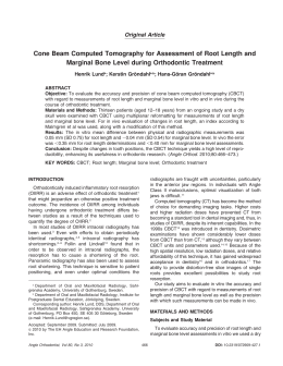
Indian of the Xicrin ethnicity; Kaiapó language from the Jê linguistic family; inhabitant of the Bacajá River, a tributary of the Xingu-Pará river.
ISSN 2176-9451
ORThODONTIcs
Dental Press Journal of
Volume 15, Number 3, May / June 2010
Dental Press International
v. 15, no. 3
Dental Press J Orthod. 2010 May-June;15(3):1-160
May/June 2010
ISSN 2176-9451
EDITOR-IN-CHIEF
Jorge Faber
Brasília - DF
ASSOCIATE EDITOR
Telma Martins de Araujo
UFBA - BA
ASSISTANT EDITOR
(Online only articles)
Daniela Gamba Garib
HRAC/FOB-USP - SP
ASSISTANT EDITOR
(Evidence-based Dentistry)
David Normando
UFPA - PA
ASSISTANT EDITOR
(Editorial review)
Flávia Artese
UERJ - RJ
PUBLISHER
Laurindo Z. Furquim
UEM - PR
EDITORIAL SCIENTIFIC BOARD
Adilson Luiz Ramos
Danilo Furquim Siqueira
Maria F. Martins-Ortiz Consolaro
UEM - PR
UNICID - SP
ACOPEM - SP
EDITORIAL REVIEW BOARD
Adriana C. da Silveira
Univ. of Illinois / Chicago - USA
Björn U. Zachrisson
Univ. of Oslo / Oslo - Norway
Clarice Nishio
Université de Montréal / Montréal - Canada
Jesús Fernández Sánchez
Univ. of Madrid / Madri - Spain
José Antônio Bósio
Marquette Univ. / Milwaukee - USA
Júlia Harfin
Univ. of Maimonides / Buenos Aires - Argentina
Larry White
AAO / Dallas - USA
Marcos Augusto Lenza
Univ.of Nebraska / Lincoln - USA
Maristela Sayuri Inoue Arai
Tokyo Medical and Dental University / Tokyo - Japan
Roberto Justus
Univ. Tecn. do México / Cid. do Mexico - Mexico
Orthodontics
Adriano de Castro
Ana Carla R. Nahás Scocate
Ana Maria Bolognese
Antônio C. O. Ruellas
Ary dos Santos-Pinto
Bruno D'Aurea Furquim
Carla D'Agostini Derech
Carla Karina S. Carvalho
Carlos A. Estevanel Tavares
Carlos H. Guimarães Jr.
Carlos Martins Coelho
Eduardo C. Almada Santos
Eduardo Silveira Ferreira
Enio Tonani Mazzieiro
Fernando César Torres
Guilherme Janson
Haroldo R. Albuquerque Jr.
Hugo Cesar P. M. Caracas
José F. C. Henriques
José Nelson Mucha
José Renato Prietsch
José Vinicius B. Maciel
Júlio de Araújo Gurgel
Karina Maria S. de Freitas
Leniana Santos Neves
Leopoldino C. Filho
Luciane M. de Menezes
Luiz G. Gandini Jr.
Luiz Sérgio Carreiro
Marcelo Bichat P. de Arruda
Márcio R. de Almeida
Marco Antônio Almeida
Marcos Alan V. Bittencourt
Maria C. Thomé Pacheco
Marília Teixeira Costa
Marinho Del Santo Jr.
Mônica T. de Souza Araújo
Orlando M. Tanaka
Oswaldo V. Vilella
Patrícia Medeiros Berto
Pedro Paulo Gondim
Renata C. F. R. de Castro
Ricardo Machado Cruz
Ricardo Moresca
Robert W. Farinazzo Vitral
Roberto Rocha
Dental Press Journal of Orthodontics
(ISSN 2176-9451) continues the
Revista Dental Press de Ortodontia e Ortopedia Facial
(ISSN 1415-5419).
Dental Press Journal of Orthodontics
(ISSN 2176-9451) is a bimonthly publication of Dental Press International
Av. Euclides da Cunha, 1.718 - Zona 5 - ZIP code: 87.015-180 - Maringá / PR, Brazil Phone: (55 044) 3031-9818 - www.dentalpress.com.br - [email protected].
DIRECTOR: Teresa R. D'Aurea Furquim - INFORMATION ANALYST:
Carlos Alexandre Venancio - EDITORIAL PRODUCER: Júnior Bianchi DESKTOP PUBLISHING: Fernando Truculo Evangelista - Gildásio Oliveira
Reis Júnior - Tatiane Comochena - REVIEW / CopyDesk: Ronis Furquim
Siqueira - IMAGE PROCESSING: Andrés Sebastián - LIBRARY: Alessandra
Valéria Ferreira - NORMALIZATION: Marlene G. Curty - DATABASE:
Adriana Azevedo Vasconcelos - E-COMMERCE: Soraia Pelloi - ARTICLES
SUBMISSION: Simone Lima Rafael Lopes - COURSES AND EVENTS: Ana
Claudia da Silva - Rachel Furquim Scattolin - INTERNET: Carlos E. Lima Saugo
- FINANCIAL DEPARTMENT: Márcia Cristina Nogueira Plonkóski Maranha
- Roseli Martins - COMMERCIAL DEPARTMENT: Roseneide Martins Garcia SECRETARY: Michaele Rezende - PRINTING: Gráfica Regente - Maringá / PR.
UCB - DF
UNICID - SP
UFRJ - RJ
UFRJ - RJ
FOAR/UNESP - SP
private practice - PR
UFSC - SC
ABO - DF
ABO - RS
ABO - DF
UFMA - MA
FOA/UNESP - SP
UFRGS - RS
PUC - MG
UMESP - SP
FOB/USP - SP
UNIFOR - CE
UNB - DF
FOB/USP - SP
UFF - RJ
UFRGS - RS
pucpr - pr
FOB/USP - SP
Uningá - PR
UFVJM - MG
HRAC/USP - SP
PUC-RS - RS
FOAR/UNESP - SP
UEL - PR
UFMS - MS
UNIMEP - SP
UERJ - RJ
UFBA - BA
UFES - ES
UFG - GO
BioLogique - SP
UFRJ - RJ
PUC-PR - PR
UFF - RJ
private practice - DF
UFPE - PE
UMESP - SP
UNIP - DF
UFPR - PR
UFJF - MG
UFSC - SC
Indexing: IBICT
Rodrigo Hermont Cançado
Sávio R. Lemos Prado
Weber José da Silva Ursi
Wellington Pacheco
Dentofacial Orthopedics
Dayse Urias
Kurt Faltin Jr.
Orthognathic Surgery
Eduardo Sant’Ana
Laudimar Alves de Oliveira
Liogi Iwaki Filho
Rogério Zambonato
Waldemar Daudt Polido
Dentistics
Maria Fidela L. Navarro
TMJ Disorder
Carlos dos Reis P. Araújo
José Luiz Villaça Avoglio
Paulo César Conti
Phonoaudiology
Esther M. G. Bianchini
Implantology
Carlos E. Francischone
Oral Biology and Pathology
Alberto Consolaro
Edvaldo Antonio R. Rosa
Victor Elias Arana-Chavez
Periodontics
Maurício G. Araújo
Prothesis
Marco Antonio Bottino
Sidney Kina
Radiology
Rejane Faria Ribeiro-Rotta
Uningá - PR
UFPA - PA
FOSJC/UNESP - SP
PUC - MG
UFG - GO
SCIENTIFIC CO-WORKERS
Adriana C. P. Sant’Ana
Ana Carla J. Pereira
Luiz Roberto Capella
Mário Taba Jr.
FOB/USP - SP
UNICOR - MG
CRO - SP
FORP - USP
PRIVATE PRACTICE - PR
UNIP - SP
FOB/USP - SP
UNIP - DF
UEM - PR
PRIVATE PRACTICE - DF
ABO/RS - RS
FOB/USP - SP
FOB/USP - SP
CTA - SP
FOB/USP - SP
CEFAC/FCMSC - SP
FOB/USP - SP
FOB/USP - SP
PUC - PR
USP - SP
UEM - PR
UNESP - SP
PRIVATE PRACTICE - PR
- CCN
Databases:
LILACS - 1998
BBO - 1998
National Library of Medicine - 1999
SciELO - 2005
Dental Press Journal of Orthodontics
Bimonthly.
ISSN 2176-9451
1. Orthodontics - Periodicals. I. Dental Press International
Table
of conTenTs
5
Editorial
12
Events Calendar
13
News
16
What’s new in Dentistry
19
Orthodontic Insight
31
Interview with Ademir Roberto Brunetto
Online Articles
46
Evaluation of the applicability of a North American cephalometric standard to
Brazilian patients subjected to orthognathic surgery
Fernando Paganeli Machado Giglio, Eduardo Sant’Ana
48
Analysis of biodegradation of orthodontic brackets using scanning electron
microscopy
Luciane Macedo de Menezes, Rodrigo Matos de Souza, Gabriel Schmidt Dolci,
Berenice Anina Dedavid
Original Articles
S
S0
52
Nasopharyngeal and facial dimensions of different morphological patterns
Murilo Fernando Neuppmann Feres, Carla Enoki,
Wilma Terezinha Anselmo-Lima, Mirian Aiko Nakane Matsumoto
62
Cephalometric evaluation of vertical and anteroposterior changes associated
with the use of bonded rapid maxillary expansion appliance
Moara De Rossi, Maria Bernadete Sasso Stuani, Léa Assed Bezerra da Silva
71
Evaluation of maxillary atresia associated with facial type
Marina Gomes Pedreira, Maria Helena Castro de Almeida,
Katia de Jesus Novello Ferrer, Renato Castro de Almeida
78
Possible etiological factors in temporomandibular disorders of articular origin
with implications for diagnosis and treatment
Aline Vettore Maydana, Ricardo de Souza Tesch, Odilon Vitor Porto Denardin,
Weber José da Silva Ursi, Samuel Franklin Dworkin
ad2
Ba
ad1
Ptm
I - Muscular Diagnoses
a - myofascial pain
b - myofascial pain with limited opening
II - Disk Displacement
a - disk displacement with reduction
b - disk displacement without reduction and with limited opening
c - disk displacement without reduction and without limited opening
III - Arthralgia, osteoarthritis and osteoarthrosis
a - arthralgia
b - temporomandibular joint (TMJ) osteoarthritis
c - temporomandibular joint (TMJ) osteoarthrosis
16%
14%
87
Factors predisposing 6 to 11-year old children in the first stage of orthodontic
treatment to temporomandibular disorders
Patrícia Porto Loddi, André Luis Ribeiro de Miranda, Marilena Manno Vieira,
Brasília Maria Chiari, Fernanda Cavicchioli Goldenberg, Savério Mandetta
94
Extraction of upper second molars for treatment of Angle Class II malocclusion
Maurício Barbieri Mezomo, Manon Pierret, Gabriella Rosenbach,
Carlos Alberto E. Tavares
106
Evaluation of shear bond strength of brackets bonded with orthodontic
fluoride-releasing composite resins
Marcia Cristina Rastelli, Ulisses Coelho, Emígdio Enrique Orellana Jimenez
114
Statement of the 1st Consensus on Temporomandibular Disorders
and Orofacial Pain
Simone Vieira Carrara, Paulo César Rodrigues Conti,
Juliana Stuginski Barbosa
121
Race versus ethnicity: Differing for better application
Diego Junior da Silva Santos, Nathália Barbosa Palomares,
David Normando, Cátia Cardoso Abdo Quintão
125
BBO Case Report
12%
10%
Female
Male
6
5
7
6
6
4%
11
11
6%
14
14
8%
2
2%
0%
Finger/paciAtypical
fier sucking swallowing
Mouth
breathing
Mixed
breathing
Bruxism
Angle Class II, Division 2 malocclusion with severe overbite
and pronounced discrepancy
Daniela Kimaid Schroeder
134
Special Article
Tooth extraction in orthodontics: an evaluation of diagnostic elements
Antônio Carlos de Oliveira Ruellas, Ricardo Martins de Oliveira Ruellas,
Fábio Lourenço Romano, Matheus Melo Pithon, Rogério Lacerda dos Santos
158
Information for authors
ediTorial
Treatment of temporomandibular disorders (TMD)
and orofacial pain
diagnosed with TMD at the beginning of followup. To simplify my reasoning, let us consider
that we have two possible treatment outcomes:
improvement and no improvement. If the final
results indicate that 35 patients improved, treatment as a whole was a success, right? The correct
answer is: wrong. We cannot conclude anything
other than that this treatment might work.
Some conditions are cyclical or transitory,
and it might be that the patients who improved
with this TMD therapy would eventually get
better anyway. Therefore, a control group should
be included, provided that the researcher finds
it ethically acceptable to deprive these people of
treatment. Thus, if the control group was included in the study and only 20 patients improved
without treatment (Table 1), we would have
a statistically significant difference between
treatment and control groups (p<0.001), with
the latter group showing more improvement
than the former. Can we now conclude that
this treatment is effective? No. At least not yet.
Furthermore, it is perfectly conceivable that a
portion of those treated improved as a result of
the placebo effect. It would be all but impossible
to include a placebo effect per se in a non-drug
therapy such as TMD. To achieve such effect, one
could implement false treatments such as, for
example, brackets bonded to teeth without de-
It is intriguing to see how information flows
in the healthcare area. It is particularly curious
to note that certain obsolete concepts and old,
threadbare themes are sometimes reinstated
and infect many practitioners. These treatment
approaches are enough to spoil the mood of any
scientifically-minded professional and—worse
still—can wreak havoc with the victims of such
treatments. The less lethal this condition, the
more susceptible to such impropriety. An article
in this issue provides a unique insight into one
of the subjects most affected by what I just
described: the treatment of temporomandibular
disorders and orofacial pain.
Consider the following questions concerning
TMD. Is your TMD treatment controversial? Is
orthodontics an integral part of TMD treatment
methods? Should TMJ CT's be routinely used to
assess the problem? Is joint space relevant to the
diagnosis and treatment goal? Is treatment aimed
at adjusting the joint spaces? If you answered yes
to one or more of these questions you must read
the article by Carrara, Conti and Barbosa.
A close relationship between dentition and
TMD was erroneously established decades ago.
The mistaken conclusions stemmed from an interpretation of retrospective case series studies.
This study design is most often performed by
practitioners in the office setting, simply because
that is where patients go for treatment. Thus, after a few years, material is collected from a series
of cases on a given subject. To better understand
why this study design is inefficient in pinpointing
solutions to the problems that confront us, let us
consider the following line of reasoning.
A hypothetical professional analyzes the
results of orthodontic treatment of 41 patients
in her office. All complained of pain and were
Dental Press J Orthod
TABLE 1 - Results of a hypothetical study that proposes an orthodontic
treatment plan for TMD.
IMPROVEMENT
5
TREATMENT
CONTROL
FAKE
TREATMENT
35
20
33
NO IMPROVEMENT
6
19
8
TOTAL
41
39
40
2010 May-June;15(3):5-6
Editorial
findings from a series of cases treated in their
offices, without realizing the complexity that
lies behind the formulation of clinical studies.
It was in an attempt to help these people,
who are part of the dental and medical communities, and also the people who suffer from
TMD and orofacial pain, that Carrara, Conti
and Barbosa wrote the Statement of the 1 st Consensus on Temporomandibular Disorders and
Orofacial Pain. This article is unique because it
not only reflects the authors' opinion, but also
that of today's leading Brazilian professionals.
They endorsed the article and proved that the
subject is not controversial.
Furthermore, the article shows that the available evidence can suggest many things: that orthodontics is not an integral part of routine TMD
treatment methods, that TMJ CT's should not
be used routinely, that joint space analysis is not
relevant to the diagnosis and that adjusting the
joint spaces is not a treatment goal, among other
conclusions. The article is a landmark in the area
and I strongly recommend that all read it in full.
livering any actual forces, or an acrylic plate that
does not cover the occlusal surfaces of the teeth.
In our hypothetical study, a Fake Treatment
was evaluated. The results showed that 33 patients improved with the fake treatment and no
difference was found between Treatment and
Fake Treatment groups (p = 0.63). Thus the
new therapy—or old therapy, if it happens to be
the new edition of an old concept—is not more
effective than the fake treatment.
The table showing the clinical trials with the
three groups, described above, gives an overview
of the process of assembling information for
clinical decision making. However, the mere
creation of the three groups is still a relatively
incomplete action and therefore insufficient.
Important issues regarding the randomness of
patient selection for treatment, the fact that it is
a prospective study, the analysis of intention to
treat, among other items relevant to the design
of a clinical trial, were not even mentioned.
Mainly because it would require many pages to
elaborate on these details.
Additionally, the sketch depicts a common
shortcoming, namely, many well-intentioned
professionals take advantage of conferences
and other channels as a platform to disseminate
Dental Press J Orthod
Jorge Faber
Editor-in-chief
[email protected]
6
2010 May-June;15(3):5-6
TM
Aquarium 2
Case presentation software
Show it. Share it.
Lingual
Brackets
Teeth
Eruption
Forsus Appliance
TAD Canine Retraction
Mandibular Advancement Surgery
Export movies to other programs
Intuitive Interface • Stunning 3D Movies • Comprehensive Library •
Personalized Images • Network-Ready • Export Movies
The second generation of Aquarium brings greatly expanded content and
capabilities. New movies such as 3rd Molar Extraction, Lip Bumper, and Lingual
Braces make this interactive patient education software more relevant than ever.
Record your own audio, export media, enlarge interface for easy viewing, and
personalize your program with thematic skins. Aquarium movies are networkready and display beautifully on most monitors and resolutions. To learn more,
visit www.renovatio3.com.br or contact us at [email protected],
fone: +55 11 3286-0300.
© 2010 Dolphin Imaging & Management Solutions
LEAVE YOUR PERSONAL TOUCH
AT THE BIGGEST DENTAL EXHIBITION OF PORTUGAL
The Expo-Dentária is the largest exhibition of dentistry performed in Portugal,
receiving in its previous edition more than 5800 visitors. Its growing success
confirms that it is the right place to create the best business opportunities
and international visibility for your company.
Leave your personal touch at Expo-Dentária 2010
For further information visit: www.omd.pt
evenTs calendar
III Congresso de Ortodontia e II Congresso de Ortopedia Funcional
Date: June 17 to 19, 2010
Location: MinasCentro - Belo Horizonte / MG, Brazil
Information: www.abomg.org.br
Mini-residência em DTM/Apneia
Date: August 14 to 22, 2010
Location: Marquette University – Wisconsin, Milwaukee/USA
Information: (55 011) 3061-5584
XVI Reunião Científica ABFCOC
Date: August 17 to 20, 2010
Location: Hotel SESC Pantanal - Cuiabá / MT, Brazil
Information: (55 011) 3031-4687
www.abfcoc.com.br
IV Congresso Sul Brasileiro de Ortodontia
Date: August 19 to 21, 2010
Location: Lajes / SC, Brazil
Information: (55 049) 3224-0838
www.oralesthetic.com
1º Straight-Wire Lingual Meeting - Diagnóstico e Planejamento em Ortodontia
Date: August 27 and 28, 2010
Location: Grand Mercure - Ibirapuera - São Paulo / SP, Brazil
Information: (55 067) 3326-0077 / (55 016) 3397-1401
[email protected]
FDI Annual World Dental Congress
Date: September 2 to 5, 2010
Location: Salvador / BA, Brazil
Information: [email protected]
17º Congresso Brasileiro de Ortodontia - SPO
Date: October 14 to 16, 2010
Location: Anhembi – São Paulo / SP, Brazil
Information: www.spo.org.br
Dental Press J Orthod
12
2010 May-June;15(3):12
News
2010 AAO Annual Session
Jorge Faber, editor-in-chief of the Dental
Press Journal of Orthodontics, was the winner of
the CDABO Case Report of the Year for the best
case report published during 2009. His article,
published in the American Journal of Orthodontics and Dentofacial Orthopedics (AJO-DO)
was voted the best case report published in 2009
by the editorial board of the Journal.
The ceremony took place during a lunch
with the College of Diplomates of the American
Board of Orthodontics (CDABO), in Washington DC, where the 110th AAO Annual Session
(Meeting of the American Association of Orthodontists) was held between April 30 and May 4.
The award was bestowed by Dr. Vincent Kokich
(next editor of the AJO-DO).
Dr. Vincent Kokich handing the award to winning
author, Dr. Jorge Faber, and coauthor, Dr. Flávia
Velasque.
Dr. David Turpin, current editor of the AJO-DO, received a copy of the Dental Press Journal of Orthodontics issue featuring Dr. Turpin’s interview.
Dr. Adilson Luiz Ramos, former editor of this Journal, and the renowned Dr. Larry White.
Dr. Orlando Tanaka and the editor of this Journal,
Dr. Telma Martins de Araujo.
Trade floor of the 110th Meeting of the American
Association of Orthodontists.
Drs. Bruno Furquim, Marcos A. Lenza and
Eduardo B. Lenza.
Dental Press J Orthod
13
2010 May-June;15(3):13-5
News
Thesis defense at UEM
The Master’s degree in Integrated Dentistry, State University of Maringá (UEM) graduated its first class
of masters. The dissertations by Paula Scheibel and Luciana Manzotti De Marchi were noteworthy. Dr.
Paula presented to the examining committee her dissertation entitled “Correlation between alveolar bone
density and apical root resorption in orthodontic patients.” Dr. Luciana defended the thesis “Aesthetic and
functional evaluation of patients with agenesis of upper lateral incisors treated with implants or space closure and dental reanatomizations.”
In photo (from left to right): Prof. Renata Corrêa Pascotto (supervisor), Prof.
Dr. Adilson Luiz Ramos (examiner), Dr. Luciana Manzotti De Marchi and Prof.
Dr. Ricardo de Lima Navarro (examiner).
In photo (from left to right): Prof. Dr. Júlio de Araújo Gurgel (examiner), Dr.
Paula C. Scheibel, Prof. Dr. Adilson Luiz Ramos (supervisor) and Prof. Dr.
Hélio Hissashi Terada (examiner).
ABOR and SBO participated in the WFO Council Meeting
of its activities in recent years and in recognition
of their outstanding performance, all associate
members were presented with a free subscription to the “World Journal of Orthodontics.” The
next IOC will be held in September 2015, in
London. Brazil was strongly encouraged to apply as a candidate to host the 2020 IOC.
Brazil was very well represented in the science grid of the 7th IOC through the participation of several renowned Brazilian orthodontists. Dr. Guilherme Janson delivered a lecture
entitled “Asymmetric malocclusion: a systematic approach to diagnosis and treatment.”
The World Federation of Orthodontists
(WFO) organizes the International Orthodontics Congress (IOC) every five years. The 7th
IOC was held between February 6 and 9, 2010
in Sydney, Australia. The Meeting of the WFO
Executive Council, where ABOR and SBO have
a right to a seat and vote, was held on February
5. Drs. Flavia Artese, Slamad Rodrigues and Eustáquio Araújo represented those two Brazilian
Associations. A highlight of this meeting was the
election of Dr. Kurt Faltin Jr. as WFO representative for Latin America with a five-year term.
The WFO representative gave a brief overview
Dental Press J Orthod
14
2010 May-June;15(3):13-5
News
ABOR and SBO participated in the WFO Council
Meeting.
Dr. Kurt Faltin Jr. addressed “The orthopedic treatment of anterior open bite with Balters’ Bionator.” The issue of “Whether or not
to abandon the early treatment of Class II” deserved a bold argument by Dr. Eustáquio Araújo. Dr. Camillo Morea gave a lecture on the
“Initial healing of hard and soft tissues around
unloaded mini-implants.” Finally, Dr. Nelson
Mucha talked about the “Long-term evaluation of anterior open bite treatment in adult
patients.”
On February 5, 2010 at the invitation of Dr.
Roberto Justus (WFO President), Board representatives from 15 countries as well as others
where a Board has not yet been established,
gathered at the Symposium on Orthodontic
Certifying Boards.
The activities started with two presentations. The first by Dr. Jeryl English of the
American Board of Orthodontics (ABO) and
the second by Dr. Robert Carter of the College of Diplomates of the American Board of
Orthodontics (CDABO).
ABO’s keynote address focused on the increased demand for Board certification, which this
year has exceeded twice the expected number of
entries (more than 520 applicants). Currently, the
ABO boasts 79% of diplomate orthodontists.
CDABO keynote address described the
functions of the American College, which include encouraging, supporting and facilitating
the process of certification for orthodontists as
Dental Press J Orthod
Brazilian orthodontists lectured at the WFO Congress.
BBO and CDBBO participated in the WFO Meeting.
well as sponsoring lectures and continuing education for its members.
Although the WFO has identified 15 countries that have a Board, few have Diplomate
Colleges, which puts Brazil, once again, at the
forefront of the orthodontic world.
15
2010 May-June;15(3):13-5
whaT´s
new in
denTisTry
Shared brain activity for aesthetic and
moral judgments: implications for the
Beauty-is-Good stereotype
Jorge Faber*, Patrícia Medeiros Berto**
region to both judgments. The orbitofrontal and
insular cortices were negatively correlated with
each other, suggesting an opposing relationship
between these regions during attractiveness and
goodness judgments.
These findings have implications for understanding the neural mechanisms of the Beautyis-Good stereotype. People judged to be physically attractive often have their personality also
judged positively, be it as a person of good conduct, virtuous or even honest. One is capable of,
at first sight, considering another human being
attractive or unattractive while at the same time
assigning values to that person. The study suggests a possible explanation for this fact since
the same neural mechanisms are activated or
deactivated during these types of assessments.
So, perhaps now, we can explain why, when a
person is seen as beautiful, they are likewise
seen as good. In other words, how beauty becomes goodness.
The Beauty-is-Good stereotype refers to the
assumption that attractive people possess sociably desirable personalities and higher moral
standards. The existence of this bias suggests
that the neural mechanisms for judging facial
attractiveness and moral goodness overlap, i.e.,
they are circumscribed to the same brain regions. The hypothesis of this overlap was investigated by Tsukiura and Cabeza1 and published
in the March 2010 issue of the Journal of Social
Cognitive and Affective Neuroscience.
The research participants were scanned with
functional magnetic resonance imaging while
they made attractiveness judgments about faces
and goodness judgments about hypothetical actions. Activity in the medial orbitofrontal cortex increased as a function of both attractiveness and goodness ratings, whereas activity in
the insular cortex decreased with both attractiveness and goodness ratings. These activations
support the idea of similar contributions of each
* Editor-in-Chief of the Dental Press Journal of Orthodontics. PhD in Biology - Morphology, Electronic Microscopy Laboratory, University of
Brasília (UnB). MSc in Orthodontics and Dentofacial Orthopedics, UFRJ.
** Specialist in Orthodontics, Federal University of Goiás (UFG). Reviewer of the Dental Press Journal of Orthodontics.
Dental Press J Orthod
16
2010 May-June;15(3):16-8
Faber J, Berto PM
Facial expressions and how the brain
decodes them
after stimulus onset—regardless of the expression or the brain hemisphere side—an information processing mechanism is triggered locally
to take motor control of the eyes. The eyes then
perform a wide zoom to process the entire face
and finally a close-up zoom on specific spots for
diagnostic purposes (e.g., eyes open in “fear”;
mouth opens in “happiness”).
A categorizing model showed that in 200
milliseconds the left and right hemispheres process enough information to predict the behavioral category of the face being analyzed. This
investigation contributes to the understanding
of how facial information is quickly processed
in the brain to identify emotions.
Research of this nature, which enhance our
understanding of how beauty is recognized,
will probably be useful in establishing treatment strategies that involve aesthetic reconstruction of the face or its subcomponents,
such as the smile.
It is a fact that sociable living beings are
able to perceive the social cues of their peers.
The same applies to humans. In primates, the
face has evolved to convey emotional states,
while the brain has simultaneously evolved to
decode the signals in the facial expressions of
others. The study by Schyns, Petro and Smith2
reviewed and integrated the evidence supporting this hypothesis.
With the aid of computer programs they
co-examined facial expressions as signals that
transmit information and the brain as a receiver
and decoder of these signals. The authors found
that facial expressions were a set of subtly correlated signals, i.e., only slightly resembling one
another. For example, the eyes can share similar
expressions of anger and happiness. Data from
EEG’s showed that the brain uses spatial frequency information that reaches the retina to
identify the expressions by breaking up their
correlations. Within 140 to 200 milliseconds
Motivation and enthusiasm over
orthognathic surgery results influence
treatment satisfaction
was to determine whether the expectations of
patients and their parents regarding their possible future appearance were correlated with
the patients’ treatment satisfaction.
A retrospective study was performed and
questionnaires were presented to 115 patients
Patients’ motivation to undergo orthognathic surgery can affect their satisfaction with
treatment outcome. Meade and Inglehart 3 investigated this relationship and published their
findings in the American Journal of Orthodontics and Dentofacial Orthopedics. The goal
Dental Press J Orthod
17
2010 May-June;15(3):16-8
What´s new in Dentistry
orthognathic surgery is strongly correlated with
their treatment satisfaction.
The findings of this study have clinical implications for maxillofacial surgeons and orthodontists. Attention to technical excellence and
the use of advanced technologies are currently
the day-to-day concerns of most practitioners.
They are indeed essential for ensuring a successful surgery. However, patient satisfaction should
be added to the technical requirements of a surgery—it is possible the coexistence of a surgery
that meets the technical criteria and a patient
dissatisfied with its results, and this would be
a scenario of failure. What the article suggests
is the need to evaluate and encourage patients
about the surgery results from the very first
appointment in the pre-operative phase. The
more motivated and focused are the patients,
the more likely they are to experience ultimate
success. Such evidence can, no doubt, be readily
applied in our daily professional practice.
(aged 13-21 years at surgery) and 117 parents
(response rates of 41% and 42% respectively),
with responses from 95 parent-patient pairs.
The patients’ motivation was evaluated before
surgery by determining how excited they were
when they envisioned themselves after surgery
and how focused they were on the results. Parents completed parallel questionnaires on their
children’s motivation. Patient satisfaction was
determined by means of a postsurgical satisfaction questionnaire. The data collected indicated
that the more excited the patients were before
surgery, the more satisfied they were with the
results. Likewise, the more these patients focused on functional and aesthetic changes, the
more satisfied they were with the results. The
assessments made by the parents regarding the
motivation of their children before surgery were
consistent with the children’s reports and correlated with patient satisfaction after the surgery.
Thus, young patients’ self-motivation towards
RefeRenCes
1.
2.
3.
Tsukiura T, Cabeza R. Shared brain activity for aesthetic
and moral judgments: implications for the Beauty-is-Good
stereotype. Soc Cogn Affect Neurosci. 2010 Mar 15.
[Epub ahead of print].
Schyns PG, Petro LS, Smith ML. Transmission of facial
expressions of emotion co-evolved with their efficient
decoding in the brain: behavioral and brain evidence. PLoS
One. 2009 May 20;4(5):e5625.
Meade EA, Inglehart MR. Young patients’ treatment motivation
and satisfaction with orthognathic surgery outcomes: the role
of possible selves. Am J Orthod Dentofacial Orthop. 2010
Jan;137(1):26-34.
Dental Press J Orthod
Contact address
Jorge Faber
Brasília Shopping Torre Sul sala 408
CEP: 70.715-900 – Brasília/DF
E-mail: [email protected]
18
2010 May-June;15(3):16-8
OrthOdOntic insight
Saucerization of osseointegrated
implants and planning of simultaneous
orthodontic clinical cases
Alberto Consolaro*, Renato Savi de Carvalho**, Carlos Eduardo Francischone Jr.***,
Maria Fernanda M.O. Consolaro****, Carlos Eduardo Francischone*****
occurrence of saucerization, should special care
be given to teeth located in the neighborhood of
osseointegrated implants when moving teeth and
finishing orthodontic cases?
The field for Orthodontics has seen significant
expansion with the advent of new diagnostic and
therapeutic approaches in all specialties, such as
medical and dental implantology, sleep medicine,
orthognathic surgery, computed tomography,
gerodontology, etc. This requires the mastery of
new concepts and technical terms typical of the
jargon used by each specific area. Such mastery
plays a key role in discussions about diagnosis
and planning of clinical cases with professionals
from other specialties.
Dental osseointegrated implants, for example,
completely changed the practice and scope of
dentistry in the last 20 years. Many adult orthodontic patients have already had one or more
osseointegrated implants installed or may be
planning, or need to do so. Many young orthodontic patients have also had osseointegrated
implants installed because of tooth loss caused
by trauma or partial anodontia.
Osseointegrated implant saucerization is a
phenomenon worthy of recognition and consideration in orthodontic planning to establish
functional and aesthetic prognosis. With this insight in mind, we intend to discuss the concept
of saucerization, with the specific purpose of
answering a few important questions. Given the
*
**
***
****
*****
The concept of osseointegration is a peculiarity of the teeth and implants in our bodies:
The importance of cervical soft tissues
Osseointegration allows the direct anchorage of an implant through bone tissue formation around the implant without the growth or
development of fibrous tissue at the bone-implant interface.3,5
Teeth are the only body structures that traverse or penetrate an epithelial lining or coverage (Figs 1, 2 and 3). By extension, dental implants also have this feature and the anchorage
provided by osseointegration is a prerequisite
for implant stability. Long-term implant survival depends on the adhesion of the epithelium
and connective tissues to the titanium surface
since a complete soft tissue cervical sealing protects the bone from the highly contaminated
oral environment.8,10,15,22,23,26
The marginal gingiva and peri-implant mucosa share many clinical and microscopic characteristics.1,2,19,20,25 The gingival mucosa around
Full Professor of Pathology, FOB-USP and at FORP-USP Postgraduate courses.
Professor of Implantology, Sacred Heart University (USC).
Professor and MSc in Implantology, USC.
Professor and PhD in Orthodontics, Postgraduate Program of Oral Biology, USC.
Full professor, FOB-USP. Full Professor of Implantology, USC.
Dental Press J Orthod
19
2010 May-June;15(3):19-30
Saucerization of osseointegrated implants and planning of simultaneous orthodontic clinical cases
connective tissue above the bone crest of the
tooth are nourished by supraperiosteal vessels
that originate in the alveolar process and periodontal ligament. In the soft and hard peri-implant tissues the mucosa region is nourished by
terminal branches of wide vessels originating
from the periosteum of the bone implant site. In
both cases the vessels built a "plexus clevicular"
lateral to the junctional epithelium. All natural
teeth in the connective portion above the crest
showed a rich vasculature, unlike the implant
sites as very few vessels were observed in this region.7 This finding reinforces the suspicion that
the peri-implant soft tissue may have a slightly
decreased ability to defend itself against external
aggression compared to the natural periodontal
tissues (Fig 1).
The mechanical resistance between the gingiva and the peri-implant mucosa was tested in
successful implants usually displays no inflammatory lesions. When lesions do occur, they are
small and located adjacent to the junctional
epithelium.1,19 Clinically, a healthy or slightly
inflamed gingiva, as well as the peri-implant mucosa, if proper oral hygiene is performed, exhibit
inflammatory infiltrates at similar locations and
with similar extension.20 Several studies have
shown similarities between the peri-implant mucosa and the gingiva in terms of their epithelial
and connective structures.9,16,17,18,24,27 However,
the absence of root cementum on the surface of
the implants change the orientation plane and
the adhesion of the fibers between teeth and implants.9 The importance of sealing the soft tissue
at implant sites to achieve functional success has
not been completely or thoroughly evaluated.
Studies on the topography of periodontal
tissue vasculature revealed that the gingiva and
CT
V
F
D
E
GE
GE
IJE
JE
CT
V
C
F
AB
IT
O
M
M
IP
P
A
B
FIGURE 1 - In the normal periodontium, at A, the collagen fibers are highlighted, extending from the gingival alveolar bone (AB) crest to the cementum (C),
gingiva and periodontal ligament (P) to form a cross-hatch pattern at the connective attachment. The rich blood vascular (V) and fibroblastic (F) components can be seen, to a lesser extent in the cervical peri-implant connective tissue (CT). B shows schematically that the bundles of collagen fibers in the
peri-implant cervical connective attachment tend to run parallel to the surface of the intermediate prosthesis (IT). GE = gingival epithelium; JE = junctional
epithelium, IJE = implant junctional epithelium; D = dentin; M = marrow space; IP = implant.
Dental Press J Orthod
20
2010 May-June;15(3):19-30
Consolaro A, Carvalho RS, Francischone CE Jr, Consolaro MFM-O, Francischone CE
E
D
E
JE
GE
GCT
JE
CA
C
Cb
B
PL
Ob
D
C
PL
B
FIGURE 2 - The tooth is the only structure of the body that crosses the
lining epithelium and interacts with the internal environment. Layout of
the periodontal structures relative to the biological distances: dentin
(D), cementum (C), alveolar bone (B), periodontal ligament (PL), junctional epithelium (JE), gingival epithelium (GE) and gingival connective tissue (GCT). The junctional epithelium has 15-30 cell layers and
as it proliferates in the apical direction it enables the contact of EGF
molecules with bone cells, thereby stimulating bone resorption and
maintenance of the biological distances. In the human body, between
the epithelium and the bone, there is always connective tissue interposition due to the presence of EGF in the underlying epithelial and
connective tissues. EGF is released by the Epithelial Rests of Malassez and keeps the alveolar bone away from the cementum through the
same mechanism and thus prevents dentoalveolar ankylosis.
FIGURE 3 - The form of the alveolar bone crest, with its rhomboidal
aspect, corresponds to the morphology of the junctional epithelium
(JE) which fosters the steady release of EGF, depicted by the arrows.
The collagen fibers of the connective attachment (CA) perpendicular
to the cementum (C) can help limit the effect of EGF on bone cells. The
cementoblasts (Cb) on the root surface have receptors for EGF and
other mediators of bone turnover, which ultimately protect teeth from
resorption. D = dentin; PL = periodontal ligament; B = alveolar bone, E
= enamel; Ob = osteoblasts.
dogs and revealed that probe penetration was
greater in implants than in teeth: 2 mm and 0.7
mm, respectively.14 In peri-implant soft tissues,
the probe displaced the junctional epithelium
and connective tissue on the implant’s adhesion
surface interface and stopped at the bone crest.
Occasionally, bleeding occurred due to vessel rupture. In the teeth, the probe stopped at
the apical portion of the junctional epithelium,
identifying the bottom of the gingival sulcus.
The bleeding was minimal, in contrast with that
of the implants.14
The effects of dental bacterial plaque after
three weeks and after three months in the gingiva and peri-implant tissues were comparatively
evaluated.6 Both tissues exhibited inflammatory lesions identical in size and composition
features. Within three months the bleeding was
similar and both inflammatory infiltrates had
the same characteristics, but the apical extent
was more pronounced in the peri-implant mucosa than in the gingiva. This finding implies
that the defense mechanisms of the gingiva are
more efficient than those of the peri-implant
tissues in preventing future spreads of sulcus microbiota.6 However, the neck of an osseointegrated dental implant tends to display
normal function and aesthetics, provided that
adequate oral hygiene is maintained. This also
applies to normal teeth.
Dental Press J Orthod
21
2010 May-June;15(3):19-30
Saucerization of osseointegrated implants and planning of simultaneous orthodontic clinical cases
Saucerization of osseointegrated implants:
Concept and Mechanism
Saucerization occurs in all osseointegrated
implants, regardless of their design, surface type,
platform, connection type, commercial brand or
patient conditions (Fig 12). Although the speed
with which it occurs can vary, its occurrence
seems to be part of the integration of implants
with epithelium and gingival connective tissue.
The cervical region of osseointegrated implants, when exposed to the oral environment,
usually exhibits some degree of bone resorption (Figs 4-11), of approximately 0.2 mm
depth.4,5,11 The plane of the resorbed osseointegrated bone surface forms an open angle with
the implant’s cervical region on nearly all of its
surfaces. Three-dimensionally, this cervical bone
resorption—observed in all types of osseointegrated implants—is in the shape of a saucer, i.e.,
it is shallow and superficial, hence "saucerization.” This process can be extended over time,
crown
gingival
connective
tissue
Intermediate prosthesis
JE
S
S
GE
crown
gingival
connective
tissue
Intermediate prosthesis
implants
Bone tissue
FIGURE 4 - The gingival stratified squamous epithelium (GE) is juxtaposed with its normal thickness soon after the placement of healing
caps or intermediate prosthesis and crown. The ulcerated epithelium
has its cell membranes exposed to mediators that interact with their
receptors. Under stress the cells increase the production of mediators.
The EGF (arrows) of the epithelial cells themselves stimulates periimplant epithelial proliferation and initiates the formation of the periimplant junctional epithelium. EGF from saliva (S) probably participates
in this process because it is greatly increased during oral surgery.
Gingival
epithelium
Gingival
epithelium
crown
gingival
connective
tissue
Intermediate prosthesis JE
implants
implants
Bone tissue
Bone tissue
FIGURE 5 - The peri-implant junctional epithelium (JE) produces new cell layers and assumes a conformation similar to the
junctional epithelium of natural teeth. This new conformation
of the peri-implant junctional epithelium brings it closer to the
osseointegrated surface, increasing the local concentration of
EGF and, as a result, accelerating bone resorption and starting
saucerization.
Dental Press J Orthod
FIGURE 6 - The peri-implant junctional epithelium (JE) conformation is similar to the junctional epithelium of natural teeth. It derives structural balance from the peri-implant connective attachment to stabilize its proliferative activity. On the bone surfaces
resorption decreases, approaching normal bone turnover. Thus,
the peri-implant bone surface undergoes corticalization, indicative of process stabilization.
22
2010 May-June;15(3):19-30
Consolaro A, Carvalho RS, Francischone CE Jr, Consolaro MFM-O, Francischone CE
A
B
FIGURE 7 - During the removal of the healing caps or intermediate prosthesis there occurs the formation of the peri-implant junctional epithelium (JE) that
covers the surface interface with the mucosa, including the gingival tissue. When it is still thin and disorganized, the peri-implant junctional epithelium tends
to show a reddish appearance and can bleed if touched, given its frailty (A). When organized and mature, the peri-implant junctional epithelium appears pink,
resembling the epithelium of the adjacent mucosa. Occasionally, the underlying microcirculation (B) can be seen as the JE becomes transparent.
years to a level even lower than that recorded
in previous studies, and that these results would
soon be reported in the literature.
Many theories and explanations have been
provided to account for saucerization but almost
all have had difficulty explaining some of its features. One of these theories attributes saucerization to the occlusal masticatory load that implants have to sustain. However, when osseointegrated implants are out of occlusion or are fitted only with the gingival healing caps for many
months or even years, without ever coming into
occlusion, saucerization is also present (Fig 13).
On the other hand, when implants remain submerged for a few months/years, the bone moves
toward the more cervical surface and may even
grow over the cover screws (Fig 12). This bone
gain requires osteotomy maneuvers in order to
place healing caps or an intermediate prosthesis.
Shortly after the placement of healing caps,
or directly from the intermediate prosthesis
and crown, the stratified squamous epithelium
consuming on average 0.1 mm of peri-implant
cervical bone tissue each year.4,5,11 In a personal
communication, Albrektsson reported that this
cervical bone loss tends to decrease over the
implant
osseointegration
Stabilization of
the corticalization process
FIGURE 8 - After saucerization, the peri-implant bone surface normalizes, with corticalization (arrows) indicative of stabilization of the pericervical bone remodeling process (toluidine blue, 10X).
Dental Press J Orthod
23
2010 May-June;15(3):19-30
Saucerization of osseointegrated implants and planning of simultaneous orthodontic clinical cases
A
B
FIGURE 9 - Clinical case of implant in the upper lateral incisor region after six years, highlighting saucerization with regular bone surface and osseointegration.
of the oral mucosa is juxtaposed to the surface
with its normal thickness (Fig 4). When an epithelium is ulcerated their cell membranes are
exposed to mediators in order to interact with
their receptors, in the same manner as in oral ulcers and surgical wounds, including in the periimplant region.
The epidermal growth factor (EGF) in the
saliva and in the epithelial cells stimulates periimplant epithelial proliferation, thereby triggering the formation of the peri-implant junctional
epithelium. The peri-implant junctional epithelium produces new cell layers and assumes a conformation similar to the junctional epithelium of
natural teeth (Fig 5). This new conformation of
the peri-implant junctional epithelium brings it
closer to the osseointegrated surface, increasing
the local concentration of EGF and, as a result,
accelerating bone resorption and starting saucerization (Fig 5). Two recent papers have reviewed
EGF functions and history.12,13
Dental Press J Orthod
A few weeks or months after the peri-implant junctional epithelium and saucerization
are formed they start moving away from each
other. A stable biological distance is then established between the implant-integrated cervical
bone and the peri-implant junctional epithelium, as occurs with natural teeth. From this
stage, saucerization balance and stabilization
are in place, allowing the bone on the cervical
surface to resume corticalization (Figs 6, 8-11).
It is probably due to this stabilization over the
years that bone loss resulting from cervical saucerization diminishes its rhythm,4,5,11 provided
that the conditions of hygiene and periodontal
health are close to ideal. This situation has been
noted in clinical cases that were followed up for
many years after placement of osseointegrated
implants (Figs 10 and 11).
The reestablishment of the junctional epithelium in the peri-implant oral mucosa may be
due to stimulation by the EGF of the mucous
24
2010 May-June;15(3):19-30
Consolaro A, Carvalho RS, Francischone CE Jr, Consolaro MFM-O, Francischone CE
A
B
C
FIGURE 10 - Implant installed in the region of tooth 21 avulsed in an accident. A shows the abutment installed over the implant. Periapical radiograph at
B shows the correct adjustment of the abutment on the implant; the height and shape of the bone tissue around the implant are highlighted. C) Prosthetic
crown cemented over the abutment.
A
B
C
FIGURE 11 - Same clinical case as in the previous figure. A is a five-year control periapical radiograph showing pericervical saucerization and corticalization
of peri-implant bone tissue. B shows 15 years of clinical control: Note normality and stability of peri-implant gingival tissue. C shows a 15-year control periapical radiograph: Note the stability of the bone around the implant and increased corticalization.
epithelium-implant integration occurs, salivary
EGF penetration ceases or is drastically reduced
and the process of cell-renewal epithelial proliferation goes back to normal.
The thickness of the gingival tissue appears to
have a considerable effect on alveolar crest bone
loss. When this thickness is 2 mm or smaller, the
cervical bone loss tends to be significantly greater.21
epithelium itself through what is known as the
autocrine effect. Although it probably takes
place throughout the mucosa, it is particularly active in ulcerated areas where this autocrine effect is compounded by salivary EGF.
As a result, a considerable increase occurs in
cell layers to the extent that the peri-implant
junctional epithelium is formed. Once the
Dental Press J Orthod
25
2010 May-June;15(3):19-30
Saucerization of osseointegrated implants and planning of simultaneous orthodontic clinical cases
crown
EJI
crown
EG
Intermediate prosthesis
Intermediate prosthesis
TCG
implant
implant
O
A
B
cone morse
intermediate
prosthesis
en bloc
implant
C
D
FIGURE 12 - Saucerization invariably occurs in all types of osseointegrated implants. The epithelial tissue has essentially a lining function and it is not very
selective as to what it chooses to line. The epithelium will line even root surfaces which, although scraped, still manage to keep LPS (lipopolysaccharide)
in its structure. LPS molecules are excessively toxic to our cells, but that does not stop the long junctional epithelium from forming, which is very important
for maintaining clinical normality.
Saucerization timing and
orthodontic treatment
In natural teeth, the union of the junctional
epithelium to the cervical enamel and surface
is performed by means of several kinds of union
structures, which effect an efficient sealing for
salivary EGF (Figs 1, 2 and 3) in the peri-implant
These results could probably be explained in light
of the EGF. The thickness of the gingival tissue at
the time of implant placement is commensurate
with the distance from the implant junctional epithelium to be formed relative to bone tissue, i.e.,
EGF molecules rise to the bone surface in lower
concentration.
Dental Press J Orthod
26
2010 May-June;15(3):19-30
Consolaro A, Carvalho RS, Francischone CE Jr, Consolaro MFM-O, Francischone CE
suture
GE
GE
GCT
GCT
implant
implant
B
Stabilization of
the corticalization process
B
B
A
implant
B
osseointegration
C
D
healing caps
GCT
GE
PJE
implant
B
FIGURE 13 - Osseointegrated Implants submerged from A to D. In this
situation saucerization does not occur. Bone repair fosters partial overlap of implant coverage (as at B, C and D) because there is no formation
of peri-implant junctional epithelium that would provide EGF molecules
(arrows) in the vicinity of the bone surface. As soon as the healing caps
are fitted, the formation of the peri-implant junctional epithelium (PJE)
begins and so does saucerization (E). GE = gingival epithelium; GCT =
gingival connective tissue; B = alveolar bone. (C: toluidine blue, 10X).
E
Dental Press J Orthod
27
2010 May-June;15(3):19-30
Saucerization of osseointegrated implants and planning of simultaneous orthodontic clinical cases
15A
14A
15B
14B
15C
FIGURE 14 - lmplant installed in the region of tooth 12. The periapical
radiograph (A) shows the proximity of the roots of teeth 11 and 13 due to
the missing lateral incisor, which renders implant placement impossible;
B shows the fixed orthodontic appliance for separation of the roots and
crowns of teeth 11 and 13, thereby creating adequate space, suitable for
implant installation in the region of tooth 12.
Dental Press J Orthod
FIGURE 15 - The same clinical case of the previous figure with abutment
mounted on the implant (A). Periapical radiograph (B) showing adequate
interradicular space between 11 and 13, which allowed the installation
of the implant in the correct position. C shows the prosthetic crown cemented onto the abutment.
28
2010 May-June;15(3):19-30
Consolaro A, Carvalho RS, Francischone CE Jr, Consolaro MFM-O, Francischone CE
junctional epithelium. This sealing, however—
provided by the epithelium-implant junction—is
less efficient and supposedly allows a constant
salivary EGF input which, in conjunction with
the EGF of the junctional epithelium and mucosa, sets in motion a process of slow and steady
approach to the cervical bone (Figs 1, 4, 5, 6, 9).
After an osseointegrated implant has been
placed, peri-implant saucerization can normally
be expected to occur, regardless of implant type
(Figs 14 and 15). So what is the average distance
that should be maintained by orthodontists between the cervical regions of neighboring natural
teeth—when using osseointegrated implants—so
that the cervical bone level of these implants is
not affected by neighboring saucerization?
This concern may be even greater in upper
anterior teeth such as, for example, lateral incisor
implants (Figs 10, 11, 14, 15) in cases of partial unilateral or bilateral anodontia. Or, again, in
cases of incisors and canines lost by accidental injury. The aesthetic and functional implications of
the gingiva should be considered in planning and
installing implants, such as the shape and size of
the papillae, as well as the maintenance of a harmonious smile line.
Can saucerization, eventually, adversely affect
the cervical hard and soft tissues of teeth located in the neighborhood of implants in patients
treated orthodontically and whose teeth were
harmoniously aligned with the implants? What
special orthodontic care would be required to
avoid or reduce the undesirable long-term consequences of osseointegrated implant saucerization
occurring in the neighborhood of natural teeth?
The more we succeed in clarifying the phe-
Dental Press J Orthod
nomena related to cell and tissue saucerization,
the more we will be able to learn about the
care, and the aesthetic and functional nuances
involved. Additional refinement and details concerning the evolution of the operative and restorative procedures of dentistry as a whole come to
light every day, dissolving boundaries or obstacles
between the most diverse specialties.
Final considerations
Orthodontists should increasingly familiarize themselves with the jargon of other clinical specialties, including implantology, as well
as their concepts and more specific issues. This
need stems from increased transdisciplinary actions undertaken by professionals in the joint
planning of clinical cases involving multiple
specialties, and whose ultimate goal is to rehabilitate the patient's mouth.
Bone saucerization around osseointegrated
implants is one such concept that forms a specific part of the implantology jargon. Orthodontists should consider the occurrence of this periimplant bone phenomenon while simultaneously placing osseointegrated implants and moving
the other teeth, realigning or relocating them
harmoniously, many a time with such proximity
to the cervical region that the condition should
be carefully evaluated for its risks and aesthetic
and functional benefits.
Further research is probably needed to answer
the following question: Given the occurrence of
saucerization, what are the special needs and
care required by teeth located in the neighborhood of osseointegrated implants when moving
teeth and finishing orthodontic cases?
29
2010 May-June;15(3):19-30
Saucerization of osseointegrated implants and planning of simultaneous orthodontic clinical cases
ReFeRenCeS
1.
2.
3.
4.
5.
6.
7.
8.
9.
10.
11.
12.
13.
14. Ericsson I, Lindhe J. Probing depth at implants and teeth.
An experimental study in the dog. J Clin Periodontol.
1993;20:623-7.
15. Gould TRL. Clinical implications of the attachment of
oral tissues to perimucosal implants. Exerpta Medica.
1985;19:253-70.
16. Gould TRL, Brunette DM, Westbury L. The attachment
mechanism of epithelial cells to titanium in vitro. J
Periodontal Res. 1981;16(6):611-6.
17. Hashimoto M, Akagawa Y, Nikai H, Tsuru H. Single-cristal
sapphire endosseous dental implant loaded with functional
stress: clinical and histological evaluation of peri-implant
tissues. J Oral Rehabil. 1988;15:65-76.
18. Jansen JA, Wijn JR, Wolters-Lutgerhorst JML, van Mullem
PJ. Ultrastructural study of epithelial cell attachment to
implant material. J Dental Res. 1985;64:891-6.
19. Lekholm U, Adell R, Lindhe J, Branemark PI, Eriksson
B, Rockler B, et al. Marginal tissue reactions at
osseointegrated titanium fixtures. A cross-sectional
retrospective study. Int J Oral Maxillofacial Surg.
1986;15:53-61.
20. Lekholm U, Eriksson B, Adell R, Slots J. The condition of
the soft tissues at tooth and fixture abutments supporting
fixed bridges. A microbiological and histological study. J
Clin Periodontol. 1986;13:558-62.
21. Linkevicius T, Apse P, Grybauskas S, Puisys A. The influence
of soft tissue thickness on crestal bone changes around
implants: a 1-year prospective controlled clinical trial. Int J
Oral Maxillofac Implants. 2009 Jul-Aug;24(4):712-9.
22. McKinney RV, Steflik DE, Koth DL. Evidence for junctional
epithelial attachment to ceramic dental implants, a
transmission electron microscope study. J Periodontol.
1985;6:425-36.
23. McKinney RV, Steflik DE, Koth DL. The epithelium-dental
implant interface. J Oral Implantol. 1988;13:622-41.
24. Schroeder A, van der Zypen E, Stich H, Sutter F. The
reaction of bone, connective tissue and epithelium to
endosteal implants with sprayed titanium surfaces.
J Maxillofacial Surg. 1981;4:191-7.
25. Seymour GJ, Gemmel E, Lenz LJ, Henry P, Bower R,
Yamazaki K. Immunohistologic analysis of the inflammatory
infiltrates associated with osseointegrated implants. Int J
Oral Maxillofac Implants. 1989;4(3):191-7.
26. Ten Cate AR. The gingival junction. In: Branemark PI,
Zarb GA, Albrektsson T, editors. Tissue-integrated
prostheses: osseointegration in clinical dentistry. Chicago:
Quintessence; 1985. p. 145-53.
27. Van Drie HJY, Beertsen W, Grevers A. Healing of the
gingiva following installment of Biotes implants in beagle
dogs. Adv Biomater. 1988;8:485-90.
Adell R, Lekholm U, Rockler B, Branemark PI, Lindhe J,
Eriksson B, et al. Marginal tissue reactions at osseointegrated
titanium fixtures (I). A 3-year longitudinal prospective study.
Int J Oral Maxillofac Surg. 1986;15:39-52.
Akagawa Y, Takata T, Matsumoto T, Nikai H, Tsuru H.
Correlation between clinical and histological evaluations
of the peri-implant gingiva around single cristal sapphire
endosseous implant. J Oral Rehabil. 1989;16:581-7.
Albrektsson T. On long-term maintenance of the
osseointegrated response. Aust Prosthodont J. 1993;7:15-24.
Albrektsson T, Brånemark PI, Hansson HA, Lindström J.
Osseointegrated titanium implants: requirements for ensuring
a long-lasting, direct bone to implant anchorage in man. Acta
Orthop Scand. 1981;52(2):155-70.
Albrektsson T, Zarb G, Worthington P, Eriksson RA. The longterm efficacy of currently used dental implants: a review and
proposed criteria of success. Int J Oral Maxillofac Implants.
1986;1(1):11-25.
Berglundh T, Lindhe J, Marinello CP, Ericsson I, Liljenberg B.
Soft tissue reactions to de novo plaque formation at implants
and teeth. An experimental study in the dog. Clin Oral
Implants Res. 1992 Mar;3(1):1-8.
Berglundh T, Lindhe J, Jonsson K, Ericsson I. The
topography of the vascular systems in the periodontal and
peri-implant tissues in the dog. J Clin Periodontol. 1994
Mar;21(3):189-93.
Branemark PI. Introduction to osseointegration. In: Branemark
PI, Zarb GA, Albrektsson T, editors. Tissue-integrated
prostheses: osseointegration in clinical dentistry. Chicago:
Quintessence; 1985. p. 11-76
Buser D, Stich H, Krekeler G, Schroeder A. Faserstrukturen
der periimplantaren mukosa bei titanimlantaten. Eine
experimentelle studie am beagle-hund. Zeitschrift fur
Zahnarztliche Implantologie. 1989;5:15-23.
Carmichael RP, Apse P, Zarg GA, McCulloch CAG. Biological,
microbiological and clinical aspects of the peri-implant
mucosa. In: Albrektsson T, Zarb GA, editors. The Branemark
osseointegrated implant. Chicago: Quintessence; 1989. p. 39-78.
Cochran DL, Nummikoski PV, Schoolfield JD, Jones AA, Oates
TW. A prospective multicenter 5-year radiographic evaluation
of crestal bone levels over time in 596 dental implants placed
in 192 patients. J Periodontol. 2009 May;80(5):725-33.
Consolaro A, Consolaro MFMO. ERM functions, EGF and
orthodontic movement or why doesn't orthodontic movement
cause alveolodental ankylosis? Dental Press J Orthod. 2010
Mar-Abr;15(2);24-32.
Consolaro A, Carvalho RS, Francischone CE Jr, Francischone
CE. Mecanismo da saucerização nos implantes
osseointegrados. Rev Dental Press Periodontia Implantol.
2009 out-dez;3(4):25-39.
Contact address
Alberto Consolaro
E-mail: [email protected]
Dental Press J Orthod
30
2010 May-June;15(3):19-30
inTerview
An interview with
Ademir Roberto Brunetto
• DDS,FederalUniversityofParanáState(UFPR),1976.
• PostgraduateOrthodonticsandDentofacialOrthopedics,University
ofCalifornia,LosAngeles,USA,1984.
• ScientificAdvisor,DentalPressJournalofOrthodontics.
• RenownedLecturerinBrazilandabroad.
• Diplomate,BrazilianBoardofOrthodonticsandDentofacial
Orthopedics(BBO),2004.
• Director,BrazilianBoardofOrthodonticsandFacialOrthopedics
(BBO).
It gives me great satisfaction and pride to conduct this interview with
Prof. Dr. Ademir Brunetto, a prominent professional in today’s Brazilian
orthodontic scenery. This longtime friend, we forged our friendship
when we sat side by side at the 1st diplomate examination of the Brazilian Board of Orthodontics and Dentofacial Orthopedics (BBO), when at the same time, we were Board candidates.
A diplomate since 2004, he was later invited to join the BBO Board, which set the stage for our frequent encounters. I
have since learned to increasingly admire his in-depth scientific knowledge—especially in the area of Orthodontics and
Facial Orthopedics—, his ethical conduct, his composure and common sense in addressing all issues, regardless of their
complexity and, last but not least, his contagious joy. Born in Concórdia, at the west end of Santa Catarina State, in
southern Brazil, where he spent his childhood and adolescence, he soon moved to Curitiba where he studied Dentistry
at the Federal University of Paraná, graduating in 1976. As a Dentistry undergraduate, he worked as a trainee in a number
of orthodontic clinics and after graduation applied for the position of assistant professor at UFPR. Since his approval in
1981 he has taught orthodontics at UFPR. Dr. Brunetto attended his postgraduate program in orthodontics at the University of California, Los Angeles, USA (UCLA) where he was awarded the title of Master in Orthodontics in 1984. He is
currently in private practice in Curitiba, Paraná State, where he seeks to apply and disseminate his extensive knowledge.
Outside his professional activities, he is a very dedicated family man and an accomplished fisherman with a predilection
for ocean fishing. In his replies to the interviewers, he has shown substantial knowledge of current state-of-the-art issues
such as Class III correction, application of new imaging techniques using cone beam tomography, absolute anchorage
and orthodontic preparation for orthognathic surgery. I am certain that our valued readers will enjoy this interview.
Deocleciano da Silva Carvalho
Dental Press J Orthod
31
2010 May-June;15(3):31-45
Interview
Regarding the early treatment of Class iii,
what is the state-of-the-art in terms of interceptive procedures and what protocol do you
adopt, specifically in maxillary reverse pull
headgear cases? What type of retainer do
you use after maxillary reverse traction?
Márcio Sobral and Luís Antonio Aidar
I first started working with palatal expansion
associated with protraction in 1982, as a UCLA
resident. The then Head of the Department of
Orthodontics, Dr. Patrick K. Turley, had just begun his work with Class III patients. Those two
residence years were rather fruitful and, although
fraught with doubts, also brought many surprises and knowledge. When I returned to Brazil in
1984, I continued within the same line of work,
making slight changes to the expander design.
A few years later, I started to use prefabricated
masks, which greatly expedited my work.
My protocol begins with ¼ turn expansion
per day for initial suture release.24 My intent is
always to control so as not to overexpand the
maxilla to prevent excessive crossbite (Brodie)
because during anterior maxillary traction we
are moving from a wider, posterior mandibular
region and as we displace the maxilla forward
and downward, we have a narrower mandible.
After the expansion, I start using the face mask
for at least 14 hours a day. I start with a force
of 250 to 300 g/side and eventually increase it
to 500 g/side.
The treatment time is approximately one
year24 and the goal is to turn the patient into a
Class II (overcorrection). When this period is
over, the expansion appliance and the face mask
are removed and the patient starts being monitored every 6 months. A new traction might be
necessary depending on the patient’s growth
pattern. The actual orthodontic treatment starts
only when cervical vertebrae7 maturation evolves
from phase 5 (maturity) to phase 6, when adolescent growth is fully established.
I don’t believe the use of a retainer after
reverse traction is necessary. As we can see in
follow-up lateral radiographs, “point A” remains
positioned exactly where it was pulled, with no
relapse10 (Table 1 and Fig 1). The problem is that
the maxilla grows slower than the mandible,16
which sometimes leads to the need for traction
to be once again performed.
TABLE 1 - Cephalometric measurements.
MEASUREMENTS
STANDARD
A
A1
A2
SNA (Steiner)
82º
82º
85º
85º
SNB (Steiner)
80º
82º
82º
83.5º
ANB (Steiner)
2º
0
+3º
+1.5º
Aug./2001
Sept./2002
Feb./2005
A
B
C
FIGURE 1 - Initial (A) and intermediate lateral radiographs (B and C).
Dental Press J Orthod
32
2010 May-June;15(3):31-45
Brunetto AR
and in these cases surgeries were performed for
mandibular reduction (maxillary surgeries were
just beginning). Therefore, in Class III cases,
even if due to maxillary deficiency, we had to
deal with a bi-retrusion issue, which caused
severe aesthetic and functional problems for
these patients. Attempts to use chin cups were
thwarted because patients only used them for
a short time—and even that took a great deal
of convincing. The literature tells us that any
changes achieved by the use of chin cups are
not sustained in the long term.19,23
Fortunately, the number of Class III patients
in our population is relatively low, around 3.3
to 4.4%,2 and the vast majority’s problems involve the maxilla.1 Therefore, the number of
Class III patients who require orthognathic surgery is negligible (Fig 2). Among patients indicated for surgery there are those with a vertical
growth pattern, like patients with severe Class
II (Fig 3) and Class I with vertical excess (long
face syndrome) (Fig 4).
The use of chin cups, although an old-time
orthodontic resource, is still advocated by
some professionals, mostly from the Japanese
school. What is your experience and opinion
on the use of chin cups in mandibular skeletal
Class iii cases, especially when patients display a marked vertical growth? Deocleciano
da Silva Carvalho and Mirian Nakane Matsumoto
When I started pursuing the orthodontic path,
there was great concern with Class III patients.
We used to keep our fingers crossed that these
cases would never show up at our offices. Preferably, these patients should seek a professional we
weren’t so keen on. There is no telling how often
professionals have been baffled to realize—during or after orthodontic treatment—that their
patient has developed a skeletal Class III.
In fact, our knowledge of long-term maxillary and mandibular development was scarce.
What we really did was a camouflage, compensating for an unbalanced basal bone with tipping. Orthognathic surgery was in its infancy
FIGURE 2 - Initial and growth control lateral radiographs; and initial intraoral photographs of a patient with Class III surgical indication.
Dental Press J Orthod
33
2010 May-June;15(3):31-45
Interview
FIGURE 3 - Initial and final lateral radiographs and intraoral photographs - Class II patient with tooth extractions (15, 25, 34,
44) and surgical advancement of the mandible.
FIGURE 4 - Initial and final lateral radiographs and intraoral photographs of a Class I patient with combined surgery (maxillary
impaction and mandibular advancement).
Dental Press J Orthod
34
2010 May-June;15(3):31-45
Brunetto AR
for post-surgical orthodontic movement, in addition
to the future need for removing these same plates.
In my view, the main difference between the
two techniques is that conventional procedure,
after dental decompensation, provides better
post-surgical occlusal stability in the short term
since the dental arches are perfectly aligned and
coordinated. ABM, on the other hand, is likely
to develop occlusal instability, hindering the stability of the fragments that remain from the recently performed surgery. This could pose future
problems involving the movement of fragments.
This shortcoming should be carefully assessed in
the new technique. It is true, though, that patient comfort is greatly enhanced, firstly because
they don’t have to go through that awkward, unsightly pre-surgical phase and secondly due to a
shortened treatment time. I believe it is a promising technique but it still requires further study
and improvement before it is properly evaluated.
in the orthodontic treatment of Class iii malocclusion in adult patients with surgical indication, the pre-surgical phase tends to “worsen” patients’ aesthetics and occlusion in order
to align the teeth, coordinate the arches and
restore the correct axial inclination of the
teeth in their supporting bone. What is your
opinion about using the Anticipated Benefit
Method (ABM) in surgical treatment? Mirian
Nakane Matsumoto and Márcio Sobral
With the protocol I used, the number of Class III
surgical patients decreased significantly, except for
patients with vertical growth pattern and adult patients who would come to me when it was already
too late (Fig 5). I have never tried surgical treatment with ABM. In my opinion it can and should be
used in specific cases, provided that the patient be
informed that it is not the conventional procedure
used in these cases and that it will entail an extra financial cost due to the placement of titanium plates
FIGURE 5 - Initial, preoperative and final phases of a Class III surgical patient.
Dental Press J Orthod
35
2010 May-June;15(3):31-45
Interview
unprecedented role in the history of orthodontics. If we want Brazilian orthodontics to develop,
however, the best possible initiative would be to
provide this software in specialization and masters programs. Even more so than in private clinics, for it would go a long way towards leveraging
our already outstanding, worldwide recognized
scientific production.
Orthodontic planning using cone beam tomography and highly sophisticated, quality
software is an undeniable reality in today’s
Dentistry. Do you believe that this diagnostic
resource is on its way to becoming a routine
in orthodontic practice? Luís Antonio Aidar
In the U.S. this routine is already in place, both
in clinics and in orthodontics, and oral and maxillofacial surgery programs. In Brazil, I have been
keeping track of this technology’s expansion and
I can tell you that it has advanced dramatically.
At conferences, I have noticed that the booths
selling this software tend to be always crowded.
Numerous professionals are purchasing and disseminating this technology in their hometowns.
Years ago, I was among the first to try my hand at
this software. After many years’ experience and
after an initial period of adjustment inherent in
any major technological change, I can say that it
has done much to raise the level of orthodontics
as it is practiced in Brazil today. Cost still stands
as the major limiting factor in our country. But
I think it’s an investment that has become increasingly vital to any professional who wishes to
avoid obsolescence. Besides, a few years ago the
number of radiological clinics that made conebeam CT scanning available to orthodontists
was extremely small. But fortunately, I see this
trend changing, with clinics increasingly acquiring these devices and offering this technology,
thereby making it more affordable to patients.
Now if you ask me whether it is feasible for a
Brazilian orthodontist to purchase a scanner for
their “own” use, like Americans are used to doing, the answer is no (due to acquisition, maintenance and infrastructure costs). Therefore,
there is no way we can turn our backs on this
technology since, above and beyond the many
benefits it already offers, it is poised to play an
Dental Press J Orthod
How do you see the gradual replacement of
conventional X-rays used in orthodontic diagnosis by cone beam computed tomography,
and what tangible clinical benefits can orthodontists derive from this technological innovation? is conventional cephalometry doomed
to fall into disuse in the short term? Márcio
Sobral and Deocleciano da Silva Carvalho
Recent scientific studies have shown that the
location of anatomical landmarks on the images
obtained through cone beam computed tomography is much more accurate11,14,20 and, therefore, better than those obtained from conventional cephalometric images. The actual benefit
accrued from CBCT is a more reliable cephalometry, with reduced measurement error, be it due
to image distortion (CT is 1:1) (Fig 6) or to a
difficulty in locating anatomical landmarks (CT
features better contrast and filters that help more
easily identify the landmarks, in both hard and
soft tissue) (Fig 7).
Even the growing number of studies in the
literature demonstrate the superiority and accuracy of cephalometric radiographs obtained with
cone beam CT compared to conventional radiographs. I do not believe that this transition will
be so rapid, though. Mainly because the former
requires more resources to do the tracing (software and hardware), while the latter does not (a
pencil and some tracing paper suffice).
36
2010 May-June;15(3):31-45
Brunetto AR
A
B
FIGURE 6 - Images of the same patient (A = conventional radiograph and B = radiograph taken from CT)
on the same date, showing differences in quality and sharpness between the two images.
patient skull in one single scan. To say nothing of
the fact that, if the patient were to suffer an accident with severe trauma to the face, we would have
on file a data set that faithfully reproduces all of the
patient’s hard and soft tissue in the face and head,
in case a surgical reconstruction is required. And,
just as important, we can detect—with greater ease
and accuracy—a tumor or lesion that might go unnoticed in conventional panoramic radiography.
I can’t say that tomography is the only diagnostic resource available for cases of impacted
teeth. What I can say, however, and with absolute confidence, is that it substantially facilitates
both diagnosis and treatment plan, especially in
cases of impacted canines (Fig 8). I take this opportunity to mention and recommend an article
by Bjerklin and Ericson,3 in which they describes
how they drew up a treatment plan for 80 patients using conventional documentation. They
then prepares new documentation with CT scans
and draws up a new treatment plan. They reports
that the plans had to be changed in almost 50%
of the cases. That is a significant percentage.
What are, in your experience, the major indications for cone beam computed tomography
in orthodontics? in cases of impacted teeth,
are CT scans the only means of diagnosis to
establish an orthodontic treatment strategy?
Mirian Nakane Matsumoto
This is a somewhat controversial issue. Some
authors recommend CT only in specific cases such
as impacted teeth or facial asymmetry cases. After
talking to some highly experienced professionals,
however, I have come to realize that the trend is
to indicate CT for all patients. The reason is simple: cost-effectiveness (not financial, but radioactive cost-effectiveness). Benefits are so significant
in terms of diagnostic tomography, especially with
respect to the accuracy of cephalometric measurements, that a slightly increased radiation—compared to conventional documentation—is fully
justified. Furthermore, with the evolution of CT
scanners that radiation tends to decrease more and
more. With the new generation of CT scanners
featuring extended field of view (eFOV, a must for
orthodontists), we can acquire a nearly complete
Dental Press J Orthod
FIGURE 7 - Software-generated maximum intensification filter.
37
2010 May-June;15(3):31-45
Interview
FIGURE 8 - Cone beam tomographic images.
have noticed very encouraging results in patients
with respiratory failure who underwent surgery
for maxillary advancement (Fig 9).
The problem is that we can have patients
with skeletally well-positioned maxilla and mandible, a condition that contra-indicates any surgical increase in the basal bone.15 In such cases we
try to address the issue in different manners (e.g.,
CPAP or mandibular repositioners) because we
can create severe functional (especially in TMJ’s)
and aesthetic problems to the patient by protruding the maxillas excessively.15 Finally, we can
never forget that obstructive sleep apnea (OSA)
syndrome requires a multidisciplinary approach
and, given its severity, we should not try to solve
the problem per se.
Have you ever made orthodontic preparation
of patients for orthognathic surgery (maxillomandibular advancement) in patients with
severe obstructive sleep apnea, regardless of
craniofacial alterations? Luís Antonio Aidar
Until recently, our concern with surgical orthodontic patients was confined to achieving aesthetic and functional results without taking into
account their breathing condition. Currently, three
factors are required to ensure adequate treatment
outcome. With the advent of cone beam CT and
advances in evaluation software, we are in a comfortable position to assess pre- and post-treatment
conditions and can now determine the volume
of air (in mm³) that is moved through a patient’s
airway. Moreover, with this type of evaluation we
Dental Press J Orthod
38
2010 May-June;15(3):31-45
Brunetto AR
lated patients (by reducing the number of indications for this type of surgery).
In most cases I use buccal devices (miniplates or micro-implants) and palatal microimplants for en-masse intrusion of posterior
teeth and apply closed Nitinol springs or silk
threads as elements of force.
Surgical procedures are reserved for patients
with a severe vertical pattern, those with vertical maxillary excess and who would benefit
from maxillary impaction surgery.
How would you advise orthodontists to
deal with orthognathic surgeons during
the planning of cases that require this type
of therapy as well as during treatment development? What is your view on the fact
that, under certain circumstances, a surgeon’s mistake or inaccuracy can result in
a failure for which the orthodontist might
eventually take the blame? Deocleciano da
Silva Carvalho
We often see patients being referred to surgeons by orthodontists to assess whether or not
it is a surgery case. Actually, it should the other
way around. It is up to the orthodontist to determine the limitations of orthodontic movement. He is the one doing all the planning while
the surgeon performs only one treatment phase.
The orthodontist is responsible for finishing the
case. Therefore, knowing who and how skillful
your surgeon is, can prove vital. I usually establish the following protocol for surgical cases:
a) First appointment and request for additional documentation.
b) Develop diagnosis and give patient an
idea of costs.
c) Referral to surgeon for further explanation of the surgery, risks, and an idea of
future costs.
d) Patient returns to the office for further
briefing on the surgical procedure. Make
if perfectly clear to the patient that there
FIGURE 9 - Air volume before and after orthognathic surgery combining maxillary and mandibular advancement.
In cases of “en-masse extrusion” of upper
posterior teeth, what are your criteria for
choosing between intrusion orthodontic
procedures and surgical procedures? Luciano Castellucci
The emergence of micro-implants and mini
titanium plates considerably improved the predictability of orthodontic movements in muti-
Dental Press J Orthod
39
2010 May-June;15(3):31-45
Interview
99% has been reported in the literature—for
the maxilla and the mandible, respectively—by
studies of short and long term support of fixed
partial dentures. These findings have led orthodontists to use these implants as orthodontic
anchorage. Because of their behavior, which resembles an ankylosis, dental implants work as
an ideal anchor point for orthodontic accessories, facilitating tooth movement and avoiding
the use of headgear.
A prospective study investigated seven
adults who used implants as rigid anchorage.
After 6 months of osseointegration, all fourteen
implants remained stable during treatment,
withstanding forces of 150 to 400g. There were
no complications. The desired orthodontic results were achieved in all cases. A three-year
follow-up has shown that rigid intraoral anchorages are predictable.9
The horizontal impact of orthodontic forces
on dental implants has been examined in several animal studies, showing no interference
with osseointegration. In particular, only small
changes can be noted in marginal bone level,
pocket depth, bone-implant contact and increased bone density.6,18
The literature describes the application of
orthodontic force to implants after a 6-month
period of osseointegration. Two years after
orthodontic treatment, the study found a survival rate of 87.1% in the maxilla and 100% in
the mandible. No significant bone loss was observed during orthodontic treatment.21
Scientific studies conducted in animals and
humans using implants for orthodontic anchorage suggest, in general, the existence of a healing period ranging from 12 weeks to 6 months
for osseointegration to occur, thus allowing
their use for orthodontic anchorage.
One of the goals of implant therapy is to
reduce the healing time and treatment period
of clinical cases through the development of
implant macro-geometry, besides physical and
is no looking back, that is, once treatment gets started, if he or she decides not
to undergo surgery, the case will likely
become worse than when he or she started treatment (treatment can only begin
with a committed patient, fully aware of
his or her responsibility).
e) Once the case is on track, the teeth have
been uprighted on the basal bone and
dental arches have been coordinated,
send the patient back to the surgeon for
a general pre-surgical assessment.
f) Request new documentation and plan
the surgery with the surgeon to optimize
the final aesthetic and functional results.
This step is very important because this
is where orthodontist and surgeon must
see eye to eye to ensure that results are
according to plan while minimizing any
future problems for those involved in
the treatment (orthodontist, surgeon
and patient).
g) Placement of surgical hooks by orthodontist within the week surgery was scheduled for. Usually 1 week to 10 days after
surgery the patient starts coming to the
office on a regular basis for monitoring
elastic use, which allows better control
and stabilization of surgical fragments.
h) Orthodontic treatment is finished.
Surely, if we follow those steps carefully, errors can be minimized and any minor discrepancies that may arise can now be corrected with
the use of micro-implants to finish the case in
the best possible way.
in your practice, in cases where you need
to use as anchorage an implant, with a provisional crown, do you usually wait for the
osseointegration period of the implant or
do you go for immediate loading? Luciano
Castellucci
A success rate ranging between 92% and
Dental Press J Orthod
40
2010 May-June;15(3):31-45
Brunetto AR
and note the different bone densities because
if an implant is installed in low density bone it
requires a longer osseointegration period than
one installed in high density bone. Finally, you
should observe the insertion torque and initial
implant stability to determine when to activate
the implant-supported anchorage.
Ordinarily, I use implants as orthodontic anchorage with two goals in mind:
1) For orthodontic anchorage.
2) To use the same implant for future oral
rehabilitation.
We now know that if we apply forces to implants through immediate loading we run the
risk of encountering future problems, such as
implant tipping, bone loss or even implant loss,
which would render our 2nd goal impossible.8
Figure 10 illustrates the use of implants for
mesial repositioning of the left lower segment
and subsequent rehabilitation of the first molar (36) in a Class II malocclusion patient, on
the left side, caused by missing molars in the
lower left segment.
chemical surface treatment. The former increases initial stability and the latter accelerates osseointegration. Efforts have been made
to develop protocols for putting the implant in
function within a 45-day period.
A 5-year prospective study assessed the early loading of 104 SLA-treated implants (sandblasting and acid etching) in 51 patients. The
study showed a 99% success rate in the application of orthodontic force to implants after a
period of six weeks of osseointegration. Clinical
parameters were similar to other clinical studies and bone crest peri-implant stability was
maintained.4 The chemical activation of the
implant surface reduced temporary appliance
installation time from 6 to 3 weeks.5
Ideally, before starting orthodontic anchorage with implants, you should consider the type
of implant to be used. You should evaluate if
the implant has some feature in its geometry
and surface that can accelerate osseointegration. It is also advisable to check the placement site, if it is in the maxilla or mandible,
FIGURE 10 - Use of dual-purpose osseointegrated implants (mesialization of the left lower segment to correct canine Class II and prosthetic rehabilitation of the first molar).
Dental Press J Orthod
41
2010 May-June;15(3):31-45
Interview
Should any tooth extractions be required
and two upper ageneses be present, we would
probably opt for upper space closure and replacement of laterals with canines, and canines
with first premolars. In these cases, I always
perform canine extrusion and first premolar
intrusion to try and improve the condition of
the gingival margins in relation to the upper
central incisors.12
As for aesthetics, we know that the sine qua
non condition for a successful implant outcome
is adequate bone condition,17 which should be
in place before implant installation along with
prior orthodontic movements or bone grafts
whenever necessary.
The truth of the matter is that dental implants had their aesthetic quality greatly improved in the late 90’s, so we are talking about
nearly 10-years’ experience, which is too short a
time period for any conclusive statements. As we
speak, I am in the process of putting together a
list of my patients who had implants placed to
replace the lateral incisors. After I have carried
out a thorough evaluation of these cases I will be
better equipped to answer this question.
Finally, the advent of skeletal anchorage has
certainly put us in a more comfortable position
to benefit patients both in the opening and closing of spaces. The Figure 11 describes a case of a
patient with molar Class I and canine Class II on
the right side with agenesis (12) and microdontic (22), increased clinical crown (22) and space
opened for implant placement (12).
in cases of agenesis of upper lateral incisors,
when do you distalize canines to place an implant in edentulous regions and when do you
mesialize canines to close spaces? Luciano
Castellucci
The answer to this question depends on an
individualized assessment of each case. Several
factors have a bearing on the decision: The age
of the patient seeking treatment, whether it’s a
teenager or an adult, the need for extractions in
the lower arch, the patient’s aesthetic requirements. You should have a very honest, up-front
chat with the patient and/or his/her legal guardians to discuss the cost-effectiveness of the different alternatives, their advantages and disadvantages in the short and long term.
Let’s try to shed a little more light on the issue: Let’s say it’s an adolescent or adult patient
who presents with agenesis of a lateral incisor and
a skeletal and dental Class I. We will try to convince him or her that the best treatment option
is the placement of an implant in the missing side
to restore symmetry, while explaining the potential future risks, such as discolored gingiva in the
implant region or even height differences due to
the extrusion of the remaining teeth, especially
when gingival exposure is an issue.
In the case of agenesis of lateral incisors given
the same skeletal and dental condition, we have
to better assess the cost-benefit analysis. In this
case, we might also have to convince him or her
to have an implant installed, explaining all future
risks, as mentioned above.
Dental Press J Orthod
42
2010 May-June;15(3):31-45
Brunetto AR
FIGURE 11 - Opening of denture space for implant (12) and clinical crown increase (22).
We constantly hear that self-ligating brackets are the future of orthodontics. What are
your views on the current scientific rationale of these appliances and your personal
experience with this subject? Deocleciano
da Silva Carvalho
I have always been against placing too much
emphasis on the role of orthodontic appliances.
In my opinion there is no such thing as a smart
appliance. It’s the mind behind the pliers that
needs to be smart. We witnessed a parade of fad
techniques before the emergence of self-ligating
brackets. There was the promise of lightning fast
results and cases would purportedly finish of their
own accord. But this is not what the literature has
shown lately. In cases of minor crowding results
have been faster. But in cases of severe crowding
almost no statistical differences have been found.22
Dental Press J Orthod
Allow me to comment on our cases treated
with self-ligating brackets:
a) The biggest advantage is for patients who
live far away in distant cities, who can only come
to the office at longer time intervals (up to 6
weeks) and whose treatment is making good
headway thanks to heat-activated archwires.
b) In patients with missing teeth requiring increased sliding mechanics the response is
indeed faster (due to reduced friction between
bracket and archwire). 13
c) I have also noticed a quicker response
when sliding-jigs are used, especially in asymmetric Class II cases (Fig 12).
d) Hygiene is improved thanks to the absence of elastic ligatures on the brackets.
e) I had some doubts regarding the response
of this appliance in surgical cases. I followed up
43
2010 May-June;15(3):31-45
Interview
no scientific study to support this claim—especially in cases that require more sliding.
h) I noted a transverse arch development
but long-term monitoring is needed to assess
stability.
The most critical part is definitely bonding,
given the need to reposition brackets during
treatment, even if your bonding was perfect.
on a surgical case hand in hand with a maxillofacial surgeon, who gave the appliance a very
positive assessment.
f) Retreatment patients who had previously
used a conventional appliance also made a favorable evaluation (less discomfort).
g) My experience shows a gain of approximately 10% in treatment time—though I have
FIGURE 12 - Jig made of 0.021 X 0.025-in SS archwire and intermediate NiTi spring for maximization effect, with medium force 3/16-in intermaxillary Class II elastic.
ACKnOWLeDGeMenTs
I am grateful to Dr. Keila Rodrigues Correia
for her assistance in organizing this interview
and Dr. Daniel P. Brunetto for his help and
support in the area of tomography and digital
documentation.
RefeRenCes
4.
1.
2.
3.
Alcan T, Keles A, Erverdi N. The effects of a modified protraction
headgear on maxilla. Am J Orthod Dentofacial Orthop. 2000
Jan;117(1):27-38.
Baptista AA, Cury SAA, Motta AFJ, Vilella OV, Mucha JN. A
prevalência de más-oclusões em escolares de Niterói. Rev Flum
Odontolol. 1998 maio-ago; 2(8):34-41.
Bjerklin K, Ericson S. How a computerized tomography
examination changed the treatment plans of 80 children with
retained and ectopically positioned maxillary canines. Angle
Orthod. 2006 Jan;76(1):43-51.
Dental Press J Orthod
5.
6.
44
Bornstein MM, Schmid B, Belser UC, Lussi A, Buser D.
Early loading of non-submerged titanium implants with a
sandblasted and acid etched surface. 5 years results of a
prospective study in partially edentulous. Clin Oral Implants
Res. 2005 Dec;16(6):631-8.
Buser D, Chen ST, Weber HP, Belser UC. Early implant
placement following single-tooth extraction in the esthetic zone:
biologic rationale and surgical procedures. Int J Periodontics
Restorative Dent. 2008 Oct;28(5):441-51.
Gotfredsen K, Berglundh T, Lindhe J. Bone reactions adjacent to
titanium implants subjected to static load of different duration.
Clin Oral Implants Res. 2001 Dec;12(6):552-8.
2010 May-June;15(3):31-45
Brunetto AR
7.
8.
9.
10.
11.
12.
13.
14.
15.
Hassel B, Farman AG. Skeletal evaluation using cervical
vertebrae. Am J Orthod Dentofacial Orthop. 1995
Jan;107(1):58-66.
Higuchi K. Osseointegration and orthodontics. In: Branemark
PI, editor. The osseointegration book: from calvarium to
calcaneus. 1. Osseointegration. Berlin: Quintessence Books;
2005. p. 251-69.
Higuchi KW, Slack JM. The use of titanium fixtures for intraoral
anchorage to facilitate orthodontic tooth movement. Int J Oral
Maxillofac Implants. 1991 Fall;6(3):338-44.
Kapust AJ, Sinclair PM, Turley PK. Cephalometric effects of
face mask/expansion therapy in Class III children: a comparison
of three age groups. Am J Orthod Dentofacial Orthop. 1998
Feb;113(2):204-12.
Kobayashi K, Shimoda S, Nakagawa Y, Yamamoto A. Accuracy
in measurement of distance using limited cone-beam
computerized tomography. Int J Oral Maxillofac Implants. 2004
Mar-Apr;19(2):228-31.
Kokich VO Jr, Kinzer GA. Managing congenitally missing
lateral incisors. Part I: canine substitution. J Esthet Restor Dent.
2005;17(1):5-10.
Krishnan M, Kalathil S, Abraham KM. Comparative evaluation
of frictional forces in active and passive self-ligating brackets
with various archwires alloys. Am J Orthod Dentofacial Orthop.
2009 Nov;136(5):675-82.
Lascala CA, Panella J, Marques MM. Analysis of the accuracy
of the linear measurements obtained by cone beam computed
tomography (CBCT-NewTom). Dentomaxillofac Radiol. 2004
Sep;33(5):291-4.
Li KK, Powell NB, Riley RW, Zonato A, Gervacio L, Guilleminault
C. Morbidly obese patients with severe obstructive sleep
apnea: is airway reconstructive surgery a viable treatment
option? Laryngoscope. 2000 Jun;110(6):982-7.
16. MacDonald KE, Kapust AJ, Turley PK. Cephalometric changes
after the correction of Class III malocclusion with maxillary
expansion/facemask therapy. Am J Orthod Dentofacial Orthop.
1999 Jul;116(1):13-24.
17. Meirelles JKS, Reis SA, Fornazari RF. Inter-relação ortodontiaimplantodontia. Terapia clínica avançada em implantodontia.
Säo Paulo: Artes Médica; 2002.
18. Melsen B. Tissue reaction to orthodontic tooth movement – a
new paradigm. Eur J Orthod. 2001 Dec;23(6):671-81.
19. Mitani H, Fukazawa H. Effects of chin cup force on the timing
and amount of mandibular growth associated with anterior
reversed occlusion (Class III malocclusion) during puberty. Am J
Orthod Dentofacial Orthop. 1986 Dec; 90(6):454-63.
20. Misch KA, Yi ES, Sarment DP. Accuracy of cone beam
computed tomography for periodontal defect measurements.
J Periodontol. 2006 Jul;77(7):1261-6.
21. Molly L. Periodontal parameters around implants anchoring
orthodontic appliances: a series of case report. J Periodontol.
2004 Jan;75(1):176-81.
22. Fleming PS, DiBiase AT, Sarri G, Lee RT. Efficiency of
mandibular arch alignment with 2 preadjusted Edgewise
appliances. Am J Orthod Dentofacial Orthop. 2009
Dec;136(6):756-7.
23. Sugawara J, Asano T, Endo N, Mitani H. Long term effects on
chin cup therapy on skeletal profile in mandibular prognathism.
Am J Orthod Dentofacial Orthop.1990; 98(2):127-33, 1990.
24. Weissheimer F, Brunetto AR, Petrelli E. Disjunção palatal e
protração maxilar: alterações cefalométricas após tratamento.
J Bras Ortodon Ortop Facial. 2003;8(44):111-21.
Deocleciano da silva Carvalho
Luciano Castellucci
-
-
DDS, USP, São Paulo.
MSc in Orthodontics, USP, São Paulo.
PhD in Pediatric Dentistry, USP, São Paulo.
Director of the Brazilian Board of Orthodontics and
Facial Orthopedics.
DDS, UFBA.
MSc and PhD in Oral Rehabilitation, FOB/USP.
Adjunct Professor, FO/UFBA.
Scientific Director and Professor, Specialization
Courses in Prosthodontics and Implant Dentistry,
ABO/BA.
Luís Antônio de Arruda Aidar
Mirian Aiko nakane Matsumoto
- DDS, UNIMES, Santos, São Paulo State.
- Specialist and MSc in Orthodontics, UMESP
(Methodist College/São Paulo).
- PhD (Otolaryngology and Head and Neck Surgery),
UNIFESP (EPM/São Paulo).
- Professor, Department of Orthodontics, School of
Dentistry, UNISANTA (Santa Cecília/Santos).
- Head of the Specialization Course in Orthodontics,
School of Dentistry, UNISANTA (Santa Cecília/
Santos).
-
DDS, FORB/USP, Ribeirão Preto/SP
MSc and PhD in Orthodontics, UFRJ.
Full Professor, FORB/USP, Ribeirão Preto/São Paulo.
Diplomate of the Brazilian Board of Orthodontics and
Facial Orthopedics (BBO).
Márcio sobral
Contact address
Ademir Roberto Brunetto
Av. 7 de Setembro, 4456 - Batel
CEP: 80.250-210 - Curitiba/PR
Email: [email protected]
- MSc in Orthodontics, UFRJ.
- Professor, Specialization Course in Orthodontics,
UFBA.
Dental Press J Orthod
45
2010 May-June;15(3):31-45
online arTicle*
Evaluation of the applicability of a North American
cephalometric standard to Brazilian patients
subjected to orthognathic surgery
Fernando Paganeli Machado Giglio**, Eduardo Sant’Ana***
Abstract
Objectives: To study the applicability of a North American cephalometric standard to
Brazilian patients subjected to orthognathic surgery by comparing the post-surgical/orthodontic treatment cephalometric tracings of 29 patients who had undergone surgery of
the maxilla and mandible with the cephalometric standard used as guidance in planning
the cases. Methods: The tracings were generated by the Dolphin Imaging 9.0 computer
program from scanned lateral cephalograms in which 48 dental, osseous and tegumentary
landmarks were defined. Thus, were obtained 26 linear and angular cephalometric measurements to be compared with normative values, considering sexual dimorphism and
possible modifications to the treatment plan to meet the individual needs of each case, as
well as any possible ethnic and racial differences. The sample data were compared with
the standard using Student’s t-test means and standard deviations. Results: The results
showed that for males, the sample means were significantly different from the standard
in five of the measurements, while for women, nine were statistically different. However, despite the similarity of the means of most measurements in both genders, the data
showed marked individual variations. Conclusions: An analysis of the results suggests
that the North American cephalometric standard is applicable as a reference for planning
orthodontic-surgical cases of Brazilian patients, provided that consideration is given to
variations in the individual needs of each patient.
Keywords: Orthognathic surgery. Facial analysis. Cephalometric standard.
* Access www.dentalpress.com.br/journal to read the full article.
** MSc and PhD in Stomatology, FOB, USP.
*** MSc in Oral Diagnosis and PhD in Periodontics, FOB, USP - Full Professor of Surgery, FOB, USP.
Dental Press J Orthod
46
2010 May-June;15(3):46-7
Giglio FPM, Sant’Ana E
editor’s summary
Authors from many regions of the world have
established cephalometric standards for hard and
soft tissue normality for their specific populations with the purpose of orienting treatment
plans according to the characteristics of each
ethnic-racial group. This study compared the
post-treatment cephalometric results of patients
who had undergone orthognathic surgery in
conformity to the normative values1 used to inform the treatment plans. The goal was to check
whether or not the use of such standard would
be feasible for this group of patients.
In both genders, a statistically significant difference was found for overbite, exposure of upper central incisor and lower lip thickness. In
these cases, the sample data values were smaller
than the standard. In men, two other measurements differed from the standard, i.e., the angle
formed by the lower central incisor and the mandibular occlusal plane, and the horizontal distance between points A’ and B’ (anteroposterior
maxillomandibular relationship of the soft tissues). In these cases, sample patient values were
significantly higher than the standard. Moreover,
for women, there were differences in the angle
formed by the upper central incisor and maxillary occlusal plane and the interlabial space
(which were smaller than the standard), whereas
upper lip height, lower lip height, height of lower facial third and total facial height were higher
than the standard. It is noteworthy, however, that
the standards should be considered as planning,
not treatment guidelines, so as to ensure the fulfillment of individual case needs.
Questions to the authors
1) What are the main cephalometric differences between north Americans and
Brazilians in terms of normal/acceptable
occlusion?
In fact, we found differences in almost all
cephalometric landmarks and magnitudes of
soft tissue profile, but the most striking finding
was that the Americans have longer faces and
more protrusive chins.
2) What can explain these differences?
This difference can be attributed to the fact
that North Americans are basically Anglo-Saxon and Brazilians, mostly Mediterranean.
3) Were you surprised by these findings?
No, the results did not surprise us because
we had already observed that with the measures
proposed by Arnett, Brazilian patients tended to
show stronger and more protrusive chins.
RefeRenCes
1.
Arnett GW, Jelic JS, Kim J, Cummings DR, Beress A, Worley
CM Jr, et al. Soft tissue cephalometric analysis: diagnostic and
treatment planning of dentofacial deformity. Am J Orthod
Dentofacial Orthop. 1999 Sep;116(3):239-53.
Contact address
Fernando Paganeli Machado Giglio
Rua André Rodrigues Benavides nº 67 ap. 403 – Pq. Campolim
CEP: 18.048-050 – Sorocaba/SP, Brazil
E-mail [email protected]
Dental Press J Orthod
47
2010 May-June;15(3):46-7
online arTicle*
Analysis of biodegradation of orthodontic brackets
using scanning electron microscopy
Luciane Macedo de Menezes**, Rodrigo Matos de Souza***, Gabriel Schmidt Dolci***, Berenice Anina Dedavid****
Abstract
Objectives: The purpose of this study was to analyze, with the aid of scanning electron mi-
croscopy (SEM), the chemical and structural changes in metal brackets subjected to an in
vitro biodegradation process. Methods: The sample was divided into three groups according
to brackets commercial brand names, i.e., Group A = Dyna-Lock, 3M/Unitek (AISI 303) and
Group B = LG standard edgewise, American Orthodontics (AISI 316L). The specimens were
simulated orthodontic appliances, which remained immersed in saline solution (0.05%) for a
period of 60 days at 37°C under agitation. The changes resulting from exposure of the brackets to the saline solution were investigated by microscopic observation (SEM) and chemical
composition analysis (EDX), performed before and after the immersion period (T0 and T5,
respectively). Results: The results showed, at T5, the formation of products of corrosion on
the surface of the brackets, especially in Group A. In addition, there were changes in the composition of the bracket alloy in both groups, whereas in group A there was a reduction in iron
and chromium ions, and in Group B a reduction in chromium ions. Conclusions: The brackets
in Group A were less resistant to in vitro biodegradation, which might be associated with the
type of steel used by the manufacturer (AISI 303).
Keywords: Corrosion. Biocompatibility. Orthodontic brackets. Nickel.
editor’s summary
The occurrence of hypersensitivity caused
by the nickel present in stainless steel alloys—
widely used in orthodontic treatment—has become increasingly common. Orthodontic brackets, bands and archwires are universally made
from this alloy, which contains about 6% to 12%
nickel and 15% to 22% chromium. Besides allergenicity, carcinogenic, mutagenic and cytotoxic
effects have been attributed to nickel and, to a
lesser extent, chromium. One of the factors that
determine the biocompatibility of alloys used in
dentistry is their resistance to corrosion. However, despite the high resistance of austenitic stainless steel—the major alloy employed in the manufacture of orthodontic brackets—several studies
have revealed the corrosion of these brackets. In
view of the wide array of factors associated with
* Access www.dentalpress.com.br/journal to read the full article.
** PhD in Orthodontics, Federal University of Rio de Janeiro (UFRJ). Professor, Master’s Program in Orthodontics, School of Dentistry, Rio
Grande do Sul Catholic University (PUCRS).
*** MSc in Orthodontics and Facial Orthopedics, PUCRS.
**** PhD in Engineering, Head of the Centre for Microscopy and Microanalysis, PUCRS.
Dental Press J Orthod
48
2010 May-June;15(3):48-51
Menezes LM, Souza RM, Dolci GS, Dedavid BA
agitation for 8 hours a day at a constant temperature of 36±1ºC (Dubnoff Bath, Nova Técnica™)
for a period of up to 60 days.
The microscopic analysis (SEM) at T0 indicated that the brackets in Group A had a better
surface finish than those of Group B. Alterations
were found on the surfaces of the brackets after a 60-day immersion in saline solution (T5).
These changes were more evident in Group A.
As shown in Figures 2 and 3, differences were
found in the composition of the metal alloy
used in the brackets before (T0) and after having remained 60 days immersed in saline solution (T5). The brackets in Group A showed a
reduction in the amount of iron and chromium
(p < 0.05) and the brackets in Group B showed
a decrease in chromium ions (p < 0.05).
It should be underscored that the use of alloys
with a lower biodegradation rate would reduce
the risk of harm to patient health.
corrosion and the susceptibility of orthodontic
brackets to this process, the purpose of this study
was to analyze, using scanning electron microscopy (SEM), the chemical and structural changes
in two brands of metal brackets subjected to a
process of biodegradation in vitro.
Two different brackets were analyzed: DynaLock Standard Edgewise (3M Unitek, Monrovia,
CA, USA) and LG Edgewise (American Orthodontics, Sheboygan, Wisconsin, USA), which were
divided into 2 experimental groups, according to
their commercial brand names. For evaluation by
SEM (Philips XL30, Eindhoven, Netherlands) 70
brackets were randomly selected and analyzed in
two stages: T0 - analyzed “as received” and T5 after 60 days immersion in saline. The specimens
were immersed in test tubes containing 10 ml of
saline solution (NaCl 0.05%, Biochemistry Department, PUCRS) and subjected to a process of
chemical-mechanical aging. They remained under
A
B
C
D
FIGURE 1 - General view (50x) of the brackets in Group A at T0 (A) and T5 (B) and general view (50x) of the brackets in group B at T0 (C) and T5 (D). Products
of corrosion can be seen at T5, notably in Group A brackets.
Group A
%
80
70
60
50
40
30
20
10
0
Group B
%
80
70
T0
T5
T0
T5
60
50
40
30
20
Iron
Nickel
10
0
Chromium
FIGURE 2 - Chemical composition (EDX) of Group A bracket alloy at T0
and T5. There was a reduction in the amount of iron (p < 0.05) and chromium (p < 0.05) ions.
Dental Press J Orthod
Iron
Nickel
Chromium
FIGURE 3 - Chemical composition (EDX) of Group B bracket alloy at T0
and T5. There was a reduction in the amount of chromium (p < 0.05) ions.
49
2010 May-June;15(3):48-51
Analysis of biodegradation of orthodontic brackets using scanning electron microscopy
Questions to the authors
cycled brackets should be avoided. This issue
was investigated by assessing the patterns of ion
release by new brackets and recycled stainless
steel brackets. To this end, the brackets were
immersed in solutions with different pH values
over a period of 48 weeks. The release of nickel,
chromium, iron, copper, cobalt and manganese
ions was analyzed by atomic absorption spectrophotometry. The results showed that recycled brackets release more ions than new brackets. This study demonstrates that although both
new and recycled brackets will suffer corrosion
in the oral environment,12 the cleaning and sterilization procedures involved in the recycling
process result in microstructural changes that
increase corrosion. We must also consider the
possibility of using alternative products, such as
nickel-free brackets, ceramic, titanium, polycarbonate or gold plated brackets.
1) How did you develop an interest in this subject matter?
Biocompatibility began to arouse our interest
because of a patient who showed an allergic reaction to the metal in his cervical headgear. At the
time, the patient came to the office reporting urticaria and rash in the neck area. A clinical examination revealed an erythematous area with vesicles
in the neck and with injuries on both sides, in the
same size and location of the headgear metal parts.
The patient’s medical history disclosed allergy to
non-gold earrings, which caused local inflammation
and skin peeling. Thus, contact dermatitis was diagnosed. The treatment consisted in removing the
stimulus (replacement of the cervical headgear by
a new one with no metal contact with the skin).
Fifteen days later, the patient returned with no
signs of allergic reaction.1 Since then we began to
study, by means of in vitro2 and in vivo3-8 studies,
the causes and consequences of the organic reactions which can manifest themselves in local or
distant regions of the human body. One of the determinants of biocompatibility of metallic alloys in
dentistry is the resistance to corrosion.6 Corrosion
is defined as metal loss or oxidation. In the humid
environment of the oral cavity all alloys undergo
corrosion, at least to a certain extent.9 A number
of factors can affect the process of ion release by an
alloy: Manufacturing method; bracket surface characteristics; features of the environment in which
brackets are inserted, such as composition, temperature, pH, bacterial flora, enzyme activity and
the presence of proteins;10 in addition to factors
such as alloy usage (aging), which may be subject
to adverse conditions such as stress, heat treatment,
recycling or reuse of components, among others.11
3) Would it be important to evaluate the cytotoxicity of chemical agents released in the corrosion of steel brackets?
Material biocompatibility entails an appropriate response by the host (organism), which,
in dentistry, means the non-occurrence of adverse reactions, or the occurrence of tolerable
adverse reactions of the organism to the presence of a given material.14 The occurrence of
any adverse reaction is what we call toxicity.
On the other hand, cytotoxicity, or assessment
of toxicity in cell culture, is a complex in vivo
phenomenon, which can manifest a wide range
of effects, from simple cell death to metabolic
aberrations, whereby cell death does not occur,
but rather changes in cell function.15
The literature contains a wealth of studies
focusing on metal ion release by orthodontic
brackets—especially iron, chromium and nickel, the main stainless steel corrosion products.
However, other metal ions present in the silver
solder used in orthodontic appliances—such as
cadmium, copper and zinc—may be released
2) What can be done to reduce the biodegradation of metal brackets?
First, we should use good quality materials
to minimize corrosion effects. The use of re-
Dental Press J Orthod
50
2010 May-June;15(3):48-51
Menezes LM, Souza RM, Dolci GS, Dedavid BA
estimated amount of nickel release of a complete orthodontic appliance is less than 10% of
the amount consumed in our daily diet17 and
can be considered negligible from a toxicological standpoint.16 Barrett, Bishara and Quinn17
emphasize the need to determine the quantity
of these corrosion products that is actually absorbed by the patient. Bergman et al18 pointed
out that they had no information on when the
dissolution of nickel alloy begins, nor when the
maximum concentration of nickel occurs in
various tissues. They also have no knowledge of
the pattern or dynamics of nickel release, and
the uptake and excretion of nickel by the organism.3 The real effects of nickel on the functioning of organs and tissues exposed to it is still unknown. Despite several studies, many questions
still remain unanswered, pointing to the need
for further research on this issue.
into the oral cavity. These are considered potentially hazardous chemicals, included in the
list of substances and processes considered
of high risk to human life. In a study on ion
release and silver solder cytotoxicity, Freitas7
observed high toxicity of this material in fibroblasts, reflecting changes in cell adhesion,
proliferation and growth. Additionally, it was
found a significant release of silver solder ions,
with high concentrations occurring immediately after appliance installation. These ions
were, in descending order, copper, silver, zinc
and cadmium, involving a risk of absorption
and retention of these ions by the human body.
An in vitro study by Kerosuo, Moe and Kleven16 found that there seems to occur detectable
release of nickel and chromium from orthodontic appliances, with the largest amounts being
released under dynamic conditions. Even so, the
RefeRenCes
1.
Menezes LM, Souza FL, Bolognese AM, Chevitarese O. Reação
alérgica em paciente ortodôntico: um caso clínico. Ortodontia
Gaúcha. 1997;1(1):51-6.
2. Dolci GS, Menezes LM, Souza RM, Dedavid BA. Biodegradação
de braquetes ortodônticos: avaliação da liberação iônica in vitro.
Rev Dental Press Ortod Ortop Facial. 2008 maio-jun;13(3):77-84.
3. Menezes LM, Campos LC, Quintão CC, Bolognese AM.
Hypersensitivity to metals in orthodontics. Am J Orthod
Dentofacial Orthop. 2004;126:58-64.
4. Menezes LM, Quintão CA, Bolognese AM. Urinary excretion
levels of nickel in orthodontic patients. Am J Orthod Dentofacial
Orthop. 2007;131:635-8.
5. Westphalen GH, Menezes LM, Pra D, Garcia GG, Schmitt
VM, Henriques JA, et al. In vivo determination of
genotoxicity induced by metals from orthodontic appliances
using micronucleus and comet assays. Genet Mol Res
2008;7:1259-66.
6. Souza RM, Menezes LM. Nickel, chromium and iron levels in the
saliva of patients with simulated fixed orthodontic appliances.
Angle Orthod. 2008;78:345-50.
7. Freitas MPM. Toxicidade da solda de prata utilizada em
Ortodontia: estudo in vitro e in situ. [dissertação]. Porto Alegre:
Pontifícia Universidade Católica do Rio Grande do Sul; 2008.
8. Menezes LM, Freitas MPM, Gonçalves TS. Biocompatibilidade
dos materiais em Ortodontia: mito ou realidade? Rev Dental
Press Ortod Ortop Facial. 2009 mar-abr;14(2):144-57.
9. Stenman E, Bergman M. Hypersensitivity reactions to dental
materials in a referred group of patients. Scand J Dent Res.
1989;97(1):76-83.
10. Staffolani N, Damiani F, Lilli C, Guerra M, Staffolani NJ, Belcastro
S, et al. Ion release from orthodontic appliances. J Dent.
1999;27(6):449-54.
Dental Press J Orthod
11. Huang TH, Yen CC, Kao CT. Comparison of ion release from new
and recycled orthodontic brackets. Am J Orthod Dentofacial
Orthop. 2001;120(1):68-75.
12. Huang TH, Ding SJ, Min Y, Kao CT. Metal ion release from new and
recycled stainless steel brackets. Eur J Orthod. 2004;26:171-7.
13. Von Fraunhofer JA. Corrosion of orthodontic devices. Semin
Orthod. 1997;3:198-205.
14. Schmalz G, Browne RM. The biological evaluation of medical
devices used in dentistry: the influence of the European Union
on the preclinical screening of dental materials. Int Dent J.
1995;45(4):275-8.
15. Estrela C. Metodologia científica. 2ª ed. São Paulo: Artmed; 2005.
16. Kerosuo H, Moe G, Kleven E. In vitro release of nickel and
chromium from different types of simulated orthodontic
appliances. Angle Orthod. 1995;65(2):111-6.
17. Barrett RD, Bishara SE, Quinn JK. Biodegradation of orthodontic
appliances. Part I. Biodegradation of nickel and chromium in
vitro. Am J Orthod Dentofacial Orthop. 1993 Jan;103(1):8-14.
18. Bergman B, Bergman M, Magnusson B, Söremark R, Toda Y. The
distribution of nickel in mice. An autoradiographic study. J Oral
Rehabil. 1980;7(4):319-24.
Contact address
Luciane Macedo de Menezes
Av. Ipiranga, 6681, prédio 6, sala 209
CEP: 90.619-900 – Porto Alegre / RS
E-mail: [email protected]
51
2010 May-June;15(3):48-51
original arTicle
Nasopharyngeal and facial dimensions of
different morphological patterns
Murilo Fernando Neuppmann Feres*, Carla Enoki**,
Wilma Terezinha Anselmo-Lima***, Mirian Aiko Nakane Matsumoto****
Abstract
Objective: The purpose of this study was to compare the dimensions of the nasopharynx
and the skeletal features—evaluated by cephalometric examination—of individuals with
different morphological patterns. Methods: Were used cephalometric radiographs of 90
patients of both genders, aged 12 to 16 years, which were divided into three distinct groups,
according to their morphological patterns, i.e., brachyfacials, mesofacials and dolichofacials. Measurements were performed of specific nasopharyngeal regions (ad1-Ptm, ad2-Ptm,
ad1-Ba, ad2-S0, (ad1-ad2-S0-Ba-ad1/Ptm-S0-Ba-Ptm) X 100, and Ptm-Ba), and relative to the
facial skeletal patterns. Results: Dolichofacial patients were found to have smaller sagittal
depth of the bony nasopharynx (Ptm-Ba) and lower nasopharyngeal airway depth (ad1-Ptm
and ad2-Ptm). Arguably, these differences are linked to a relatively more posterior position
of the maxilla, typical of these patients. No differences were found, however, in the soft
tissue thickness of the posterior nasopharyngeal wall (ad1-Ba and ad2-S0), or their proportion in the whole area bounded by the nasopharynx [(ad1-ad2-S0-Ba-ad1/Ptm-S0-Ba-Ptm) X
100]. Conclusions: We therefore suggest that the excessively vertical facial features found
in dolichofacial patients may be the result, among other factors, of nasopharyngeal airway
obstruction, since such dimensions were shown to be smaller in dolichofacials.
Keywords: Mouth breathing. Nasopharynx. Cephalometry.
* MSc in Orthodontics, Pontific Catholic University of Minas Gerais (PUC - MG). PhD student at the Federal University of São Paulo (EPM - UNIFESP).
** PhD in Experimental Pathology, Ribeirão Preto School of Medicine (FMRP - USP). Professor of the Specialization Course in Orthodontics, Ribeirão Preto Dentistry Foundation (FUNORP).
*** PhD in Otorhinolaryngology, Ribeirão Preto School of Medicine (FMRP - USP). Associate Professor, Department of Ophthalmology, Otorhinolaryngology and Head and Neck Surgery.
**** PhD in Orthodontics, School of Dentistry, Federal University of Rio de Janeiro (FO - UFRJ). Associate Professor, Children’s Clinic Department,
Ribeirão Preto School of Dentistry, USP.
Dental Press J Orthod
52
2010 May-June;15(3):52-61
Feres MFN, Enoki C, Anselmo-Lima WT, Matsumoto MAN
period prior to when the radiographs were taken were excluded from the final sample.
Once selected, the radiographs were divided
into three groups consisting of 30 subjects each,
according to the morphological patterns displayed by the patients (brachyfacial, mesofacial
and dolichofacial). The criterion used to divide
the sample into groups was the measurement of
the facial axis (BaN.PtGn), indicative of mandibular growth direction, whose normal value is
90°.19 The groups were defined taking into account the 3º variation proposed by McNamara,17
as explained below.
- Brachyfacials: facial axis below 87º.
- Mesofacials: facial axis equal to or above
87º and equal to or below 93º.
- Dolichofacials: facial axis above 93º.
We subsequently took the angular (NSBa,
SN.GoGn, NSGn, SNA, SNB, and ANB) and
inTRODuCTiOn
A major difficulty encountered by researchers has been to determine the true role of airway obstruction in the development of craniofacial features. Experimental evidence suggests
a strong correlation between mouth breathing
and vertical face development.8,12,14,20 Nonetheless, opinions differ when an attempt is
made to establish a direct cause and effect link
between these two variables.
While some authors5,16,28 believe that
mouth breathing is the major etiological factor
in the development of “long face syndrome”,
others20,24 ascribe to heredity the expression
of these facial features, suggesting that mouth
breathing may not be regarded as a cause, but
rather an aggravating factor in a context that
is already peculiar to individuals with a dolichofacial pattern. After evaluating the studies
published hitherto,5,8,12,14,16,20,23,24,26,28 one cannot state with any degree of certainty whether
a specific facial pattern is directly related to an
individual’s respiratory capacity.
We therefore need to investigate whether
or not patients with different facial patterns
can display different nasopharyngeal dimensions. In view of the need to uncover new evidence to contribute to and assist in addressing
this complex issue, this study aimed to compare different facial patterns in terms of nasopharyngeal dimensions and skeletal features as
demonstrated by cephalometric examination.
S
S0
ad2
MATeRiAL AnD MeTHODs
This is a cross-sectional, comparative and
descriptive study previously approved by the
Ethics in Research Committee of the institution where it was conducted (File No. 2003. 1.
1045. 58. 4).
We used lateral cephalometric radiographs
of patients of both genders aged between 12
and 16 years. Patients who had undergone adenoidectomy or orthodontic treatment in the
Dental Press J Orthod
Ba
ad1
Ptm
FIGURE 1 - Nasopharyngeal measurements.
53
2010 May-June;15(3):52-61
Nasopharyngeal and facial dimensions of different morphological patterns
linear (N-Me, ENA-Me, S-Go) skeletal cephalometric measurements.
The indices derived from the linear measurements were calculated as shown below.
• iAF (S-Go/N-Me): facial height index,
• iAFA (ENA-Me/N-Me): anterior facial
height index.
Measurements of the nasopharyngeal dimensions15 were taken by scanning the images
into digital files for later perusal of the formation using Cad Overlay 2000 (Autodesk, USA)
computer software (Fig 1):
• ad1-Ptm: Depth of the airway through the
nasopharynx.
• ad2-Ptm: Depth of the airway through the
nasopharynx.
• ad1-Ba: Thickness of soft tissue in the
posterior wall of the nasopharynx through the
Ptm-Ba line.
• ad2-S0: Thickness of soft tissue in the posterior wall of the nasopharynx through the
Ptm-S line.
• (ad1-ad2-S0-Ba-ad1/Ptm-S0-Ba-Ptm) X 100:
Area of soft tissue in the bony nasopharyngeal area.
• Ptm-Ba: Sagittal depth of the bony nasopharynx.
Measurements were performed by a single
orthodontist trained for this purpose, who did
not know to which group each of radiograph
belonged.
TABLE 1 - Characterization of children’s gender groups relative to their
morphological patterns.
TABLE 2 - Children’s age groups relative to their morphological patterns.
Gender (n / %)
Morphological
pattern
female
male
Meso
12 (40.0%)
18 (60%)
Dolicho
13 (43.3%)
17 (56.7%)
Brachy
14 (46.7%)
16 (53.3%)
statistical analysis
Group characterization was conducted
through descriptive data analysis. To check
data normality the Shapiro-Wilks test was applied since there were fewer than 50 cases in
each group. Due to the presence of normal distribution of data, parametric tests were used
for inferential analysis.
Once assessed, the measurement values
were compared between the groups. To assess
the differences in sample characterization in
terms of gender (categorical variable), the Chisquare test was applied, and for age (quantitative variable), analysis of variance (ANOVA).
Comparisons between each of the cephalometric measurements (quantitative variable) and
groups (categorical variable) were analyzed
using ANOVA. For variables whose ANOVA
value was significant (p < 0.05), we used the
Tukey test for multiple comparison analysis.
The level of significance set for statistical tests
was 5% (α ≤ 0.05). All tests were performed
with a computer program (SPSS 10.0 for Windows, Statistical Package for Social Sciences,
version 10.0, 1999 – SPSS Inc., USA).
Chi-square
(p-value)
0.873
Level of significance = 5%.
Age (years)
Morphological pattern
minimum
maximum
mean
s.d.
Meso
12
16
13.73
1.39
Dolicho
12
16
13.43
1.28
Brachy
12
16
13.37
1.50
Level of significance = 5%.
Dental Press J Orthod
54
2010 May-June;15(3):52-61
ANOVA
(p-value)
0.555
Feres MFN, Enoki C, Anselmo-Lima WT, Matsumoto MAN
TABLE 3 - Comparison between morphological patterns in terms of angular measurements (degrees).
ANOVA
Angular
measurements
Morphological
pattern
mean
s.d.
minimum
maximum
MESO
128.417
6.435
119.0
143.0
NSBa
DOLICHO
126.317
5.439
116.5
134.0
BRACHY
128.700
5.154
115.0
137.0
MESO
31.317
7.023
24.0
60.0
M-D
DOLICHO
36.617
3.662
28.5
42.0
M-B
0.002
BRACHY
26.750
3.674
17.0
33.0
B-D
< 0.001
MESO
66.800
2.996
63.0
74.0
M-D
< 0.001
SNGoGn
NSGn
SNA
SNB
ANB
Tukey
(p-value)
0.215
< 0.001
-
< 0.001
DOLICHO
71.033
3.000
65.5
76.0
M-B
< 0.001
BRACHY
62.450
2.440
56.0
67.0
B-D
< 0.001
MESO
81.883
4.586
70.5
91.0
M-D
0.137
M-B
0.253
< 0.001
DOLICHO
79.667
3.909
70.5
85.0
BRACHY
83.717
4.815
73.5
92.0
B-D
0.002
MESO
79.317
3.800
72.0
86.5
M-D
0.001
DOLICHO
75.983
3.019
68.0
82.5
M-B
0.001
BRACHY
82.817
3.497
75.0
91.0
B-D
< 0.001
0.003
< 0.001
MESO
2.733
1.700
-0.5
7.0
M-D
0.294
DOLICHO
3.683
2.419
-1.0
8.0
M-B
0.013
BRACHY
0.900
2.995
-6.5
5.0
B-D
< 0.001
< 0.001
Level of significance = 5%.
All mandibular plane angulation differences
showed considerable statistical significance.
The SNA values in the brachyfacial group
were even higher. Mesofacials showed intermediate values, whereas dolichofacial patients
exhibited the lowest relative values. However,
these differences could only be considered statistically significant when two groups at opposite extremes (brachyfacial and dolichofacial)
were compared.
Regarding the anteroposterior position of the
mandible (SNB) statistically significant differences were found in all pairwise comparisons. Once
again, brachyfacials attained the highest values,
followed by mesofacials and dolichofacials.
As regards the ANB angle, we detected a
significant difference between mesofacials and
brachyfacials, since the latter’s values were lower
ResuLTs
The three groups comprised a majority of
male subjects aged between 13 and 14 years.
They did not differ significantly from each other, both in terms of composition by gender or
age (Tables 1 and 2).
Angular cephalometric
measurements (Table 3)
Although the three facial patterns did not
display statistically discrepant cranial base inclination angles (NSBa), they differed significantly from each other regarding SN.GoGn
and NSGn. In this analysis, the dolichofacial
group exhibited the greatest mandibular inclination, followed by the mesofacial patients.
Compared with the other groups, brachyfacials
had a significantly smaller mandibular angle.
Dental Press J Orthod
55
2010 May-June;15(3):52-61
Nasopharyngeal and facial dimensions of different morphological patterns
TABLE 4 - Comparison between morphological patterns in terms of linear measurements (mm) and facial indices.
Linear
Measurements
N-Me
ENA-Me
S-Go
iAF
S-Go/N-Me
iAFA
ENA-Me/N-Me
Morphological
Pattern
mean
s.d.
minimum
maximum
MESO
119.067
7.011
108.0
136.0
DOLICHO
123.500
6.994
109.0
134.0
BRACHY
115.300
7.011
102.0
132.0
MESO
66.800
6.400
58.0
85.0
DOLICHO
71.600
4.773
61.500
82.0
BRACHY
64.450
5.297
55.000
75.0
MESO
78.433
6.285
66.5
90.0
DOLICHO
77.333
4.973
67.0
89.0
BRACHY
78.150
6.367
66.5
90.0
ANOVA
Tukey
(p-value)
< 0.001
< 0.001
M-D
0.043
M-B
0.099
B-D
< 0.001
M-D
0.003
M-B
0.232
B-D
< 0.001
0.756
-
MESO
0.65894
0.392
0.569
0.716
M-D
0.003
DOLICHO
0.62665
0.308
0.583
0.609
M-B
0.119
BRACHY
0.67789
0.392
0.605
0.776
B-D
< 0.001
MESO
0.56037
0.302
0.504
0.627
M-D
0.012
DOLICHO
0.57983
0.222
0.535
0.628
BRACHY
0.55865
0.237
0.509
0.607
< 0.001
0.003
M-B
0.964
B-D
0.005
Level of significance = 5%.
facial indices (Table 4)
Dolichofacials’ facial height (iAF and iAFA)
indices differed from both mesofacials’ and
brachyfacials’. They showed lower iAF values
and higher iAFA values. Nevertheless, mesofacials and brachyfacials exhibited no differences
with regard to both indices.
than the former’s. Brachyfacials also displayed
significantly lower ANB values when compared
with dolichofacials. The latter, however, showed
no differences with respect to mesofacials.
Linear cephalometric
measurements (Table 4)
Mesofacials and brachyfacials were found to
have no significant differences regarding total
anterior facial height (N-Me). Dolichofacials,
however, displayed considerably higher averages than the other two groups.
In a separate comparison with the other
two groups, dolichofacial patients’ lower anterior facial height (ENA-Me) again proved to be
significantly higher. Once again, however, mesofacials and brachyfacials did not differ from
each other significantly.
No statistically significant differences were
found between the groups with respect to total
posterior facial height (S-Go).
Dental Press J Orthod
nasopharyngeal measurements (Table 5)
The groups did not differ in terms of soft
tissue thickness in the posterior nasopharyngeal wall (ad1-Ba and ad2-S0). Nor did they
show any differences with respect to the soft
tissue area in the bony nasopharyngeal region
[(ad1-ad2-S0-Ba-ad1/Ptm-S0-Ba-Ptm) X 100].
When dolichofacials were compared with
brachyfacials in terms of ad2-Ptm (airway depth
through the nasopharynx), the discrepancy was
found to be statistically significant. Regarding
ad1-Ptm (airway depth through the nasopharynx), a significant difference was found when
56
2010 May-June;15(3):52-61
Feres MFN, Enoki C, Anselmo-Lima WT, Matsumoto MAN
TABLE 5 - Comparison between morphological patterns in terms of nasopharyngeal measurements (mm).
Nasopharyngeal
measurements
Morphological
pattern
MESO
ad1-Ptm
DOLICHO
BRACHY
MESO
ad2-Ptm
ad1-Ba
ad2-S0
mean
ANOVA
s.d.
minimum
maximum
25.2202
2.8125
16.8
29.5
M-D
0.071
22.9436
4.4868
9.2
28.9
M-B
0.985
25.0527
4.3306
16.5
32.8
B-D
0.102
19.2648
2.7616
14.4
24.4
M-D
0.124
M-B
0.886
B-D
0.043
DOLICHO
17.5871
4.0816
8.4
25.2
BRACHY
19.6630
2.8402
14.1
24.7
MESO
22.4617
2.8870
18.4
30.7
DOLICHO
22.4550
5.7209
13.3
36.8
BRACHY
24.0539
4.0864
17.1
35.7
MESO
23.1262
3.0702
16.8
28.7
DOLICHO
22.7854
4.5983
13.0
31.0
BRACHY
23.4690
3.54160
17.6
32.3
MESO
74.7063
6.6228
55.3
87.1
Tukey
(p-value)
0.050
0.039
0.272
-
0.784
-
0.793
-
(ad1-ad2-S0-Baad1/Ptm-S0-BaPtm) X 100
DOLICHO
75.7773
10.9547
51.0
95.4
BRACHY
76.1653
7.5959
60.7
88.7
MESO
47.6820
3.4734
40.1
54.1
M-D
0.034
Ptm-Ba
DOLICHO
45.3987
3.4158
41.4
55.3
M-B
0.263
BRACHY
49.0927
3.5361
43.040
56.680
B-D
< 0.001
< 0.001
Level of significance = 5%.
cephalometric examination, although this is
a two-dimensional test.13 The cephalometric
method is simple and yields satisfactory results
in children of all ages.4,29 Authors such as Jakhi
and Karjodkar7 and Wu et al27 regard cephalometric radiography as an easy, affordable and
appropriate exam that provides useful information about the nasopharynx. Moreover, it is a
routine diagnostic tool and should therefore be
considered a viable instrument for this study.
It should be acknowledged, however, that the
absence of an X-ray measurement method error test limits this study and does not allow its
data to be extrapolated for purposes other than
group comparison.
The data revealed that the criterion used for
sample division (BaN.PtGn) should be considered an appropriate tool for the morphological
comparing the three groups in conjunction. In
pairwise comparison, the difference was considered more meaningful, although not statistically significant when dolichofacials were
compared with mesofacials. Mesofacials and
brachyfacials did not differ with respect to
both airway depth measurements.
As regards the sagittal depth of the bony nasopharynx (Ptm-Ba), dolichofacial patients had
statistically lower means than mesofacials and
brachyfacials. The latter two groups, however,
did not differ significantly from each other.
DisCussiOn
The results obtained with posterior rhinoscopy when evaluating the size of the adenoids
in the posterior wall of the nasopharynx are
highly correlated with data derived from the
Dental Press J Orthod
57
2010 May-June;15(3):52-61
Nasopharyngeal and facial dimensions of different morphological patterns
in the cranial base plane inclination (NSBa).
Tourné,24 in turn, argued that the cranial base
angle seems to exert less influence on the development of the vertical face than is commonly assumed.
Since the anterior cranial base angle did
not undergo any significant differences between the groups, we would suggest maxillary anteroposterior positioning as a potential
mechanism to justify the decreased sagittal dimension of the bony nasopharynx in dolichofacials. An analysis of the averages provided by
the antagonist facial pattern group (brachyfacials) disclosed that dolichofacials—who had
significantly smaller SNA values—also had
the lowest bony nasopharynx depth. On the
other hand, brachyfacials had higher SNA values and significantly greater bony nasopharynx
depth compared with dolichofacials. Sosa et
al22 agrees with this theory and suggests that
patients with a larger pharyngeal area and
larger bony nasopharynx tend to have a more
anteriorly positioned maxilla and mandible. It
is therefore assumed that a more posteriorly
positioned maxilla (which entails point Ptm)
might have influenced the dolichofacials’ bony
nasopharynx depth since, the more posteriorly
located is point Ptm, the smaller is its distance
to point Ba.
The dolichofacials’ more posteriorly positioned maxilla was accompanied, on an even
larger scale, by a mandibular displacement in
the same direction. The reduced SNB values
found for this facial group may have resulted
from a clockwise rotation of the mandible, as
evidenced by high NSGn and SN.GoGn values. The opposite occurred with brachyfacials,
who responded with an anterior displacement
not only of the mandible, but of both maxillary bones. It is also likely that this group’s anteriorly positioned mandible may result from
a counterclockwise rotation, as suggested by
the group’s lower NSGn and SNGoGn values.
classification of patients, since the groups determined by this criterion—especially those
with extreme facial features (brachyfacials and
dolichofacials)—showed differences in most of
the facial parameters measured. Although no
significant differences were found with respect
to posterior facial height (S-Go), dolichofacials
showed higher values compared to the other
facial groups regarding total anterior (N-Me)
and lower anterior (ANS-Me) facial height.
Thus, iAF (S-Go/N-Me) was considerably lower for dolichofacials when compared with the
other two groups separately. The anterior facial
height (ANS-Me/N-Me) index also differed
significantly when comparing brachyfacials
with dolichofacials, and between the latter
and mesofacials. The index was higher for the
long faced patients. Moreover, the three groups
classified according to the aforementioned criterion distinguished themselves in terms of
mandibular inclination levels (SN.GoGn and
NSGn). We therefore consider the measuring
of the facial axis angle a suitable parameter to
differentiate the facial groups, particularly to
recognize dolichofacials among the other morphological patterns.
After analyzing the data, we found that
the measurement corresponding to the sagittal depth of the bony nasopharynx (Ptm-Ba)
showed significant variation between the specific facial groups, being significantly lower in
dolichofacials. Bergland2 found a positive correlation between the angle of inclination of the
anterior cranial base (NSBa) and nasopharyngeal depth. According to him, the more obtuse
the angle of the cranial base, the greater is the
sagittal depth of the bony nasopharynx (PtmBa). Although dolichofacials produced significantly lower Ptm-Ba values, the inclination of
the anterior cranial base angle did not change
significantly in the group comparisons. Other
authors3,9,10 further substantiate this finding,
as they did not indicate any group differences
Dental Press J Orthod
58
2010 May-June;15(3):52-61
Feres MFN, Enoki C, Anselmo-Lima WT, Matsumoto MAN
groups in terms of ad1-Ptm, a statistically significant difference was found in a joint comparison
of the three groups. Furthermore, hyperdivergent patients had the lowest mean for this measurement. Additionally, long-faced patients distinguished themselves effectively with respect
to their opposites, in terms of ad2-Ptm.
This “reduction” of the nasopharyngeal airway among dolichofacials cannot be attributed
to the larger adenoids or the presence of soft
tissue in the posterior nasopharyngeal region.
The reason for this is that the groups did not
differ with respect to soft tissue thickness in
the posterior nasopharyngeal wall (ad2-S0 and
ad1-Ba), nor with regard to their proportion
relative to the entire area bounded by the
nasopharynx [(ad1-ad2-S0-Ba-ad1/Ptm-S0-BaPtm) X 100]. The results indicate that the
volume of soft tissue, including the adenoid, is
constant for all facial groups, both in linear and
proportional terms.
Therefore, the fact that dolichofacial patients display a smaller airway cannot be attributed to adenoid size. Dolichofacials’ reduced
airway may be the result of factors not fully accounted for—although perhaps suggested—by
this research. The data mentioned above have
led us to suspect that because dolichofacials
exhibit a more posteriorly positioned maxilla,
this condition may narrow the nasopharyngeal
airway passage.
We therefore suggest that the excessively
vertical facial features found in dolichofacial
patients may be the result, among other factors,
of nasopharyngeal airway obstruction, since
such dimensions were shown to be smaller in
dolichofacials. These considerations, therefore,
are designed to motivate dentists to alert the
parents and legal guardians of patients with
typically dolichofacial features. These patients
may be more prone to mouth breathing as a
result of their relatively diminished nasopharyngeal dimensions.
This combined “movement” of both maxillary
bones, sometimes towards the posterior, as in
the case of dolichofacials, sometimes anteriorly, like in the brachyfacial group, was also
noted by Joseph et al8 when comparing normodivergent and hyperdivergent individuals. This
factor may have caused ANB values to remain
within a pattern of relative normality since
their means ranged from 0.9° to 3.6°, which
is considered normal by advocates of this standard.6,21 The mandibular movement “in response” to the maxillary movement may also
have caused the changes observed in anterior
facial heights and in the indices of the skeletal
features described above.
Mergen and Jacobs, 18 Kerr 11 and Trotman et
al 25 believe that the aforesaid dolichofacials’
anteriorly repositioned maxilla and mandible
may also be associated with a reduced sagittal
dimension of the nasopharyngeal airway. Some
studies 1,8,9,10 also reinforce the hypothesis that
dolichofacials exhibit smaller nasopharyngeal
airways. Joseph et al 8 found a narrowing of the
pharyngeal airway in hyperdivergent patients,
as indicated by a significantly lower ad 1-Ptm.
Conversely, ad 2-Ptm did not differ significantly between groups. Kawashima et al 9 reported
a narrower pharyngeal space in patients with
pronounced vertical features, when compared
to control patients. Akcam, Toygar and Wada 1
observed that patients with posterior mandibular rotation showed a decreased upper airway
space. Kawashima et al 10 assessed three groups
that were similar to the ones in the present
study with respect to the aforesaid airway
measurements. Although the authors did not
detect any significant differences in ad 1-Ptm
and ad 2-Ptm, they noted lower means in the
group with predominantly vertical faces.
These data, in a sense, confirm the findings
of this investigation on the effective size of the
airway passage. Although dolichofacials were
not statistically differentiated from the other
Dental Press J Orthod
59
2010 May-June;15(3):52-61
Nasopharyngeal and facial dimensions of different morphological patterns
rotation, sometimes clockwise, as in the case
of dolichofacial, sometimes counterclockwise
(brachyfacials). Such mandibular rotation influenced the facial heights and indices, ensuring an appropriate maxillomandibular interrelationship, irrespective of facial pattern. Based
on our review and the findings evidenced by
the results, it would be plausible to ascribe the
decreased size of dolichofacials’ nasopharyngeal airway to their characteristically vertical
facial pattern.
COnCLusiOns
Based on the assessment of the facial pattern data produced in this study, we found that
dolichofacial patients had smaller bone depth
sagitally as well as smaller nasopharyngeal airway depth, when compared with the distinct
facial patterns of other patients. It could be argued that this difference is due to a distally positioned maxilla, typical of long-faced patients.
Maxillary position, which proved different for
each group, was accompanied by mandibular
RefeRenCes
1.
2.
3.
4.
5.
6.
7.
8.
Joseph AA, Elbaum J, Cisneros GJ, Eisig SB. A cephalometric
comparative study of the soft tissue airway dimensions in persons with hyperdivergent and normodivergent facial patterns.
J Oral Maxillofac Surg. 1998 Feb;56(2):135-9.
9. Kawashima S, Niikuni N, Chia-hung L, Takahasi Y, Kohno
M, Nakajima I. Cephalometric comparisons of craniofacial
and upper airway structures in young children with obstructive sleep apnea syndrome. Ear Nose Throat J. 2000
Jul;79(7):499-502, 505-6.
10. Kawashima S, Peltomäki T, Laine J, Rönning O. Cephalometric
evaluation of facial types in preschool children without sleeprelated breathing disorder. Int J Pediatr Otorhinolaryngol. 2002
Apr 25;63(2):119-27.
11. Kerr WJ. The nasopharynx, face height, and overbite. Angle
Orthod. 1985 Jan;55(1):31-6.
12. Lessa FCR, Enoki C, Feres MFN, Valera FCP, Lima WTA,
Matsumoto MN. Breathing mode influence in craniofacial development. Rev Bras Otorrinolaringol. 2005 marabr;71(2):156-60.
Akcam MO, Toygar TU, Wada T. Longitudinal investigation of
soft palate and nasopharyngeal airway relations in different
rotation types. Angle Orthod. 2002 Dec;72(6):521-6.
Bergland O. The bony nasopharynx. A roentgen-craniometric
study. Acta Odontol Scand. 1963;21:Suppl 35:1-137.
Fields HW, Proffit WR, Nixon WL, Phillips C, Stanek E. Facial
pattern differences in long-faced children and adults. Am J
Orthod. 1984 Mar;85(3):217-23.
Gay I, Breslaw Z. Diagnosis of adenoid hypertrophy by means
of lateral radiograph of naso-pharynx. Isr Med J. 1960 JulAug;19:185-7.
Harvold EP, Chierici G, Vargervik K. Experiments on the
development of dental malocclusion. Am J Orthod. 1972
Jan;61(1):38-44.
Holdaway RA. Changes in relationship of points A and B during
orthodontic treatment. Am J Orthod. 1956 Mar;42(3):176-93.
Jakhi SA, Karjodkar FR. Use of cephalometry in diagnosing
resonance disorders. Am J Orthod Dentofacial Orthop. 1990
Oct;98(4):323-32.
Dental Press J Orthod
60
2010 May-June;15(3):52-61
Feres MFN, Enoki C, Anselmo-Lima WT, Matsumoto MAN
13. Linder-Aronson S. Adenoids. Their effect on mode of breathing and nasal airflow and their relationship to characteristics
of the facial skeleton and the dentition. Acta Otolaryngol
Suppl. 1970;265:1-132.
14. Linder-Aronson S. Respiratory function in relation to facial morphology and the dentition. Br J Orthod. 1979 Apr;6(2):59-71.
15. Linder-Aronson S, Leighton BC. A longitudinal study on the
development of the posterior nasopharyngeal wall between 3
and 16 years of age. Eur J Orthod. 1983 Feb;5(1):47-58.
16. Lopatiene K, Babarskas A. Malocclusion and upper airway
obstruction. Medicina (Kaunas). 2002;38(3):277-83.
17. McNamara JA Jr. A method of cephalometric evaluation. Am J
Orthod. 1984 Dec;86(6):449-69.
18. Mergen DC, Jacobs RM. The size of nasopharynx associated
with normal occlusion and Class II malocclusion. Angle Orthod.
1970 Oct;40(4):342-6.
19. Ricketts RM. A foundation for cephalometric communication.
Am J Orthod. 1960 May;46(5):330-57.
20. Rickets RM. Respiratory obstruction syndrome. Am J Orthod.
1968 Jul;54(7):495-507.
21. Riedel R. The relation of maxillary structures to cranium in
malocclusion and in normal occlusion. Angle Orthod. 1952
Jul;22(3):142-5.
22. Sosa FA, Graber TM, Muller TP. Postpharyngeal lymphoid tissue
in Angle Class I and Class II malocclusions. Am J Orthod. 1982
Apr;81(4):299-309.
23. Subtelny JD. Effects of diseases of tonsils and adenoids on
dentofacial morphology. Ann Otol Rhinol Laryngol. 1975 MarApr;84(2):50-4.
24. Tourné LP. Growth of the pharynx and its physiologic implications. Am J Orthod Dentofacial Orthop. 1991 Feb;99(2):129-39.
25. Trotman CA, McNamara JA Jr, Dibbets JM, Van der Weele LT.
Association of lip posture and the dimensions of the tonsils
and sagittal airway with facial morphology. Angle Orthod.
1997;67(6):425-32.
26. Warren DW. Effect of airway obstruction upon facial growth.
Otolaryngol Clin North Am. 1990 Aug;23(4):699-712.
27. Wu JT, Huang GF, Huang CS, Noordhoff MS. Nasopharyngoscopic evaluation and cephalometric analysis of velopharynx
in normal and cleft palate patients. Ann Plast Surg. 1996
Feb;36(2):117-22.
28. Yamada T, Tanne K, Miyamoto K, Yamauchi K. Influences of nasal
respiratory obstruction on craniofacial growth in young Macaca
fuscata monkeys. Am J Orthod Dentofacial Orthop. 1997
Jan;111(1):38-43.
29. Zwiefach E. The radiographic examination of the adenoid
mass and the upper air passages. J Laryngol Otol. 1954
Nov;68(11):758-64.
Submitted: August 2008
Revised and accepted: November 2008
Contact address
Murilo Fernando Neuppmann Feres
Rua Rui Barbosa, nº 261, apto. 74 – Centro
CEP: 14.015-120 – Ribeirão Preto/SP, Brazil
E-mail: [email protected]
Dental Press J Orthod
61
2010 May-June;15(3):52-61
original arTicle
Cephalometric evaluation of vertical
and anteroposterior changes associated
with the use of bonded rapid maxillary
expansion appliance
Moara De Rossi*, Maria Bernadete Sasso Stuani**, Léa Assed Bezerra da Silva***
Abstract
introduction: Bonded rapid maxillary expansion appliances have been suggested to control
increases in the vertical dimension of the face after rapid maxillary expansion but there is
still no consensus in the literature concerning its actual effectiveness. Objective: The purpose
of this study was to evaluate the vertical and anteroposterior cephalometric changes associated with maxillary expansion performed using bonded rapid maxillary expansion appliances.
Methods: The sample consisted of 25 children of both genders, aged between 6 and 10 years
old, with skeletal posterior crossbite. After maxillary expansion, the expansion appliance itself
was used for fixed retention. Were analyzed lateral teleradiographs taken prior to treatment
onset and after removal of the expansion appliance. Conclusion: Based on the results, it can
be concluded that the use of bonded rapid maxillary expansion appliance did not significantly
alter the children’s vertical and anteroposterior cephalometric measurements.
Keywords: Bonded rapid maxillary expansion appliance. Rapid maxillary expansion. Cephalometry.
inTRODuCTiOn
Rapid maxillary expansion (RME) is a widely accepted procedure recommended for the
correction of maxillary atresia related to posterior crossbite.7,8 The opening of the midpalatal
suture causes increases in maxillary width and
dental arch perimeter, allowing the coordination of the upper and lower basal bones and
crossbite correction. As well as the correction of
transverse discrepancy, however, RME also promotes changes such as inferior displacement of
the maxilla, extrusion and inclination of maxillary and mandibular molars, clockwise rotation
of the mandible, with a resulting increase in facial height and anterior open bite.4,14,15,20,21,26
In 1860, Angell1 reported the first maxillary
expansion case using an appliance with a screw
placed across the maxilla. Since then, different
appliances have been suggested for hemi maxillary separation, all featuring modifications, especially in the type of material and anchoring,
and different activation modes.5,10,12,14,18,22,23
* PhD in Pediatric Dentistry, FOP / UNICAMP. MSc in Pediatric Dentistry, FORP / USP.
** Professor of Orthodontics, FORP / USP.
*** Professor and Chair of the Department of Child, Preventive and Social Dentistry, FORP / USP.
Dental Press J Orthod
62
2010 May-June;15(3):62-70
Rossi M, Stuani MBS, Silva LAB
had erupted and were in occlusion. The orthodontic documentation comprised panoramic
and occlusal X-rays, lateral and frontal cephalometric radiographs, intraoral photographs
and study models.
Bonded rapid maxillary expansion appliance
have been proposed to control the side effects
of RME, which may be associated with adverse
increases in anterior facial height, especially
in individuals with a predominantly vertical
growth pattern and a tendency towards open
bite.2,10,17,18,20,22,24 No consensus has been found
in the literature, however, concerning the RMErelated vertical and anteroposterior effects produced with this type of appliance.2,7,9,13,19,20,24,25
The purpose of this study was to evaluate
lateral teleradiographs for possible vertical and
anteroposterior changes resulting from the use
of bonded rapid maxillary expansion appliance
for the correction of skeletal posterior crossbite in children.
Rapid maxillary expansion
RME was performed using bonded rapid
maxillary expansion appliance, made from colorless acrylic resin covering the posterior teeth
(Jet; Artigos Odontológicos Clássico Ltda, São
Paulo, SP, Brazil) and a palatal expansion screw
(split screw, 9 mm, code 65.05.011; Dental
Morelli, Sorocaba, SP, Brazil) positioned on
the midpalatine raphe at about 2 mm from the
palate and between the primary second molars
(Fig 1). The appliance was adjusted in the patient’s mouth in order to ensure as many bilateral occlusal contacts as possible, and was then
attached using dual-curing acrylic resin cement
adhesive (Rely X: 3M do Brasil Ltda., Produtos
Dentários, Sumaré, SP, Brazil).
MATeRiAL AnD MeTHODs
sample
The sample comprised 25 children (13 girls
and 12 boys), irrespective of gender, race or
social class, with a mean age of 8 years and 5
months (ranging from 6 years and 11 months to
10 years and 11 months) presenting with maxillary atresia and either unilateral or bilateral posterior crossbite, indicated for maxillary expansion as the first stage of orthodontic treatment.
Maxillary atresia was detected based on clinical parameters characterized by the presence
of posterior crossbite associated with a deep
palate, “V”-shaped maxillary arch and reduced
transverse maxillary dimensions compared with
the mandible. This study was approved by the
Research Ethics Committee of the Ribeirão Preto School of Dentistry, University of São Paulo
(FORP / USP - Case No 2003.1.1067.58.8), and
the children’s parents and/or guardians signed a
consent form, according to resolution 196/96 of
the Brazilian Health Council.
The children included in the sample had
received no previous orthodontic treatment
and exhibited good general and oral health.
Their upper and lower first permanent molars
Dental Press J Orthod
FIGURE 1 - Bonded rapid maxillary expansion appliance.
63
2010 May-June;15(3):62-70
Cephalometric evaluation of vertical and anteroposterior changes associated with the use of bonded rapid maxillary expansion appliance
• Posterior Nasal Spine Point (PNS): Located at the posterior end of the maxilla.
• Basion (Ba): Lowest point of the image of
the anterior margin of the foramen magnum.
• Pterygoid Point (Pt): Posterior-most and
superior-most point in the upper contour
of the pterygomaxillary fissure.
• Pogonion (Pg): Anterior-most point of the
bony chin.
• Gnathion (Gn): The anterior-most and
inferior-most point of the mandibular
symphysis, as determined by bisecting the
angle formed by the lower margin of the
mandibular body and the facial line (NPg).
• Menton (Me): Located at the intersection
of the outer contour of the mandibular
symphysis and the inferior margin of the
mandibular body.
• Gonion (Go): Located in the outer contour
of the gonial angle, determined by bisecting
the angle between the mandibular ramus and
the lower margin of the mandibular body.
Activation was carried out by the children’s
parents and/or guardians and amounted to ¼
turn of the screw every 12 hours, starting one
week after appliance installation. When crossbite overcorrection was observed, i.e., when the
palatal cusps of the upper posterior teeth were
occluding on the buccal cusps of the lower posterior teeth, the expander screw was fixed with
acrylic resin and a new occlusal adjustment was
made. The average interval time between activations was 20 days (ranging between 14 and
26 days) and the appliance remained in the patients’ mouth as fixed retention for a minimum
of 90 days (107 days average, ranging from 90
to 124 days). After this period, the appliance
was removed and patients wore a removable retainer (acrylic plate with a Hawley labial clasp
and retention clasps) for 6 months.
Cephalometric evaluation
Lateral teleradiographs were taken before
treatment onset (T1) and after removal of the
expansion appliance (T2). The cephalometric
radiographs were performed in standardized
fashion by a single technician in the Laboratory
of Analysis and Control of Dental Radiographic
Images (LACIRO), at FORP-USP.
The cephalometric tracings were performed
manually by the same experienced and calibrated examiner. The following cephalometric landmarks were located and marked on the lateral
cephalograms (Fig 2):
• Sella (S): Virtual point located at the geometric center of the sella turcica.
• Nasion (N): The anterior-most point of
the frontonasal suture.
• Subspinal Point (A): The deepest point of
the subspinal concavity.
• Supramental Point (B): The deepest point
of the supramental concavity.
• Anterior Nasal Spine Point (ANS): Located at the anterosuperior end of the
maxilla.
Dental Press J Orthod
N
S
Pt
ANS
PNS
A
S1
Ba
Go
B
Me
Pg
Gn
FIGURE 2 - Lateral cephalogram and location of cephalometric landmarks.
64
2010 May-June;15(3):62-70
Rossi M, Stuani MBS, Silva LAB
• Point S1: Connection point between a line
drawn from Point S—perpendicularly to
the SN line—and the palatal plane (junction of ANS and PNS).
After locating and marking the landmarks
the following lines and planes of orientation
were traced:
• S-N Line: Connecting S to N.
• N-A Line: Connecting N to A.
• N-B Line: Connecting N to B.
• S-Gn Line: Connecting S to Gn.
• Ba-N Line: Connecting Ba to N.
• Pt-Gn Line: Connecting Pt to Gn.
• N-ANS Line: Connecting N to ANS.
• ANS-Me Line: Connecting ANS to Me.
• N-Me Line: Connecting N to Me.
• Steiner’s mandibular plane (GoGn): Determined by Go and Gn.
• Palatal plane (PP): Determined by ANS
and PNS.
• Occlusal Plane (Ploc): Determined by
intersecting the landmarks of the first
permanent molars and intersecting the
upper and lower incisors.
To assess the anteroposterior behavior of the
apical bases, the following cephalometric measurements were used (Fig 3):
• SNA Angle: Formed by intersecting the
SN and NA lines. Measures the position
of the maxilla relative to the anterior cranial base.
• SNB Angle: Formed by intersecting the
SN and NB lines. It measures the position
of the mandible relative to the anterior
cranial base.
• ANB Angle: Determined by the difference between SNA and SNB. It measures
the anteroposterior relationship between
maxilla and mandible.
To assess the vertical behavior of the apical bases, we used the following cephalometric
measurements (Fig 3 and 4):
• S-S1: linear measurement determined by
the junction of the S and S1 landmarks.
2
8
6
1
11
4
9
10
3
7
5
13
12
FIGURE 3 - Lateral cephalogram and location of the vertical and anteroposterior angular cephalometric measurements: (1) SNA angle, (2) SNB
angle, (3) ANB angle, (4) SN.PP angle, (5) PP.GoGn angle, (6) SN.GoGn
angle, (7) SN.Ploc angle; (8) SN.Gn angle; (9) Facial Axis.
Dental Press J Orthod
FIGURE 4 - Lateral cephalogram and location of linear cephalometric measurements: (10) Linear S-S1 measurement, (11) Linear N-ANS
measurement, (12) Linear ANS-Me measurement, (13) Linear N-Me
measurement.
65
2010 May-June;15(3):62-70
Cephalometric evaluation of vertical and anteroposterior changes associated with the use of bonded rapid maxillary expansion appliance
• SN.PP Angle: Formed by intersecting the
PP plane with the SN line. Reflects the
degree of inclination of the maxilla relative to the anterior skull base.
• PP.GoGn Angle: Formed by intersecting
the PP plane with the GoGn line. Reflects
the inclination of the mandible relative to
the palatal plane.
• SN.GoGn Angle: Formed by intersecting
the GoGn plane with the SN line. Reflects
the degree of inclination of the mandible
relative to the anterior cranial base.
• SN.Ploc Angle: Formed by intersecting the
SN line with the occlusal plane. Reflects
the degree of inclination of the maxilla
relative to the anterior cranial base.
• SN.Gn Angle: “Y”-growth axis, formed by
intersecting the SN and SGn lines, shows
the direction of mandibular growth.
• Facial Axis (BaN.PtGn Angle): Formed by
intersecting the BaN and PtGn lines. Shows
the direction of mandibular growth.
• N-ANS: Linear measurement determined
by the junction of the N and ANS landmarks. Reflects the anterior superior
height of the face.
• ANS-Me: Linear measurement determined by the junction of the Me and ANS
landmarks. Reflects the anteroinferior
height of the face.
• N-Me: Linear measurement determined
by the junction of the N and Me landmarks. Reflects the anterior facial height.
Data analysis and statistics
The cephalometric data were statistically
analyzed using SPSS software version 10.0 for
Windows (SPSS Inc., Chicago, IL, USA) and
the paired t-test was used to compare pre and
post-expansion.
TABLE 1 - Mean, standard deviation and statistical significance of the cephalometric variables before and after expansion (n = 25).
Pre-expansion
(T1)
MEASUREMENTS
Post-expansion
(T2)
Difference
(T2-T1)
Paired
t-test
mean
s.d.
mean
s.d.
mean
s.d.
variation
“p” values
SNA (degrees)
80.76
4.40
81.12
4.31
0.36
1.93
-0.43 to 1.15
0.361
SNB (degrees)
77.24
4.77
77.44
4.69
0.20
1.32
-0.34 to 0.74
0.457
ANB (degrees)
3.52
2.48
3.68
2.86
0.16
1.46
-0.44 to 0.76
0.590
Anteroposterior
Vertical
SN.PP (degrees)
7.88
3.44
7.40
3.31
-0.48
1.75
-1.20 to 0.24
0.158
PP.GoGn (degrees)
29.40
4.17
29.92
3.35
0.52
2.16
-0.37 to 1.41
0.241
SN.GoGn (degrees)
37.28
5.31
37.36
4.79
0.08
1.60
-0.58 to 0.74
0.805
SN.Ploc (degrees)
19.24
3.97
19.00
4.67
-0.24
2.87
-1.42 to 0.94
0.680
SN.Gn (degrees)
68.88
4.52
68.92
4.61
0.04
1.05
-0.39 to 0.47
0.852
Facial Axis (degrees)
85.16
3.28
85.04
4.01
-0.12
2.12
-0.99 to 0.75
0.780
N-ANS (mm)
45.96
2.92
46.52
3.76
0.56
1.41
-0.02 to 1.14
0.060
ANS-Me (mm)
63.08
4.06
63.72
3.92
0.64
1.97
-0.17 to 1.45
0.119
N-Me (mm)
106.72
5.07
107.76
5.24
1.04
1.83
0.28 to 1.79
0.009*
* Statistical significance: p < 0.01.
Dental Press J Orthod
66
2010 May-June;15(3):62-70
Rossi M, Stuani MBS, Silva LAB
and anteroposterior cephalometric changes associated with the opening of the sutures using different types of appliances. Currently, in
view of RME’s positive and proven results, it
has become a widely accepted procedure used
to increase the transverse dimension of the
maxilla. On the other hand, the literature is
not unanimous about the actual vertical and
anteroposterior orthopedic effects associated
with the RME and its potential benefits or
harm in orthodontic treatment.
This study showed that, with the exception
of N-Me, no vertical change exceeded 1° or 1
mm. Thus, in addition to a lack of statistical significance, the vertical changes occurring after
RME—when using the bonded rapid maxillary
expansion appliance—are also devoid of clinical
significance. Although the 1.04 mm increase in
anterior face height (N-Me) was statistically significant (p < 0.01), this change does not cause
any clinical losses. Moreover, such change may
be related to the method error, which was 0.8
mm and proved significant (p < 0.05) for the
anterior face height measurement (N-Me).
Thus, it was found that RME—when performed using the bonded rapid maxillary expansion appliances—did not cause posteroinferior
mandibular displacement, nor did it increase
the children’s anterior facial height. Contrary to
these findings, studies conducted with Haas and
Hyrax style appliances show that RME fosters
inferior displacement of the maxilla, alveolar
process inclination, extrusion and buccal inclination of posterior teeth, which result in posteroinferior mandibular rotation and increased
lower anterior facial height.4,14,15,21,26
Bonded rapid maxillary expansion appliances
have been proposed by different authors, who
have reported that anteroinferior facial height
control may result from intrusion, inhibition of
alveolar growth and eruption of posterior teeth,
decreased axial inclination and extrusion of encapsulated teeth in comparison to what occurs
To obtain method error, 10 radiographs were
retraced of 10 different, randomly selected patients after a minimum three month interval
time. Dahlberg’s formula11 was applied to estimate error magnitude and the paired t-test to
detect statistical significance.
ResuLTs
The values (mean and standard deviation) of
each cephalometric variable measured before
treatment (T1) and after expansion and removal of the expansion appliance (T2) are shown in
Table 1. The mean, standard deviation, variation
in the difference between the values of T1 and
T2 and statistical significance (“p” values) can
be found in Table 1.
In assessing the anteroposterior behavior of
the apical bases after maxillary expansion an increase in the means of the SNA (0.36°), SNB
(0.20°) and ANB (0.16°) angles was observed,
although the changes were not statistically significant (p > 0.01).
In assessing the vertical behavior of the apical bases after maxillary expansion an increase
in the means of variables PP.GoGn (0.52°),
SN.GoGn (0.08°) and SN.Gn (0.04°) and a
decrease in SN.PP (-0.48°), SN.Ploc (-0.24°)
and Facial Axis (-0.12°) were observed. These
changes, however, were not statistically significant (p > 0.01).
As for the behavior of the facial heights,
after maxillary expansion an increase in the
means of variables N-ANS (0.56 mm), ANSMe (0.64 mm) and N-Me (1.04 mm) was noted, with a statistically significant increase (p <
0.01) only for N-Me.
Method error was greater than 0.5 mm and
statistically significant (p < 0.05) only for the
anterior facial height measurement (N-Me).
DisCussiOn
Since the RME early studies, several investigations have evaluated transverse, vertical
Dental Press J Orthod
67
2010 May-June;15(3):62-70
Cephalometric evaluation of vertical and anteroposterior changes associated with the use of bonded rapid maxillary expansion appliance
Contrary to these results, Sarver and Johnston20 and Asanza et al2 reported posterior
maxillary displacement after the use of bonded
rapid maxillary expansion appliances. In this
study, although SNA increased in most patients, there were cases where SNA decreased
and cases where SNA remained stable (ranging
from 1.15° to -0.43°), as must have been the
case with Sarver and Johnston,20 who found
an average 0.75º decrease in SNA, and Asanza
et al,2 whose average SNA decrease was 0.66°
(ranging from -3.6º to 1.7º). Thus, any divergence in the results can be explained by the
variability of the samples used in each study.
Haas14,15 and Biederman5 reported anterior
maxillary displacement after RME, which aids
in the correction of skeletal Class III malocclusion and anterior crossbite. After the retention
period, however, values tend to revert close to
those found at the start.7,9,13,14 The relapse of anteroposterior cephalometric changes after RME
using Haas-type appliance was also found using
Hyrax-type and bonded rapid maxillary expansion appliances.7,9,19 The maxilla is projected anteriorly as an immediate response to therapy, but
throughout the retention period it tends to return to the starting position, which may explain
the fact that anterior maxillary displacement
was significant in some studies where analysis
was carried out immediately after expander activation3,5,8,14,15 and not in others where, similarly to the present study, assessments were made
after the retention period.7,9,13,19,21
Based on the results of this study, where
increases in SNA, SNB and ANB were not
significant, RME, by itself, should not be performed with the purpose of accruing any possible benefits from anteroposterior changes in
the maxilla and/or mandible. In cases where, in
addition to RME, maxillary advancement also
proves necessary, treatment should include the
use of specific appliances for maxillary protraction after the phase of expander activation.
with conventional Haas and Hyrax type expanders2,10,12,17,18,20,22,23. In agreement with the present
study, Asanza et al2 did not see a significant increase in anteroinferior facial height (ANS-Me)
after RME had been performed using bonded
rapid maxillary expansion appliances. According to the authors, both inferior displacement of
the maxilla and mandibular plane inclination are
greater with Hyrax-type appliances. In Sarver and
Johnston’s view,20 inferior displacement of the
maxilla and mandible is decreased when bonded
rapid maxillary expansion appliances are used due
to the action of the levator muscles and stretching
of soft tissues provided by the occlusal acrylic.
As regards anteroposterior skeletal changes
after RME, anterior maxillary displacement was
observed by several authors who used conventional expansion appliance (like Haas and Hyrax) and bonded rapid maxillary expansion appliances.2,6-9,13,14,15,21,26 Bramante and Almeida7
found no significant differences in anteroposterior changes with the use of Haas/Hyrax-type
appliances or bonded rapid maxillary expansion
appliances. Sarver and Johnston20 and Johnson et
al,16 on the other hand, found that anterior maxillary displacement increased when the appliance
was used with orthodontic bands, suggesting the
use of bonded rapid maxillary expansion appliances to restrict maxillary movement, which is
undesirable in patients presenting with skeletal
Class II malocclusion.
In the present study it was observed that,
following RME, a slight displacement of the
maxilla and mandible occurred as could be attested by an increase of 0.36° in the SNA angle
and 0.20° in SNB. Clockwise mandibular rotation was negligible and insufficient to displace
point B posteriorly, which justifies the fact that
the SNB did not decrease. The fact that SNA
underwent a considerable increment relative
to SNB caused a 0.16º increase in ANB. Skeletal anteroposterior changes, however, were not
statistically significant.
Dental Press J Orthod
68
2010 May-June;15(3):62-70
Rossi M, Stuani MBS, Silva LAB
maxillary transverse dimension and we did not
take into account any aspects related to growth
pattern and maxillomandibular sagittal relationship. Further investigation is therefore needed
involving a sample that is standardized according to growth pattern and maxillomandibular
relationship with the aim of raising awareness
about the possible benefits brought by bonded
rapid maxillary expansion appliances to Class II
and hyperdivergent patients.
Similarly, although vertical changes were not
significant, in cases of transverse discrepancy
associated with a predominance of vertical
growth, the latter should be treated with orthopedic appliances for this specific purpose
during the active phase of RME.
Cephalometric variations found in this study
were small and may have been caused by measurement errors or normal changes expected
during growth. We therefore believe that expansion bonded rapid maxillary expansion appliances present an option for the correction of posterior crossbite and maxillary atresia, regardless of
vertical problems and the patient’s facial pattern.
By not using bands clinical work is reduced, facilitating the preparation and installation of the
bonded rapid maxillary expansion appliance.
However, one should pay special attention to occlusal adjustment to ensure that the contact of
the acrylic with the lower teeth is bilateral and
balanced, thereby preventing the appliance from
falling while reducing patient discomfort.
Finally, it should be underscored that our
sample was selected based only on reduced
COnCLusiOns
In view of the specific conditions of this
study, it can be concluded that rapid maxillary
expansion performed in children using bonded
rapid maxillary expansion appliance did not
bring about any vertical or anteroposterior
cephalometric changes.
ACKnOWLeDGeMenTs
We wish to thank Dental Morelli, and Mr.
José Damian in particular, for donating the
materials needed for fabrication of the expansion appliances.
RefeRenCes
1.
2.
3.
4.
5.
6.
7.
8.
Angell EH. Treatment of irregularity of the permanent or
adult teeth. Dental Cosmos. 1860 May;1(1):540-4.
Asanza S, Cisneros GJ, Nieberg LG. Comparison of
Hyrax and bonded expansion appliances. Angle Orthod.
1997;67(1):15-22.
Basciftci FA, Karaman AI. Effects of a modified acrylic
bonded rapid maxillary expansion appliance and vertical
chin cap on dentofacial structures. Angle Orthod. 2002
Feb;72(1):61-71.
Berlocher WC, Mueller BH, Tinanoff N. The effect of
maxillary palatal expansion on the primary dental arch
circumference. Pediatr Dent. 1980 Mar;2(1):27-30.
Biederman W. A hygienic appliance for rapid expansion.
J Pract Orthod. 1968 Feb;2(2):67-70.
Biederman W. Rapid correction of Class III malocclusion by
midpalatal expansion. Am J Orthod. 1973;63(1):47-55.
Bramante FS, Almeida RR. Estudo cefalométrico em norma
lateral das alterações dentoesqueléticas produzidas por três
expansores: colado, tipo Haas e Hyrax. Rev Dental Press
Ortod Ortop Facial. 2002 nov-dez;7(6):19-41.
Chung CH, Font B. Skeletal and dental changes in the
sagittal, vertical, and transverse dimensions after rapid
palatal expansion. Am J Orthod Dentofacial Orthop. 2004
Nov;126(5):569-75.
Dental Press J Orthod
9.
10.
11.
12.
13.
14.
15.
16.
69
Claro CAA, Ursi W, Chagas RV, Almeida G. Alterações
ortopédicas ântero-posteriores decorrentes da disjunção maxilar
com expansor colado. Rev Dental Press Ortod Ortop Facial.
2003 set-out;8(5):35-47.
Cohen M, Silverman E. A new and simple palate splitting device.
J Clin Orthod. 1973 Jun;7(6):368-9.
Dahlberg G. Statistical methods for medical and biological
students. London: Grorge Allen and Unwin; 1940.
Faltin K Jr., Moscatiello VAM, Barros EC. Alterações dentofaciais
decorrentes da disjunção da sutura palatina mediana. Rev
Dental Press Ortod Ortop Facial. 1999 jul-ago;4(4):5-13.
Galon GM, Calçada F, Ursi W, Queiroz GV, Atta J, Almeida GA.
Comparação cefalométrica entre os aparelhos de ERM bandado
e colado com recobrimento oclusal. Rev Dental Press Ortod
Ortop Facial. 2003 maio-jun; 8(3):49-59.
Haas AJ. Rapid expansion of the maxillary dental arch and
nasal cavity by opening the midpalatal suture. Angle Orthod.
1961;31:73-9.
Haas AJ. The treatment of maxillary deficiency by opening the
midpalatal suture. Angle Orthod. 1965 Jul;35:200-17.
Johnson GD, Killiany DM, Ferguson DJ. Skeletal changes
following rapid maxillary expansion in the mixed dentition
using a bonded expansion appliance. J Dent Res. 2000;
79:326-9.
2010 May-June;15(3):62-70
Cephalometric evaluation of vertical and anteroposterior changes associated with the use of bonded rapid maxillary expansion appliance
23. Steiman H. Visual aid for bonded acrylic rapid palatal
expander. J Clin Orthod. 1997 May;31(5):327.
24. Ursi W, Dale RCXS, Claro CA, Chagas RV, Almeida G.
Alterações transversais produzidas pelo aparelho de expansão
maxilar com cobertura oclusal, avaliada pelas telerradiografias
póstero-anteriores. Ortodontia. 2001;34:43-55.
25. Vardakas MH, Ursi W, Calçada F, Queiroz GV, Atta J, Almeida
GA. Alterações cefalométricas verticais produzidas pelo
aparelho de expansão rápida maxilar colado com cobertura
oclusal, em pacientes em crescimento. Rev Dental Press Ortod
Ortop Facial. 2003 set-out;8(5):69-93.
26. Wertz RA. Skeletal and dental changes accompanying rapid
midpalatal suture opening. Am J Orthod. 1970 Jul;58(1):41-66.
17. McNamara JA Jr., Brudon WL. Bonded rapid maxillary expansion
appliance. 5th ed. Ann Arbor: Needham Press, 1995.
18. Mondro JF, Litt RA. An improved direct bonded palatal
expansion appliance. J Clin Orthod. 1977 Mar;11(3):203-6.
19. Reed N, Ghosh J, Nanda RS. Comparison of treatment
outcomes with banded and bonded rapid palatal expansion
appliances. Am J Orthod Dentofacial Orthop. 1999
Jul;116(1):31-40.
20. Sarver DM, Johnston MW. Skeletal changes in vertical and
anterior displacement of the maxilla with bonded rapid palatal
expansion appliances. Am J Orthod Dentofacial Orthop. 1989
Jun;95(6):462-6.
21. Silva Filho OG, Boas MC, Capelozza Filho L. Rapid
maxillary expansion in the primary and mixed dentitions: a
cephalometric evaluation. Am J Orthod Dentofacial Orthop.
1991 Aug;100(2):171-9.
22. Spolyar JL. The design, fabrication, and use of a full coverage
bonded rapid maxillary expansion appliance. Am J Orthod.
1984 Aug;86(2):136-45.
Submitted: March 2007
Revised and accepted: November 2007
Contact address
Moara De Rossi
Rua Ipê Ouro, 633, Condomínio Rio das Pedras
CEP: 13.085-135 – Barão Geraldo – Campinas/SP, Brazil
E-mail: [email protected]
Dental Press J Orthod
70
2010 May-June;15(3):62-70
original arTicle
Evaluation of maxillary atresia associated
with facial type
Marina Gomes Pedreira*, Maria Helena Castro de Almeida**,
Katia de Jesus Novello Ferrer***, Renato Castro de Almeida****
Abstract
Objectives: To associate maxillary atresia with facial types, investigating whether dimorphism
occurs between males and females and evaluating the percentage of such dimorphism according to gender and facial type. Methods: Initially, the sample consisted of 258 lateral cephalometric radiographs. After analyzing Ricketts’ VERT index, 108 radiographs were excluded
for not meeting the selection criteria. Therefore, the sample consisted of 150 lateral cephalometric radiographs and 150 models of 150 Caucasian individuals aged 14 years to 18 years
and 11 months, regardless of malocclusion type. The sample was divided into 50 mesofacials,
50 brachyfacials and 50 dolichofacials. The Schwarz’s analysis was applied to all 150 models.
Results: The presence of maxillary atresia in the sample consisted of 64% in dolichofacials,
58% in brachyfacials and 52% in mesofacials. Conclusions: There was no evidence showing
that atresia is in any way associated with facial type. Gender dimorphism was proportionally
greater in dolichofacial males while females did not exhibit different proportions.
Keywords: Maxillary atresia. Schwarz’s analysis. Facial types.
inTRODuCTiOn AnD LiTeRATuRe
ReVieW
Dental arch shape is essential for the diagnosis
of malocclusion given the fact that ideal stability
and function require perfect dental intercuspation.
Maxillary atresia is a dentofacial deformity
characterized by a discrepancy in the maxilla/
mandible relationship in the transverse plane,
which may exhibit unilateral or bilateral posterior crossbite. It consists of a narrowing of the
upper arch with a deep gothic palate often associated with respiratory dysfunction.
* MSc in Orthodontics, CPO São Leopoldo Mandic. Head and Professor of Specialization and Improvement in the area of Orthodontics,
Funorte/SOEBRÁS, Alfenas/MG.
** Specialist in Orthodontics, CFO. Professor of Orthodontics, FOP/UNICAMP (retired). Professor of the Masters in Dentistry Program CPO
São Leopoldo Mandic.
*** Specialist in Orthodontics, UNICASTELO. MSc in Dentistry in the area of Orthodontics, UNICASTELO. PhD in Orthodontics, FOP / UNICAMP. Professor of the Masters in Dentistry Program, CPO São Leopoldo Mandic.
**** Specialist in Orthodontics, CFO. Specialist in Radiology, FOP/UNICAMP. MSc and PhD in Orthodontics, FOP/UNICAMP. Professor and
Head of the Masters in Dentistry Program in the Orthodontics area, CPO São Leopoldo Mandic.
Dental Press J Orthod
71
2010 May-June;15(3):71-7
Evaluation of maxillary atresia associated with facial type
facial types.
• Gender dimorphism, considering these facial types.
• Association of atresia with these facial types.
It may be hidden due to the sagittal position
of the maxilla and mandible with no apparent
transverse deficiency.³
Witzig and Spahl 10 affirm that Pont, in
1909, after assessing Basque individuals of
southern France, established a fixed constant
for the ideal shape of the dental arches in the
premolar (80 mm) and molar (64 mm) regions using the formula: SI x 100 divided by
80 or 64, respectively.
Later, however, in disagreement with Pont,
Schwarz and Gratzinger12 developed a formula
for each facial type.
For a better diagnosis of maxillary atresia
Schwarz’s analysis system is commonly used to
determine the magnitude of the discrepancy, in
millimeters, by measuring the actual arch width
versus the ideal width of the upper and lower dentitions, thus indicating whether there is
more need for anterior or posterior expansion.12
Arch morphology can assume different
forms given their relationship with face width.
Brachyfacials feature a larger transverse axis
than do dolichofacials, whose faces are longer
and narrower.4
The combined analysis of models and facial
pattern can assist in choosing the mechanical procedure to be adopted by professionals,
thereby optimizing the chances of a successful
treatment.
By analyzing the maxilla transversely using
Ricketts analysis and Schwarz’s analysis, we realized it is possible to contribute with more evidence to orthodontic treatment diagnosis and
planning, thereby increasing the likelihood of
stability and successful results.
MATeRiAL
Initially, our sample consisted of 258 lateral
cephalometric radiographs. When performing
cephalometry using Ricketts (VERT) analysis we
selected 150 lateral cephalometric radiographs,
i.e., 50 of brachyfacials, 50 of mesofacials and 50
of dolichofacials. Inclusion criteria required that
all subjects should have complete permanent dentition with no agenesis, supernumerary teeth, extractions or extensive restorations.
The sample also comprised 150 stone casts of
maxillary arches of 150 Caucasian individuals of
both genders, aged 14 years to 18 years and 11
months, regardless of malocclusion type.
The models were analyzed using Schwarz’s
analysis to determine the extent of maxillary atresia.
MeTHODs
On the lateral cephalometric radiographs we
highlighted the landmarks to perform Ricketts’
(VERT) analysis and determine the facial pattern
of each individual in the sample.
The following measurements were evaluated
(Fig 1, Tables 1 and 2): lower facial height (angle formed by lines Xi-ENA and Xi-Pm), facial
axis (posterior angle formed by the basion-nasion line and Pt-Gn), facial depth (angle formed
by the intersection of the facial and Frankfurt
planes), mandibular plane angle (formed by the
intersection of the Frankfurt and mandibular
planes), and the mandibular arch [obtained by
extending the Xi-Pm and Xi-DC lines (condyle
axis)]. With the resulting measurements we calculated the VERT index using the age standard,
obtained according to the growth prediction
method used by Ricketts to determine normal
values for 9 year-old children.
The cephalometric analysis was performed
OBJeCTiVe
The purpose of this study was to employ
Ricketts vertical growth (VERT) analysis and
Schwarz’s model analysis to evaluate:
• The percentage of maxillary atresia in the
dolichofacial, mesofacial and brachyfacial
Dental Press J Orthod
72
2010 May-June;15(3):71-7
Pedreira MG, Almeida MHC, Ferrer KJN, Almeida RC
in a Radiology Center with the aid of a computer program (CFX 2000, Cuiabá, Mato
Grosso, Brazil).
In maxillary arch dental casts a pencil was used
to mark landmarks on the occlusal surfaces of the
following teeth: distal fossae of the first premolars
and distal fossae of the first molars (Fig 2).
A bow divider was positioned over the landmarks on the first right and left premolars and
subsequently, on the landmarks of the first right
and left molars (Fig 5). The measurements (in
mm) were recorded. With this procedure we obtained the transverse measurements between the
first premolars and first molars in order to determine the presence of maxillary atresia.
Using a bow divider we measured the mesiodistal widths of the central and lateral maxillary
incisors (in mm) (Fig 3 and 4).
The sum total of the mesiodistal diameters
of the four incisors was represented by SI. The
standard formulas of Schwarz used to compare
models and cephalometric radiographs were:
SI+6 = ideal premolar width and SI+12 = for molars (for leptoprosopics or dolichofacials), SI+7
= ideal premolar width and SI+14 = for molars
(for mesoprosopics or mesofacials), SI+8 = ideal
premolar width and SI+16 = for molars (for euriprosopics or brachyfacials).
The value of SI, added to the value for each
facial type, resulted in the ideal width of the
transverse distances between first maxillary premolars and first maxillary molars.
Ub and um acronyms were used: the optimal
distance measured in a linear fashion directly on
the arch between the distal fossae of the first premolars was represented by ub and the ideal arch
distance between the central fossae of the first
molar was defined as um.
The actual distances between the distal fossae
of the first premolars and the distal fossae of the
first molars were measured with a bow divider.
The actual values were subtracted from the
ideal values. When ub and um were identical in
2
1
B
A
9
8
7
3
E
10
D
4
C
6
5
FIGURE 1 - Ricketts cephalometric analysis with lines, planes and angles
in the CFX 2000 software.
Angles
A
Lower facial height
B
Facial axis
C
Facial depth
D
Mandibular plane angle
E
Mandibular arch
TABLE 1 - Ricketts’ VERT angles.
Lines and Planes
1
Horizontal Frankfurt plane
2
Cranium-base plane
3
Xi-ENA line
4
Occlusal plane
5
Mandibular plane
6
Axis of the mandibular body
7
Facial axis
8
Long axis of the upper incisors
9
Facial plane
10
Aesthetic plane (line E)
TABLE 2 - Lines and planes in Ricketts’ cephalogram.
Dental Press J Orthod
73
2010 May-June;15(3):71-7
Evaluation of maxillary atresia associated with facial type
FIGURE 2 - Landmarks (distal fossa of the first
premolars and distal fossa of the first upper
molars).
FIGURE 3 - Measurement of mesiodistal widths of upper central incisors.
FIGURE 4 - Measurement of mesiodistal widths of upper lateral incisors.
FIGURE 5 - Bow divider measuring the actual inter first premolar and intermolar widths.
On the other hand, when the two discrepancies
equaled zero, or when the actual distance was
greater than the ideal distance, such discrepancies were not defined as maxillary atresia.
To investigate the association of atresia and
gender with facial type the Pearson’s chi-square
test was used. For the comparison between the
terms of discrepancies, it indicated that they required identical lateral expansion of the maxillary arch, when discrepancy ub>um it indicated
that it required further anterior lateral expansion,
and when discrepancy ub<um it indicated that
it required more posterior lateral expansion. All
of these results were defined as maxillary atresia.
Dental Press J Orthod
74
2010 May-June;15(3):71-7
Pedreira MG, Almeida MHC, Ferrer KJN, Almeida RC
TABLE 4 - Atresia in males and facial types.
TABLE 3 - Facial types and atresia.
Facial Types
Atresia
Male Atresia
Total (%)
Facial Types
18 (36.00)
50 (100.00)
Dolichofacial
19 (70.37)
8 (29.63)
27 (100.00)
24 (48.00)
50 (100.00)
Mesofacial
10 (38.46)
16 (61.54)
26 (100.00)
21 (42.00)
50 (100.00)
Brachyfacial
11 (44.00)
14 (56.00)
25 (100.00)
63 (42.00)
150 (100.00)
Total
40 (51.28)
38 (48.72)
78 (100.00)
Yes (%)
No (%)
Dolichofacial
32 (64.00)
Mesofacial
26 (52.00)
Brachyfacial
29 (58.00)
Total
87 (58.00)
mean deviations of the premolars and molars in
relation to gender for each facial type the Student’s t test was used when the data approached
a normal distribution (Shapiro-Wilk test) and the
Mann-Whitney U test was used for data without
normal distribution. P < 0.05 values were considered significant.
As reference the computer software Statistica (version 6, from StatSoft Inc., 2001, www.
statsoft.com) was employed.
The presence of maxillary atresia in the sample consisted of 64% in dolichofacials, 58% in
brachyfacials and 52% in mesofacials.
No evidence was found (p = 0.4776) of any
association between atresia and facial type
(Table 3).
Regarding gender dimorphism, however, Table 4 shows that the presence of atresia in men
is proportionally higher in dolichofacials (p =
0.0455), while women, as shown in Table 5, did
not show different proportions (p = 0.5229).
No (%)
Total (%)
TABLE 5 - Atresia in females and facial types.
Facial Types
Female Atresia
Total (%)
Yes (%)
No (%)
Dolichofacial
13 (56.52)
10 (43.48)
23 (100.00)
Mesofacial
16 (66.67)
8 (33.33)
24 (100.00)
Brachyfacial
18 (72.00)
7 (28.00)
25 (100.00)
Total
47 (65.28)
25 (34.72)
72 (100.00)
between the three facial types in a study7 that
used transverse maxillary measurements. A later
study8 eventually found no correlation between
the asymmetry of the maxillary hemiarches and
the three facial types, and no statistical difference between the asymmetries.
By comparing Pont’s index with mesofacials
and dolichofacials, no differences were found in
the interpremolar and intermolar widths associated with the facial types. These findings, however, disagreed with the report5 in which the
transverse measurements were correlated with
the mandibular plane angle because it was found
that any increase in this angle (in dolichofacials)
contributed to a higher incidence of atretic
arches. It was also observed that in dolichofacial
individuals with nasal obstruction there was a
greater prevalence of maxillary atresia.9
When distributing the sample by gender
(Figs 7 and 8) we found that 51.28% presented
with maxillary atresia with a significant proportion of dolichofacials (70.37%). This disagrees
with the study1 in which the Class I and Class
II male dolichofacial groups had significantly
increased interpremolar and intermolar widths
DisCussiOn
In this study we found 32 dolichofacial individuals with maxillary atresia, 26 mesofacials
with maxillary atresia and 29 brachyfacials with
maxillary atresia (Fig 6) in a total of 50 individuals for each facial type. We found that 64%
of dolichofacials, 52% of mesofacials and 58%
of brachyfacials presented with maxillary atresia. However, there was no evidence indicating
that maxillary atresia is in any way associated
with facial type. These results confirm findings
showing no statistically significant differences
Dental Press J Orthod
Yes (%)
75
2010 May-June;15(3):71-7
amount of individuals
Evaluation of maxillary atresia associated with facial type
35
30
25
20
15
10
5
0
significant difference when comparing the maxilla
of the mesofacial and dolichofacial groups (males
and females). The male group showed larger dimensions than the female, while in brachyfacials
no significant differences were found.
A thorough analysis of the three facial
types disclosed that 62.28% of females and
51.28% of males presented with maxillary
atresia. No different proportions were found
between the genders.
Regarding the presence of maxillary atresia
associated with gender,11 the results confirmed
the aforementioned study since we demonstrated that there is a difference in maxillary
interpremolar and intermolar widths, which
are smaller—indicating maxillary atresia—for
both males and females, with no differences
between them.6
Therefore the study sample did not show
an association between maxillary atresia and
facial type, but in dolichofacial males, where
a statistically significant value was found, it
became clear that measuring the transverse
width of the maxilla—in both genders—is of
paramount importance since it contributes to
diagnosis and planning, thereby avoiding unnecessary expansion and ensuring improved
orthodontic treatment results.
atresia
with
without
Dolichofacial Mesofacial Brachyfacial
amount of individuals
FIGURE 6 - Association of maxillary atresia with facial type.
20
18
16
14
12
10
8
6
4
2
0
Male
atresia
with
without
Dolichofacial Mesofacial Brachyfacial
amount of individuals
FIGURE 7 - Association of maxillary atresia with facial types in males.
18
16
14
12
10
8
6
4
2
0
Female
atresia
with
without
Dolichofacial
COnCLusiOns
The results and discussion of this study indicate that:
1. In our sample, 64% of dolichofacials, 58%
of brachyfacials and 52% of mesofacials presented with maxillary atresia.
2. There was no gender dimorphism in terms
of facial types and presence of atresia, but in
males the percentage of dolichofacials presenting
with atresia was proportionally higher. Women,
on the other hand, did not show different proportions between facial types.
3. No association was found between maxillary atresia and facial types.
Mesofacial Brachyfacial
FIGURE 8 - Association of maxillary atresia with facial types in females.
when compared with females. The transverse,
intercanine, interpremolar and inter-first-molar dimensions of the male patients exhibited
higher values than females.2
A total of 65.28% of female patients had
maxillary atresia, although different proportions
were not found in terms of facial types, which
disagrees with a study7 which found a statistically
Dental Press J Orthod
76
2010 May-June;15(3):71-7
Pedreira MG, Almeida MHC, Ferrer KJN, Almeida RC
RefeRenCes
1.
2.
3.
4.
5.
6.
7.
8.
Kanashiro LK, Vigorito JW. Estudo comparativo das dimensões transversais dos hemi-arcos dentários superiores nas
maloclusões de Classe II divisão 1ª, em diferentes tipos faciais.
Ortodontia. 2004;37(2):8-13.
9. Mocellin M, Fugmann EA, Gavazzoni FB, Ataíde AL, Ouriques FL, Herrero F. Estudo cefalométrico-radiográfico e
otorrinolaringológico correlacionando o grau de obstrução
nasal e o padrão de crescimento facial em pacientes não
tratados ortodonticamente. Rev Bras Otorrinolaringol. 2000;
66(2):116-20.
10. Witzig JW, Spahl TJ. Ortopedia maxilofacial clínica e aparelhos.
3ª ed. São Paulo: Ed. Santos; 1995. p. 286-93.
11. Rejman R, Martins DR, Scavone H, Ferreira FAC, Ferreira FV.
Estudo comparativo das dimensões transversais dos arcos
dentários entre jovens com oclusão normal e má oclusão de
Classe II, 1ª divisão. Rev Dental Press Ortod Ortop Facial.
2006;11(4):118-25.
12. Schwarz AM, Gratzinger M. Removable orthodontic appliances.
Philadelphia: WB Saunders; 1966. p. 61-83.
Albuquerque CM, Vigorito JW. Estudo comparativo do índice
de Pont com os tipos faciais, em brasileiros apresentando
oclusão normal e maloclusão de Classe I e de Classe II divisão
1ª. [dissertação]. São Paulo: Universidade de São Paulo; 1995.
Araújo AM, Ursi WJS. Estudo comparativo das dimensões
transversais em más-oclusões de Classe I e II, de Angle. Rev
Dental Press Ortod Ortop Facial. 1997 nov-dez;2(6):69-74.
Capelozza Filho L, Silva Filho OG. Expansão rápida da maxila:
considerações e aplicações clínicas. In: Interlandi S. Ortodontia:
bases para a iniciação. 4ª ed. São Paulo: Artes Médicas; 1999.
p. 285-328.
Filho LA. Arcos dentais. In: Madeira MC. Anatomia do dente.
São Paulo: Sarvier; 2001. p.17-9.
Howes AE. Arch width in the premolar region - still the major
problem in orthodontics. Am J Orthod. 1957;43(1):5-31.
Kageyama T, Domínguez-Rodríguez GC, Vigorito JW, Deguchi
T. A morphological study of the relationship between arch dimensions and craniofacial structures in adolescents with Class II
division 1 malocclusions and various facial types. Am J Orthod
Dentofacial Orthop. 2006 Mar;129(3):368-75.
Kanashiro LK, Vigorito JW. Estudo das formas e dimensões
das arcadas dentárias superiores e inferiores em leucodermas,
brasileiros, com maloclusão de Classe II, divisão 1ª e diferentes
tipos faciais. Ortodontia. 2000;33(2):8-18.
Submitted: August 2008
Revised and accepted: October 2009
Contact address
Marina Gomes Pedreira
Rua Amélio da Silva Gomes, 106, Centro
CEP: 37.130-000 – Alfenas / MG, Brazil
E-mail: [email protected]
Dental Press J Orthod
77
2010 May-June;15(3):71-7
original arTicle
Possible etiological factors in temporomandibular
disorders of articular origin with implications for
diagnosis and treatment
Aline Vettore Maydana*, Ricardo de Souza Tesch**, Odilon Vitor Porto Denardin***,
Weber José da Silva Ursi****, Samuel Franklin Dworkin*****
Abstract
The authors reviewed the factors involved in the etiology, diagnosis and treatment of
temporomandibular joint disorders (TMD). Although essential, specific criteria for inclusion and exclusion in TMD diagnosis have shown limited usefulness. Currently, the
Research Diagnostic Criteria for Temporomandibular Disorders (RDC/TMD) offer the
best evidence-based classification for the most common TMD subgroups. The RDC/
TMD includes not only methods for physical diagnostic classification, comprised in Axis
I, but also methods to assess the intensity and severity of chronic pain and the levels of
non-specific depressive and physical symptoms, in Axis II. Although historically malocclusions have been identified as risk factors for the development of TMD—including
those predominantly joint-related—in many cases the association established between
these variables seems to have taken opposite directions. Regarding internal TMJ derangements, the results of studies on the induced shortening of the mandibular ramus, secondary to anterior articular disk displacement, indicate that repositioning the displaced disk
in children or young adolescents may make more sense than previously imagined. The
therapeutic use of dietary supplements, such as glucosamine sulfate, seems to be a safe
alternative to the anti-inflammatory drugs commonly used to control pain associated
with TMJ osteoarthritis, although evidence of its effectiveness for most TMD patients
has yet to be fully established.
Keywords: Temporomandibular disorders. RDC/TMD. Disk displacement.
Osteoarthritis. Malocclusion.
*
**
***
****
*****
TMD and Orofacial Pain Specialist - Petrópolis School of Medicine / ABO, Petrópolis. Specialist in Orthodontics - ABO, Petrópolis.
Head of the Department of TMD and Orofacial Pain, Petrópolis School of Medicine. Specialist in Orthodontics.
Associate Professor, Department of Head and Neck Surgery, Heliópolis Hospital.
Associate Professor, Department of Orthodontics, University of São Paulo - São José dos Campos.
Professor Emeritus. Department of Oral Medicine, School of Dentistry. Department of Psychiatric and Behavioral Sciences, School of
Medicine. University of Washington.
Dental Press J Orthod
78
2010 May-June;15(3):78-86
Maydana AV, Tesch RS, Denardin OVP, Ursi WJS, Dworkin SF
(RDC/TMD) provides the best evidence-based
classification for the most frequent TMD subgroups,6 i.e., those subgroups which experts now
agree are different, based on criteria that can
be replicated and scientifically evaluated. Thus,
the RDC/TMD, a dual axis diagnosis and classification system designed for clinical research
on TMD, comprises methods for the physical
classification of TMD diagnoses (Axis I) as well
as methods to assess the intensity and severity
of chronic pain and levels of non-specific depressive and physical symptoms (Axis II). RDC/
TMD reliability has been tested and found to be
satisfactory in adult populations,7,8 whereas in
children and adolescents29 its validity and clinical utility has been demonstrated for Axis I but
not completely for Axis II (although extensive
studies by the National Institutes of Health/
NIH are currently well underway to examine
the validity of all RDC/TMD components).
RDC/TMD Axis I addresses the physical
conditions of TMD and aims to establish standardized diagnostic criteria for use in scientific
research. The suggested system is hierarchical,
allowing not only group diagnosis but also the
possibility of multiple diagnoses for the same
individual. It is thus divided into three major
groups representing the vast majority of clinical
TMD cases, i.e.: myofascial pain; articular disk
displacement; and arthralgia, osteoarthritis and
osteoarthrosis (Table 1).
The purpose of this study was to address
possible etiologic factors involved in the development of temporomandibular disorders of articular origin (groups II and III according to the
RDC/TMD) and suggest implications for diagnosis and treatment.
inTRODuCTiOn
Temporomandibular disorders (TMD) refers
to a set of conditions that affect the masticatory
muscles and/or the temporomandibular joint
(TMJ).30 These conditions have failed to demonstrate a common etiology or biological basis
in terms of clear signs and symptoms and, therefore, are considered a heterogeneous group of
health problems related to chronic pain. Characteristic symptoms such as muscle and/or joint
pain and/or pain on palpation, limited mandibular function and joint noises may be prevalent
in isolation or in association, with a prevalence
of up to 75% in the adult population.15 Nevertheless, the emergence of some symptoms, such
as joint noises, does not appear to be related—in
the majority of the population—to pain or other
important risk factors that require treatment.
Epidemiological studies suggest that the
prevalence of symptoms such as pain and restricted movement range from 5-15%, with
most cases occurring in young adults aged between 20 and 40 years, especially in females.15
The low prevalence of TMD among older age
groups, as seen in cross-sectional and longitudinal studies,18 is consistent with the typically
limiting nature of the symptoms.
The current classification is largely descriptive, based more on the presence of signs and
symptoms than on etiology, mainly due to the
fact that a full understanding of the relationship
between etiological factors and pathophysiological mechanisms has not yet been achieved.
From a clinical standpoint, however, it is probably irrelevant to extend the division of so-called
diagnostic subgroups if all disorders within the
same subgroup can be controlled using similar
therapeutic procedures.
Therefore, specific inclusion and exclusion
criteria for the diagnosis of these disorders
would only prove crucial if tested to determine
their validity. Currently, the Research Diagnostic Criteria for Temporomandibular Disorders
Dental Press J Orthod
inTeRnAL TMJ DeRAnGeMenTs
Internal TMJ derangement is an orthopedic
term defined as a mechanical failure related
to improper positioning of the TMJ articular
disk combined with an interference in normal
79
2010 May-June;15(3):78-86
Possible etiological factors in temporomandibular disorders of articular origin with implications for diagnosis and treatment
and IIc)—is relatively rare, with occurrence frequency ranging from 1-5% according to studies
conducted in TMD clinics around the world.30
In some animal studies, where anterior displacements of the articular disk were surgically
created in rabbits—keeping the ligament intact
in the posterior condyle—their mandibles became significantly smaller in the side where the
disk had been displaced, resulting in a midline
shift in the affected side. Mandibular asymmetry was not observed in the group that had their
articular disk displaced.16 These results suggest
that displacement of the articular disk may precede the development of mandibular asymmetry and can therefore be considered as a risk factor for the development of transverse malocclusion. Whether or not this sequence of events is
relevant to the growth and development of the
human mandible has not yet been established.
For appropriate treatment protocols to be
implemented, however, it is first necessary to
determine under what conditions and for which
individuals it might prove wise to control and
prevent these diseases. Future investigations are
required, preferably focusing on the study of
the biomechanical and biochemical events that
can trigger disk displacement, such as changes
in joint lubricating,22,23 to determine whether
there are specific conditions for the emergence
of specific malocclusions.
Biomechanical analyses of TMJ hard and
soft tissues have revealed that these tissues are
normally capable of withstanding and adapting
to the functional loads and pressures that occur during physiological mandibular movement.
These tissues, however, cannot withstand compression for a long period of time, such as that
associated with clenching in some individuals
and at certain levels.22
In assessing the levels of intra-articular pressure in the TMJ of awake patients undergoing
arthrocentesis procedures, Nitzan22 found that
voluntary clenching produced high levels of
TABLE 1 - Categories of clinical TMD conditions according to the
RDC/TMD.
I - Muscular Diagnoses
a - myofascial pain
b - myofascial pain with limited opening
II - Disk Displacement
a - disk displacement with reduction
b - disk displacement without reduction and with limited opening
c - disk displacement without reduction and without limited opening
III - Arthralgia, osteoarthritis and osteoarthrosis
a - arthralgia
b - temporomandibular joint (TMJ) osteoarthritis
c - temporomandibular joint (TMJ) osteoarthrosis
mandibular movements. Articular disk displacement is only a subset of these disorders. When
it is called articular disk displacement with
reduction, it can be recognized by a ‘pop’ or
‘click’ sound in opening and closing the mouth,
which only subsides when the mouth is open
and maintained at maximum protrusion (RDC/
TMD Axis I, Group IIa).
Patients presenting with articular disk displacement have been characterized in terms of
occlusion by the presence of unilateral posterior crossbite and long shifts from centric relation (CR) to maximal habitual intercuspation
(MHI).26 This correlation, however, was established without sufficient and unequivocal evidence to support the fact that this malocclusion
is a risk factor for disk displacement.
Whereas the anterior articular disk displacement asymptomatic and unaccompanied by
any other TMD indication (RDC/TMD Axis I,
Group IIa) is quite common, with a prevalence
of 20-35% of the population, on the other hand,
disk displacement without reduction—which
need not necessarily to be associated with
pain, but may be associated with limitations in
mouth opening (RDC/TMD Axis I groups IIb
Dental Press J Orthod
80
2010 May-June;15(3):78-86
Maydana AV, Tesch RS, Denardin OVP, Ursi WJS, Dworkin SF
intra-articular pressure (as high as 200 mm Hg).
Intra-articular pressure above 40 mmHg exceeds
peripheral capillary pressure and can cause temporary intra-articular hypoxia followed by reoxygenation as soon as the compression subsides,
resulting in the release of free radicals.
A variety of effects caused by free radicals in
articular tissue has been described22, including
the degradation of hyaluronic acid, which, once
degraded, loses the ability to inhibit enzyme
phospholipase A2 and break the active surface
of phospholipids, which are primarily responsible for the process of TMJ lubrication. Potentially, any increase in friction accompanied by a
lack of proper lubrication is aimed at preventing
the smooth functioning of the articular disk in
conjunction with the mandibular condyle during
normal functional movements. This condition
may hypothetically trigger the anterior displacement of the articular disk, as described in detail
by Nitzan23. However, these hypotheses have not
hitherto been scientifically confirmed.
Theories and clinical observation have ascribed to articular disk displacement the occurrence of joint pain, limited mandibular
movement, joint noises and degenerative TMJ
changes. These reports are not at present supported by longitudinal data of any kind and
suggest the possibility that the articular disk
effectively protects the underlying tissues and
that its displacement might expose these tissues to an additional, excessive pressure, thereby causing degenerative changes. This assumed
sequence of events has led to the use of surgical procedures seeking to restore normal mandibular anatomy and movements, often resulting in serious complications20 and eventually
forcing professionals to question their belief in
a necessary relationship between articular disk
displacement and TMD related pain.5
Clinical observation has shown that articular disk displacement may be present in asymptomatic as well as symptomatic patients. 14
Dental Press J Orthod
Likewise, the drainage of the upper TMJ compartment during arthrocentesis—in the presence of articular disk displacement without
reduction—proved, in the short term, to be
able to relieve pain and restore function without modifying the mandibular relationship between condyle and articular disk.24
Thus, as the symptoms associated with disk
displacement are not always the outcome of
this internal TMJ derangement, the concept
of second stage therapy—whereby irreversible
changes such as occlusion adjustment, prosthetics, orthodontics or orthognathic surgery are indicated—does not appear justified at this time2.
TMJ DeGeneRATiVe CHAnGes
TMJ degenerative changes are characterized
by the presence of clinical signs of continuous
crackling noises (crepitus) in the joint. According to the RDC/TMD, crackling may be accompanied by arthralgia. It is named osteoarthritis
or, in the absence of pain, osteoarthrosis.6 Temporomandibular arthralgia is characterized by
spontaneous pre-auricular pain or palpation
and/or function induced pain, which is occasionally referred to the temporal region.
Patients with osteoarthritis are more consistently characterized by long shifts from CR to
MHI, increased overjet and a tendency towards
anterior open bite. An increased risk for these disorders is predominantly associated with extremes
of these conditions.26 Practitioners, however, are
confronted with a dilemma to determine whether
these malocclusions are etiological factors or consequences of dysfunctional joint remodeling.
It should be underscored that while osteoarthritis is a prevalent joint disease affecting multiple joints in the body with increasing prevalence
in old age, TMJ osteoarthritis is a rare disorder
according to epidemiological studies. Spontaneous pain in the TMJ decreases in prevalence
with advancing age, especially in men over 55-60
years of age, where the prevalence of TMJ pain is
81
2010 May-June;15(3):78-86
Possible etiological factors in temporomandibular disorders of articular origin with implications for diagnosis and treatment
position and may cause specific malocclusions,
such as, for example, anterior open bite.
The balance between anabolic and catabolic
events appears to be highly individual and subject to a wide range of functional and genetic
factors.17 There is a need, however, to enhance
the understanding of normal, biological and biomechanical TMJ function, including the identification of variables associated with changes and
increases in joint pressure levels. These variables
can lead to microtraumatic stimuli to the tissue
and, consequently, can trigger a series of events
that could lead to degeneration and joint pain.
Proinflammatory cytokines have been isolated in samples of synovial fluid drawn from
the TMJ of symptomatic patients, since recent
evidence shows that free radicals can stimulate
the synthesis of cellular proteins by increased
expression of specific genes.27
The cytokines predominantly involved in intraarticular degenerative processes are interleukin-1
beta (IL-1beta), interleukin-6 (IL-6) and tumor
necrosis factor alpha (TNF-alpha).21 Together,
these cytokines stimulate the breakdown of arachidonic acid thus causing a major proinflammatory effect and triggering the synthesis and activation of metalloproteinases, which are responsible
for the breakdown of extracellular structure, accelerating the joint degeneration process.
extremely low. The possible relationship between
osteoarthritis and anterior open bite does not
seem to be frequent but may be a clinical finding
that does not necessarily correlate to TMJ pain.
Morphological changes in the TMJ that are
not associated with any significant change in
joint dynamics or occlusion are features of functional remodeling. This remodeling becomes
dysfunctional when it adversely affects mechanical joint function or occlusion and is therefore
characterized by reduced condyle head volume,
ramus size decrease, progressive mandibular retrusion in adult patients or perhaps a reduction
in growth rate between children. This condition
can be generated by excessive mechanical stress
applied to or sustained by joint structures to the
extent that the pressure exceeds the joint’s ability to adapt to such changes.1
Again, although there is radiological evidence
of extensive TMJ remodeling, this remodeling
may be within a normal biological variation because the occurrence of pain or TMJ pathology
requiring treatment is a relatively rare phenomenon in older people.
In some cases, extensive remodeling of the
mandible can lead to occlusal instability reflected in open bite, increased overjet and sometimes, in cases where the mandibular muscles
manage to secure an MHI position, an increase
in the distance between this position and the
so-call centric relation position. These relations
were demonstrated by Pullinger and Seligman,26
although the hypothesis that the degenerative
process is an etiological factor for malocclusion
still remains inconclusive.
Multiple variables, including genetic and environmental factors, such as behaviors or harmful
breathing habits, have been shown to influence
facial growth rate.12 The data mentioned above
suggest that dysfunctional remodeling can also
produce defects in mandibular growth, which together with the other variables mentioned, could
be contributing factors to the final mandibular
Dental Press J Orthod
THeRApeuTiC iMpLiCATiOns
As regards therapies, clinical trials are especially useful and, therefore, required by the
U.S. NIH as the gold standard to evaluate treatment effectiveness. Clinical trials play an even
more important part in conditions such as TMD,
where pain intensity can vary over time and placebo and nonspecific effects can be just as important as in other chronic pain conditions.13
Dworkin et al8,9 conducted randomized
clinical trials which compared standard, conservative TMD treatment with self-control interventions and cognitive-behavioral techniques.
82
2010 May-June;15(3):78-86
Maydana AV, Tesch RS, Denardin OVP, Ursi WJS, Dworkin SF
however, continue to be recommended for the
treatment of TMJ arthralgia, although they still
require further clarification as to the physiological mechanisms involved in their therapeutic
effect, such as the reduction of parafunctionrelated mechanical stress.
Another study22 that evaluated intra-articular
pressure during functional and parafunctional
movements also investigated 22 patients for
intra-articular pressure against an interocclusal
device, which uniformly increased the occlusion
plane, reducing the force applied to the TMJ. A
decrease in intra-articular pressure was observed
at around 80% within a range of 0-40 mmHg.
The functional integrity of articular cartilage is determined by the balance between the
synthesis of extracellular structure by chondrocytes and the breakdown of said structure. Glucosamine is normally found in human tissues
and is directly involved in the synthesis of substances that are essential to maintaining joint
function integrity, such as glycosaminoglycans,
proteoglycans and hyaluronic acid,19 although
the precise mechanism behind this function has
not yet been determined.
In osteoarthritis, this balance is disrupted by
the increased presence of enzymes such as metalloproteinases, which are capable of breaking down
the extracellular structure. Preliminary results of
laboratory experiments4 indicate that the dietary
supplement glucosamine sulfate can stimulate the
protein levels of the extracellular structure while
simultaneously inhibiting the enzymatic production and activity of metalloproteinases in the
chondrocytes of osteoarthritic joints.
Glucosamines were evaluated for their effectiveness in reducing pain associated with
osteoarthritis in joints other than the temporomandibular joint and for its potential to change
the course of the disease. In short-term clinical
trials, symptom improvement was achieved in
patients with osteoarthritis as well as promising results in altering disease progression after
After monitoring the groups for one year, both
showed improvements in all clinical categories
as well as those observed by the patients themselves. Patients undergoing alternative treatment programs, however, exhibited a more satisfactory response, defined as greater reduction
in (a) pain intensity, (b) level of interference in
daily activities and (c) number of masticatory
muscles painful to palpation.
These results indicate that the use of psychosocial assessment criteria such as, for example, those included in Axis II of the RDC/
TMD, can contribute to the success of clinical
decision making regarding the control of TMD,
especially muscle generated TMD. Conversely,
predominantly articular disorders appear to suffer greater influence of localized phenomena.
In light of the wide array of studies that evaluate the efficacy of stabilizing plates in TMD
pain control, Ekberg et al11 argues that the differences raised in these studies may be due to
the inclusion of different painful TMD subgroups, such as myofascial pain3 and temporomandibular arthralgia.11 The latter group has
been shown to achieve significant therapeutic
results in short11 and long-term10 follow-up.
In the study by Dao et al3, a randomized
group used stabilizing plates only in the dental
office during consultations. No significant effect
was found on any clinical parameter that could
distinguish it from other groups in the randomized study, i.e.: one group that used a stabilizing
plate 24 hours a day and another that used a
plate with no flat occlusion surface. In a randomized clinical trial scheduled for publication
in the near future, a comparison was made between a group using a flat acrylic plate, another
using a prefabricated soft device and a control
group with no plates. No difference was found
between the groups in course of pain, mandibular function or emergence of side effects after a
one year follow-up.
Flat surface stabilizing acrylic plates may,
Dental Press J Orthod
83
2010 May-June;15(3):78-86
Possible etiological factors in temporomandibular disorders of articular origin with implications for diagnosis and treatment
established between these variables seems to
wide of the mark. Thus, prospective clinical
and laboratory investigations addressing issues
related to the etiology of these conditions, especially in the early stages of development, can
shed light on the future of therapy.
According to Legrell and Isberg,16 the findings on induced mandibular ramus reduction—
secondary to articular disk displacement—indicate that the repositioning of the disk in children and young adolescents may make more
sense than previously believed.
In view of the above, the use of orthopedic
devices for mandibular advancement, such as
the Herbst appliance, which has demonstrated
effectiveness in improving the prior positioning
of disks displaced in the early stages of this process,25 should be tested by means of appropriate
randomized clinical trials.
Whereas the therapeutic use of dietary supplements such as glucosamine sulfate seems to
be a safe alternative to the use of anti-inflammatory drugs commonly used to control pain
associated with TMJ osteoarthritis—in the same
fashion as stabilizing plates—the evidence of
their effectiveness for most TMD patients has
not yet been fully established.
three years of follow-up,19 although these findings have not yet been carefully evaluated.
Thie et al28 compared the therapeutic potential of glucosamine sulfate with ibuprofen in
patients with TMJ osteoarthritis. Both groups
showed a significant improvement in the variables studied when these data were compared
with those at the beginning of treatment. A
comparison between these two groups showed
that during the time period that patients used
glucosamine sulfate they had a significant pain
reduction in the affected joint and a decreased
influence of pain on the patients’ daily activities, thus reducing their related disability.
The specific effects of pain relief associated with the use of glucosamine sulfate are
probably due to their anabolic properties in
the cartilage. These effects, which change the
degenerative condition of the disease, are not
observed with the use of routine analgesics and
can yield substantial benefits.
COnCLusiOns
Although historically malocclusions have
been identified as risk factors for the development of TMD—including those predominantly
joint-related—in many cases the association
Dental Press J Orthod
84
2010 May-June;15(3):78-86
Maydana AV, Tesch RS, Denardin OVP, Ursi WJS, Dworkin SF
ReferEncEs
1.
2.
3.
4.
5.
6.
7.
8.
9.
10.
11.
12.
13. Forssell H, Kalso E. Application of principles of evidencebased medicine to occlusal treatment for temporomandibular
disorders: are there lessons to be learned? J Orofac Pain. 2004
Winter;18(1):9-22.
14. Kircos LT, Ortendahl DA, Mark AS, Arakawa M. Magnetic resonance imaging of the TMJ disc in asymptomatic volunteers.
J Oral Maxillofac Surg. 1987;45(10):852-4.
15. Le Resche L. Epidemiology of temporomandibular disorder:
implications for the investigation of etiologic factors. Crit Rev
Oral Biol Med. 1997;8:291-305.
16. Legrell PE, Isberg A. Mandibular length and midline asymmetry after experimentally induced temporomandibular joint
disk displacement in rabbits. Am J Orthod Dentofacial Orthop.
1999 Mar;115(3):247-53.
17. Lobbezoo F, Drangsholt M, Peck C, Sato H, Kopp S, Svensson
P. Topical review: new insights into the pathology and diagnosis of disorders of the temporomandibular joint. J Orofac Pain.
2004 Summer;18(3):181-91.
18. Magnusson T, Egermark I, Carlsson GEA. Longitudinal epidemiologic study of signs and symptoms of temporomandibular
disorders from 15 to 35 years of age. J Orofac Pain. 2000
Fall;14(4):310-9.
19. Matheson AJ, Perry CM. Glucosamine: a review of its use in the
management of osteoarthritis. Drugs Aging. 2003;14:1041-60.
20. Mercuri LG, Wolford LM, Sanders B, White RD, Hurder A, Henderson W. Custom CAD/CAM total temporomandibular joint
reconstruction system: preliminary multicenter report. J Oral
Maxillofac Surg. 1995 Feb;53(2):106-15.
21. Milam SB, Zardeneta G, Schmitz JP. Oxidative stress and
degenerative temporomandibular joint disease: a proposed
hypothesis. J Oral Maxillofac Surg. 1998 Feb;56(2):214-23.
22. Nitzan DW. Intraarticular pressure in the functioning human temporomandibular joint and its alteration by uniform
elevation of the occlusal plane. J Oral Maxillofac Surg. 1994
Jul;52(7):671-9.
23. Nitzan DW. The process of lubrication impairment and its
involvement in temporomandibular joint disc displacement: a theoretical concept. J Oral Maxillofac Surg. 2001
Jan;59(1):36-45.
24. Nitzan DW, Samson B, Better H. Long-term outcome of
arthrocentesis for sudden-onset, persistent, severe closed lock
of the temporomandibular joint. J Oral Maxillofac Surg. 1997
Feb;55(2):151-7.
Arnett GW, Milam SB, Gottesman L. Progressive mandibular
retrusion-idiopathic condylar resorption. Part II. Am J Orthod Dentofacial Orthop. 1996 Aug;110(2):117-27.
Dao TT, Lavigne GJ. Oral splints: the crutches for temporomandibular disorders and bruxism? Crit Rev Oral Biol Med.
1998;9(3):345-61.
Dao TT, Lavigne GJ, Charbonneau A, Feine JS, Lund JP.
The efficacy of oral splints in the treatment of myofascial
pain and jaw muscles: a controlled clinical trial. Pain. 1994
Jun;56(1):85-94.
Dodge GR, Jimenez SA. Glucosamine sulfate modulates
the levels of aggrecan and matrix metalloproteinase-3
synthesized by cultured human osteoarthritis articular chondrocytes. Osteoarthritis Cartilage. 2003 Jun;11(6):424-32.
Dolwick MF. Intra-articular disc displacement. Part I: its
questionable role in temporomandibular joint pathology.
J Oral Maxillofac Surg. 1995 Sep;53(9):1069-72.
Dworkin SF, Le Resche L. Research diagnostic criteria for
temporomandibular disorders: review, criteria, examinations and specifications, critique. J Craniomandib Disord.
1992; 6:301-55.
Dworkin SF, Le Resche L, De Rouen T, Von Korff M. Assessing clinical signs of temporomandibular disorders:
reliability of clinical examiners. J Prosthet Dent. 1990
May;63(5):574-9.
Dworkin SF, Sherman J, Mancl L, Ohrbach R, Le Resche L,
Truelove E. Reliability, validity, and clinical utility of the research diagnostic criteria for temporomandibular disorders
axis II scales: depression, non-specific physical symptoms,
and graded chronic pain. J Orofac Pain. 2002;6:207-20.
Dworkin SF, Turner JA, Mancl L, Wilson L, Massoth D, Huggins
KH, et al. A randomized clinical trial of a tailored comprehensive care treatment program for temporomandibular disorders.
J Orofac Pain. 2002;16:259-76.
Ekberg E, Nilner M. A 6- and 12-month follow-up of appliance
therapy in TMD patients: a follow-up of a controlled trial. Int J
Prosthodont. 2002 Nov-Dec;15(6):564-70.
Ekberg EC, Vallon D, Nilner M. Occlusal appliance therapy in
patients with temporomandibular disorders. A double-blind
controlled study in a short-term perspective. Acta Odontol
Scand. 1998 Apr;56(2):122-8.
English JD. Early treatment of skeletal open bite malocclusions.
Am J Orthod Dentofacial Orthop. 2002 Jun;121(6):563-5.
Dental Press J Orthod
85
2010 May-June;15(3):78-86
Possible etiological factors in temporomandibular disorders of articular origin with implications for diagnosis and treatment
25. Popowich K, Nebbe B, Major PW. Effect of Herbst treatment on temporomandibular joint morphology: a systematic
literature review. Am J Orthod Dentofacial Orthop. 2003
Apr;123(4):388-94.
26. Pullinger AG, Seligman DA. Quantification and validation of
predictive values of occlusal variables in temporomandibular
disorders using a multifactorial analysis. J Prosthet Dent. 2000
Jan; 83(1):66-75.
27. Remacle J, Raes M, Toussaint O, Renard P, Rao G. Low levels of
reactive oxygen species as modulators of cell function. Mutat
Res. 1995 Feb;316(3):103-22.
28. Thie NM, Prasad NG, Major PW. Evaluation of glucosamine
sulfate compared to ibuprofen for the treatment of temporomandibular joint osteoarthritis: a randomized double
blind controlled 3 month clinical trial. J Rheumatol. 2001
Jun;28(6):1347-55.
29. Wahlund K, List T, Dworkin SF. Temporomandibular disorders
in children and adolescents: reliability of a questionnaire,
clinical examination, and diagnosis. J Orofac Pain. 1998 Winter;12(1):42-51.
30. Yap AU, Dworkin SF, Chua EK, List T, Tan KB, Tan HH. Prevalence of temporomandibular disorder subtypes, psychologic
distress, and psychosocial dysfunction in Asian patients.
J Orofac Pain. 2003 Winter;17(1):21-8.
Submitted: September 2006
Revised and accepted: November 2008
Contact address
Aline Vettore Maydana
Rua Marechal Deodoro 46 sala 207 – Centro
CEP: 25.620-150 – Petrópolis / RJ, Brazil
E-mail: [email protected]
Dental Press J Orthod
86
2010 May-June;15(3):78-86
original arTicle
Factors predisposing 6 to 11-year
old children in the first stage of
orthodontic treatment to
temporomandibular disorders
Patrícia Porto Loddi*, André Luis Ribeiro de Miranda*, Marilena Manno Vieira**, Brasília Maria Chiari***,
Fernanda Cavicchioli Goldenberg****, Savério Mandetta*****
Abstract
introduction: The etiology of temporomandibular disorders (TMD’s) is currently con-
sidered multifactorial, involving psychological factors, oral parafunctions, morphological and functional malocclusion. Objectives: In keeping with this reasoning, we evaluated children who seek preventive orthodontic treatment, to better understand their
grievances and to assess the prevalence of TMD signs and symptoms in these patients.
Methods: Two examiners evaluated 65 children aged 6 to 11 years. Results: In our
sample, bruxism featured the highest prevalence rate, whereas atypical swallowing displayed the highest rate among predisposing factors. Conclusion: We therefore recommend that the evaluation of possible TMD signs and symptoms in children be adopted
as routine in the initial clinical examination.
Keywords: Temporomandibular joint disorders/diagnosis. Temporomandibular Joint Dysfunction
Syndrome. Epidemiology. Children.
inTRODuCTiOn
Temporomandibular disorder (TMD) is a
generic term that encompasses signs and symptoms involving the masticatory muscles, temporomandibular joint and associated structures.
TMD etiology is currently considered multifactorial, involving psychological factors, oral
parafunctions, morphological and functional
malocclusion. There is growing evidence that
temporomandibular joint (TMJ) dysfunctions
may originate in early craniofacial development and that early signs and symptoms of TMJ
problems are frequently associated with morphological malocclusions.10
* PhD in Health Sciences, UNIFESP-EPM. MSc and Specialist in Orthodontics, Methodist University of São Paulo (UMESP). Professor of Preventive Orthodontics, School
of Dentistry, UMESP.
** Adjunct Professor, Department of Human Communication Disorders; Head of the Course on Improvement/Specialization in Speech Pathology, UNIFESP-EPM.
*** Chair Professor, Department of Speech Pathology; Head of the DCH Postgraduate Program, UNIFESP-EPM.
**** Professor, PhD, Head of the Department of Orthodontics, School of Preventive Dentistry and Postgraduate Program in Dentistry, Area of Concentration: Orthodontics,
Methodist University of São Paulo.
***** Adjunct Professor, PhD, Postgraduate Department, School of Dentistry, Methodist University of São Paulo; Dean of the School of Dentistry, Methodist University
of São Paulo.
Dental Press J Orthod
87
2010 May-June;15(3):87-93
Factors predisposing 6 to 11-year old children in the first stage of orthodontic treatment to temporomandibular disorders
the children, the habit of gritting or grinding
teeth (bruxism), in 35%, followed by headache
(22.5%), TMJ noises (18.7%) and earaches or
pain in the TMJ region (13.7%). The most frequently found malocclusions were anterior open
bite (56.2%) and posterior crossbite (38.7%).15
Although the factors underlying these conditions, such as occlusal problems, parafunctions and emotional state are well known,
we cannot as yet determine to what extent
each of these, alone or in combination, may
indicate that the patient will develop temporomandibular disorder. Be it as it may, the
examination of children and adults for signs
and symptoms of TMJ dysfunction should be
adopted as a routine procedure in the initial
clinical examination. 14,15,16
Therefore, our goal is to contribute to the existing knowledge on TMD in children by monitoring its development in order to better understand its origins and predispositions.
TMJ dysfunction studies have always been
more geared towards adult diagnosis and treatment, with all this adult information being extrapolated to children. Although some conditions are similar major differences exist, such
as the stage of craniofacial growth and development and the extreme ability exhibited by children in adapting to changes in the masticatory
system.11 Some conditions such as malocclusion, bruxism, sucking habits and psychological
behavior may be related to TMJ dysfunction
symptoms and signs. The dysfunction is more
common in tense/nervous children. Recurrent
headaches may be indicative of this problem,
whereas certain malocclusions and sucking habits can cause dysfunction symptoms.4
Open bite patients have been positively associated with muscle tension, and patients with
crossbite, negative or excessively positive overjet
are related to joint noises. These occlusal characteristics have a statistically significant correlation
with TMD signs and symptoms, and this correlation is greater in young adults.13
Professionals are strongly advised to perform
an anamnesis with all patients who come to the
office, regardless of their apparent need or lack of
need for treatment, in order to identify subclinical TMD signs and symptoms.
Children evaluations performed by means
of a clinical examination and patient history
have revealed a 16% to 27% prevalence1,2,12 of
temporomandibular disorders and the presence
of symptoms such as headache, earache and/
or tinnitus, and ear clicks in most children,2,5,14
as well as a high prevalence of parafunctional
habits, especially mouth breathing and bruxism.3,15 Therefore, any factor capable of interfering with the optimal functioning of the stomatognathic system can cause the emergence
of one or more signs or symptoms.2,3
More recently, it was found that in any
given group of children the habit of nail biting (onychophagy) can be found in 47.5% of
Dental Press J Orthod
MATeRiAL AnD MeTHODs
Our sample consisted of 65 male and female
patients whose ages ranged from 6 to 11 years,
selected from among the patients applying for
orthodontic treatment in the Children’s Clinic
of the School of Dentistry, UMESP.
To allow us to gather data on the presence
of TMD signs, all patients were identified and
evaluated by means of a standardized clinical
examination. Evaluations were performed by 2
examiners. All examinations were performed at
the Clinic of the School of Dentistry, UMESP.
All participants in this study underwent an
evaluation that consisted of the following:
1) Anamnesis (patient history).
2) Clinical Examination.
Anamnesis
Anamnesis or patient history is an interview
conducted with the purpose of learning about
the patient’s symptoms. Since it is a subjective
88
2010 May-June;15(3):87-93
Loddi PP, Miranda ALR, Vieira MM, Chiari BM, Goldenberg FC, Mandetta S
analysis, which depends on the patient’s cognition and his or her age group, the assessment
was performed using a literature-based questionnaire12 administered to the subjects’ parents or
legal guardians (Table 1).
Clinical examination
The physical examination consisted in evaluating the malocclusion features, palpating the
masticatory muscles and the TMJ, TMJ auscultation, measuring the degree of mouth opening
and observing any midline shifts (Table 2).
Inspection
The clinical examination revealed the morphofunctional characteristics of the occlusion,
such as malocclusion classification according to
Angle, crossbite, open bite, early tooth loss, tooth
crowding, oral habits such as sucking, swallowing
and phonation.
Methodist University of São Paulo
Children´s Clinic (2004)
Patient history form for TMD diagnosis
Name:____________________age_____
Address:_______________________________
Telephone No.:_______________________________
Palpation
I) Muscle palpation
The following regions were palpated in a
systematic manner: Deep masseter, superficial
masseter, anterior and posterior portions of the
temporal muscle. Palpation was performed by
applying digital pressure, using the middle fingers of the left and right hands and palpating the
muscles on both sides simultaneously. Muscle
pain on palpation was recorded only if palpation
produced a sharp reaction in the patient, or if
the patient reported that the palpated area felt
distinctly more sensitive than the corresponding
structures on the opposite side.
1) Do you have difficulty opening the mouth?
( ) Yes
( ) No
2) Do you find it difficult to move your mandible
sideways?
( ) Yes
( ) No
3) Do you feel any discomfort or muscle pain when chewing?
( ) Yes
( ) No
4) Do you have frequent headaches?
( ) Yes
( ) No
5) Do you feel pain in the neck and/or shoulders?
( ) Yes
( ) No
6) Do you feel earaches or pain near the ear?
( ) Yes
( ) No
7) Have you noticed any noises in the TMJ?
( ) Yes
( ) No
8) Do you consider your bite “normal”?
( ) Yes
II) TMJ palpation
The temporomandibular joints were palpated laterally, at first with the patient’s mouth
closed and shortly thereafter, while the patient
was opening and closing the mouth. Palpation
was performed using the middle fingers of both
hands on the lateral portions of the two joints
simultaneously. Only the sharp reactions of patients to palpation were recorded.
( ) No
9) When chewing food, do you use only one side of your mouth?
( ) Yes
( ) No
10) Do you feel pain in your face when you wake up in the morning?
( ) Yes
( ) No
11) Have you ever felt your jaw “lock up” or “dislocate”?
( ) Yes
( ) No
12) Have you ever been treated for unexplained facial pain
or any TMJ problem?
( ) Yes
( ) No
TMJ auscultation
Joint noises were evaluated without the aid
of a stethoscope during the opening and closing
13) Do you grind your teeth? (bruxism)
( ) Yes
( ) No
TABLE 1 - Patient history form.
Dental Press J Orthod
89
2010 May-June;15(3):87-93
Factors predisposing 6 to 11-year old children in the first stage of orthodontic treatment to temporomandibular disorders
Patient____________________________________________________
ID________
Age____________
Gender_______
Address :____________________________________________________
Phone No.:_________________________________________________________
1 - Muscle palpation:
a - Deep masseter
(0) _ _ _
(1) _ _ _
(2) _ _ _
(3) _ _ _
b - Superficial masseter
(0) _ _ _
(1) _ _ _
(2) _ _ _
(3) _ _ _
c - Anterior temporal muscle
(0) _ _ _
(1) _ _ _
(2) _ _ _
(3) _ _ _
d - Midtemporal muscle
(0) _ _ _
(1) _ _ _
(2) _ _ _
(3) _ _ _
e - Posterior temporal muscle
(0) _ _ _
(1) _ _ _
(2) _ _ _
(3) _ _ _
f - Medial pterygoid
(0) _ _ _
(1) _ _ _
(2) _ _ _
(3) _ _ _
g - Upper lateral pterygoid
(0) _ _ _
(1) _ _ _
(2) _ _ _
(3) _ _ _
h - Lower lateral pterygoid
(0) _ _ _
(1) _ _ _
(2) _ _ _
(3) _ _ _
i - TMJ
(0) _ _ _
(1) _ _ _
(2) _ _ _
(3) _ _ _
click ( )
opening ( )
right laterality ( )
left laterality ( )
protrusive ( )
crepitation ( )
opening ( )
right laterality ( )
left laterality ( )
protrusive ( )
2 - TMJ auscultation
normal ( )
3 - Maximum Opening
>40 mm _ _ _ _
<40 mm _ _ _ _
pain:
Yes ( )
No ( )
• Shift centralized at maximum opening ( )
Right ( )
Left ( )
• Shift accentuated at maximum opening ( )
Right ( )
Left ( )
4 - Mandibular opening path
• No shift ( )
TABLE 2 - TMD physical examination form.
During this phase we also noted their mandible opening and closing pattern and only recorded midline shifts greater than or equal to 2 mm.
movements of the mouth, as well as the right and
left lateral movements and mandible protrusion.
Recording the movement of mouth opening
We used a millimeter ruler (DesetecTM) to
record the linear measurements of maximum
mouth opening, measured from maximum habitual intercuspation (MHI). Maximum mouth
opening was measured by instructing patients to
open their mouth to the fullest, and by measuring the distance between the incisal edges of the
opposite upper and lower incisors.
Patients were inquired whether they felt any
pain during these movements, but we only recorded the presence of pain when it was clearly
identified by the patient.
Dental Press J Orthod
ResuLTs AnD DisCussiOn
Data were tabulated and distributed in graphs
(Figs 1, 2, 3 and 4) and data prevalence was evaluated using a percentage rate.
The study was conducted with children who
applied for orthodontic treatment at the School
of Dentistry, UMESP. Sixty-five patients were selected, consisting of 38 female (58.46%) and 27
male (41.54%) subjects.
Among the symptoms reported, headache
was the most frequently found (55.38%), corroborating other authors,2,3,5 with 38.46% of females
90
2010 May-June;15(3):87-93
Loddi PP, Miranda ALR, Vieira MM, Chiari BM, Goldenberg FC, Mandetta S
being due to the faster development and heightened tension experienced by the female gender.
Similar to other findings, the least frequently reported signs were difficulty in opening
the mouth (1.54%) and moving the mandible
(3.07%). It is highly likely that the absence of
these signs is due to the adaptability of the child
at a stage of primary and mixed dentition, when
the stomatognathic system is undergoing development and major changes impact on the oral cavity.
Two cases (3%) of mandibular locking were
reported. A similar number was found by Almeida et al2 (4%). However, Egermark-Erikson
et al4 found luxation or locking in only 1% of 402
children tested.
The mean maximum extent of mouth opening among the children was 45.4 mm, a finding
similar to that of Almeida et al2 (43 mm).
As regards the opening movement, 21 patients (32.30%) displayed midline shifts. Seventeen of them (26.15%) centered their upper
and lower midlines at maximum opening while
6.15% did not.
Among the risk factors we found a high
prevalence of parafunctional habits (57.57%),
contradicting reports from other studies. The
habit of atypical swallowing was the most common, affecting 38.46% of patients, followed by
reporting this condition, compared with 16.94%
of males. The second most frequent complaint
was earache (23.07%). These data are difficult to
compare because the concept of headache and
earache may be related to other pathologies. This
study did not investigate the source of such pain,
which can result from a series of problems other
than TMJ dysfunction.
The prevalence of tenderness to palpation of
masticatory muscles was 52.30%, which is high
compared to the findings of Almeida et al.2 Twenty percent of the sample exhibited sensitivity in
the masseter and 4.61% in the temporal muscle.
Upon lateral palpation, 20% of the patients
reported TMJ pain, a finding that was similar
to that of Almeida et al2 (21.7%), lower than
Guedes and Bonfante5 (30%) and higher than
Cyrano et al3 (5.55%).
Joint noises, typical of TMJ dysfunction, affected 16.9% of the sample, i.e., 6 female (9.23%)
and 5 male (7.6%) patients.
Bruxism was reported by 38.46% of the sample (21.53% female and 16.9% male subjects).
These data are similar to the findings of Cyrano
et al,3 but slightly higher than other studies that
found rates ranging between 7% and 20%. Prevalence of this habit was foremost among girls. This
finding has been justified by several authors as
45%
45%
40%
40%
35%
Male
25%
0%
pain
in the
TMJ
Male gender
TMJ noises
FIGURE 1 - Graphical representation of TMD symptoms.
Dental Press J Orthod
Female gender
Bruxism
16.92%
13.86%
6.15%
5%
6.15%
16.92%
9.23%
pain
earache
pain
pain
in the
in the
in the
shoulders
masseter temporal
muscle muscle
10%
9.23%
21.53%
10.76%
Headache
1.54%
3.07%
0%
9.23%
10.76%
5%
15%
18.46%
4.61%
10%
20%
12.32%
7.70%
15%
16.94%
20%
Both genders
Discomfort when chewing
FIGURE 2 - Graphical representation of TMD signs.
91
2010 May-June;15(3):87-93
20%
30%
25%
38.46%
Female
38.46%
35%
30%
Factors predisposing 6 to 11-year old children in the first stage of orthodontic treatment to temporomandibular disorders
16%
16%
14%
14%
12%
12%
10%
2%
2%
4
0
Shift not
centralized
on opening
Locking
Difficulty
opening
14
14
5
2
2
0%
0
0
1
0
2
3
Shift
centralized
on opening
11
4%
Male
6
4%
7
6
6%
6
6%
0%
Female
8%
Male
14
8%
Female
11
10%
Difficulty
moving
Finger/paciAtypical
fier sucking swallowing
Mouth
breathing
Mixed
breathing
Bruxism
FIGURE 3 - Number of female and male patients with mandibular alterations.
FIGURE 4 - Number of females and males patients with TMD predisposing factors.
mouth breathing (36.9%) and sucking habits
(12%). Although usually not included in TMD
studies, these factors deserve special attention
because they are linked to the development
of malocclusion, which can be correlated with
TMD signs and symptoms.
The surveyed data include only TMD predisposing signs and symptoms. The findings of this
study should raise dental surgeons’ awareness of
the need for a detailed patient history (anamnesis) and a thorough review of the stomatognathic
system in children—in view of the likelihood of
TMD—as well as the need to monitor patients
with evidence of any TMJ alterations, thereby
preventing the development of severe dysfunction or major sequelae in future.
COnCLusiOns
Based on the results of this study we have
concluded that because some TMD signs and/or
symptoms exhibited high prevalence, it is of paramount importance to evaluate the data with caution to rule out any association with other diseases. Professionals are also advised not to make their
final diagnosis based on one single factor since we
now know that TMD has a multifactorial etiology.
Bruxism displayed the highest prevalence
rate of all signs and atypical swallowing the highest rate among predisposing factors.
It is recommended that the evaluation of
possible signs and symptoms of TMD in children be adopted as routine during the initial
clinical examination.
Dental Press J Orthod
92
2010 May-June;15(3):87-93
Loddi PP, Miranda ALR, Vieira MM, Chiari BM, Goldenberg FC, Mandetta S
RefeRenCes
1.
2.
3.
4.
5.
6.
7.
8.
9.
10. Moyers RE. Análise da musculatura mandibular e bucofacial. In:
Moyers RE, editor. Ortodontia. 4ª ed. Rio de Janeiro: Guanabara Koogan; 1991. p. 183.
11. Okeson JP. Temporomandibular disorders in children. Pediatr
Dent. 1989 Dec; 11(4):325-33.
12. Okeson JP. Tratamento das desordens temporomandibulares e
oclusão. 4ª ed. São Paulo: Artes Médicas; 2000.
13. Oliveira RSMF. Prevalência de sinais e sintomas e grau de severidade clínica de distúrbios temporomandibulares em crianças
e adolescentes, antes do tratamento ortodôntico, e sua relação
com a classificação de Angle e algumas características das más
oclusões. [dissertação]. São Bernardo do Campo: Universidade
Metodista de São Paulo; 2000.
14. Riolo ML, Brandt D, TenHave TR. Associations between occlusal characteristics and signs and symptoms of TMJ dysfunction
in children and young adults. Am J Orthod Dentofacial Orthop.
1987 Dec;92(6):467-77.
15. Santos ECA, Mendonça MR, Cuoghi OA, Pignatta LMB,
Magalhães MVP, Bertoz AP. Disfunção temporomandibular em
crianças: etiologia, diagnóstico e abordagens terapêuticas. Rev
Assoc Paul. 2003 jul-set;1(3):15-20.
16. Santos ECA, Bertoz FA, Pignatta LMB, Arantes FA. Avaliação
clínica de sinais e sintomas da disfunção temporomandibular
em crianças. Rev Dental Press Ortodod Ortop Facial. 2006 janabr;11(2):29-34.
17. Soviero VM, Gama FVA, Castro LA, Bastos EPS, Souza IPR. Disfunção da articulação têmporo-mandibular em crianças: revisão
de literatura. JBO. 1997 maio-jun;2(9):49-52.
Alamoudi N, Farsi N, Salako NO, Feteih R. Temporomandibular
disorders among school children. J Clin Pediatr Dent. 1998
Summer;22(4):323-8.
Almeida IC, Silva RHHR, Cardoso AC. Disfunção do sistema
estomatognático, dor e disfunção miofacial em escolares na
faixa etária de 7 a 12 anos. RGO. 1989 jul-ago;37(4):251-4.
Cirano GR, Rodrigues CRMD, Oliveira MDM, Lopes LF.
Disfunção de ATM em crianças de 4 a 7 anos: prevalência de
sintomas e correlação destes com fatores predisponentes.
RPG. 2000 jan-mar; 7(1):14-21.
Egermark-Erikson I, Carlsson GE, Ingerval B. Prevalence of
mandibular dysfunction and orofacial parafunction in 7-11 and
15 years-old Swedish children. Eur J Orthod. 1981;3(3):163-72.
Guedes FA Jr., Bonfante G. Desordens temporomandibulares
em crianças. J Bras Oclusão ATM, Dor Orofac. 2001 janmar;1(1): 39-43.
Keeling SD, McGorray S, Wheeler TT, King GJ. Risk factors associated with temporomandibular joint sounds in children 6 to 12
years of age. Am J Orthod Dentofacial Orthop 1994;105: 279-87.
Lemos JBD, Amorim MG, Correia FAZ, Procópio ASF. Incidência de sinais e sintomas de disfunção da articulação temporomandibular em pacientes que procuram tratamento ortodôntico. RPG. 1997 out-dez; 4(4):306.
Mintz SS. Craniomandibular dysfunction in children and adolescents: a review. Cranio. 1993 Jul;11(3):224-31.
Motegi E, Miyazaki H, Ogura I, Konishi H, Sebata M. An
orthodontic study of temporomandibular joint disorders. Part
1: Epidemiological research in Japanese 6-18 years old. Angle
Orthod. 1992 Winter;62(4):249-56.
Submitted: September 2006
Revised and accepted: September 2008
Contact address
Patrícia Porto Loddi
Rua Conselheiro Lafayete, 760 Barcelona
CEP: 09.550-000 – São Caetano do Sul/SP, Brazil
E-mail: [email protected]
Dental Press J Orthod
93
2010 May-June;15(3):87-93
original arTicle
Extraction of upper second molars for treatment
of Angle Class II malocclusion
Maurício Barbieri Mezomo*, Manon Pierret**, Gabriella Rosenbach***, Carlos Alberto E. Tavares****
Abstract
The purpose of this article is to present an alternative approach to the orthodontic treatment of Angle Class II malocclusion. According to a literature review it was observed that
the extraction of upper second molars has proven to be a viable alternative for the treatment of this type of malocclusion. This therapeutic option enables faster first molar retraction and requires less patient compliance. However, the level of development, intraosseous
position and morphology of the third molar should be carefully evaluated to ensure its
correct positioning in place of the extracted second molar. Two clinical case reports will
demonstrate that the sequence of diagnosis and treatment used with this mechanics yields
satisfactory functional and aesthetic results.
Keywords: Orthodontic treatment. Second molars. Extractions. Class II.
* Specialist in Orthodontics, Brazilian Orthodontics Association, Rio Grande do Sul State (ABO/RS). MSc in Orthodontics, PUC/RS.
Professor, School of Dentistry, UNIFRA-Santa Maria/RS.
** Specialist in Orthodontics ABO-RS.
*** Specialist and MSc in Orthodontics, UERJ. Professor, Specialization Course in Orthodontics, ABO/RS.
**** MSc and PhD in Orthodontics, UFRJ. Professor, Specialization Course in Orthodontics, ABO/RS.
Dental Press J Orthod
94
2010 May-June;15(3):94-105
Mezomo MB, Pierret M, Rosenbach G, Tavares CAE
LiTeRATuRe ReVieW
The extraction of permanent teeth as part of
the orthodontic treatment has given rise to conflicting opinions since it was first performed by
Angle and Tweed. Currently, the extraction of
premolars, especially the first, is a routine part
of orthodontic planning. Such tooth extractions
are indicated in cases of crowding, biprotrusion
and presence of an unsightly profile (when the
retraction of anterior teeth is desirable). These
teeth are positioned near the center of each
arch quadrant and usually near the site of the
crowding. Under certain circumstances, however, extracting other teeth may prove more appropriate and convenient.
Molar extractions are not a recent practice.
As early as 1939, Chapin6 suggested the removal of these teeth as an alternative to premolar
extraction. Several authors have recommended
the removal of the second molar for the correction of Class II, division 1 malocclusions with
excessive buccal inclination of the incisors, no
diastema, minimal overjet and the presence of
conveniently positioned and shaped third molars.3,8 Patients with dolichocephalic facial pattern, a tendency towards vertical growth and
the need for first molar retraction particularly
benefit from second molar extraction thanks to
a decreased likelihood of open bites.22
Despite clear indications for this treatment approach, some criteria must be satisfied.
The presence of third molars is vital and these
teeth must feature appropriate size and shape,
with crowns partly or wholly formed and cusps
clearly identified. Adequate axial inclination is
also required to allow for proper tooth eruption.
The best age to assess these teeth is between
12 and 14 years when their crowns are almost
completely calcified and their position relative
to the second molar has been established. The
ideal procedure to ensure compliance with these
requirements is a radiographic analysis since in
most cases third molars have not yet erupted at
Dental Press J Orthod
the beginning of treatment, thereby rendering
impossible any clinical assessment.1,7,16,18,20,22,25
Second molars may also be indicated for extraction in the case of existing pathologies—such
as buccal eruption, crown or root anomalies,
caries or extensive restorations and enamel defects—and be replaced by healthy third molars.20
Extraction timing
The findings of most studies agree about the
right time to carry out the extractions. The best
outcomes are achieved when second molars are
removed and third molars are in a stage of development where the crown is fully developed, with
little or no root formation.3,5,7,16,18,20,22,25
Advantages
Second molar extraction is followed by distalization of the first molars of the same arch to
achieve a Class I relationship. Some authors have
reported that this distalization movement is rendered easier after second molar extraction.18,28
Besides facilitating first molar distalization,
because this is a bodily movement (translation) it requires the delivery of lighter forces. 2,18 Intraoral mechanics can be used in first
molar distalization and rapid correction of
molar relationship. 11
One of the concerns of orthodontic treatment is with the effects of orthodontic mechanics on the patient’s profile. It is a known fact
that tooth movement has effect on it, especially
after anterior segment retraction or projection.
When second molars are extracted, the impact
on patient profile is minimal compared with
conventional treatments performed with first
premolar extraction.11,13,15,17,18,20,21,25,26,28
Some authors, however, have noted the occurrence of upper incisor retraction, causing
significant changes and affecting soft tissue profile. They asserted that the upper lips had undergone retraction although the second molars
were posteriorly positioned.3,24
95
2010 May-June;15(3):94-105
Extraction of upper second molars for treatment of Angle Class II malocclusion
Third molar eruption is facilitated by second
molar extraction. This fact is widely discussed
in the literature and can be regarded as a major advantage of this treatment approach. When
the second molar is extracted and the possibility
of third molar impaction is decreased, the third
molar usually comes into occlusion and in most
cases spontaneously assumes a favorable position relative to the first molar.3,5,14,17,19,20,22
One of the goals of any orthodontic treatment is ensuring the stability of the results obtained at the end of therapy. The authors agree
that second molar extraction provides stability
that is unequaled by other forms of treatment.
Since there is no need for space closure in this
treatment modality, the issue of space reopening (relapse) in the middle of the arch is successfully addressed.14,16,20,21,25,29 Some authors,
after comparing groups with and without second molar extraction, ascribed their result stability to the fact that—unlike the non-extraction group—no lower incisor proclination was
observed in the extraction group.27
Second molar extraction for the correction of Class II, division 1 malocclusions often
streamlines therapy and significantly shortens
treatment time by making first molar distalization easier and faster.4,9,16,18,28
Overbite control is facilitated when second
molar extraction is performed. The increment
pattern of facial height is in opposition to the
mechanics deployed, i.e., even though the posterior teeth move distally, facial height is decreased,
rather than increased, as would be expected.3,28
When orthodontic treatment is completed,
the third molar, which will take up the position
previously occupied by the extracted second molar, is usually not yet erupted. After the eruption
of this tooth, should it be in a position considered
less than ideal for a satisfactory occlusion from
the functional point of view, resumption of the
orthodontic treatment is required in order to ensure successful treatment results.3,4,11,13,16,20,21,25,28
Basdra, Stellzig and Komposch3, after analyzing models of cases treated with second molar extractions, found that all reexamined third
molars had erupted with a mesial contact point,
adequate mesiodistal axial inclination and no
periodontal damage.
Some authors argue that second molar extraction creates space away from the region
where crowding is common, and that this might
be a disadvantage.2,10,21
Haas10 remarked that the extraction of these
teeth creates much more space than is necessary
to solve crowding problems. However, the space
created by extraction is not entirely used by first
molar distalization. The first molar is moved distally only to the extent that molar relationship is
corrected and the remaining space is occupied by
the subsequent third molar eruption.3,9
patient compliance
Patient compliance is of paramount importance during orthodontic treatment. Treatment
requires patient participation in all its different
aspects and, in cases where maxillary first molar
distalization is needed, headgear use requires patient compliance, especially in the early treatment
stages.13 In view of this factor, some authors have
proposed the use of intraoral distalization devices
to achieve first molar distalization since these devices do not rely on patient compliance.11,22
However, considering that first molar distalization is easier and faster when extracting the
second molar, patient cooperation is needed for
only a short period of time.18
Disadvantages
Supraeruption of the second molar can occur
while third molar eruption is still on its way. This
problem is mainly related to the distal portion of
these teeth, which have no contact with the first
molar. The use of a fixed orthodontic appliance, a
lingual arch or a removable plate can prevent this
undesirable lower second molar movement.2,9,23
Dental Press J Orthod
96
2010 May-June;15(3):94-105
Mezomo MB, Pierret M, Rosenbach G, Tavares CAE
deficiency and the possibility of third molar
eruption failure. Additionally, patients with severe anterior space deficiency or patients with
minimal space problem and patients with pronounced incisor protrusion.4,7,20
Risks
One of the major risks of this alternative treatment lies in the possible non-eruption of the third
molar or its improper root formation.2,5,13,18,21,25
It should be emphasized that predicting
third molar eruption with absolute certainty is
a daunting task. Moreover, the ideal time to extract the second molar is when the crown of the
third molar is fully developed but the root is not
formed, which implies the risk of small, too short
or malformed roots that can compromise the replacement of the extracted tooth.12
Haas10 found that the third molar may
erupt with irregular size and shape. Haas also
mentioned the limitation of bone growth in
this region as yet another problem arising from
second molar extraction.
CLiniCAL CAse sTuDY 1
Female patient aged 17 years and 01 month,
who sought orthodontic treatment complaining
of lack of space for her canines.
Diagnosis
A clinical examination showed a slightly
asymmetrical face; lip asymmetry (increased
muscle contraction on the left side); lip seal at
rest; a low smile line and asymmetry when raising the lips; mesocephalic facial pattern; balanced facial thirds; and convex profile (Fig 1).
An intraoral examination revealed parabolic
shaped arches; Class II relationship of molars
and canines; 4 mm overjet; 50% overbite; teeth
25 and 34 in crossbite; light curve of Spee; lower midline shifted 0.5 mm to the right; severe
crowding in the upper arch (-11 mm discrepancy) and crowding in the lower arch (-5 mm
discrepancy) (Fig 2).
Contraindications
Contraindications for second molar extraction are as follows: Third molars with small or
malformed roots; exceedingly large-sized third
molars; missing third molars; the possibility
of third molars involving the sinus area; horizontally positioned third molars; congenital
absence of premolars or incisors; severe space
FIGURE 1 - Initial facial photographs.
Dental Press J Orthod
97
2010 May-June;15(3):94-105
Extraction of upper second molars for treatment of Angle Class II malocclusion
FIGURE 2 - Initial intraoral photographs.
FIGURE 3 - Initial panoramic radiograph.
FIGURE 4 - Initial lateral cephalometric radiograph.
Measurements
Pre-treatment
values
Post-treatment
values
SNA
84º
81º
SNB
77º
76º
ANB
7º
5º
SND
73º
73º
1.NA
19º
19º
1-NA
4.5 mm
3 mm
1.NB
42º
37º
1-NB
10.5 mm
7 mm
Pog-NB
0
1.5
Pog-1NB
10.5 mm
5.5 mm
1:1
112º
118º
Ocl:SN
22º
22º
GoGn:SN
35º
34º
S – Ls
1 mm
-3 mm
S – Li
1 mm
-2.5 mm
Y axis
58º
58º
Facial Angle
88º
87º
Convexity Angle
17º
9º
Wits
3 mm
1 mm
FMA
29º
24º
FMIA
41º
50º
IMPA
110º
106º
TABLE 1 - Pre and post-treatment cephalometric data of patient (clinical case study 1).
Dental Press J Orthod
98
2010 May-June;15(3):94-105
Mezomo MB, Pierret M, Rosenbach G, Tavares CAE
The radiographs confirmed the presence of
intraosseous third molars with normal anatomy.
The upper third molars had fully formed crowns
with two-thirds of root formation. The lower
third molars were impacted. Supernumerary
teeth were also present (Fourth right and left
lower molars, and fourth right upper molar),
and visible lack of space for correct positioning
of the upper canines (Fig 3).
Cephalometric analysis revealed a skeletal
Class II (ANB = 7º; Wits = 3 mm); a predominantly vertical facial growth pattern (Ocl-SN = 22º;
GoGn-SN = 35º); mandibular deficiency (SNB =
77º); proclined lower incisors (1.NB = 42º; IMPA
= 110º); and dental double protrusion (1-NA =
4.5 mm, 1-NB = 10.5 mm) (Fig 4 and Table 1).
of the crowding. We used 0.016-in Multiloop
“Tweed” style archwires to correct canine mesiobuccal inclination.
After alignment and leveling, the canines
were retracted with chain elastics. Brackets
were then bonded to the lateral incisors followed by realignment and releveling.
Any residual space was then closed by retraction of the upper and lower incisors using
rectangular archwires with bull loops.
Twenty-two months after the extraction of
the second molars, third molars were erupted
and ready for banding or bonding.
After treatment completion, an upper
wraparound removable appliance and a fixed
lower canine-to-canine lingual arch were installed for retention.
Treatment
In order to establish a Class I molar relationship as soon as possible and because the
patient did not exhibit any growth potential,
we opted for upper second molar extraction to
facilitate distalization of the upper first molar
and Class II correction.
Additionally, we also extracted the lower
third molars that were impacted and the lower supernumerary teeth. We decided against
extracting the upper supernumerary molar
given the possibility of damage to the third
molar when doing so. The extraction of this
tooth was postponed to a future, more convenient occasion.
After extraction, the upper first molars
were banded and a cervical traction headgear
was installed (350 g - 16 h / day) for first molar
distalization, which was achieved after a period of four months.
The first upper and lower premolars were extracted to address the severe crowding and the
protrusion. Subsequently, brackets were bonded to the lower second premolars, canines and
central incisors. Brackets were not bonded to
the upper and lower lateral incisors on account
Dental Press J Orthod
Results
The patient’s extraoral aspect remained as it
was initially (Fig 5), except for her profile, which
had its convexity reduced.
Intraorally, a Class I relationship was achieved
for molars and canines as well as appropriate
overbite and overjet. The crossbite was corrected,
the curve of Spee leveled and the lower midline
corrected, with the upper and lower midlines coinciding with the facial midline. Both upper and
lower crowding were eliminated (Fig 6).
The radiographs disclosed adequate root
parallelism. Moreover, upper third molars were
found to be appropriately positioned. At this
time the removal of the supernumerary upper
molar was performed (Fig 7).
From a cephalometric standpoint, the skeletal pattern was maintained. The most significant
changes occurred in the upper and lower incisors and lips. The upper and lower incisors were
retracted. Thus, correction of the dental double
protrusion was achieved by moving the incisors to
their original position. Due to these dental changes, the lips were retracted, reducing the patient’s
profile convexity (Figs 5 and 8 and Table 1).
99
2010 May-June;15(3):94-105
Extraction of upper second molars for treatment of Angle Class II malocclusion
FIGURE 5 - Final facial photographs.
FIGURE 6 - Final intraoral photographs.
FIGURE 7 - Final panoramic radiograph.
FIGURE 8 - Final lateral radiograph.
Dental Press J Orthod
100
2010 May-June;15(3):94-105
Mezomo MB, Pierret M, Rosenbach G, Tavares CAE
The upper third molars had fully formed crowns
with two-thirds of root formation. Space was
also lacking for the correct positioning of the
upper canines (Fig 11).
The cephalometric analysis revealed a skeletal
Class I (ANB = 2º; Wits = 2 mm), horizontal facial
growth pattern (GoGN-SN = 24º); mandibular
deficiency (SNB = 78º) compensated by maxillary
retrusion; incisor proclination (1.NB = 33º; IMPA
= 110º); and dental double protrusion (1-NA = 10
mm; 1-NB = 6 mm) (Fig 12 and Table 2).
CLiniCAL CAse sTuDY 2
Male patient aged 16 years and 05 months,
who sought orthodontic treatment complaining of unsightly smile caused by the position
of the canines.
Diagnosis
A clinical examination revealed a symmetrical face. The patient’s nearly expressionless smile
reduced his upper incisor exposure. He had a
brachycephalic facial pattern, well balanced facial thirds and convex profile (Fig 9).
The intraoral examination revealed parabolic
shaped arches; Class II canine and molar relationship; 5.5 mm overjet; 30% overbite; reverse
crossbite between teeth 17 and 47; mild curve of
Spee; lower midline shifted 0.5 mm to the left;
severe crowding in the upper arch (discrepancy
of -11 mm) and moderate crowding in the lower
arch (discrepancy of -6 mm) (Fig 10).
The radiographs confirmed the presence of
intraosseous third molars with normal anatomy.
Treatment
Since the patient had low growth potential, we opted for extracting the upper second
molars to facilitate first molar distalization and
Class II correction.
After extraction, the upper first molars
were banded and a cervical traction headgear
was installed (350 g - 16 h / day) for first molar
distalization, which was achieved after a period of five months.
FIGURE 9 - Initial facial photographs.
Dental Press J Orthod
101
2010 May-June;15(3):94-105
Extraction of upper second molars for treatment of Angle Class II malocclusion
FIGURE 10 - Initial intraoral photographs.
FIGURE 11 - Initial panoramic radiograph.
Pre-treatment
values
Post-treatment
values
SNA
78º
77.5º
SNB
76º
78º
ANB
2º
-0.5º
SND
74º
76º
1:NA
34º
23º
1-NA
10 mm
6 mm
1:NB
33º
20º
1-NB
6 mm
2 mm
Pog-NB
1.5
1.5
Pog-1NB
4.5 mm
0.5 mm
1:1
110º
135º
Ocl:SN
15º
15º
GoGn:SN
24º
24º
S – Ls
2 mm
-2 mm
S – Li
5 mm
0 mm
Y Axis
58º
56º
Facial Angle
89º
89º
Convexity Angle
3º
-3º
Wits
2 mm
2 mm
FMA
14º
14º
FMIA
56º
69º
IMPA
110º
97º
TABLE 2 - Pre and post-treatment cephalometric data of patient (clinical
case study 2).
FIGURE 12 - Initial lateral cephalometric radiograph.
Dental Press J Orthod
Measurements
102
2010 May-June;15(3):94-105
Mezomo MB, Pierret M, Rosenbach G, Tavares CAE
Results
Extraorally we observed significant changes in
the patient’s expression when smiling, with proper
exposure of the upper incisors and significant improvement in the appearance of the profile (Fig 13).
Intraorally, a Class I relationship was achieved
for molars and canines as well as appropriate overbite and overjet. The crossbite was corrected, the
curve of Spee leveled and the lower midline corrected, with the upper and lower midlines coinciding with the facial midline (Fig 14).
The first upper and lower premolars were extracted because of the severe upper crowding and
the lower protrusion and proceeded to bond the
lower fixed appliance. Initially, no brackets were
bonded to the upper incisors. Firstly, the canines
were retracted to create enough space to accommodate all teeth in the arch.
After treatment completion, an upper wraparound removable appliance and a fixed lower
canine-to-canine lingual arch were installed for
retention.
FIGURE 13 - Final facial photographs.
FIGURE 14 - Final intraoral photographs.
Dental Press J Orthod
103
2010 May-June;15(3):94-105
Extraction of upper second molars for treatment of Angle Class II malocclusion
FIGURE 15 - Final panoramic radiograph.
FIGURE 16 - Final lateral cephalometric radiograph.
The radiographs presented adequate root parallelism. Moreover, upper third molars were found
to be properly positioned. Tooth 48 was extracted
and tooth 38 had already been removed (Fig 15).
From a cephalometric standpoint, we observed
a small retraction of point A due to a retraction in
the upper incisors while the mandible (point B)
advanced by 2º, which decreased facial convexity.
The upper and lower incisors were moved back to
their original sites, which improved lip positioning
(Fig 13 and 16 and Table 2).
simplify treatment mechanics. It is essential,
however, that all available diagnostic resources
be used for an accurate selection of cases best
suited for this kind of therapy.
In the clinical cases presented in this article,
second molar extraction was performed to enable
first molar distalization and, consequently, Class
II correction in patients not undergoing facial
growth. First molar extraction was performed to
improve the facial profile and correction of anterior discrepancy caused by either severe crowding or excessive protrusion of the incisors.
These clinical cases serve as examples of how
a proper diagnosis coupled with a compliant
patient can result in a treatment that enhances
both the patient’s aesthetics and function.
finAL COnsiDeRATiOns
When properly indicated, second molar extraction can prove a beneficial treatment option
for patients. It can shorten treatment time and
RefeRenCes
1.
2.
3.
4.
Aras A. Class II correction with the modified sagittal appliance
and maxillary second molar extraction. Angle Orthod. 2000
Aug;70(4):332-8.
Basdra EK, Komposch G. Maxillary second molar extraction
treatment. J Clin Orthod. 1994 Aug;28(8):476-81.
Basdra EK, Stellzig A, Komposch G. Extraction of maxillary second
molars in the treatment of Class II malocclusion. Angle Orthod.
1996;66(4):287-91.
Bishara SE, Burkey PS. Second molar extractions: a review. Am J
Orthod. 1986 May;89(5):415-24.
Dental Press J Orthod
5.
6.
7.
8.
9.
104
Cavanaugh JJ. Third molar changes following second molar
extractions. Angle Orthod. 1985 Jan;55(1):70-6.
Chapin WC. The extraction of maxillary second molars to reduce
growth stimulation. Am J Orthod Oral Surg. 1939;11:1072-8.
Chipman MR. Second and third molars: their role in orthodontic
therapy. Am J Orthod. 1961 Jul;47(7):498-520.
Graber TM. The role of upper second molar extraction in
orthodontic treatment. Am J Orthod. 1955;41:354-61.
Graber TM. Maxillary second molar extraction in Class II
malocclusion. Am J Orthod. 1969 Oct; 56(4):331-53.
2010 May-June;15(3):94-105
Mezomo MB, Pierret M, Rosenbach G, Tavares CAE
10. Haas AJ. Let’s take a rational look at permanent second molar
extraction. Am J Orthod Dentofacial Orthop. 1986 Nov;90(5):361-3.
11. Harnick DJ. Case report: Class II correction using a modified
Wilson bimetric distalizing arch and maxillary second molar
extraction. Angle Orthod. 1998 Jun; 68(3):275-80.
12. Henriques JFC, Janson G, Hayasaki SM. Parâmetros para a
extração de molares no tratamento ortodôntico: considerações
gerais e apresentação de um caso clínico. Rev Dental Press Ortod
Ortop Facial. 2002 jan-fev;7(1):57-64.
13. Jäger A, El-Kabarity A, Singelmann C. Evaluation of orthodontic
treatment with early extraction of four second molars. J Orofac
Orthop. 1997 Feb; 58(1):30-43.
14. Jones H. Second molar extraction therapy - two case reports.
Funct Orthod. 2000 Winter;17(1):17-20.
15. Liddle DW. Second molar extraction in orthodontic treatment. Am
J Orthod. 1977 Dec;72(6):599-616.
16. Light A. Second molar extractions in orthodontic therapy. Penn
Dent J. 1986;86(1):14-6.
17. Little RM. Stability and relapse of mandibular anterior alignment:
University of Washington Studies. Seminars Orthod. 1999
Sep;5(3):191-204.
18. Magness WB. Extraction of second molars. J Clin Orthod. 1986
Aug; 20(8):519-22.
19. Orton-Gibbs S, Crow V, Orton HS. Eruption of third permanent
molars after the extraction of second permanent molars. Part
1: assessment of third molar position and size. Am J Orthod
Dentofacial Orthop. 2001 Mar;119(3):226-37.
20. Quinn GW. Extraction of four second molars. Angle Orthod. 1985
Jan;55(1):58-69.
21. Romanides N, Servoss JM, Kleinrock S, Lohner J. Anterior and
posterior dental changes in second molar extraction cases. J Clin
Orthod. 1990 Sep;24(9):559-63.
22. Rondeau BH. Second molar extraction technique: overrated or
under utilized? Funct Orthod. 1999 Oct-Dec;16(4):4-14.
23. Smith R. The effects of extracting upper second permanent
molars on lower second permanent molar position. Br J Orthod.
1996 May;23(2):109-14.
24. Staggers JA. A comparison of results of second molar and first
premolar extraction treatment. Am J Orthod Dentofacial Orthop.
1990 Nov;98(5):430-6.
25. Stellzig A, Basdra EK, Komposch G. Skeletal and dentoalveolar
changes after extraction of the second molars in the upper jaw.
J Orofac Orthop. 1996 Oct;57(5):288-7.
26. Thomas P. Second molar extraction. Br Dent J. 1994 Nov;
177(9):324.
27. Waters D, Harris EF. Cephalometric comparison of maxillary
second molar extraction and nonextraction treatments in patients
with Class II malocclusions. Am J Orthod Dentofacial Orthop.
2001 Dec;120(6):608-13.
28. Whitney EF, Sinclair PM. An evaluation of combination second
molar extraction and functional appliance therapy. Am J Orthod
Dentofacial Orthop. 1987 Mar;91(3):183-92.
29. Zanelato RC, Trevisi HJ, Zanelato ACT. Extração dos segundos
molares superiores. Uma nova abordagem para os tratamentos
da Classe II, em pacientes adolescentes. Rev Dental Press Ortod
Ortop Facial. 2000 mar-abr;5(2):64-75.
Submitted: December 2006
Revised and accepted: September 2009
Contact address
Maurício Barbieri Mezomo
Rua Francisco Manuel 28 / 404
CEP: 97.015-260 – Santa Maria/RS, Brazil
E-mail: [email protected]
Dental Press J Orthod
105
2010 May-June;15(3):94-105
original arTicle
Evaluation of shear bond strength
of brackets bonded with orthodontic
fluoride-releasing composite resins
Marcia Cristina Rastelli*, Ulisses Coelho**, Emígdio Enrique Orellana Jimenez***
Abstract
Objective: To evaluate the shear bond strength of stainless steel brackets bonded with fluo-
ride releasing composite resins, comparing them with a conventional resin and to analyze
the amount of resin left on the enamel surface. Methods: Sixty premolars were randomly
divided into three groups: Group I – Concise (3M), Group II – Ultrabond (Aditek do Brasil)
and Group III – Rely-a-Bond (Reliance). After bonding, the samples were thermocycled
(500 cycles) at 5ºC and 55ºC temperatures. After 48 hours they were subjected to shear
bond strength testing, in the occluso-gingival direction, using an MTS 810 Universal Testing
Machine with load speed of 0.5 mm/min. Results: The results demonstrated a mean shear
bond strength of 24.54 ± 6.98 MPa for Group I, 11.53 ± 6.20 MPa for Group II, and 16.46 ±
5.72 MPa for Group III. Analysis of Variance (ANOVA) determined a statistical difference
in the mean shear bond strengths between groups (p < 0.001). The Tukey test evidenced
that the averages of the three groups were significantly different (p < 0.05), with the highest
values for Group I and the lowest for Group II. The Kruskal-Wallis test did not show significant differences in the amount of resin left on the enamel in any of the three groups (p
= 0.361). Conclusion: All materials exhibited adequate adhesive bond strength for clinical
use. Concise exhibited the highest degree of shear bond strength but no significant differences were found in Adhesive Remnant Index (ARI) between the groups.
Keywords: Shear bond strength. Brackets. Composite fluoride resin.
* MSc inGeneral Practice, Universidade Estadual de Ponta Grossa – PR.
** MSc and PhD in Orthodontics, School of Dentistry, Araraquara – UNESP. Post-Doctor of Bioengeneering, Universidade Federal Tecnológica do
Paraná. Associate Professor in Orthodontics, Universidade Estadual de Ponta Grossa. Professor of Orthodontics and Dentofacial Orthopedics,
Escola de Aperfeiçoamento Profissional da Associação Brasileira de Odontologia de Ponta Grossa.
*** MSc in General Practice, Universidade Estadual de Ponta Grossa – PR. Doctoral Student in Orthodontics, Catholic University of Curitiba - Paraná
State (PR). Head Professor, Universidade Estadual de Ponta Grossa e Head of the Specialty Course in Orthodontics and Dentofacial Orthopedics,
Escola de Aperfeiçoamento Profissional da Associação Brasileira de Odontologia de Ponta Grossa.
Dental Press J Orthod
106
2010 May-June;15(3):106-13
Rastelli MC, Coelho U, Jimenez EEO
releasing efficacy, which has been confirmed
by several studies.9,16,23,26,28,29
Fluoride inhibits bacterial activity and can
remineralize enamel.25 However, such materials
are relatively new and the need therefore arises
to ascertain that the bond strength is sufficient
to meet clinical needs, and also whether or not
the fluoride comprised in these materials decreases its strength. For these reasons, the authors set out to evaluate the shear bond strength
of stainless steel brackets bonded with fluoridereleasing resins, compare them with conventional resins and assess the amount of adhesive
left on the enamel surface.
inTRODuCTiOn
Several advances have contributed to the
improvement of the technique of bonding orthodontic accessories, such as, the introduction of
enamel acid etching by Buonocore,7 and its association with composite resins based on bisphenol A glycidyl methacrylate (Bis-GMA). As a result, this technique has become the method of
choice for bonding orthodontic accessories.11,12
However, during treatment with fixed orthodontic appliances certain problems may occur,
such as: (1) fractures or even loss of enamel,
which may be related to the pretreatment of the
enamel surface during prophylaxis27 and/or during phosphoric acid etching;6 (2) additional loss
of enamel during bracket debonding, removal of
debris from the tooth, or rebonding procedures;2
and (3) decalcification of the enamel around the
brackets, which is considered the most common
problem in patients undergoing orthodontic
treatment with fixed appliances.2,4,14,17,23,26
The presence of brackets and resin predisposes to a greater accumulation of plaque around
the brackets,2 which can cause white spot lesions likely to occur after the first four weeks of
orthodontic treatment.17 These changes appear
mainly in the cervical region of upper incisors.1
The risk of demineralization can be countered by performing plaque control and fluoride application.1,17 However, it has been found
that the fluoride toothpaste brushing program did not prevent enamel decalcification
around the brackets because the effectiveness
of plaque control depends on the daily routine
followed by the patient.16
Given the fact that it is difficult to secure
patient compliance in plaque control and the
use of fluoride, and due to the inconvenient
effects caused by the unsightly white spots,
researchers started to develop adhesives with
the addition of fluoride to prevent enamel demineralization around the brackets.25 These
materials were investigated for their fluoride
Dental Press J Orthod
MATeRiAL AnD MeTHODs
This study used 60 freshly extracted permanent premolars—all extractions indicated for
orthodontic purposes—of patients aged between
12 and 14 years. The design of this study was
submitted to and approved by the Ethics Committee of the Ponta Grossa State University.
After extraction, the teeth were cleaned with
a scalpel blade n° 11, spatula LeCron and a spray
of bicarbonate, washed and stored in chilled distilled water changed weekly. Prior to the preparation of the specimens, the teeth were immersed
in a 0.5% chloramine solution for disinfection
for 48 hours in a closed container, as directed
by the ISO/TS 11405 (2003) standard. A 6.5X
magnification stereomicroscope was used to select teeth with the following characteristics: A
healthy enamel surface or at least an intact facial
surface, i.e., should not present decay, decalcification, restorations, cracks, fractures, and should
not have undergone any treatment with chemical agents, such as formaldehyde, hydrogen peroxide, alcohol or thymol. The teeth selected for
this study were healthy and free of any flaws
that might impair adhesion.
For the preparation of the samples an acrylic square was used to standardize the position
of the teeth on a PVC tube. This square was
107
2010 May-June;15(3):106-13
Evaluation of shear bond strength of brackets bonded with orthodontic fluoride-releasing composite resins
with 37% phosphoric acid gel (Dentalville, Joinville, Brazil) for 30 seconds in all groups. The
buccal surfaces were then washed with air and
water sprays for 20 seconds and dried with moisture-free air sprays for 10 seconds.
Premolar stainless steel brackets (Morelli, Lot
No. 664362) were bonded with the following
orthodontic resins: Concise (3M/ESPE, Dental
Products Division, St. Paul, Minnesota, USA - Lot
No. 17093), Ultrabond with fluoride (Aditek do
Brazil, Cravinhos, São Paulo, Lot No. 9776) and
Rely-a-Bond with fluoride (Reliance Orthodontic Products, Itasca, Illinois, Lot No. 046602).
The brackets were pre-adjusted with -7º torque,
0° angulation and had a 13.02 mm2 base area,
which was automatically obtained using Solidworks software (SolidWorks Corp., USA), according to the manufacturer’s instructions. The
samples were divided into three groups with
twenty sampling units, according to the orthodontic resin that was used.
After etching the enamel, a sealant—specific
for each group—was applied, followed by the
resin. Bonding was then performed according to
manufacturer’s recommendations.
During bonding, an ABZ-0179 (Ormco
Corp., USA) positioner was used at a distance of
4 mm from the occlusal surface to the bracket
slot to standardize bracket positioning. A standard seating pressure of 300 grams was used
throughout bonding of all teeth, with the aid
of a Correx dynamometer (Haag-Streit, Switzerland).3,4,5 Excess resin was removed with an
explorer probe prior to polymerization.
After bonding, the samples were stored for
24 hours in distilled water at room temperature in sealed plastic containers and labeled
according to each group. The samples were
then subjected to thermocycling in an MSCT-3
machine (Marcelo Nucci ME, Brazil), applying
500 cycles at 5°C (± 3°C) and 55°C (± 3°C)
temperatures. Each cycle was performed for 20
seconds with 7-second intervals.
FIGURE 1 - Tooth-square set bonded to PVC tube with sticky wax.
made from two 2 mm thick acrylic sheets. Each
acrylic sheet was 5 mm wide, one measuring
10 mm in length and one 20 mm. These sheets
were glued with universal instant adhesive.
Each tooth was attached to the acrylic square
with sticky wax while keeping the buccal surface parallel to the surface of the square and
the cemento-enamel junction was used as the
lower limit. The tooth-square set was bonded
with sticky wax to a PVC tube measuring 25
mm in diameter and 35 mm in height (Fig 1).
The crown was centered and the root completely inserted inside the tube, which was
filled with hard plaster type IV (SS White, Rio
de Janeiro, Brazil). After the hard plaster had
set the square was removed. The bonding area
ran perpendicular to the base of the PVC tube
to keep the buccal surface parallel to the force
during the shear bond strength test. All traces
of wax and plaster were removed from the samples, which were stored in distilled water for 24
hours in a closed container.
Prior to bonding, buccal surface prophylaxis
was performed using a rubber cup and pumice
and water, ensuring that the rubber cup was replaced following five prophylaxis procedures.
The teeth were washed with water sprays for
15 seconds and dried with moisture-free air
sprays for 15 seconds.12,24
Buccal surface enamel etching was performed
Dental Press J Orthod
108
2010 May-June;15(3):106-13
Rastelli MC, Coelho U, Jimenez EEO
that the variances or standard deviations of the
bond strength measurements be equivalent
across the three experimental groups, was tested
using Levene statistics. Normality of Residuals,
which can be defined as estimates of experimental errors determined by the difference between
each bond strength measurement and the average of the group to which each measurement
belongs, was tested using Shapiro-Wilk statistics. A 5% significance level was adopted.
Analysis of Variance was utilized to assess
shear bond strength of brackets bonded with
two resins, both containing fluoride (Ultrabond and Rely-a-Bond) and a conventional
resin (Concise). Analysis was complemented
by the Tukey test for multiple comparison of
means in pairs.
In addition, 95% confidence intervals were
constructed for the population means of the
experimental groups. These intervals allow researchers to quantify the differences between
the means since the tests only indicate whether
or not there is evidence that these differences
are significant at 5%.
The Kruskal-Wallis nonparametric test was
used—at 5% significance level—to evaluate the
adhesive remnant index.
After a 48-hour interval, counted from the end
of thermocycling, the samples were subjected to
shear bond strength tests in the occluso-cervical
direction and with the chisel positioned at the
tooth-bracket interface. The tests conformed to
the ISO/TS 11405 (2003) standard and were performed with a universal electronic machine for
mechanical tests (MTS 810, MTS Systems Corp.,
USA), with 1 kN load cell, and crosshead speed of
0.5 mm/min. The breaking loads were recorded in
Newtons and converted to Megapascal. This conversion was carried out automatically by the test
machine itself, or else it could have been calculated using the following formula: R = F/A, where
R = shear bond strength in Megapascal, F = breaking load or debonding force in Newtons, and A =
bracket base area in mm2.
After debonding, the teeth with their respective brackets were stored in individual plastic
bags for later analysis of the amount of adhesive
remnant. The teeth and brackets were examined
with the help of a stereomicroscope using 40X
magnification and classified according to the adhesive remnant index (ARI) proposed by Artun
and Bergland1, with scores of 0 to 3, indicating:
• Score 0 = no adhesive remnant left on the
tooth.
• Score 1 = less than 50% adhesive remnant
left on the tooth.
• Score 2 = more than 50% adhesive remnant left on the tooth.
• Score 3 = 100% adhesive remnant left on
the tooth.
ResuLTs
Table 1 shows the means and standard deviations in MPa, according to the experimental
groups analyzed: Group I - Concise (3M/ESPE),
Group II - Ultrabond with fluoride (Aditek do
Brasil) and Group III - Rely-a-Bond with fluoride (Reliance Orthodontic Products).
The result of the Levene Statistics (p = 0.366)
and the result of the Shapiro-Wilk Statistics (p =
0.164) demonstrated that there was homogeneity
of variance and normality of residuals since the
p values are greater than 0.05 (Table 2), which
ensured that analysis of variance could be applied.
Analysis of variance (Table 2) showed compelling evidence of significant differences be-
statistical analysis
Analysis of Variance (ANOVA) is a useful
statistical procedure, provided that certain conditions are met, such as: (1) data should be obtained randomly and independently—which is
true on this study; (2) there should be homogeneity of variance between experimental groups
and residuals should be within a normal range.
Homogeneity of Variance, i.e., the requirement
Dental Press J Orthod
109
2010 May-June;15(3):106-13
Evaluation of shear bond strength of brackets bonded with orthodontic fluoride-releasing composite resins
groups were significantly different. Group
I (Concise) had a significantly higher mean
than the means of the fluoride-releasing resin
Groups (p < 0.001), while group III (Rely-aBond) had a significantly higher mean (p =
0.044) than group II (Ultrabond).
Figure 2 presents the observed frequencies
of ARI scores for each resin used for bonding.
There was no score 3 and only one or two scores
2. Although Ultrabond showed a tendency to
have more scores 1 (and consequently fewer
scores 0) compared with other resins, the Kruskal-Wallis test showed no statistically significant difference between the three procedures in
terms of debonding (p = 0.361).
TABLE 1 - Mean and standard deviation by experimental group.
GROUP
SAMPLE
I
II
III
mean
24.54
11.53
16.46
standard deviation
6.98
6.20
5.72
TABLE 2 - Summary of analysis of variance applied to compare the study
groups in terms of shear bond strength.
EFFECT
DEgREES OF
FREEDOM
RMS
F
p
Group
2
862.66
21.59
< 0.001
Residuals
57
39.95
Homogeneity of variances: p = 0.366 (Levene).
Normality of residuals: p = 0.164 (Shapiro-Wilk).
DisCussiOn
Many researchers have investigated alternative materials to the use of conventional resins
with the purpose of preventing enamel decalcification around the brackets—through the
release of fluoride for a prolonged period of
time—thus increasing enamel strength and promoting its remineralization. These authors have
also investigated whether these materials have
an adequate shear bond strength.3,4,8,10,11,13,14,24,25
Fluoride-releasing resins are a new generation
of preventive orthodontic materials for bracket
bonding, which combine the appropriate enamel-bonding physical properties and fluoride releasing agents. They also provide clinically desirable shear bond strength features, easy cleaning
after bonding and easily removable residual materials in debonding procedures.25
Practitioners should be aware of the properties of resins used for bracket bonding, especially with respect to their efficiency during
accessory placement.3 This feature is essential
as an orthodontic resin must be capable of
keeping accessories firmly adhered to the teeth
throughout treatment, resisting masticatory
forces as well as those generated by orthodontic mechanics.21,24 The minimum shear strength
TABLE 3 - p values of the Tukey test for comparison of shear bond
strength means between groups.
GROUP
GROUP
I
I
II
< 0.001
III
0.001
II
III
< 0.001
0.001
0.044
0.044
1
2
3
2
12
18
1
11
0
7
7
2
Concise
Ultrabond
Rely-a-Bond
FIGURE 2 - Graphical representation of the frequencies of ARI scores.
tween the means of shear bond strength between the groups (p < 0.001).
The p values of the Tukey test, for comparing the means in pairs, were all lower than 0.05
(Table 3), showing that the means of the three
Dental Press J Orthod
110
2010 May-June;15(3):106-13
Rastelli MC, Coelho U, Jimenez EEO
premolar-specific brackets given their better fit
to the tooth surface.
Concise exhibited the highest shear bond
strength due to its high filler content since the
content of inorganic particles directly influences the resistance of composite resins.12 The
results found by Correr Sobrinho et al10 (after
10 min = 6.22 ± 0.28 MPa and after 24 hours =
7.73 ± 0.21 MPa) were lower than those found
in this study. This is probably due to the shorter time taken to debond the brackets, which
delayed polymerization. Nevertheless, Concise
still showed higher shear bond strength compared with the other materials.
Group III (Rely-a-Bond = 16.46 ± 5.72
MPa) showed a significantly higher shear bond
strength mean than Group II (Ultrabond =
11.53 ± 6.20 MPa). This difference becomes
more pronounced when these two groups (II
and III) were compared with Group I (Concise
= 24.54 ± 6.98 MPa).
The results of Ultrabond (Group II) and Rely-a-Bond (Group III) were smaller and could
be explained as follows. Since these are 1-paste
resins the catalyst is applied to the tooth and
to the base of the brackets while the paste is
placed on the base of the brackets. Since these
are chemical polymerization materials and are
not manipulated prior to use the catalyst is
mixed with the base paste only by the seating
pressure exerted on the bracket during bracket
placement, this procedure can lead to incomplete polymerization of some portions of the
material, which compromises its strength and
makes it difficult to attain the homogeneity of
results for this bonding system.
When the results for the fluoride-releasing
resins used in this study were observed—Ultrabond (Group II = 11.53 ± 6.20 MPa) and
Rely-a-Bond (Group III = 16.46 ± 5.72 MPa)—
they were found to be similar to those obtained
by Sinha et al,25 who used a fluoride-releasing
self-curing resin (Rely-a-Bond = 19.0 MPa).
of any adhesive should be between 60 Kgf/cm 2
(5.88 MPa) and 80 Kgf/cm2 (7.84 MPa) if it
is to meet clinical needs. 21,22 When the results
of this study were compared with the values
of reference,21,22 all adhesives showed strength
values suitable for clinical use.
Several factors can affect the final outcome
of shear bond strength tests. Therefore, in an
attempt to achieve more reliable results the
methods were standardized according to the
ISO/TS11405 (2003) standard, which is specific for shear tests and recommends that to obtain
a pure shear stress it is necessary that the action
of the force be parallel to the tooth surface.
This study compared two fluoride-releasing
composite resins (Ultrabond and Rely-a-bond)
and a conventional composite resin (Concise).
All were employed as per manufacturer’s recommendations. It is a known fact that improper
manipulation and/or the use of inadequate quantities of resin may affect shear bond test results.
The results show that the three groups are
significantly different from one another. Group
I (Concise = 24.54 ± 6.98 MPa) had the highest
shear bond strength mean compared with the
other groups. These findings corroborate the
work of Kawakami et al13 (48 hours = 20.10
± 1.44 MPa and 10 days = 20.62 ± 1.53 MPa),
and Meister15 (29.99 ± 15.89 MPa), which also
found higher shear bond strength values when
using Concise.
Kawakami et al13 evaluated Concise using
48-hour and 10-day periods after the polymerization of the material. They related their results to the time used for acid etching, whether
or not etching had been performed and the time
consumed in debonding brackets, since full polymerization does not occur before a period of
24 hours has elapsed. Within 10 days there was
an increase in shear bond strength but for Concise no statistically significant difference was
found in both periods. Meister15 ascribed their
results to method standardization and the use of
Dental Press J Orthod
111
2010 May-June;15(3):106-13
Evaluation of shear bond strength of brackets bonded with orthodontic fluoride-releasing composite resins
bonding materials allow for a greater amount
of adhesive to be left on the tooth surface after bracket removal as this will provide greater
security and maintain tooth integrity while
preventing enamel damage. Removal of resin
remnants is not a difficult procedure. It is part
and parcel of the orthodontic office routine.
Nevertheless, it does require skill as it can also
damage the enamel.
Simplício24 also found similar results when using a self-curing resin (Rely-a-Bond = 13.16 ±
4.87 MPa). Komori and Ishikawa,14 however,
found a different result for the same self-curing
resin (Rely-a-Bond = 25.7 ± 3.6 MPa).
As regards the adhesive remnant index,
bonding failures were found to occur more frequently at the adhesive-enamel interface in all
three groups assessed since there was little or
no adhesive left on the teeth after debonding.
Moreover, there was no damage to the enamel
surface after debonding, with the exception
of two samples of Group 1 (Concise), which
showed fractures on the enamel. Penido et al18
also noted a greater number of fractures at the
adhesive-enamel interface in an in vitro study.
However, in an in vivo study, Penido et al18
found that bonding failures occurred at the adhesive-bracket interface, and remarked that this
type of fracture, often found in clinical practice, is the most desirable since any fracture at
the adhesive-enamel interface can damage the
enamel. This is due to the entanglement of the
resin in the bracket mesh, which makes this
area more brittle. Pithon et al19,20 found that
the fracture occurred at the adhesive-bracket
interface and underscored the importance that
COnCLusiOns
A careful review of the results yields the following conclusions:
1. All materials tested in this investigation
have adequate shear bond strength to meet clinical needs, i.e., sufficient strength to withstand
the stresses generated by orthodontic mechanics
and chewing. However, Concise showed greater
resistance than the other two resins (Rely-aBond and Ultrabond).
2. Regarding the adhesive remnant index, no
difference was found between the groups, and
although the fractures occurred at the adhesive-enamel interface, no damage was found to
have been caused to the enamel surface after
debonding, except in two samples of Group 1
(Concise), which exhibited enamel fractures.
RefeRenCes
1.
2.
3.
4.
Årtun J, Bergland S. Clinical trials with crystal growth conditioning
as an alternative to acid-etch enamel pretreatment. Am J Orthod.
1984 Apr;85(4):333-40.
Årtun J, Brobakken BO. Prevalence of carious white spots after
orthodontic treatment with multiband appliances. Eur J Orthod.
1986 Nov; 8(4):229-34.
Bishara SE, Vonwald L, Laffoon JF, Jakobsen JR. Effect of altering
the type of enamel conditioner on the shear bond strength of a
resin-reinforced glass ionomer adhesive. Am J Orthod Dentofacial
Orthop. 2000 Sep;118(3):288-94.
Bishara SE, Soliman M, Laffoon J, Warren JJ. Effect of antimicrobial
monomer-containing adhesive on shear bond strength of
orthodontic brackets. Angle Orthod. 2005 May;75(3):397-9.
Dental Press J Orthod
5.
6.
7.
8.
112
Bishara SE, Soliman M, Laffoon J, Warren JJ. Effect of changing a
test parameter on the shear bond strength of orthodontic brackets.
Angle Orthod. 2005 Sep;75(5):832-5.
Brown CR, Way DC. Enamel loss during orthodontic bonding and
subsequent loss during removal of filled and unfilled adhesives. Am
J Orthod. 1978 Dec;74(6):663-71.
Buonocore MG. A simple method of increasing the adhesion
of acrylic filling material to enamel surface. J Dent Res. 1955
Dec;34(6):849-53.
Cacciafesta V, Sfondrini MF, Calvi D, Scribante A. Effect of fluoride
application on shear bond strength of brackets bonded with a resinmodified glass-ionomer. Am J Orthod Dentofacial Orthop. 2005
May;127(5):580-3.
2010 May-June;15(3):106-13
Rastelli MC, Coelho U, Jimenez EEO
9.
10.
11.
12.
13.
14.
15.
16.
17.
18.
Cohen WJ, Wiltshire WA, Dawes C, Lavelle CLB. Long-term in vitro
fluoride release and rerelease from orthodontic bonding materials
containing fluoride. Am J Orthod Dentofacial Orthop. 2003
Nov;124(5):571-6.
Correr Sobrinho L, Correr GM, Consani S, Sinhoreti MAC,
Consani RLX. Influência do tempo pós-fixação na resistência ao
cisalhamento de braquetes colados com diferentes materiais.
Pesqui Odontol Bras. 2002 jan-mar;16(1):43-9.
Graf I, Jacobi BE. Bond strength of various fluoride-releasing
orthodontic bonding systems – Experimental study. J Orofac
Orthop. 2000 May;61(3):191-8.
Ianni Filho D, Silva TBC, Simplício AHM, Loffredo LCM, Ribeiro RP.
Avaliação in vitro da força de adesão de materiais de colagem em
Ortodontia: ensaios mecânicos de cisalhamento. Rev Dental Press
Ortod Ortop Facial. 2004 jan-fev;9(1):39-48.
Kawakami RY, Pinto AS, Gonçalves JR, Sakima MT, Gandini LG.
Avaliação “in vitro” do padrão de descolagem na interface de
fixação de materiais adesivos ortodônticos ao esmalte de dentes
inclusos: resistência ao cisalhamento após 48 horas e 10 dias. Rev
Dental Press Ortod Ortop Facial. 2003 nov-dez;8(6):43-61.
Komori A, Ishikawa H. Evaluation of a resin-reinforced glass
ionomer cement for use as an orthodontic bonding agent. Angle
Orthod. 1997 Jun;67(3):189-96.
Meister ER. Avaliação “in vitro” da resistência adesiva ao
cisalhamento na colagem de braquetes usando dois tipos de
resinas. [tese]. Ponta Grossa: Universidade Estadual de Ponta
Grossa; 2004.
Øgaard B, Rezk-Lega F, Ruben J, Arends J. Cariostatic effect and
fluoride release from a visible light-curing adhesive for bonding
of orthodontics brackets. Am J Orthod Dentofacial Orthop. 1992
Apr;101(4):303-7.
O’Reilly MM, Featherstone JDB. Demineralization and
remineralization around orthodontic appliances: an in vivo study.
Am J Orthod Dentofacial Orthop. 1987 Jul;92(1):33-40.
Penido SMMO, Penido CVSR, Pinto AS, Sakima T, Fontana
CR. Estudo in vivo e in vitro com e sem termociclagem, da
resistência ao cisalhamento de braquetes colados com fonte de
luz halógena. Rev Dental Press Ortod Ortop Facial. 2008 maiojun;13(3):66-76.
19. Pithon MM, Santos RL, Oliveira MV, Ruellas ACO. Estudo
comparativo in vitro da resistência ao cisalhamento da colagem
e do índice de remanescente adesivo entre os compósitos
Concise e Fill Magic. Rev Dental Press Ortod Ortop Facial. 2006
jul-ago;11(4):76-80.
20. Pithon MM, Bernardes LAA, Ruellas ACO, Romano FL. Avaliação
da resistência ao cisalhamento do compósito Right-On em
diferentes condições de esmalte. Rev Dental Press Ortod Ortop
Facial. 2008 maio-jun;13(3):60-5.
21. Reynolds IR. A review of direct orthodontic bonding. Br J Orthod.
1975;2(3):171-8.
22. Reynolds IR, von Fraunhofer JA. Direct bonding in orthodontics:
a comparison of attachments. Br J Orthod. 1977 Apr;4(2):65-9.
23. Rix D, Foley TF, Banting D, Mamandras A. A comparison of
fluoride release by resin-modified GIC and polyacid modified
composite resin. Am J Orthod Dentofacial Orthop. 2001
Oct;120(4):398-405.
24. Simplício AHM. Avaliação in vitro de materiais utilizados para
colagem ortodôntica – potencial cariostático, resistência ao
cisalhamento e padrão de descolagem. [tese]. Araraquara:
Universidade Estadual Paulista Júlio de Mesquita Filho; 2000.
25. Sinha PK, Nanda RS, Duncanson MG Jr, Hosier MJ. In vitro
evaluation of matrix-bound fluoride-releasing orthodontic
bonding adhesives. Am J Orthod Dentofacial Orthop. 1997
Mar;111(3):276-82.
26. Staley RN, Mack SJ, Wefel JS, Vargas MA, Jakobsen JR. Effect
of brushing on fluoride from 3 bracket adhesives. Am J Orthod
Dentofacial Orthop. 2004 Sep;126(3):331-6.
27. Thompson RE, Way DC. Enamel loss due to prophylaxis and
multiple bonding/debonding of orthodontic attachments. Am J
Orthod. 1981 Mar;79(3):282-95.
28. Wheeler AW, Foley TF, Mamandras A. Comparison of fluoride
release protocols for in-vitro testing of 3 orthodontic adhesives.
Am J Orthod Dentofacial Orthop. 2002 Mar;121(3):301-9.
29. Wilson RM, Donly KJ. Demineralization around orthodontic
brackets bonded with resin-modified glass. Pediatr Dent. 2001
May-Jun;23(3):255-9.
Submitted: December 2006
Revised and accepted: September 2009
Contact address
Marcia Cristina Rastelli
Rua Santana, 276, Centro
CEP: 84.010-320 – Ponta Grossa / PR, Brazil
E-mail: [email protected]
Dental Press J Orthod
113
2010 May-June;15(3):106-13
original arTicle
Statement of the 1st Consensus on
Temporomandibular Disorders and Orofacial Pain
Simone Vieira Carrara**, Paulo César Rodrigues Conti***, Juliana Stuginski Barbosa****
Abstract
This Statement of the 1st Consensus on Temporomandibular Disorders and Orofacial
Pain* was created with the purpose of substituting controversies for scientific evidence
within this specialty field of dentistry. The document provides clear and well-grounded
guidance to dentists and other health professionals about the care required by patients
both in the process of differential diagnosis and during the stage when they undergo treatment to control pain and dysfunction. The Statement was approved in January 2010 at a
meeting held during the International Dental Congress of São Paulo and draws together
the views of Brazil’s most respected professionals in the specialty of Temporomandibular
Disorders and Orofacial Pain.
Keywords: Bruxism. TMJ. Temporomandibular joint disorders. Headache. Dentistry.
Cervicalgia (neck pain).
inTRODuCTiOn
By definition, orofacial pain is any pain associated with soft and mineralized tissues (skin,
blood vessels, bones, teeth, glands or muscles)
of the oral cavity and face. This pain can usually
be referred to the head and/or neck region or
even be associated with cervicalgia (neck pain),
primary headaches and rheumatic diseases such
as fibromyalgia and rheumatoid arthritis.1
The main sources of orofacial pain are odon-
togenic problems, headaches, neurogenic diseases, musculoskeletal pain, psychogenic pain,
cancer, infections, autoimmune phenomena
and tissue trauma.1
Historically, dentistry has been geared
primarily to the diagnosis and treatment of
odontogenic—pulp and periodontal—pain.
We should not, however, neglect to identify
other sources of orofacial pain, such as typical
inflammatory processes (sinusitis, parotitis),
* Note from the rapporteurs: Although the Federal Council of Dentistry designates the specialty, in Portuguese, with the term “Têmporo-mandibular”, its
correct spelling is still under debate. A query on the website of the Brazilian Academy of Letters (www.academia.org.br) yielded the alternative “Temporomandibular” and no mention of the hyphenated spelling. For this reason, this is the term used throughout the Portuguese version of this document, as
we anticipate that, in future, it will go into force as the official designation.
** Specialist in TMD and Orofacial Pain.
*** Associate Professor, Department of Prosthodontics, School of Dentistry, Bauru, USP. Head of Postgraduate Programs in Applied Dental Sciences, FOB,
USP. Diplomate, American Board of Orofacial Pain.
**** Specialist in TMD and Orofacial Pain. MSc in Neurosciences, School of Medicine, Ribeirão Preto, USP.
Dental Press J Orthod
114
2010 May-June;15(3):114-20
Carrara SV, Conti PCR, Barbosa JS
continuous or intermittent neuropathic pain
(neuralgia, deafferentation pain, sympathetically maintained pain), headache and temporomandibular disorder.
Referring orofacial pain patients, as speedily
as possible, to the appropriate therapist is an
integral part of the quality of care provided by
health professionals. Any professional willing
to treat these patients must possess an in-depth
knowledge of the differential diagnosis of orofacial pain and its subtypes, and apply evidencebased techniques to control the symptoms.
Orofacial pain is highly prevalent in the
population. It causes patients great suffering
and can, moreover, stem from life-threatening diseases. Hence the crucial importance of
dentists in conducting an appropriate diagnostic process.
It has been estimated that approximately
22% of the population presented with at least
one type of orofacial pain in the 6 months prior
to data collection.2 The most frequent cause of
orofacial pain pointed out in that study had an
odontogenic origin (12.2%), followed by temporomandibular disorders (TMD), found in
5.3% of the population.
From now on, this Statement will be focusing on the discussion of temporomandibular
disorder.
The signs are primarily muscle and TMJ
tenderness to palpation, limitation and/or incoordination of mandibular movements and
joint noises. 1
epiDeMiOLOGY
Epidemiological studies estimate that 40%
to 75% of the population have at least one TMD
sign, such as TMJ noises, and 33%, at least one
symptom such as pain in the face or TMJ.1
Few studies in Brazil have assessed the
prevalence of TMD signs and symptoms in
population samples. A recent study found that
37.5% of the population had at least one TMD
symptom.3 An estimated 41.3% to 68.6% of
college students showed at least one TMD sign
or symptom.4-7
There is a difference between the prevalence
of TMD signs and symptoms in the population
and the actual need to treat these individuals.
In a systematic review and meta-analysis published recently, the prevalence of treatment
need for TMD in the adult population was estimated at 15.6%, while the estimates for the
younger population, 19 to 45 years, was higher
than for older adults (above 46 years old).8
Factors such as a dearth of studies, the diversity of features found in the samples and the
methodology used to determine TMD signs and
symptoms preclude the extrapolation of results
to the entire Brazilian population. It is important that a national study with appropriate
methodology be conducted to gain knowledge
of the actual situation. It would be of vital importance to include TMD and other non-dental
diseases whose symptoms are characterized by
orofacial pain in the “Survey of oral health conditions among the Brazilian population”, conducted by the Ministry of Health.
DefiniTiOn Of TeMpOROMAnDiBuLAR
DisORDeR (TMD)
According to the American Academy of
Orofacial Pain, TMD is defined as a group of
disorders involving the masticatory muscles,
the temporomandibular joint (TMJ) and associated structures.1
The symptoms most often reported by
patients include pain in the face, TMJ, masticatory muscles and pain in the head and ear.
Other symptoms reported by patients are ear
manifestations such as tinnitus, ear fullness
and vertigo. 1
Dental Press J Orthod
DiAGnOsis
No reliable method currently exists that
can be unconditionally used by researchers
115
2010 May-June;15(3):114-20
Statement of the 1st Consensus on Temporomandibular Disorders and Orofacial Pain
and clinicians to diagnose and measure the
presence and severity of temporomandibular
disorders. For diagnosis of individual cases,
patient history (anamnesis) remains the most
important step in formulating the initial diagnostic impression.
Physical examination, comprising muscle
and TMJ palpation, measurement of active
mandibular movements and joint noise analysis—when performed by calibrated, welltrained professionals—is an invaluable instrument in the diagnosis and therapy planning, as
well as in monitoring the efficacy of proposed
treatments. 1
Ancillary diagnostic methods such as polysomnography (PSG) and TMJ images are
considered auxiliary means that prove useful
only in some individual cases and in research
work.9,10,11 No direct association has been made,
however, between the results of such tests and
the presence of TMD signs and symptoms.
In clinical practice, the initial evaluation
questionnaire should include some questions concerning TMD signs and symptoms.
Any positive response to these questions may
signal the need for thorough evaluation by a
professional specialized in TMD and Orofacial Pain (Table 1).
1 - Do you have trouble, pain or both when opening the mouth, to
yawn for example?
2 - Does your jaw get “locked”, “stuck” or does it “drop”?
3 - Do you have difficulty, pain or both, when chewing, talking or
using the jaws?
4 - Have you noticed any noises in the jaw joints?
5 - Do you usually feel your jaw tired, stiff or tense?
6 - Do you have any pain in the ears, temples or cheeks?
7 - Do you often have headaches, neck pain or
toothache?
8 - Did you recently suffer any trauma to the head,
neck or jaw?
9 - Have you noticed any recent change in your bite?
10 - Have you received any previous treatment for unexplained facial
pain or a jaw joint problem?
TABLE 1 - Examples of questions to screen patients for possible signs
and symptoms of temporomandibular disorder.
Source: Leeuw1, 2010.
11.7.1.1 - Disc derangement disorders
11.7.1.1.1 - Disc displacement with reduction
11.7.1.1.2 - Disc displacement without reduction
11.7.1.2 - TMJ displacements
11.7.1.3 - Inflammatory disorders
11.7.1.3.1 - Synovitis and capsulitis
11.7.1.3.2 - Polyarthritis
11.7.1.4 - Non-inflammatory disorders
11.7.1.4.1 - Primary osteoarthritis
11.7.1.4.2 - Secondary osteoarthritis
11.7.1.5 - Ankylosis
11.7.1.6 - Fracture (condylar process)
DiAGnOsTiC CLAssifiCATiOn Of TMD’s
The American Academy of Orofacial Pain
(AAOP) recently established, in the 4th edition
of its manual, new guidelines for the diagnosis
and classification of different forms of TMD,
which are divided into two major groups (Muscular TMD and Articular TMD) with their respective subdivisions (Tables 2 and 3).1
The International Classification of Headache Disorders (ICH) of the International
Headache Society (IHS) includes a specific
type of headache secondary to TMD in its 11th
class (IHS 11.7 – Headache or facial pain attributed to TMJ disorder).12
Dental Press J Orthod
TABLE 2 - Recommended changes in the IHS 11.7.1 diagnostic classification: Headache or facial pain attributed to TMJ dysfunction.
Source: Leeuw1, 2010.
11.7.2.1 - Local myalgia
11.7.2.2 - Myofascial pain
11.7.2.3 - Centrally mediated myalgia
11.7.2.4 - Miospasms
11.7.2.5 - Myositis
11.7.2.6 - Myofibrotic contracture
11.7.2.7 - Neoplasia
TABLE 3 - Recommended changes in the IHS 11.7.2 diagnostic classification: Headache or facial pain attributed to masticatory muscle dysfunction.
Source: Leeuw1, 2010.
116
2010 May-June;15(3):114-20
Carrara SV, Conti PCR, Barbosa JS
dental occlusion can no longer be considered
a primary factor in the etiology of TMD.13-17
Some occlusal relationship factors are cited as
predisposing to TMD. These studies, however,
show that the correction of these factors in
symptomatic individuals has shown little effectiveness in controlling TMD.18,19,20
This scientific fact, however, does not diminish the importance of occlusion in the
practice of dentistry. Occlusal pathologies
produce significant aesthetic and functional
effects on the masticatory apparatus. Dental
surgeons must pay special attention to occlusion when performing physical examination or
any clinical procedure.
However, this seems incomplete because it does
not address the two major TMD groups and their
subtypes, as described in the AAOP classification.
In this regard, it is noteworthy that the AAOP has
issued a proposal to the IHS to modify that ICH
item (Tables 2 and 3), so far unsuccessfully.
eTiOLOGY
The attempt to identify a clear and universal TMD cause has not as yet proved successful. Recent studies have concluded that TMD’s
have a multifactorial origin.
To be complete, a medical history should
identify predisposing factors (which increase
the risk of TMD), trigger factors (which cause
the installation of TMD) and perpetuating
factors (which interfere with TMD control).
Among these factors we will mention those
that are, in principle, more relevant.1
TReATMenT
Scientific advances in this area require professionals to be continually upgrading their
knowledge. Inappropriate therapies can cause
iatrogenic complications, allow chronicity of
pain and induce patients to mistakenly believe
that their disease should be treated by a professional from another specialty.
The goal of TMD treatment is to control
pain, restore masticatory apparatus function,
re-educate patients and minimize adverse loads
that perpetuate the problem.
The fact that the etiology of TMD is unknown and its character self-limiting recommends the initial use of noninvasive and reversible therapies, whose efficiency has proved
extremely high in TMD patients.
Some studies report the control of signs
and symptoms in more than 90% of patients
receiving conservative treatment. Patient education, self-management, behavioral intervention, use of drugs, interocclusal splints, physical therapy, postural training and exercises
make up the list of options applicable to almost all TMD cases. 21-25
The practice of Evidence-Based Dentistry
(EBD) does not support the prescription of
Trauma
•Directtraumaormacrotrauma.
•Indirecttrauma:Representedbywhiplash
injuries.
• Microtrauma: Caused by minor trauma
performed repetitively, such as parafunctional
habits (bruxism, teeth clenching, etc.).
psychosocial factors
•Anxiety, depression, etc.
physiopathological factors
•Systemicfactors:degenerative,endocrine,
infectious, metabolic, neoplastic, neurological,
vascular and rheumatological diseases.
•Localfactors:changeinsynovialfluidviscosity, increased intra-articular pressure, oxidative stress, etc.
•Geneticfactors:presenceofhaplotypesassociated with soreness.
Researchers and clinicians specializing in
orofacial pain have reached consensus that
Dental Press J Orthod
117
2010 May-June;15(3):114-20
Statement of the 1st Consensus on Temporomandibular Disorders and Orofacial Pain
health plans. This omission can undermine
the relationship between professionals and
patients as well as hinder the dissemination of
appropriate treatment techniques to professionals in other specialties.
Regarding service provider liability in the
field of orofacial pain, agreements enforce obligations to provide therapeutic means but not
necessarily results. The reason being that even
when a professional makes use of all resources
available in the scientific literature, these may
not produce the desired results. The existence
of refractory patients is quite common in the
management of chronic diseases.
Service provision proposals, however, must
inform patients that the resources are aimed
at reducing levels of pain, improving quality of
life and restoring function.
techniques that promote complex and irreversible changes such as occlusal adjustment by
selective grinding, orthodontic therapy, functional orthopedics, orthognathic surgery or
prosthetic oral rehabilitation techniques, in the
treatment of temporomandibular disorder.19
TMJ surgery can prove necessary in a few specific cases, such as ankylosis, fractures and certain
congenital or developmental disorders. In exceptional cases, it can be applied to complement the
treatment of internal TMJ disorders.1,26
RespOnsiBiLiTies TOWARDs
TMD pATienTs
Some factors can clearly explain the reasons
why more attention should be given to temporomandibular disorders: high prevalence in
the population, significant social cost and, especially, substantial personal cost.
Currently, TMD and orofacial pain are not
mandatory topics of discussion in the curriculum of educational institutions. Such disregard
leads to the inadequate training of dental surgeons in recognizing and guiding TMD patients.
An incomplete semiology denies patients the
opportunity to have an appropriate treatment
with improvement in their quality of life.
Few public policies are currently aimed at
raising awareness of TMD and treating TMD
patients. In this respect, the health care service
provided by the state is negligible. This lack of
assistance and information invariably frustrates
patients, leading them to a wild goose chase for
other specialties that treat similar symptoms,
but do not promote proper control of TMD.
The specialty called Temporomandibular Disorders and Orofacial Pain, regulated by the
Federal Council of Dentistry, has been all but
forgotten within the scope of oral health.
It is also important to underscore that the
procedures geared to the treatment of TMD
are not included in the fee schedules published by unions, dentistry associations and
Dental Press J Orthod
finAL COnsiDeRATiOns
The TMD and Orofacial Pain specialty was
created in 2002 by the Brazilian Federal Council of Dentistry. Nonetheless, even among health
professionals this specialty is still quite unknown.
The need to include the TMD and Orofacial Pain
discipline in the curriculum of undergraduate
Dentistry courses is not only vital but urgent.
The acknowledgement and support of the authorities that manage public health policies are
necessary if primary care to patients with orofacial pain is to be effectively implemented. These
measures will reduce the suffering and financial
burden of these individuals.
Protocols or continuing education courses
that support the use of occlusal therapy as a
form of definitive treatment to control the
signs and symptoms of TMD should be regarded as unscientific practice.
Research on orofacial pain has contributed
to improve treatments, but it is essential that
new studies elucidate important issues and that
the other dental specialties absorb and support
these new achievements.
118
2010 May-June;15(3):114-20
Carrara SV, Conti PCR, Barbosa JS
ReferEncEs
1.
2.
3.
4.
5.
6.
7.
8.
9.
10.
11.
12.
13.
14.
15. Egermark I, Magnusson T, Carlsson GE. A 20-year
follow-up of signs and symptoms of temporomandibular
disorders and malocclusions in subjects with and without
orthodontic treatment in childhood. Angle Orthod.
2003;73(2):109-15.
16. McNamara JA Jr, Türp JC. Orthodontic treatment and
temporomandibular disorders: is there a relationship? Part
1: Clinical studies. J Orofac Orthop. 1997;58(2):74-89.
17. Mohlin BO, Derweduwen K, Pilley R, Kingdon A, Shaw
WC, Kenealy P. Malocclusion and temporomandibular
disorder: a comparison of adolescents with moderate
to severe dysfunction with those without signs and
symptoms of temporomandibular disorder an their
further development to 30 years of age. Angle Orthod.
2004;74:319-27.
18. Egermark I, Carlsson GE, Magnusson T. A prospective
long-term study of signs and symptoms of
temporomandibular disorders in patients who received
orthodontic treatment in childhood. Angle Orthod. 2005;
75(4):645-50.
19. Koh H, Robinson PG. Occlusal adjustment for treating
and preventing temporomandibular joint disorders. J Oral
Rehabil. 2004;31(4):287-92.
20. Wadhwa L, Utreja A, Tewari A. A study of clinical signs and
symptoms of temporomandibular dysfunction in subjects
normal occlusion, untreated, and treated malocclusions.
Am J Orthod Dentofacial Orthop. 1993;103:54-61.
21. De Laat A, Stappaerts K, Papy S. Counseling and
physical therapy as treatment for myofascial pain of the
masticatory system. J Orofac Pain. 2003;17(1):42-9.
22. Michelotti A, Steenks MH, Farella M, Parisini F, Cimino
R, Martina R. The additional value of a home physical
therapy regimen versus patient education only for the
treatment of myofascial pain of the jaw muscles: shortterm results of a randomized clinical trial. J Orofac Pain.
2004;18(2):114-25
23. Nicolakis P, Erdogmus B, Kopf A, Nicolakis M, Piehslinger
E, Fialka-Moser V. Effectiveness of exercise therapy in
patients with myofascial pain dysfunction syndrome. J Oral
Rehabil. 2002;29(4):362-8.
24. Schiffman EL, Look JO, Hodges JS, Swift JQ, Decker KL,
Hathaway KM, et al. Randomized effectiveness study of four
therapeutic strategies for TMJ closed lock. J Dent Res. 2007
Jan;86(1):58-63.
25. Yuasa H, Kurita K. Treatment group on temporomandibular
disorders randomized clinical trial of primary treatment for
temporomandibular joint disk displacement without reduction
and without osseous changes: a combination of NSAIDs and
mouth-opening exercise versus no treatment. Oral Surg Oral
Med Oral Pathol Oral Radiol Endod. 2001 Jun;91(6):671-5.
26. American Association of Oral and Maxillofacial Surgeons.
Parameters of care for oral and maxillofacial surgery. A guide
for practice, monitoring and evaluation. J Oral Maxillofac Surg.
1992 Jul;50(7 Suppl 2):i-xvi,1-174.
Leeuw R. Dor orofacial: guia de avaliação, diagnóstico e
tratamento. 4ª ed. São Paulo: Quintessence; 2010.
Lipton JA, Ship JA, Larach-Robinson D. Estimated
prevalence and distribution of reported orofacial pain in the
United States. J Am Dent Assoc. 1993;124:115-21.
Gonçalves DA, Speciali JG, Jales LC, Camparis CM, Bigal
ME. Temporomandibular symptoms, migraine and chronic
daily headaches in the population. Neurology. 2009 Aug;
25;73(8):645-6.
Bonjardim LR, Lopes-Filho RJ, Amado G, Albuquerque
RL Jr, Gonçalves SR. Association between symptoms of
temporomandibular disorders and gender, morphological
occlusion and psychological factors in a group of university
students. Indian J Dent Res. 2009 Apr-Jun;20(2):190-4.
Conti PC, Ferreira PM, Pegoraro LF, Conti JV, Salvador
MC. A cross-sectional study of prevalence and etiology of
signs and symptoms of temporomandibular disorders in
high school and university students. J Orofac Pain. 1996
Summer;10(3):254-62.
Oliveira AS, Bevilaqua-Grossi D, Dias EM. Sinais e sintomas
de disfunção temporomandibular nas diferentes regiões
brasileiras. Fisioter Pesq. 2008 out-dez;15(4):392-7.
Pedroni CR, Oliveira AS, Guaratini MI. Prevalence study of
signs and symptoms of temporomandibular disorders in
university students. J Oral Rehabil. 2003 Mar;30(3):283-9.
Al-Jundi MA, John MT, Setz JM, Szentpétery A, Kuss O.
Meta-analysis of treatment need for temporomandibular
disorders in adult nonpatients. J Orofac Pain. 2008
Spring;22(2):97-107.
Ahmad M, Hollender L, Anderson Q, Kartha K, Ohrbach
R, Truelove EL, et al. Research diagnostic criteria for
temporomandibular disorders (RDC/TMD): development
of image analysis criteria and examiner reliability for image
analysis. Oral Surg Oral Med Oral Pathol Oral Radiol Endod.
2009 Jun;107(6):844-60.
Hugger A, Hugger S, Schindler HJ. Surface
electromyography of the masticatory muscles for
application in dental practice. Current evidence and future
developments. Int J Comput Dent. 2008;11(2):81-106.
Rossetti LM, Araujo CRP, Rossetti PH, Conti PC. Association
between rhythmic masticatory muscle activity during sleep
and masticatory myofascial pain: a polysomnographic study.
J Orofac Pain. 2008 Summer;22(3):190-200.
Subcomitê de Classificação das Cefaléias da Sociedade
Internacional de Cefaléia. Classificação internacional das
cefaléias. 2ª ed. São Paulo: Segmento Farma; 2004.
Magnusson T, Carlsson GE, Egermak I. Changes in clinical
signs of craniomandibular disorders from the age of 15-25
years. J Orofac Pain. 1994;8:207-15.
Seligman DA, Pullinger A. Analysis of occlusal variables,
dental attrition, and age for distinguishing healthy
controls from female patients with intracapsular
temporomandibular disorders. J Prothet Dent.
2000;83:76-82.
Dental Press J Orthod
119
2010 May-June;15(3):114-20
Statement of the 1st Consensus on Temporomandibular Disorders and Orofacial Pain
enDORseRs
•
•
•
•
•
•
•
•
•
•
•
•
•
•
•
•
•
Ana Cristina Lotaif - MSc in TMD and Orofacial Pain, University of
California (UCLA). Diplomate of the American Board of Orofacial
Pain. Former Assistant Professor, Clinic of Orofacial Pain and Oral
Medicine, University of Southern California.
Carlos dos Reis Pereira de Araújo - PhD and MSc in Dental /
Oral Rehabilitation (USP-Bauru). Specialist in Implants (Universitat
Frankfurt, Germany). Specialist in Orofacial Pain (Rutgers, The State
University of New Jersey / USA). Specialist in Dentistry / Prosthodontics (University of Washington, USA). Specialist in Temporomandibular Disorders (University of Rochester, USA). Professor of
graduate and postgraduate studies, USP-Bauru.
Cinara Maria Camparis - MSc and PhD in Restorative Dentistry,
São Paulo State University. Postdoctoral Fellow in Orofacial
Pain, Clinics Hospital-USP and Sleep Institute-UNIFESP. Associate Professor, Julio de Mesquita Filho São Paulo State University.
Head of the Group of Assistance, Research and Study on Orofacial Pain and Headache (GAPEDOC), School of Dentistry of
Araraquara, UNESP.
Daniela Aparecida de Godói Gonçalves - Specialist in TMD and
Orofacial Pain. MSc in Neuroscience, USP, Ribeirão Preto. PhD in
Oral Rehabilitation, School of Dentistry of Araraquara.
Denise Cahnfeld - Specialist in TMD and Orofacial Pain.
Eleutério Araújo Martins - Head of the Specialization Course in
TMD and Orofacial Pain ABO / RS.
francisco José pereira Junior - MSc and PhD in TMD and Orofacial Pain, University of Lund / Sweden.
Guiovaldo paiva - Former President and founding member of the
Brazilian Society of TMJ and Orofacial Pain (SOBRADE). Specialist
in Dental Prosthesis and Periodontology. Postgraduate studies in
occlusion, Center for Teaching and Research in Oral Rehabilitation
(CIER, Mexico, DF).
João Henrique Krahenbuhl padula - Specialist in Restorative
Dentistry, UMESP. Specialization Course in Morphology, Disorders
of the TMJ and Masticatory Muscles, UNIFESP. Specialist in Temporomandibular Disorders and Orofacial Pain, CFO.
Jorge Von Zuben - MSc in TMD and Orofacial Pain, UNIFESP. Specialist in TMD and Orofacial Pain, CFO. Specialist in Dental Prosthesis, CFO. Head of the Improvement and Specialization courses
in TMD and Orofacial Pain, ACDC Campinas / SP.
José Luiz Peixoto Filho - Specialist in Orthodontics, UERJ. Specialist in TMD and Orofacial Pain, Brazilian Army Dental Clinic / RJ.
José Tadeu Tesseroli de siqueira - PhD in Pharmacology, Institute
of Biomedical Sciences, USP and post-doc, Department of Psychobiology (Sleep Medicine), UNIFESP. Supervisor, Improvement Courses
in Hospital Dentistry, area of Orofacial Pain, PAP / FUNDAP Clinics
Hospital, FMUSP. Researcher and Advisor, Department of Neurology
and Program of Experimental Pathophysiology, FMUSP. Member
of the International Association for the Study of Pain (IASP). Board
Member of the Brazilian Society for the Study of Pain. Visiting Professor and accredited supervisor of the Campinas State University.
Member of the editorial board of the Journal of Oral Rehabilitation,
the Journal of the EAP / APCD and the Pain Journal (São Paulo).
Juliana s. Barbosa - Specialist in TMD and Orofacial Pain and
MSc in Neuroscience, School of Medicine of Ribeirão Preto / SP.
Member of the Brazilian Headache Society (SBCe) and the Brazilian
Society for the Study of Pain (SBED).
Lílian C. Gionnasi Marson - PhD in Biomedical Engineering /
Sleep Disorders. MSc in Biomedical Engineering / Treatment of
sleep apnea with intra-oral appliances. Member of the Brazilian
Sleep Association (ABS). Specialist in Restorative Dentistry, UNICAMP. Specialist in Orthodontics and Functional Orthopedics (São
José dos Campos / SP).
Marta Rampan solange - Specialist in Prosthodontics and Specialist in Orofacial Pain and Temporomandibular Disorders.
paulo César Conti - PhD in Dentistry (Oral Rehabilitation), University of São Paulo and Post-doctoral Fellow, University of Medicine and Dentistry of New Jersey, USA. Professor, University of São
Paulo; Head of Postgraduate Studies in Oral Rehabilitation and
Vice Chairman of the Postgraduate Commission, University of São
Paulo. Diplomate of the American Board of Orofacial Pain.
Dental Press J Orthod
•
•
•
•
•
•
•
•
Renata Campi de Andrade pizzo - Specialist in TMD and Orofacial Pain and PhD, Department of Neurosciences, Clinics Hospital, University of São Paulo. President of the Orofacial Pain Commission, Brazilian Headache Society (SBCe).
Renata silva Melo fernandes - Assistant Professor, School of
Dentistry, Federal University of Pernambuco. Head of the course
on TMD and Orofacial Pain, Campinas Association of Dental Surgeons.
Reynaldo Leite Martins Jr - Dental Course Professor, Várzea
Grande University Center / MT (UNIVAG). Member of the clinical staff, Department of Dentistry, Mato Grosso Cancer Hospital.
Ricardo de souza Tesch - Specialist in Orthodontics, Campinas
Association of Dental Surgeons. MSc in Health Sciences, Heliopolis
Hospital of São Paulo. Professor, Course of Specialization in Orthodontics, ABO - Sections of Petrópolis and Duque de Caxias,
RJ. Head of the Specialization Course in TMD and Orofacial Pain,
Brazilian Dental Association - Section of Petrópolis.
Rodrigo Wendel dos santos - Specialist and MSc, UNIFESP. Participated in an examining board at the CRO to certify TMD and
OFP specialists.
sandra Helena dos santos - PhD in Radiology - UNESP SJC,
Division of Dentistry, General Command for Aerospace Technology - CTA.
sérgio nakazone Jr - MSc and PhD in Dental Prosthesis, USPSP. Specialist in Temporomandibular Disorders and Orofacial Pain,
CFO. Specialist in Functional Orthopedics, CFO. Former President
of the Brazilian Academy of Cranio-oro-cervical Pathophysiology
(ABDCOC). Member of the Occlusion and TMJ Service, FOUSP
(SOA-USP). Head of the Specialization Course in Oral Rehabilitation, CIODONTO.
simone Vieira Carrara - Specialist in Temporomandibular Disorders and Orofacial Pain. Member of the Brazilian Headache
Society (SBCe). Member of the Brazilian Society for the Study
of Pain (SBED).
Wagner de Oliveira - MSc and PhD, FOSJC - UNESP. Specialist
in Prosthetics and TMD, and Orofacial Pain. Head of the Center for
Occlusion and TMJ, (COAT), FOSJC. Author of the book: TEMPOROMANDIBULAR DISORDERS. EAP Series / APCD São Paulo.
Faculty of the Specialization Course in Acupuncture, IOT / FMUSP.
Submitted: February 2010
Revised and accepted: March 2010
Contact Address
Simone Vieira Carrara
SHLS 716, Bl. E, nº 503 – Asa Sul
CEP: 70.390-700 – Brasília/DF, Brazil
E-mail: [email protected]
120
2010 May-June;15(3):114-20
original arTicle
Race versus ethnicity: Differing for better
application
Diego Junior da Silva Santos*, Nathália Barbosa Palomares*, David Normando**,
Cátia Cardoso Abdo Quintão***
Abstract
Studies involving populations are often questioned as to the homogeneity of their samples
relative to race and ethnicity. Such questioning is justified because sample heterogeneity can increase the variability of and even mask results. These two concepts (race and
ethnicity) are often confused despite their subtle differences. Race includes phenotypic
characteristics such as skin color, whereas ethnicity also encompasses cultural factors such
as nationality, tribal affiliation, religion, language and traditions of a particular group.
Despite the widespread use of the term “race”, geneticists are increasingly convinced that
race is much more a social than a scientific construct.
Keywords: Ethnicity and health. Distribution by race or ethnicity. Ethnic groups.
particular study.12 In orthodontics, the attempt
to identify a racial group in a sample is, in actuality, an attempt to control the various facial
features specific to certain racial groups.
The purpose of this article is twofold:
(1) Clarify the conceptual difference between
race and ethnicity.
(2) Clarify the racial categories established by
some studies.
inTRODuCTiOn
Although categorizing individuals according
to race and ethnicity is common practice both
in diagnosis and scientific research, the meanings of these words are often confused or even
unknown in the academic environment.
The custom of using race as a distinguishing
characteristic in populations or individuals seeking medical assistance is perfectly acceptable in
the health care setting. Despite the fact that this
practice is grounded in deep-rooted prejudices,
its current use has been advocated as a useful
means of improving diagnosis and therapy.7
Race classification can be used to check
whether or not randomized trials have proved
successful. It can also be useful for readers as a
description of the population participating in a
HisTORY Of THe TeRM “RACe”
The first racial classification of humans can
be found in the Nouvelle division de la terre
par les différents espèces ou races qui l’habitent
(New division of land by the different species
or races which inhabit it) by Francois Bernier,
published in 1684.11
* Students attending the Course of Specialization in Orthodontics, Rio de Janeiro State University (UERJ).
** MSc in Integrated Clinic, School of Dentistry, University of São Paulo (USP). Specialist in Orthodontics, University of São Paulo (USP-Bauru). Adjunct Professor of Orthodontics, School of Dentistry, Pará State Federal University (UFPA). PhD student in Dentistry, Rio de Janeiro State University (UERJ).
*** MSc and PhD in Orthodontics, Rio de Janeiro Federal University (UFRJ). Adjunct Professor of Orthodontics, (UERJ).
Dental Press J Orthod
121
2010 May-June;15(3):121-4
Race versus ethnicity: Differing for better application
biologically or socially inferior race: “The cross
between a white and a Indian is a Indian, a cross
between a white and a black is a black, a cross
between a white and a Hindu is a Hindu, and
the cross between a European and a Jew is a
Jew.” In some countries, a 1/8 or 1/16 rule was
established to properly determine the racial
identity of individuals born from miscegenation. Under these rules, if an individual’s lines
of descent is 1/8 or only 1/16 black (uniform
black), such individual is also black.11
In 1790, the first North American census classified the population as composed of free white
men, free white women and other people (Native Americans and slaves). The 1890 census, in
turn, classified the population using terms such
as white, black, Chinese, Japanese and Indians.3
Carolus Linnaeus (1758), creator of modern
taxonomy and the term Homo sapiens, recognized four varieties of humans:
1) American (Homo sapiens americanus:
red, ill-tempered, subduable).
2) European (europaeus: white, serious,
strong).
3) Asian (Homo sapiens asiaticus: yellow,
melancholy, greedy).
4) African (Homo sapiens afer: black, listless, lazy).
Linnaeus also recognized a fifth race without geographical definition, the Monster
(Homo sapiens monstrosus), comprised of various real types (e.g., Patagonians from South
America, Canadians Flatheads) and other types
contrived by the imagination that did not fit
into the four ‘normal’ categories. Linnaeus’ biased classification assigned to each race specific
physical and moral characteristics.11
In 1775, the Linnaeus’ successor, J. F. Blumenbach, recognized “four varieties of mankind”:
1) European, East Asian, and part of North
America.
2) Australian.
3) African.
4) The rest of the New World.
Blumenbach’s vision continued to evolve and
in 1795, resulted in five varieties—Caucasian,
Mongolian, Ethiopian, American and Malayan—,
which differed from the previous groups, whereby Eskimos began to be classified together with
Eastern Asians.11
In 1916, Marvin Harris described the theory of hypodescence, useful in classifying the
offspring of two different races. According to
his theory, this offspring would belong to a
Dental Press J Orthod
is THeRe A DiffeRenCe BeTWeen
“RACe” AnD “eTHniCiTY”?
The term race has a wide array of definitions
commonly used to describe a group of people
who share certain morphological characteristics.
Most authors have learned that race is an unscientific term, which can only have a biological
meaning when the human being is fully homogeneous or ‘thoroughbred’, as in some animal species. These conditions, however, are never found
in humans.13 The human genome is composed
of 25,000 genes. The most apparent differences
(skin color, hair texture, shape of nose) are determined by a handful of genes. The differences between a black African and a white Nordic comprise only 0.005% of the human genome. There
is widespread agreement among anthropologists
and human geneticists that, from a biological
standpoint, human races do not exist.1
Historically, the word ethnicity stems from
the Greek adjective Ethnikos and means “heathen.” The adjective is derived from the noun
ethnos, which means foreign people or nation.
It is a multifaceted concept, which builds the
identity of an individual through: kinship, religion, language, shared territory and nationality,
and physical appearance.4,9
In Brazil, indigenous peoples constitute a racial identity. However, because of different socio-cultural characteristics, groups are defined by
ethnicity. In the state of Amazonas, for example,
122
2010 May-June;15(3):121-4
Santos DJS, Palomares NB, Normando D, Quintão CCA
developed in the 1970’s, standardized data on
racial and ethnic categories.3
The U.S. census of 2000 increased the
number of race categories to five: American Indians or Alaska Natives, Caucasians or
whites, blacks or African-Americans, Native
Hawaiians, and Asians. 3
In Brazil, according to the Brazilian Institute of Geography and Statistics (IBGE), the
census of 2000 surveyed the race or color of
the Brazilian population through self-classification, thus: White, black, mulatto, Indian
(indigenous) or yellow.6 Although there is a
wealth of literature on racial classifications, it
is inherently contradictory.
A recent study used a questionnaire to compare the accuracy of the classification of race
and ethnicity through the respondents’ selfreport and the researchers’ perception. The
results showed that the researchers’ perception of the respondents’ race was more accurate for blacks and whites, while for other races,
in many cases, researchers were often in doubt
about an individual’s race and classified him or
her as “unknown.” Thus, we concluded that the
race and/or ethnicity of an individual should
be obtained by self-report and not through the
view of the researcher since self-reported ethno-racial classification proved more accurate.2
Numerous orthodontic studies in Brazil have
attempted to define the race based on skin color
and terms such as leucoderms, xantoderms and
melanoderms are often employed, referring to
whites, Asians and blacks, respectively. Skin color does not determine even the ancestry. This
is especially true of the Brazilian people owing
to widespread racial interbreeding, aptly named
miscegenation. A study on the genetics of the
Brazilian population found that 27% of blacks
in a small town in Minas Gerais state had genes
which were predominantly of non-African ancestry. Meanwhile, 87% of white Brazilians have
at least 10% African ancestry.10
FIGURE 1 - Indian of the Xicrin ethnicity; Kaiapó language from the Jê linguistic family; inhabitant of the Bacajá River, a tributary of the Xingu-Pará
river. One of the cultural characteristics of this ethnic group is the gift of
oratory exhibited by the tribesmen. The hair is shaved across the middle
of the head and dyes are used by women and children.
home to more than 80,000 Indians, there are 65
ethnic (indigenous) groups.5 Although the concept of race is often associated with ethnicity, the
terms are not synonymous. Race includes phenotypic characteristics such as skin color, whereas
ethnicity also encompasses cultural factors such
as nationality, tribal affiliation, religion, language
and traditions of a particular group (Fig 1).8
ARe RACiAL CATeGORies pROpeRLY
ATTRiBuTeD?
One of the best known classifications to
collect data on race is the U.S. Office of Management and Budget, whose guideline No. 15,
Dental Press J Orthod
123
2010 May-June;15(3):121-4
Race versus ethnicity: Differing for better application
features that define a race. Despite its frequent
use in orthodontics, a new concept is beginning to
take shape grounded in the belief that skin color
does not determine ancestry, mainly among such
racially mixed people as the Brazilian population.
Ethnicity lies within the cultural realm. An
ethnic community is determined by linguistic
and cultural affinities and genetic similarities.
These communities often claim to have a distinct
social and political structure, and a territory.
COnCLusiOns
The concepts of race and ethnicity belong to
two different realms.
Race is related to the biological realm. In reference to humans, this term has been historically
used to identify socially defined human categories.
The most common differences refer to skin color,
hair type, face and skull shape, and genetic ancestry. Therefore, skin color, although extensively described as a racial characteristic, is only one of the
RefeRenCes
1.
2.
3.
4.
5.
6.
American Anthropological Association. Statement on Race
[Internet]. Arlington: American Anthropological Association;
1998. [acesso 2010 fev 12]. Disponível em: www.aaanet.org/
stmts/racepp.htm.
Baker DW, Cameron KA, Feinglass J, Thompson JA, Georgas
P, Foster S, et al. A system for rapidly and accurately
collecting patients race and ethnicity. Am J Public Health.
2006 Mar;96(3):532-7.
Bussey-Jones J, Genao I, St. George DM, Corbie-Smith G.
The meaning of race: use of race in the clinical setting. J Lab
Clin Med. 2005 Oct;146(4):205-9.
Dein S. Race, culture and ethnicity in minority research: a
critical discussion. J Cult Divers. 2006 Summer;13(2):68-75.
Fundação Nacional do Índio. Grupos indígenas-Amazonas
[Internet]. Brasília, DF: FUNAI; 2009. [acesso 2009 jul 31].
Disponível em: www.funai.gov.br/mapas/etnia/etn_am.htm.
Instituto Brasileiro de Geografia e Estatística. Censo
demográfico 2000 [Internet]. [acesso 2009 jul 2009].
Disponível em: www.ibge.gov.br/home/estatistica/populacao/
censo2000/populacao/censo2000_populacao.pdf
7.
8.
9.
10.
11.
12.
13.
Jay NC. The use of race and ethnicity in medicine: lessons
from the African American heart failure trial. J Law Med
Ethics. 2006 Fall;34(3):552-4.
Lott J. Do United States racial/ethnic categories still fit?
Popul Today. 1993 Jan;21(1):6-7.
Meteos P. A review of name-based ethnicity classification
methods and their potential in population studies. Popul
Space Place. 2007;13:243-63.
Parra FC, Amado RC, Lambertucci JR, Rocha J, Antunes CM,
Pena SDJ. Color and genomic ancestry in Brazilians. Proc
Natl Acad Sci USA. 2003 Jan 7;100(1):177-82.
Silva JC Jr, organizador. Raça e etnia [internet]. Amazonas:
Afroamazonas; 2005. [acesso 2009 jun 15]. Disponível em:
www.movimentoafro.amazonida.com/raca_e_etnia.htm.
Winker MA. Race and ethnicity in medical research:
requirements meet reality. J Law Med Ethics. 2006;34(3):520-5.
Witzig R. The medicalization of race: scientific
legitimation of a flawed social construct. Ann Intern Med.
1996;125(8):675-9.
Submitted: August 2009
Revised and accepted: September 2009
Contact address
Diego Junior da Silva Santos
Av. Rui Barbosa, 340 ap. 701, Liberdade
CEP: 27.521-190 – Resende/SP, Brazil
E-mail: [email protected]
Dental Press J Orthod
124
2010 May-June;15(3):121-4
bbo case reporT
Angle Class II, division 2 malocclusion with severe
overbite and pronounced discrepancy*
Daniela Kimaid Schroeder**
Abstract
This article reports the treatment of a young patient at 13.8 years of age who presented with
an Angle Class II, division 2 malocclusion, prolonged retention of deciduous teeth, dental
crossbite and severe overbite, among other abnormalities. At first, the approach involved rapid
maxillary expansion followed by the use of Kloehn headgear and fixed orthodontic appliance.
Treatment results demonstrate the importance of careful diagnosis and planning as well as the
need for patient compliance during treatment. This case was presented to the Brazilian Board
of Orthodontics and Facial Orthopedics (BBO). It is representative of the free category and
fulfills part of the requirements for obtaining the BBO Diploma.
Keywords: Class II, division 2. Crossbite. Severe overbite. Prolonged retention of deciduous teeth.
DiAGnOsis
Her dental pattern (Fig 1, 2) was an Angle
Class II, division 2, right subdivision, excessively upright upper and lower incisors, severe deep
bite (100%), upper and lower midlines shifted
3 mm to the right, lack of space for eruption of
tooth 13 and alignment of other teeth, dental
crossbites and atretic arches.
She displayed skeletal harmony, with ANB
equal to 4º, and adequate maxillary and mandibular positioning. As mentioned, the upper
and lower incisors were excessively upright with
HisTORY AnD eTiOLOGY
The patient sought orthodontic treatment at
13.8 years of age. Her main complaint was the
fact that her teeth took too long to fall and she
was ashamed to smile. No significant information was found in her past medical and dental
records. Her malocclusion, mainly presented
lack of space for the alignment of certain teeth,
which compromised her facial aesthetics significantly (Fig 1), and had as major etiological factor the prolonged retention of deciduous teeth.
Her menarche had occurred at age 12.
* Case report, free category - approved by the Brazilian Board of Orthodontics.
** MSc in Orthodontics, Federal University of Rio de Janeiro (UFRJ). Diplomate of the Brazilian Board of Orthodontics.
Dental Press J Orthod
125
2010 May-June;15(3):125-33
Angle Class II, division 2 malocclusion with severe overbite and pronounced discrepancy
FIGURE 1 - Initial facial and intraoral photographs.
FIGURE 2 - Initial dental casts.
Dental Press J Orthod
126
2010 May-June;15(3):125-33
Schroeder DK
an interincisal angle of 157°, IMPA of 75°, 1-NA
of 7º and 2.5 mm, and 1-NB of 12º and 4 mm.
These features can be seen in figure 4 and table 1.
An analysis of the periapical and panoramic
radiographs (Fig 3) reassured that the patient
did not present with any condition that might
compromise her orthodontic treatment.
The patient had a slightly convex profile and
an unpleasant smile due to crowding and incorrect tooth inclinations (Figs 1 and 4).
TReATMenT GOALs
In the anteroposterior direction, the aim
was to establish an Angle Class I relationship
and improve upper and lower incisor inclination. In the vertical direction, it would be
necessary to reduce the severe overbite by
leveling the upper and lower arches. In the
transverse direction, upper and lower arch
expansion was performed to increase intercanine width.
A
B
FIGURE 3 - Initial panoramic (A) and periapical (B) radiographs.
A
B
FIGURE 4 - Initial profile cephalometric radiograph (A) and cephalometric tracing (B).
Dental Press J Orthod
127
2010 May-June;15(3):125-33
Angle Class II, division 2 malocclusion with severe overbite and pronounced discrepancy
used as a retainer for 6 months. The maxilla was
expanded, which enhanced the form of the upper arch and consequently of the lower arch.
After removing the expansion screw, the
asymmetric AKHG was adjusted by keeping its
external right arm longer and open, with a force
of 350g, to be worn for approximately 14 hours/
day. This corrected the molar relationship and
helped to make space for upper tooth alignment.
Slot 0.022 x 0.028-in standard edgewise
metal brackets with no torques or angulations
were used. The orthodontic appliance was initially installed on the upper arch. It was only after adequate space and height had been achieved
that the lower arch appliance was bonded.
On the upper arch, 0.014-in to 0.020-in
archwires were used for alignment and leveling
and from the moment that 0.018-in archwires
began to be used, an open spring was compressed between teeth 12 and 14 to help create
space for positioning tooth 13 and subsequent
midline correction. After alignment and leveling of all teeth, individualized 0.019 x 0.025in stainless steel archwires were inserted on the
upper arch to finish the case.
The same alignment and leveling procedures
used for the upper arch were also performed
on the lower, although the archwires were contoured in order to expand the lower arch by uprighting the canines and premolars and allowing
protrusion of the incisors, which were retroclined before treatment. This enabled a correct
alignment, leveling and midline correction. To
finish the case, a 0.019 x 0.025-in stainless steel
archwire with custom-made bends was used.
After ensuring that all the intended goals
had been achieved, the fixed orthodontic appliance was removed and the retention phase begun. An upper wraparound-type retention plate
and an 0.028-in stainless steel lower intercanine
arch were used. The patient was instructed to
wear the retainer plate full time during the first
six months and then only for nighttime use.
With this, it was expected that crossbites
would be eliminated, and adequate overbite and
upper and lower midline correction would be
achieved, significantly improving smile aesthetics.
TReATMenT pLAn
A treatment plan was established, starting
with palatal expansion to increase the transverse maxillary dimension and make room for
tooth alignment.
After removing the expansion appliance, an
asymmetric Kloehn headgear (AKHG) would
be used with the purpose of correcting the
molar relationship and creating space. Concurrently with the AKHG, upper orthodontic
appliance would be installed, alignment and
leveling started in this arch, and only when
the amount of overbite permited, the lower
orthodontic appliance would be bonded. To
improve the form of the lower arch and make
room for alignment and leveling of the lower
teeth, the plan was to use archwires featuring
greater intercanine width, since the canine lingual inclination and an atretic arch would allow such expansion.
To assist in opening space for tooth 13 and
thus correct the upper midline, a compressed
open spring would be placed between teeth 12
and 14, starting from the 0.018-in archwire.
To finish the case, the use of upper and lower 0.019 x 0.025-in archwires would be coordinated, with first and third order bends, and
individualized intermaxillary elastic mechanics would be applied, according to the needs of
this particular case.
After the active treatment phase, an upper
wraparound-type retention plate would be used
and, in the lower arch, a 0.028-in intercanine arch.
TReATMenT pROGRess
To expand the palate a modified Haas appliance was employed with activation of 2/4 turn
of the screw once a day. The same appliance was
Dental Press J Orthod
128
2010 May-June;15(3):125-33
Schroeder DK
The teeth exhibited adequate alignment and improved incisor inclination. The overbite was also
corrected and intercanine width increased by 11
mm, as initially planned, while the intermolar
width was maintained.
In the mandible, a clockwise rotation occurred
as the FMA angle (Tweed) increased from 28º
to 32º (Figs 8, 9 and Table 1) due to the use of
the headgear as well as leveling. From a dental
standpoint, adequate alignment was achieved, the
curve of Spee was leveled and the incisors were
protruded with an increase in the IMPA angle
(Tweed) from 75º to 90º (Figs 8, 9 and Table 1).
The patient had her upper and lower third molars extracted.
TReATMenT ResuLTs
In reviewing the patient’s final records, it became clear that the major goals set at the beginning of treatment were attained (Figs 5, 6, 8).
In the maxilla, ANB was reduced by 2º and the
position of the maxilla relative to the overall profile improved considerably, reducing the angle of
convexity from 8º to 1º. In addition, there was
adequate vertical control and considerable enhancement of the upper arch form (Figs 5, 6, 8).
FIGURE 5 - Final facial and intraoral photographs.
Dental Press J Orthod
129
2010 May-June;15(3):125-33
Angle Class II, division 2 malocclusion with severe overbite and pronounced discrepancy
FIGURE 6 - Final casts.
A
B
FIGURE 7 - Final panoramic (A) and interproximal periapical (B) radiographs.
in the Figure 7B is compatible with the amount
of movement produced. The profile cephalometric radiograph (Fig 8A) shows improved overbite
and interlabial relationship.
Due to the correction of the asymmetries
and severe overbite, a significant improvement
in smile aesthetics was achieved, which also
benefited the patient’s face (Fig 5).
Regarding occlusion, the dental midlines
were coincident to the facial midline, the molars
and canines came into normal occlusion, vertical
overbite became appropriate and disocclusion
guides satisfactory.
The panoramic radiograph (Fig 7A) revealed
adequate root parallelism. The gentle rounding
of the apices of the upper incisor roots observed
Dental Press J Orthod
130
2010 May-June;15(3):125-33
Schroeder DK
A
B
FIGURE 8 - Final profile cephalometric radiograph (A) and cephalometric tracing (B).
A
B
FIGURE 9 - Total (A) and partial (B) superimposition of initial (black) and final (red) cephalometric tracing.
Dental Press J Orthod
131
2010 May-June;15(3):125-33
Angle Class II, division 2 malocclusion with severe overbite and pronounced discrepancy
TABLE 1 - Summary of cephalometric measurements.
Normal
A
B
DIFERENCE
A-B
SNA (Steiner)
82º
84º
82º
2
SNB (Steiner)
80º
80º
80º
0
ANB (Steiner)
2º
4º
2º
2
Convexity Angle (Downs)
0º
8º
1º
7
Y axis (Downs)
59º
59º
63º
4
Facial Angle (Downs)
87º
89º
86º
3
SN – GoGn (Steiner)
32º
34º
36º
2
FMA (Tweed)
25º
29º
32º
3
IMPA (Tweed)
90º
75º
90º
15
–1 – NA (degrees) (Steiner)
22º
7º
28º
21
4 mm
2.5 mm
8 mm
5.5
25º
12º
28º
16
–
1 – NB (mm) (Steiner)
4 mm
4 mm
7 mm
3
–1
1 - Interincisal angle (Downs)
130º
157º
121º
36
–
1 – APo (mm) (Ricketts)
1 mm
1.5 mm
5 mm
3.5
Upper Lip – S Line (Steiner)
0 mm
2 mm
1 mm
1
Lower Lip – S Line (Steiner)
0 mm
1 mm
1 mm
0
Profile
Dental Pattern
Skeletal Pattern
MEASUREMENTS
–1 – NA (mm) (Steiner)
–
1 – NB (degrees) (Steiner)
finAL COnsiDeRATiOns
At first, the possibility of treating this case
with tooth extractions was raised due to an apparent lack of space for the upper and lower
teeth. However, the lack of space was the result
of altered axial inclinations, tooth migration and
atresia of the dental arches. The patient’s age
allowed these problems to be corrected using
orthodontic resources, whereby space was created without compromising periodontal support, esthetics and function.2,3,4,5 Stability is yet
Dental Press J Orthod
another factor that should be taken into account
when protruding teeth and expanding dental
arches. It is believed that because intercanine
distances were widened by correcting upper and
lower canine position and not by bringing the
teeth out of their bone bases, it is highly likely
that stability will be maintained after correction.1 Even so, retention was carefully planned
and half-yearly follow-up visits scheduled.
Treatment was expected to take up 30
months. However, the patient had to relocate
132
2010 May-June;15(3):125-33
Schroeder DK
of total collaboration with others of sheer
negligence, despite our constant reminders
and encouragement. As can be seen in the final records, the overall result was considered
adequate in terms of occlusion and facial and
dental aesthetics.
to another town for two years, for educational
purposes. During this period, she missed too
many appointments, significantly increasing
treatment time to 48 months.
The patient’s compliance in wearing the
headgear was unstable, alternating moments
RefeRenCes
1.
2.
3.
4.
Giannely A. Evidence-based therapy: an orthodontic dilemma.
Am J Orthod Dentofacial Orthop. 2006 May;129(5):596-8.
Haas AJ. Palatal expansion: just the beginning of dentofacial
orthopedics. Am J Orthod. 1970 Mar;57(3):219-55.
Haas AJ. Long-term post-treatment evaluation of rapid palatal
expansion. Angle Orthod. 1980 Jul;50(3):189-217.
5.
Hershey H, Houghton CW, Burstone CJ. Unilateral face-bows:
a theoretical and laboratory analysis. Am J Orthod. 1981
Mar;79(3):229-49.
Turpin DL. Correcting the Class II subdivision malocclusion. Am
J Orthod Dentofacial Orthop. 2005 Nov;128(5):555-6.
Submitted: March 2010
Revised and accepted: April 2010
Contact address
Daniela Kimaid Schroeder
Rua Visconde de Pirajá, 444, sala 205 – Ipanema
CEP: 22.410-002 – Rio de Janeiro/RJ, Brazil
E-mail: [email protected]
Dental Press J Orthod
133
2010 May-June;15(3):125-33
special arTicle
Tooth extraction in orthodontics:
an evaluation of diagnostic elements
Antônio Carlos de Oliveira Ruellas*, Ricardo Martins de Oliveira Ruellas**, Fábio Lourenço Romano***,
Matheus Melo Pithon**, Rogério Lacerda dos Santos**
Abstract
Certain malocclusions require orthodontists to be capable of establishing a diagnosis in
order to determine the best approach to treatment. The purpose of this article was to
present clinical cases and discuss some diagnostic elements used in drawing up a treatment plan to support tooth extraction. All diagnostic elements have been highlighted:
Issues concerning compliance, tooth-arch discrepancy, cephalometric discrepancy and
facial profile, skeletal age (growth) and anteroposterior relationships, dental asymmetry,
facial pattern and pathologies. We suggest that sound decision-making is dependent on
the factors mentioned above. Sometimes, however, one single characteristic can, by itself, determine a treatment plan.
Keywords: Corrective Orthodontics. Diagnosis. Tooth extraction. Orthodontic planning.
inTRODuCTiOn
Since the early days of orthodontics the need
for tooth extractions in certain orthodontic situations has been discussed. In the early twentieth century, Angle favored non-extraction orthodontic treatment based on the concept of the
occlusion line.23 He believed it possible to correctly position all of the 32 teeth in the dental
arches and, as a result, the adjacent tissues (tegument, bone and muscle) would adapt to this
new position. Grounded in this belief, he taught
his students and treated numerous cases.24
One of Angle’s chief opponents was Calvin
Case, who advocated orthodontic treatment
with extraction in some cases. He asserted that
dental extractions should never be undertaken
in order to facilitate orthodontic mechanics but
rather to provide the best possible treatment for
the patient.2
Tweed, one of Angle’s brightest disciples
faithfully followed his master’s recommendation to perform treatment without extractions.
Tweed was a judicious clinician who soon noted that many of his cases relapsed, particularly
* PhD in Orthodontics, Federal University of Rio de Janeiro (UFRJ). Associate Professor, Department of Orthodontics, UFRJ.
** MSc in Orthodontics, Federal University of Rio de Janeiro (UFRJ).
*** PhD in Orthodontics, University of Campinas (UNICAMP). Professor of Orthodontics, School of Dentistry, Ribeirão Preto, University of São
Paulo (USP).
Dental Press J Orthod
134
2010 May-June;15(3):134-57
Ruellas ACO, Ruellas RMO, Romano FL, Pithon MM, Santos RL
at the expense of moving posterior teeth distally
can also compromise aesthetics by making the
lower facial third longer, which can make it more
difficult to achieve adequate lip closure. We set
out to evaluate seven issues to help us make the
right decision and to serve as qualitative guides.
In other words, it does not mean that the presence of six favorable items will determine an
extraction, since there are cases where only one
item can be crucial to the decision.
those in which the lower incisors did not end in
a vertical position relative to its bony base. In
such cases, he re-treated patients by extracting
four premolars, thereby achieving better functional and aesthetic results. Tweed went from
staunch follower to strong opponent of Angle’s
non-extractionist ideas, despite sustaining heavy
criticism by his peers.23
This dichotomy remains to this day. The diagnosis of some malocclusions can be ambiguous in terms of the need for extractions. According to Dewel,7 the challenge of orthodontic
diagnosis is not in those cases that reportedly
require extractions or those that clearly do not,
but in a large group known as borderline cases.
The literature is not consistent with respect to
the value of negative discrepancy in the lower
arch, a feature that would characterize such cases. Total discrepancy variations ranging between
-3 mm and -6 mm are, however, acceptable to
define the case as borderline. Keedy11 remarked
that diagnosis is determined by muscle tension
and post-treatment stability. Williams26 noted
that in most borderline cases patients exhibit an
appropriate and acceptable skeletal pattern and
adequate soft tissue balance, a condition that is
often indicated for extraction—in 5% to 87% of
cases—by different professionals.
In any malocclusion, and particularly in a
borderline case, it is necessary to evaluate the
patient’s dental, facial and skeletal characteristics to establish a correct diagnosis and effective
treatment plan. We will discuss some of these
characteristics, known as diagnostic elements,
which must be carefully considered in deciding
whether or not to perform extractions in orthodontic treatment planning.
Deciding on extraction involves more than
just the need to obtain space in the arches, be it
designed to align teeth or retract anterior teeth.
Sometimes, an extraction made to align teeth
can compromise facial esthetics, rendering the
profile more concave. However, obtaining space
Dental Press J Orthod
COMpLiAnCe
All orthodontic treatment requires patient
compliance in, for example, maintaining adequate oral hygiene, not breaking or damaging the orthodontic accessories, or simply attending regular appointments. Certain types
of malocclusion, however, require additional
compliance to ensure treatment success. To
correct certain types of Class II malocclusion,
especially those of a skeletal origin, patients
must wear a headgear. Moreover, in the treatment of Class III malocclusion with maxillary
deficiency (patient with growth potential), the
use of maxillary protraction face mask is also
indicated.18 In most treatments, the regular use
of intermaxillary elastics as an aid in the correction of malocclusion or in the final treatment stage—for intercuspation—also requires
patient compliance. All the resources mentioned above pose patient compliance difficulties involving potential aesthetic concerns.
At first, it is extremely difficult to determine
whether or not a patient will cooperate, but by
observing certain criteria, such as patient behavior in the office, the nature of their relationship
with their escort and through an interview with
the parents, we can venture some predictions
regarding compliance. These remarks apply
mainly to adolescent patients. Overall, adult patients are more compliant than youths because
they are more emotionally mature and can,
therefore, better understand the importance of
135
2010 May-June;15(3):134-57
Tooth extraction in orthodontics: an evaluation of diagnostic elements
The clinical case 1 illustrates the situation
of using leeway space to avoid extractions. The
9 year-old patient had a negative discrepancy
in the upper and lower arches (Fig 1). To solve
this case, we could choose for upper and lower
premolar extractions. Although the profile was
slightly convex, we opted for treatment using
leeway space in the lower arch, placement of
lingual arch during the mixed dentition (Fig
1G) and rapid maxillary expansion in the upper arch. With this therapeutic approach we
achieved tooth alignment without the need
to perform extractions and obtained a straight
profile, which probably would have been in
worse shape if the case had been conducted
with tooth extractions (Figs 2 and 3).
Another situation typical of negative discrepancy cases is when the need arises to perform
tooth extractions but no changes can be made
to the facial profile. In the clinical case 2, the
patient’s facial profile was straight with negative
discrepancy in the upper and lower arches and
asymmetry in the lower arch (Fig 4) with lower
midline shift to the right. To solve this case we
chose to extract three premolars (14, 24 and 34).
To avoid excessive retraction of anterior teeth
towards lingual and deepening of the profile,
we used resistant torque in the upper and lower
teeth during retraction and avoiding incisor uprighting. The result at the end of treatment was
dental harmony in the existent space, with maintenance of the facial profile (Fig 5).
Zero or positive model discrepancies require that treatment be performed without
extractions, unless the patient has some other
associated problem that indicates extraction.
Proffit and Fields16 developed a guide of
contemporary procedures for evaluating extraction in Class I cases with crowding and/
or protrusion. The authors reported that in
negative lower arch discrepancies below 4 mm
tooth extraction is rarely required, except in
cases of incisor protrusion or posterior vertical
this factor in their treatment. When significant
cooperation is required it is suggested that a
restudy be conducted after a certain period of
time since, if compliance is indeed an issue, the
orthodontist will not be able to fully rely on this
factor to resolve borderline cases.
Sometimes lack of compliance can extend
treatment time and even lead to reviews of the
initial planning, requiring dental extractions.
Class II malocclusions with an adequate
lower arch can be corrected by moving the
upper teeth distally with the use of elastics
or headgear. Both require substantial patient
compliance. Alternatively, distal movement
can be achieved with mini-implant support,
or orthodontic correction can be accomplished
by extracting upper premolars, which requires
virtually no patient cooperation.
Some treatment plans can achieve similar
results whether conducted with or without
extractions (especially borderline cases). However, others may have their treatment outcome
jeopardized if planning was based on patientdependent mechanics and the patient failed to
respond accordingly.
TOOTH-ARCH DisCRepAnCY
This discrepancy should be evaluated in
both the upper and lower arches. But for diagnostic purposes, the lower arch is a priority
because of greater difficulty in obtaining space.
When orthodontists are faced with a marked
negative tooth-arch discrepancy (TAD) in the
lower arch, they will be hard pressed to treat the
patient by performing tooth extractions. Small
negative discrepancies can, in most cases, be
treated without extractions. Thus, space can be
obtained by using leeway space (if still possible),
stripping, correction of pronounced mesial tipping of lower posterior teeth and small expansions and/or protrusions with the goal of restoring
normal tipping to the lower teeth, especially if accompanied by rapid maxillary expansion (RME).
Dental Press J Orthod
136
2010 May-June;15(3):134-57
Ruellas ACO, Ruellas RMO, Romano FL, Pithon MM, Santos RL
When deciding to solve a TAD with extractions, changes in the profile due to retraction of
anterior teeth and likely decrease in the lower
face should be considered. But if the decision is
for addressing the negative TAD without extractions, the likelihood of an increased lower face
caused by the distal movement of posterior teeth
in order to create space should be taken into account. These mechanisms are directly related to
the facial pattern, as discussed below.
discrepancy. Negative discrepancies in the lower arch between 5 mm and 9 mm allow treatment to be performed with or without extractions, depending on the characteristics of the
patient and the orthodontic mechanotherapy
that was used. Finally, for negative discrepancies of more than 10 mm extraction is almost
always required, preferably of first premolars
because second premolar extraction is not suitable for large discrepancies.
E
A
B
C
D
F
G
FIGURE 1 - Clinical case 1: initial photographs: A, B) facial, C to F) intraoral; G) lingual arch installed to use leeway space.
Dental Press J Orthod
137
2010 May-June;15(3):134-57
Tooth extraction in orthodontics: an evaluation of diagnostic elements
FIGURE 2 - Clinical case 1: final facial and intraoral photographs.
A
B
C
FIGURE 3 - Profile photographs: Pre (A) and post-treatment (B), and 3 years after case completion (C).
Dental Press J Orthod
138
2010 May-June;15(3):134-57
Ruellas ACO, Ruellas RMO, Romano FL, Pithon MM, Santos RL
FIGURE 4 - Clinical case 2: initial facial and intraoral photographs.
CepHALOMeTRiC DisCRepAnCY (CD)
AnD fACiAL pROfiLe
In situations of pronounced labial tipping
of the incisors with a high CD and expressive
facial convexity, extractions are often necessary to retract these incisors, improving the
patient’s profile.
The current trend in orthodontic diagnosis is
to focus more on facial features and rely less on
cephalometric measurements. Therefore, sometimes a case is finished with protrusive incisors
Dental Press J Orthod
so as not to alter a satisfactory profile, whereas
one can resort to stripping to create spaces that
would allow these teeth to be slightly uprighted.
Certain profile changes expected during
orthodontic treatment do not always occur.
Boley et al3 studied 50 patients undergoing
orthodontic treatment with and without extractions. Extraoral photographs of patients
before and after treatment were sent to US orthodontists and practitioners inquiring to what
kind of treatment they had been subjected.
139
2010 May-June;15(3):134-57
Tooth extraction in orthodontics: an evaluation of diagnostic elements
FIGURE 5 - Clinical case 2: final facial and intraoral photographs.
Subsequently, the changes in the patients’ profile were evaluated using cephalometric measurements. There were no significant differences in both evaluations, which led the authors to
conclude that changes in the profile were not
as evident for each type of treatment.
Patients can have different degrees of concave
or convex profiles (strong, moderate or mild) or
straight profiles. According to the profile type,
one can determine the need for extractions in
orthodontic treatment because the profile will
respond to the changes effected in the teeth. According to Ramos et al,17 for each 1 mm of retraction of the upper incisor the upper lip retracts
FIGURE 6 - Total superimposition.
Dental Press J Orthod
140
2010 May-June;15(3):134-57
Ruellas ACO, Ruellas RMO, Romano FL, Pithon MM, Santos RL
dental relations (Figs 9 and 10). The final profile
was not fully repositioned and was finished with
a slight protrusion in order to avoid the premature aging of the patient.
0.75 mm. Other authors found lower values for
this ratio (1/0.64 - Talass et al;20 1/0.5 - Massahud and Totti14). Regarding the lower lip, for
every 1 mm of lower incisor retraction, it retracts
0.6 mm12 or 0.78 mm14. Thus, space closure performed by retracting anterior teeth tends to render the profile more concave.
There are situations where although the
facial profile is concave, orthodontic planning
indicates extraction in order to address issues
of crowding and/or anteroposterior dental
asymmetries.
It is noteworthy that facial esthetics is increasingly valued by patients and that facial
profile becomes more concave with age. Cases
should therefore be preferably finished with
slightly protruding profiles to prevent them
from becoming concave in future. Adult patients should avoid excessive relocation of anterior teeth towards lingual for it may highlight
creases and wrinkles, and impart an immediate
perception of facial aging.
Figures 7 and 8 (clinical case 3) show a
patient aged 11 years, convex profile, skeletal
Class II (ANB = 6º), dental Class I, zero lower TAD, 2 mm overjet, 3 mm open bite, well
positioned upper incisor (1. SN = 103º) and
protruding lower incisor (IMPA = 110º). As
aggravating factors, the patient presented with
mouth breathing and difficulty in sealing the
lips. Also noticeable were an increased lower
facial third and lack of space for eruption of
maxillary canines.
Based on these assessments, we opted for
orthodontic treatment combined with extractions of teeth 14 and 24 with the goal of aligning and leveling the upper canines and teeth 35
and 45 for lower incisor retraction and mesial
movement of teeth 36 and 46. A vertical chin
cup was also used during nighttime for vertical
control, thereby avoiding extrusions.
At the end of treatment there was improvement in the facial profile and correction of
Dental Press J Orthod
sKeLeTAL AGe (GROWTH) AnD
AnTeROpOsTeRiOR ReLATiOnsHips
In malocclusions with skeletal discrepancies
it is crucial—for the diagnosis and prognosis of
the case—to check whether the patient is still
undergoing significant facial growth. Maximum
pubertal growth spurt occurs approximately
at around 11-12 years in girls and 13-14 years
in boys, subject to individual variations.16 The
most widely used method for assessing skeletal
age is through a hand and wrist radiograph, by
analyzing the size of the epiphyses relative to
the diaphyses.9 If a patient is in his/her development period it is not possible to correct a skeletal dysplasia with the use of appliances that
produce orthopedic effects.
If a malocclusion can be corrected with
growth response (growth redirection), clinicians can handle the case without extractions.
Figures 11 and 12 show a case with these
characteristics. We achieved skeletal and dental correction using headgear associated with
a fixed orthodontic appliance. Initially, this 11
year-old patient had a convex profile, Skeletal
Class II (ANB = 8º), Angle Class II, division 1,
2 mm lower TAD, 8 mm overjet, 5% overbite,
well positioned upper incisors (1.SN = 101º),
protruding lower teeth (IMPA = 99) and increased lower facial third. As an aggravating
factor, the patient had a thumb-sucking habit,
mouth breathing and a predominantly vertical
resultant growth (SN.GoGn = 40º).
In this case, we opted for the use of combined pull headgear with a greater vertical component to correct the Class II by differential
anterior displacement of the mandible (due to
growth) associated with the use of Class III elastics to reposition the lower incisors.
141
2010 May-June;15(3):134-57
Tooth extraction in orthodontics: an evaluation of diagnostic elements
FIGURE 7 - Clinical case 3: initial facial and intraoral photographs.
1.SN = 103º
ANB = 6º
IMPA = 110º
FIGURE 8 - Initial cephalometric tracing.
Dental Press J Orthod
142
2010 May-June;15(3):134-57
Ruellas ACO, Ruellas RMO, Romano FL, Pithon MM, Santos RL
FIGURE 9 - Clinical case 3: final facial and intraoral photographs.
1.SN = 100º
ANB = 4º
IMPA = 102º
A
B
FIGURE 10 - A) Final cephalometric tracing. B) Comparison of initial and final profiles.
Dental Press J Orthod
143
2010 May-June;15(3):134-57
Tooth extraction in orthodontics: an evaluation of diagnostic elements
FIGURE 11 - Clinical case 4: initial facial and casts photographs.
1.SN = 101º
At the end of treatment we achieved the
correction of dental and skeletal relationships
(ANB = 3º) at the expense of restricting the
anteroposterior and vertical maxillary growth,
in addition to the distal movement of the upper teeth and adequate anterior mandibular
growth response.
As a result of a better dental and skeletal
positioning the patient developed a passive lip
seal (Figs 13 and 14).
In adult patients, who obviously do not exhibit sufficient growth to correct skeletal problems8
ANB = 8º
SN.GoGn = 40º
IMPA = 99º
FIGURE 12 - Initial cephalometric tracing.
Dental Press J Orthod
144
2010 May-June;15(3):134-57
Ruellas ACO, Ruellas RMO, Romano FL, Pithon MM, Santos RL
FIGURE 13 - Clinical case 4: final facial and intraoral photographs.
1.SN = 95º
ANB = 3º
SN.GoGn = 39º
IMPA = 93º
A
B
FIGURE 14 - A) Final cephalometric tracing. B) Partial superimpositions - maxilla and mandible.
Dental Press J Orthod
145
2010 May-June;15(3):134-57
Tooth extraction in orthodontics: an evaluation of diagnostic elements
extraction of teeth 18, 38 and 48, impaction of
the maxilla, mandibular advancement and genioplasty.
The results included harmonic occlusal relationships with adequate positioning of the teeth
in their bony bases and correction of skeletal
disharmonies (Figs 17 and 18).
a viable alternative would be the extraction of
teeth to solve occlusal disorders, which would
mask the skeletal problem, or otherwise perform
orthognathic surgery.
Orthodontic retreatment often occurs because the correction of the skeletal problem,
which could have been performed during the
growth spurt period, sometimes is not appropriately addressed. Therefore, during retreatment,
extractions arise as a possible solution to solve
anteroposterior discrepancies. Retreatment can
become more complex due to some usual limitations: the best option has already been wasted,
teeth have been extracted, root resorption may
be present, the patient is under emotional distress and is no longer growing.
When a first treatment was performed in
which growth was not been used for malocclusion correction and dental extractions
were made, one approach to be discussed is
the orthodontic treatment combined with orthognathic surgery. Clinical case 5 clearly illustrates this situation.
Figures 15 and 16 show a 26 year-old female patient with a convex profile, skeletal
Class II, Angle Class II, division 2 malocclusion, zero lower TAD, 4 mm overjet, 40%
overbite, excessive exposure of maxillary incisors, increased lower facial third, teeth 35 and
45 congenitally missing, teeth 14 and 24 extracted in a previous treatment. The patient’s
main complaint regarded her dental and facial
aesthetics. The two possible solutions to this
case would be either to distalize some upper
teeth to achieve dental correction only, which
would probably worsen her facial aesthetics, or to eliminate any dental tipping used as
compensation, subsequently performing orthognathic surgery with maxillary impaction
and mandibular advancement.
Based on the patient’s complaint, we opted
for the surgical treatment with leveling and
alignment, elimination of dental compensations,
Dental Press J Orthod
DenTAL AsYMMeTRY
The assessment of dental and facial aesthetic is an important factor in the process
of orthodontic diagnosis and treatment planning. One of the biggest challenges in these
two tasks is the correct positioning of the upper and lower dental midlines relative to each
other and to the face. 4
According to Strang, 19 the harmonic positioning of the midlines relative to each other
and to the face is what characterizes normal
occlusion, and any variation in this combination is indicative of improper relationship between the teeth or dental arches. This requires
a careful diagnosis because properly assessing
the causes behind midline shifts allows professionals to use unique mechanics and asymmetric extractions. 21
According to Lewis,13 several methods are
proposed for diagnosing midline shifts. Chiche
and Pinault6 reported that assessment should be
based on three factors: the center of the upper
lip, the position of the papilla and central incisor
tipping. The diagnosis can also be accomplished
using well-molded plaster casts,5 marking two
or three points in the posterior-most region of
the midpalatal raphe and positioning the reticulate plate over these points.16
In Class II malocclusions, in subdivisions
with bony base symmetry but dental asymmetry, orthodontists must determine which dental
segment deviation is responsible for the shift
and evaluate the dental midline in relation to
the face in order to prepare a treatment plan
that is compatible with the situation.25
146
2010 May-June;15(3):134-57
Ruellas ACO, Ruellas RMO, Romano FL, Pithon MM, Santos RL
FIGURE 15 - Clinical case 5: initial facial and intraoral photographs.
1.SN = 94º ANB = 7º
SN.GoGn = 46º
IMPA = 82º
FIGURE 16 - Initial cephalometric tracing.
Dental Press J Orthod
147
2010 May-June;15(3):134-57
Tooth extraction in orthodontics: an evaluation of diagnostic elements
FIGURE 17 - Clinical case 5: final facial and intraoral photographs.
1.SN = 102º ANB = 4º
SN.GoGn = 42º
IMPA = 86º
A
B
FIGURE 18 - A) Final cephalometric tracing. B) Total superimposition.
Dental Press J Orthod
148
2010 May-June;15(3):134-57
Ruellas ACO, Ruellas RMO, Romano FL, Pithon MM, Santos RL
crown in the lower incisors and omega loops
that were well adjusted to the second molar
tubes so as to avoid the lingual repositioning
of the lower incisors, as well as mini-implant
support to lose anchorage in the lower right
hemi-arch. By following the procedures described above we were able to complete treatment having achieved the correction of the
Class II malocclusion without compromising
the facial profile (Figs 21 and 22).
It should be emphasized that after treatment
completion, the patient underwent a rhinoplasty to further improve her profile aesthetics.
Patients presenting with severe dental midline deviation relative to the face (especially
in the lower arch) require tooth extractions.
Small asymmetries can be corrected with intermaxillary elastics or mini-implants (in some
cases, unilateral mechanics), asymmetric extractions, stripping, and in a few situations,
orthodontists will have to settle for completing orthodontic treatment with a little midline
deviation. The lack of coincidence between the
dental and facial midlines is more noticeable in
the upper arch and is unsightly. This deviation
can be the main reason for many patients to
seek orthodontic treatment.
To illustrate this situation we will discuss
clinical case 6, an 18 year-old female patient,
who had a skeletal Class II malocclusion (ANB
= 8º), upper and lower incisors well positioned
(1.SN = 104º and IMPA = 92º), straight facial
profile (UL-S = 2 mm and LL-S = 1 mm). Regarding the dental relationship, the case presented with a large lower asymmetry due to a
prior treatment which had extracted tooth 44
only, a -3 mm lower TAD, 2 mm overjet, 50%
overbite (Figs 19 and 20).
Based on these diagnostic data, we opted for
extracting tooth 34 to correct the lower asymmetry. Although the extraction of this tooth
alone would correct the lower asymmetry it
would also cause the left canine relationship to
go into Class II. To avoid this undesired effect,
the upper second premolars had to be extracted
(teeth 15 and 25). The extraction of tooth 25
enabled the maintenance of normal occlusal relationship in the left canines, and of tooth 15
maintained the upper arch symmetry.
Initially, a question may still remain unanswered when evaluating this clinical case.
How can we prevent dental extractions from
worsening the profile of this patient, which
looked so appropriate at the start of treatment? To avoid worsening the profile, we used
mechanical resistant torque resources, labial
Dental Press J Orthod
fACiAL pATTeRn
Patients with different facial patterns require
different mechanics, and responses to orthodontic treatment are not similar. Dolichofacial patients feature increased facial height relative to
the width, exhibiting a long, narrow and protruding face. Furthermore, they have hypotonic
facial muscles in the vertical direction and can
therefore present with anterior overbite.8 These
patients normally suffer from greater anchorage
loss, which helps in closing spaces. Greater control should be exercised, however, in order to
avoid excessive anchorage loss and the consequent lack of space to ensure the planned correction. Extrusive mechanics should be avoided,
as well as distal tooth movement.
Brachyfacial patients’ facial width is greater than their facial height, displaying a broad,
short and globular face.8 These patients are
not as prone to anchorage loss due to certain
muscle characteristics (hypertonic masticatory
muscles) that hinder tooth movement. Many
patients have brachycephalic overbite. Since in
these cases tooth extractions tend to worsen the
vertical overlap, adequate mechanical control is
required. Although normally dolichocephalics
experience greater anchorage loss than brachycephalics, this is not always the case. Therefore,
extra care must be taken during space closure.
149
2010 May-June;15(3):134-57
Tooth extraction in orthodontics: an evaluation of diagnostic elements
FIGURE 19 - Clinical case 6: initial facial and intraoral photographs.
1.SN = 104º
ANB = 8º
IMPA = 92º
FIGURE 20 - Initial cephalometric tracing.
Dental Press J Orthod
150
2010 May-June;15(3):134-57
Ruellas ACO, Ruellas RMO, Romano FL, Pithon MM, Santos RL
FIGURE 21 - Clinical case 6: final facial and intraoral photographs.
ANB = 6º
1.SN = 97º
IMPA = 92º
A
B
FIGURE 22 - A) Final cephalometric tracing. B) Total superimposition.
Dental Press J Orthod
151
2010 May-June;15(3):134-57
Tooth extraction in orthodontics: an evaluation of diagnostic elements
pATHOLOGies
Some pathologies play a key role in defining orthodontic treatment planning. Patients
can have half-formed teeth, ageneses, ectopias,
abnormal shapes or even carious processes, and
endodontic lesions that indicate tooth extraction. During diagnosis these conditions should
be considered as they may change—in certain
situations—the choice of the tooth or teeth to
be extracted.
In patients with an indication for premolar
extraction due to a sharp negative model discrepancy, but with extensive decay in the first permanent molars, these teeth are a viable extraction
alternative for the premolars.22 In asymmetric
malocclusions, where only one tooth must be extracted, if the patient happens to have an anomalous tooth, this tooth should be selected for
extraction. Many other pathological conditions
such as cysts, abnormal roots and periodontal
problems indicate the extraction of teeth. Thus,
the different pathologies greatly contribute to
orthodontic treatments involving extraction.
Clinical case 8 is of a female 10 year-old patient and illustrates the importance of pathologies in deciding which tooth to extract. She was
in the mixed dentition phase and had an Angle
Class I malocclusion, 3 mm anterior open bite,
mouth breathing, upper midline shifted due to
a missing tooth (21) and skeletal Class II relationship. The maxilla was slightly contracted
with no crossbite and she had a 6 mm lower
arch model discrepancy (Figs 26 and 27).
An analysis of the lateral radiograph (Fig.
27B) showed skeletal Class II (ANB = 6º), vertical facial growth pattern (SNGoGn = 42º and
Y axis-SN = 74º), upper incisors retroclined (1.
NA = 16º) and linguoversion (1-NA = 3 mm)
and lower incisors protruding and in labioversion (1. NB = 29º and 1-NB = 5 mm), although
the latter were well established in the mandible (IMPA = 89°). The profile was straight
(S-UL = +1 / S-LL = +1).
The literature suggests the removal of posterior permanent teeth first, with subsequent
loss of anchorage, to correct anterior open bite
by means of counterclockwise rotation of the
mandible.1,15 Moreover, some authors10 question this association between growth reduction
and vertical extractions.
However, clinical experience shows that
moving the posterior teeth distally tends to cause
the opening of the mandibular plane, especially
in patients who have already gone through the
growth spurt or those who exhibit an unfavorable growth pattern (predominantly vertical),
which leads to the need for more extractions. On
the other hand, extractions performed in association with vertical control (use of vertical chin
cup, high-pull headgear, mini-implants, non-use
of extrusive mechanics) may result in the closure
of the mandibular plane and/or control of vertical facial growth, with decreased lower facial
third, improving lip seal (Figs 7-10).
To clarify this situation we present clinical
case 7 (Fig 23), where we performed orthodontic treatment in a patient with a vertical facial
pattern. The clinical examination revealed anterior and posterior open bite. According to the
treatment plan there was an indication for the
extraction of upper second molars, preserving
teeth 18 and 28, besides the placement of orthodontic mini-implants to intrude the maxillary
molars, moving them distally while maintaining anchorage during retraction. Mandibular
crowding was resolved by stripping, especially
incisors with a triangular shape and with the
presence of black spaces, when aligned. The results achieved in this case were the correction
of the Class II dental relationship with bite closure by intrusion of the upper molars (Fig 24).
The superimposition shows the total intrusion
of the upper molars, a decreased mandibular
plane as a result of the counterclockwise rotation of the mandible, and the consequent open
bite closure (Fig 25).
Dental Press J Orthod
152
2010 May-June;15(3):134-57
Ruellas ACO, Ruellas RMO, Romano FL, Pithon MM, Santos RL
FIGURE 23 - Clinical case 7: initial facial and intraoral photographs.
and was maintained thereafter by the anterior
posture of the tongue.
The excessive vertical pattern and negative
TAD were regarded as the decisive factors to
determine the extraction of the four premolars.
However, the pathology (ectopia and laceration) of tooth 21 determined the need for its
extraction instead of tooth 24. We carried out
the transposition of tooth 23 to the location of
The panoramic radiograph (Fig 27A) disclosed an inverted (intraosseous) position of
tooth 21 with an irregularity in the root portion suggestive of laceration. The lateral cephalometric radiograph showed an angle of approximately 90º between the root and crown
of the central incisor.
The patient had a prior habit of thumb sucking, which accounted for the anterior open bite
Dental Press J Orthod
153
2010 May-June;15(3):134-57
Tooth extraction in orthodontics: an evaluation of diagnostic elements
FIGURE 24 - Clinical case 7: final facial and intraoral photographs.
FIGURE 25 - Total superimposition.
Dental Press J Orthod
154
2010 May-June;15(3):134-57
Ruellas ACO, Ruellas RMO, Romano FL, Pithon MM, Santos RL
FIGURE 26 - Clinical case 8: initial facial and intraoral photographs.
1.SN = 100º
ANB = 6º
SN.GoGn = 42º
IMPA = 89º
A
B
FIGURE 27 - A) Initial panoramic radiograph. B) Initial cephalometric tracing.
headgear, and minimizing—with this mechanics—the extrusive vector. The headgear improved the anteroposterior relationship of
the bony bases (ANB = 2º), changing the case
from a skeletal Class II to a Class I relationship
(Figs 28 and 29).
tooth 21. Thus, the case was treated with the
extraction of teeth 14, 21, 34 and 44.
At the end of treatment, the patient’s vertical pattern was maintained (SNGoGn = 40º /
YSn axis = 73°) thanks to the dental extractions and use of a combined extraoral traction
Dental Press J Orthod
155
2010 May-June;15(3):134-57
Tooth extraction in orthodontics: an evaluation of diagnostic elements
FIGURE 28 - Clinical case 8: final facial and intraoral photographs.
1.SN = 104º
ANB = 2º
SN.GoGn = 40º
IMPA = 89º
A
B
FIGURE 29 - A) Final cephalometric tracing. B) Total superimposition.
Dental Press J Orthod
156
2010 May-June;15(3):134-57
Ruellas ACO, Ruellas RMO, Romano FL, Pithon MM, Santos RL
space in the dental arches. Other issues should
be evaluated in order to achieve proper malocclusion correction, maintenance or improvement of facial aesthetics and result stability.
COnCLusiOns
Any decision regarding the need for extraction of teeth during orthodontic therapy is not
only dependent on the presence or absence of
RefeRenCes
1.
2.
3.
4.
5.
6.
7.
8.
9.
10.
11.
12.
13.
14.
Aras A. Vertical changes following orthodontic treatment in
skeletal open bite subjects. Eur J Orthod. 2002;24(2):407-16.
Bernstein L, Edward H. Angle versus Calvin S. Case: extraction
versus nonextraction. Historical revisionism. Part II. Am J
Orthod Dentofacial Orthop. 1992;102(7):546-61.
Boley JC, Pontier JP, Smith S, Fulbright M. Facial changes
in extraction and nonextraction patients. Angle Orthod.
1998;68(1):539-46.
Burstone CJ. Diagnosis and treatment planning of patients with
asymmetries. Semin Orthod. 1998;4(4):153-64.
Camargo ES, Mucha JN. Moldagem e modelagem em
Ortodontia. Rev Dental Press Ortod Ortop Facial. 1999;4(2):37-50.
Chiche GJ, Pinault A. Estética em próteses fixas anteriores. São
Paulo: Quintessence; 1996. 202 p.
Dewel BF. Second premolar extraction in orthodontics.
Principles procedures and case analysis. Am J Orthod.
1955;41(2):107-20.
Enlow DH. Crescimento facial. 3ª ed. São Paulo: Artes Médicas;
1993. 553 p.
Fishman LS. Radiographic evaluation of skeletal maturation.
A clinically oriented method based on hand-wrist films. Angle
Orthod. 1982;52(3):88-112.
Hans MG, Groisser G, Damon C, Amberman D, Nelson S,
Palomo JM. Cephalometric changes in overbite and vertical
facial height after removal of 4 first molars or first premolars.
Am J Orthod Dentofacial Orthop. 2006;130(6):183-8.
Keedy LR. Indications and contra indications for extraction in
orthodontics treatment. Am J Orthod. 1975;68(1):554-63.
Kusnoto J, Kusnoto H. The effect of anterior tooth retraction on
lip position of orthodontically treated adult Indonesians. Am J
Orthod Dentofacial Orthop. 2001;120(2):304-7.
Lewis P. The deviated midline. Am J Orthod. 1976;70(3):601-18.
Massahud NV, Totti JIS. Estudo cefalométrico comparativo
das alterações no perfil mole facial pré e pós-tratamento
ortodôntico com extrações de pré-molares. J Bras Ortodon
Ortop Facial. 2004;9(2):109-19.
15. Moreira TC. A frequência de exodontias em tratamentos
ortodônticos realizados na clínica do curso de mestrado
em Ortodontia da Faculdade de Odontologia da UFRJ.
[dissertação]. Rio de Janeiro: Faculdade de Odontologia da
Universidade Federal do Rio de Janeiro, 1993.
16. Proffit WR, Fields JRW. Ortodontia contemporânea. 3ª ed. Rio de
Janeiro: Guanabara Koogan; 1995.
17. Ramos AL, Sakima MT, Pinto AS, Bowman J. Upper lip changes
correlated to maxillary incisor retraction – a metallic implant study.
Angle Orthod. 2005;75(3):435-41.
18. Roberts CA, Subtelny JD. Use of the face mask in treatment of
maxillary skeletal retrusion. Am J Orthod Dentofacial Orthop.
1988;93(4):388-94.
19. Strang RHW. A text-book of Orthodontia. 3rd ed. Philadelphia:
Lea & Febiger; 1950. 825 p.
20. Talass MF, Tollaae L, Baker RC. Soft-tissue profile changes
resulting from retraction of maxillary incisor. Am J Orthod
Dentofacial Orthop. 1987;91(7):385-94.
21. Tanaka OM. Avaliação e comparação de métodos de diagnóstico
do posicionamento das linhas medianas dentárias no exame
clínico e nos modelos em gesso ortodôntico. [tese]. Curitiba:
Pontifícia Universidade Católica do Paraná, 2000.
22. Telles CS, Urrea BEE, Barbosa CAT, Jorge EVF, Prietsch JR,
Menezes LM, et al. Diferentes extrações em Ortodontia (sinopse).
Rev SBO. 1995;2(2):194-9.
23. Vaden JL, Dale JG, Klontz HA. O aparelho tipo Edgewise de
Tweed-Merrifield: filosofia, diagnóstico e tratamento. In: Graber
TM, Vanarsdall RL. Ortodontia: princípios e técnicas atuais. Rio de
Janeiro: Guanabara Koogan; 1996. 897 p.
24. Vilella OV. Manual de cefalometria. Rio de Janeiro: Guanabara
Koogan; 1995.
25. Wertz RA. Diagnosis and treatment planning of unilateral Class II
malocclusions. Angle Orthod. 1975;45(4):85-94.
26. Williams DR. The effect of different extraction sites upon incisor
retraction. Am J Orthod. 1976;69(2):388-410.
Posted on: March 2010
Revised and accepted: April 2010
Contact address
Antônio Carlos de Oliveira Ruellas
Rua Expedicionários nº 437, ap. 51 – Centro
CEP: 37.701-041 – Poços de Caldas / MG
Email: [email protected]
Dental Press J Orthod
157
2010 May-June;15(3):134-57
Original Article
Evaluation of the applicability of a North American
cephalometric standard to Brazilian patients
subjected to orthognathic surgery
Fernando Paganeli Machado Giglio*, Eduardo Sant’Ana**
Abstract
Objectives: To study the applicability of a North American cephalometric standard to
Brazilian patients subjected to orthognathic surgery by comparing the post-surgical/orthodontic treatment cephalometric tracings of 29 patients who had undergone surgery of
the maxilla and mandible with the cephalometric standard used as guidance in planning
the cases. Methods: The tracings were generated by the Dolphin Imaging 9.0 computer
program from scanned lateral cephalograms in which 48 dental, osseous and tegumentary
landmarks were defined. Thus, were obtained 26 linear and angular cephalometric measurements to be compared with normative values, considering sexual dimorphism and
possible modifications to the treatment plan to meet the individual needs of each case, as
well as any possible ethnic and racial differences. The sample data were compared with
the standard using Student’s t-test means and standard deviations. Results: The results
showed that for males, the sample means were significantly different from the standard
in five of the measurements, while for women, nine were statistically different. However, despite the similarity of the means of most measurements in both genders, the data
showed marked individual variations. Conclusions: An analysis of the results suggests
that the North American cephalometric standard is applicable as a reference for planning
orthodontic-surgical cases of Brazilian patients, provided that consideration is given to
variations in the individual needs of each patient.
Keywords: Orthognathic surgery. Facial analysis. Cephalometric standard.
*MSc and PhD in Stomatology, FOB, USP.
**MSc in Oral Diagnosis and PhD in Periodontics, FOB, USP. Full Professor of Surgery, FOB, USP.
Dental Press J Orthod
46.e1
2010 May-June;15(3):46.e1-46.e11
Evaluation of the applicability of a North American cephalometric standard to Brazilian patients subjected to orthognathic surgery
INTRODUCTION
Recent years have seen an increase in the
demand for orthodontic treatment and surgical
correction of severe skeletal discrepancies. The
main reasons for this phenomenon are a growing aesthetic concern, a large number of adult
patients in need of occlusal correction, and improvements in surgical techniques.7
The treatment plan for performing facial
changes is complex, especially due to the need
to integrate them with occlusal correction. It
should include clinical judgment, familiarity
with the functional relationship between hard
and soft tissues, knowledge of tegumentary responses to dentoskeletal movements, experienced professionals and the patients’ willingness
to undergo treatment. As a result, occlusion and
facial aesthetics should become interdependent
and be treated as concurrent treatment goals.18
Cephalometric analyses based on lateral radiographs play an important part in diagnosis, planning, prognosis and follow-up of cases involving
orthodontics and orthognathic surgery.21 Some
of these analyses aim to qualify and/or quantify
aesthetic facial profiles. Diagnoses based only on
cephalometry, however, may not produce satisfactory cosmetic results as they focus predominantly
on dental and skeletal structures, with little or no
attention to overlying soft tissue.4
Given their paramount importance, normative cephalometric values have been sought
to guide diagnoses and decisions pertaining
to bone and tooth movements.2 However, although such values contribute to determining
the goals of treatment, it should be noted that
the appearance of soft tissues is only partially dependent on the underlying hard tissues.
Several authors have therefore suggested the
need for a detailed analysis of soft tissues to
guide the treatment of malocclusion and facial
aesthetic changes, in combination with radiographs, photographs and models.3
Authors from many regions of the world
Dental Press J Orthod
have established cephalometric standards for
hard and soft tissue normality for their specific
populations with the purpose of orienting treatment plans according to the characteristics of
each ethnic-racial group.17 Arnett et al,5 for example, launched their soft tissue cephalometric
analysis based on the clinical examination of lateral and frontal facial features.2,3 It was designed
to serve as a guide as well as a planning and
diagnostic tool for orthodontists and surgeons
to use in patients with malocclusions associated
or not with skeletal discrepancies. The authors
used a true vertical line20 (TVL) as the main
parameter for determining anteroposterior relationships. This line is perpendicular to the horizontal plane, as determined by the natural head
position, passing through the subnasal point, as
illustrated in Figure 1, a radiograph used to determine method error. One of the peculiarities
of this analysis is an objective approach to the
final positioning of the soft tissues that comprise the profile for subsequent planning of the
dental and skeletal changes needed to achieve
those aesthetic goals. It is one of the most comprehensive analyses currently employed in orthognathic surgery and it is based on normative
cephalometric values proposed by the authors,
which were obtained from a population in the
State of California, USA.
The purpose of this study is to assess the applicability of this North American cephalometric
standard5 to Brazilian patients subjected to orthognathic surgery, taking into account any adjustments made to the plan owing to possible differences between populations, and finally comparing
the postoperative results with the cephalometric
standard employed in the treatment plan.
MATERIAL AND METHODS
Sample
The sample was selected among adult Caucasian patients who had undergone surgicalorthodontic treatment with bimaxillary surgery.
46.e2
2010 May-June;15(3):46.e1-46.e11
Giglio FPM, Sant’Ana E
pattern. Surgical procedures included: Le Fort
I osteotomy for the maxilla, with or without
multisegmentation; bilateral sagittal split osteotomy of the mandible, with or without midline osteotomy; and mentoplasty.
Cephalometric tracing preparation
Dolphin Imaging 9.0 is a program used for
the analysis and generation of facial cephalometric tracings for diagnosis, planning, prognosis and follow-up of orthodontic and/or surgical
patients. It allows the insertion and comparison
of intra and extraoral photographs and models,
working as a case storage and management tool
in a convenient and orderly fashion.10,16
For inclusion in the program, the radiographs were scanned on a HP Scanjet 4C/T
scanner with 300 dpi of resolution and processed in Adobe Photoshop 7.0 for brightness
and contrast adjustments, thereby improving
the visualization of the structures of interest.
Following the steps outlined by the program,
we used the mouse to determine the 48 dental,
osseous and tegumentary cephalometric landmarks for preparation of the cephalometric
tracing, namely: porion, orbital, pterygomaxillary, saddle, nasion, basion, soft glabella, soft
nasion, nose tip, bridge of the nose, subnasal,
soft “A”, upper lip, upper stomion, lower stomion, lower lip, soft “B”, soft pogonion, soft
menton, soft gnathion, neck/mandible, “B”, pogonion, menton, gnathion, gonion, mandibular
ramus, medium third of ramus, sigmoid notch,
articular, condyle, anterior nasal spine, “A”, posterior nasal spine, upper first molar occlusal
(Mx6), lower first molar occlusal (Md6), Mx6
distal, Mx6 mesial, Md6 distal, Md6 mesial,
amelocemental junction (ACJ), labial of the
lower central incisor (Md1), Md1 incisal, Md1
root apex, lingual ACJ of Md1, labial ACJ of
upper central incisor (Mx1), Mx1 incisal, root
apex of Mx1 and lingual ACJ of Mx1. All tracings were made by the same professional.
FigurE 1 - Representation of the true vertical line (TVL).
They were analyzed and planned with the aid of
Dolphin Imaging 9.0 software (Dolphin Imaging Systems) following the cephalometric standard proposed by Arnett et al.5 The sample included 29 lateral cephalograms taken after orthodontic treatment had been completed. To be
eligible, radiographs had to be of good quality,
allowing proper identification of cephalometric
landmarks of interest and had to be taken with
the head in a natural position, in centric occlusal relation and lips at rest.2
The sample consisted of 14 male and 15 female patients aged between 16 and 44 years
(mean of 27.2). All patients were of Mediterranean stock and hailed from different cities
located in São Paulo and Paraná States, Brazil.
They were treated by four experienced orthodontists and operated on by the same surgeon. Thirteen patients underwent maxilla,
mandible and chin surgery, while the other 16
patients had no chin intervention. It is worth
noting that all patients were treated without
premolar extraction and no sample inclusion
criteria were adopted with respect to facial
Dental Press J Orthod
46.e3
2010 May-June;15(3):46.e1-46.e11
Evaluation of the applicability of a North American cephalometric standard to Brazilian patients subjected to orthognathic surgery
FigurE 2 - Completed cephalometric tracing.
FigurE 3 - All listed measurements.
After all landmarks had been determined
the program brought them together to draw
the tracing (Fig 2). The values corresponding to
each linear or angular measurement appeared
automatically on the radiographic image. The
program’s measuring tool provides a list of all
cephalometric data measured and compared
with the standard and its corresponding standard deviation (Fig 3).
between values obtained on both occasions, using
25 linear cephalometric measurements. Systematic (paired t-test) and casual (Dahlberg) errors
were calculated. Method error calculation results
are summarized in Table 1 and show no statistically significant differences between the tracings,
suggesting that the error inherent in the method
did not influence the results.
Cephalometric measurements
The next step consisted in interpreting the
data on hand. To this end, we used the following
26 cephalometric measurements to compare the
sample’s postoperative results with the standard
used in planning:
1. Angle between Mx1 and the maxillary occlusal plane.
2. Projection of Mx1 onto TVL.
3. Angle between Md1 and the mandibular
occlusal plane.
4. Projection of Md1 onto TVL.
5. Overjet.
6. Overbite.
7. Anterior maxillary height (Sn-Mx1).
8. Anterior mandibular height (Md1-Me’).
9. Upper lip height.
10. Interlabial space.
11. Lower lip height.
12. Height of the lower facial third (Sn-Me’).
Method error
Dolphin Imaging, as described, requires the
operator to use the mouse to mark reference
points of interest in the radiograph for the tracing.
Despite the clear definition of each of the points,
the tracing may still be biased by subjectivity.
With the purpose of checking for the presence
or absence of such variations, it was necessary to
evaluate the error or reliability of the method.
To calculate the error of the method, 24 lateral
cephalograms were randomly selected from the
archives of the discipline of Surgery at the School
of Dentistry of Bauru, University of São Paulo, according to one single criterion: adequate image
quality. Once again, all radiographs were scanned
and processed with the computer program to obtain two cephalometric tracings with an interval of
15 days between the two. Determination of method error consisted in an analysis of differences
Dental Press J Orthod
46.e4
2010 May-June;15(3):46.e1-46.e11
Giglio FPM, Sant’Ana E
13.
14.
15.
16.
17.
18.
19.
20.
21.
22.
23.
24.
25. Tegumentary maxillomandibular distance (A’-B’).
26. Horizontal distance between the upper
and lower lips.
Student’s t-test was applied to compare the
patients’ postoperative means with the standard
for each cephalometric measurement, taking
into account sexual dimorphism.
Total facial height (Na’-Me’).
Mx1 exposure.
Upper lip thickness.
Lower lip thickness.
Mentum thickness (Pog-Pog’).
Nasal projection onto TVL.
Projection of point A’ onto TVL.
Projection of upper lip onto TVL.
Nasolabial angle.
Projection of lower lip onto TVL.
Projection of point B’ onto TVL.
Projection of point Pog’ onto TVL.
RESULTS
Tables 2 and 3 show the results for male and
female subjects, respectively.
tablE 1 - Method error (systematic and casual errors - values in mm).
Radiographs
1st tracing
2nd tracing
t
p*
error
mean
s.d.
mean
s.d.
X-ray 1
18.96
35.16
18.66
35.29
1.831
0.077
0.54
X-ray 2
16.95
28.30
16.40
28.51
1.856
0.074
0.98
X-ray 3
17.33
33.92
17.82
33.78
1.804
0.082
0.91
X-ray 4
20.48
30.64
20.64
30.63
2.027
0.052
0.27
X-ray 5
17.61
32.73
18.06
32.86
1.586
0.124
0.93
X-ray 6
16.38
29.89
16.44
30.84
0.228
0.821
0.78
X-ray 7
15.45
31.23
15.34
31.54
0.428
0.672
0.83
X-ray 8
19.43
32.79
19.50
33.07
0.414
0.682
0.55
X-ray 9
17.55
33.89
17.53
33.20
0.117
0.907
0.65
X-ray 10
16.92
31.04
17.16
30.94
1.270
0.214
0.62
X-ray 11
20.56
35.70
20.01
35.50
1.967
0.059
0.96
X-ray 12
14.61
30.73
14.65
30.26
0.161
0.873
0.71
X-ray 13
13.10
30.28
13.23
31.01
0.428
0.672
0.95
X-ray 14
17.07
35.04
16.68
35.15
1.339
0.191
0.95
X-ray 15
15.20
30.27
15.58
30.66
1.742
0.092
0.72
X-ray 16
16.24
32.11
16.48
32.66
1.206
0.238
0.67
X-ray 17
16.03
35.11
15.68
35.69
1.519
0.140
0.75
X-ray 18
15.52
36.10
15.77
36.00
0.811
0.424
0.98
X-ray 19
16.75
35.24
17.23
34.90
1.892
0.068
0.85
X-ray 20
17.16
30.15
16.96
30.22
1.850
0.075
0.36
X-ray 21
15.65
29.54
15.56
29.59
0.650
0.521
0.41
X-ray 22
12.02
26.77
12.05
26.79
0.113
0.911
0.67
X-ray 23
17.92
32.66
17.93
33.47
0.027
0.979
0.94
X-ray 24
15.67
30.91
15.62
30.17
0.208
0.837
0.73
* Significance: for p < 0.05 and Dahlberg > 1.
Dental Press J Orthod
46.e5
2010 May-June;15(3):46.e1-46.e11
(Dahlberg)*
Evaluation of the applicability of a North American cephalometric standard to Brazilian patients subjected to orthognathic surgery
tablE 2 - Results for males.
Measurement
Sample
s.d.
Standard
s.d.
Difference
Classification*
t
p**
1
55.2
6.2
57.8
3
2.6
0
1.630
0.1130
2
-12.3
4.8
-12.1
1.8
0.2
0
-0.171
0.8654
3
70.1
7.5
64
4
6.1
+++
3.078
0.0043
4
-14.9
4.5
-15.4
1.9
0.5
0
0.446
0.6589
5
2.6
1.2
3.2
0.6
0.6
0
-1.927
0.0630
6
1.8
1
3.2
0.7
1.4
---
-4.812
0.0000
7
28.3
4.5
28.4
3.2
0.1
0
-0.076
0.9400
8
54.4
2.1
56
3
1.6
0
-1.719
0.0953
9
25.7
3.6
24.4
2.5
1.3
0
1.245
0.2221
10
2.1
1.7
2.4
1.1
0.3
0
-0.626
0.5359
11
53.3
3.1
54.3
2.4
1
0
-1.060
0.2969
12
81
5.6
81.1
4.7
0.1
0
-0.056
0.9553
13
138.7
7.8
138
6.5
0.7
0
0.285
0.7777
14
2.6
1.8
3.9
1.2
1.3
---
-2.532
0.0165
15
14.9
2.5
14.8
1.4
0.1
0
-0.149
0.8824
16
11.9
1.4
15.1
1.2
3.2
---
7.146
0.0000
17
14.5
2.5
13.5
2.3
1
0
-1.204
0.2374
18
17.2
1.8
17
1.7
0.2
0
-0.330
0.7438
19
0.5
1.9
-0.3
1
0.8
0
-1.599
0.1196
20
3.8
2.6
3.3
1.7
0.5
0
-0.679
0.5019
21
103.2
8.7
106
7.7
2.8
0
0.989
0.3299
22
0.6
5.4
1
2.2
0.4
0
-0.299
0.7667
23
-8.2
7.2
-7.1
1.6
1.3
0
0.785
0.4382
24
-3.7
8.8
-3.5
1.8
0.2
0
0.099
0.9215
25
9
6.1
6.8
1.5
2.2
+++
-1.556
0.1294
26
3.1
3.4
2.3
1.2
0.8
0
-0.974
0.3372
* --- = below standard, 0 = standard, +++ = above standard.
** Statistically significant difference for p < 0.05.
is important for facial harmony it does not
mean that once it has been achieved the profile
will always be balanced. A balanced facial contour can often be found even if a malocclusion
is present and vice versa.15 This has led orthodontists and maxillofacial surgeons to invest in
studies and resources to provide their patients
with improved diagnosis and treatment. In this
context, advanced computer programs have
been developed that allow treatment planning
DISCUSSION
Diagnosis, treatment plan and treatment implementation are the three steps of malocclusion
care.2 This triad is interdependent, so that failure in one of the steps can lead to case failure. It
should be emphasized that the goal should not
focus on malocclusion correction alone but also
on enhancing or maintaining the components
of facial aesthetics, as determined by bone, soft
tissue and teeth. Although a normal occlusion
Dental Press J Orthod
46.e6
2010 May-June;15(3):46.e1-46.e11
Giglio FPM, Sant’Ana E
tablE 3 - Results for females.
Measurement
Sample
s.d.
Standard
s.d.
Difference
Classification*
t
p**
1
54.1
7
56.8
2.5
2.7
---
1.792
0.0809
2
-9.2
3.5
-9.2
2.2
0
0
0.000
1.000
3
67
6.7
64.3
3.2
2.7
0
1.749
0.0882
4
-12.1
3.2
-12.4
2.2
0.3
0
0.355
0.7242
5
2.9
1.1
3.2
0.4
0.3
0
-1.263
0.2142
6
2.2
0.7
3.2
0.7
1
---
-4.406
0.0001
7
26.6
3.9
25.7
2.1
0.9
0
0.964
0.3409
8
50.4
4.2
48.6
2.4
1.8
0
1.753
0.0874
9
23.6
3.1
21
1.9
2.6
+++
3.340
0.0019
10
1.7
1.3
3.3
1.3
1.6
---
-3.796
0.0005
11
49.5
5.1
46.9
2.3
2.6
+++
2.248
0.0303
12
74.8
7.2
71.1
3.5
3.7
+++
2.218
0.0324
13
130.1
8.5
125
4.7
5.1
+++
2.484
0.0174
14
2.9
2.5
4.7
1.6
1.8
---
-2.817
0.0076
15
12.6
2.2
12.6
1.8
0.6
0
0.000
1.000
16
11.2
1.5
13.6
1.4
2.4
---
5.152
0.0000
17
12.9
2.2
11.8
1.5
1.1
0
-1.903
0.0645
18
17.3
1.9
16
1.4
1.3
0
-2.510
0.0163
19
-0.2
1.9
-0.1
1
0.1
0
0.222
0.8258
20
2.9
2.4
3.7
1.2
0.8
0
1.427
0.1616
21
105.6
8.2
104
6.8
1.6
0
-0.673
0.5050
22
1.3
3.8
1.9
1.4
0.6
0
0.729
0.4702
23
-6.1
3.8
-5.3
1.5
0.8
0
0.959
0.3437
24
-2.2
4.5
-2.6
1.9
0.4
0
-0.399
0.6924
25
5.8
3
5.2
1.6
0.6
0
-0.838
0.4069
26
1.6
1.7
1.8
1
0.2
0
0.476
0.6366
* --- = below standard, 0 = standard, +++ = above standard.
** Statistically significant difference for p < 0.05.
overlapping, time savings, convenient selection and exchange of cephalometric analyses,
speedy superimposition of serial radiographs,
streamlined data storage and retrieval as well as
the ability to promptly compare data for retrospective studies.16
This study compared the post-treatment
cephalometric results of patients who had
and visualization. These programs are becoming increasingly useful in the communication
between patients and professionals before and
during treatment,14 especially when it comes
to predicting results. Among the advantages of
computerized methods are the ability to manipulate the images, allowing enhanced viewing of areas with low resolution or too much
Dental Press J Orthod
46.e7
2010 May-June;15(3):46.e1-46.e11
Evaluation of the applicability of a North American cephalometric standard to Brazilian patients subjected to orthognathic surgery
plastic surgery were excluded from the sample.
Determining the extent of the discrepancy
found between the treatment plan or the cephalometric standard and the final treatment results
in patients subjected to orthognathic surgery is
a challenging task due to the numerous potential sources of inaccuracy, such as: Landmark
identification, radiographic scanning method,
accuracy in the transfer of planned movements
to the articulator, accuracy in the model surgery
and in fabricating the surgical guide, implementation of the surgical technique, the team’s skill
and experience (orthodontists and surgeons),
settlement of the soft tissues on the dental
and skeletal movements and relapse.8 It is also
important to bear in mind that most planning
methods use two-dimensional representations
of three-dimensional structures.19 The method
used in this study aimed to eliminate or at least
minimize these shortcomings.
Another noteworthy factor is that as the
extent of the surgical movements increases, so
does the potential inaccuracy of the results.1
In this study, all patients underwent maxillary
and mandibular surgery, with or without mentoplasty. Therefore, they experienced significant
spatial changes in teeth, bones and soft tissues,
thereby increasing the likelihood of inaccurate—especially long-term—results. Surgeries
involving only the maxilla or only the mandible
enable greater predictability and easier achievement of planned results.9
The period of patient follow-up also seems
to influence interpretation of the results. Studies that use immediate postoperative radiographs tend to display more accurate data and
the longer the interval between surgery and
final radiographs, the greater the inaccuracies
between treatment plan and final profile. For
proper evaluation of the results, a follow-up
period of at least 18 months is necessary to ensure that the data collected are stable. Shortterm data are prone to considerable variability
undergone orthognathic surgery in conformity to the normative values used to inform the
treatment plans. The goal was to check whether
or not the use of such standard would be feasible for this group of patients. This study did
not aim to assess the prognostic accuracy of the
results, although such results can be extrapolated to the extent that the treatment followed
certain normative values. We therefore expected the results to be within the scope of these
values, which became our “gold standard” prognosis and—subject to any changes required for
each specific case—can be used as a communication tool between patients and professionals.
The Dolphin Imaging computer program,
version 9.0 (Dolphin Imaging Systems) was
used to generate cephalometric tracings by
marking a series of dental, osseous and tegumentary landmarks on previously scanned radiographic images. This program was chosen
because it is one of the most comprehensive
available in the market today. Despite all the
advantages and the fact that nowadays such
software plays a key role in the treatment of
malocclusion, it does have certain limitations,
which are also present in manual methods, such
as a potential inaccuracy in identifying reference points (landmarks), leading to distortion
in the tracings.6 By calculating the method error
the tracings became more reliable by ensuring
that the investigator who marked the reference
points was duly calibrated.
In studies of this nature, the uniformity of
patient features is extremely important. Ethnic
and racial differences, sexual dimorphism, inclusion of young patients with growth potential after treatment, or patients with cleft lip and palate, can compromise the outcome. Our sample
for this study comprised Caucasian individuals
hailing from the states of São Paulo and Paraná,
of Mediterranean stock, separated into groups
according to gender. Patients who had undergone any type of corrective or reconstructive
Dental Press J Orthod
46.e8
2010 May-June;15(3):46.e1-46.e11
Giglio FPM, Sant’Ana E
higher than the standard.
Despite a high correlation found between
result means and the standard, there was great
individual variation, which can be explained
by the high standard deviation values of the
sample. One likely source of variation between
our data and the standard stems from the fact
that although the treatment plans followed
a specific cephalometric standard, they were
not standardized among themselves. This may
mean that plans were subject to variations
geared to meeting the needs of each specific
case and achieving the best possible result, i.e.,
after the treatment plans had been prepared
based on the normative values advocated by
Arnett et al,5 these plans could be modified so
as to ensure a better outcome in a particular
area of the facial profile.
This was precisely the purpose of this study,
namely, to evaluate the feasibility of using a
North American cephalometric standard to
plan the orthognathic surgery of Brazilian patients, taking into consideration possible changes in the plans to suit the specific needs of each
case. In short, we sought to assess whether the
racial/ethnic differences between these two
populations—although already intensely intermingled—are sufficient to contraindicate
the use of cephalometric standards adopted by
one population in planning the treatment of
the other population’s patients. It is noteworthy, however, that the standards should be considered as planning guidelines, not treatment
guidelines, so as to ensure the fulfillment of
individual case needs.
The use of three-dimensional facial reconstruction using CAT scans and facial scanners
are currently under study. Hopefully, in the
near future the two will combine definitively
or even replace the current two-dimensional
models so that orthognathic surgery planning
and treatment predictability can be further refined, especially with regard to soft tissues.22,23
in spatial changes between hard and soft tissues,
occurring over time. This is due to tissue adaptations following abrupt changes in bone caused
by the surgery.13 To minimize this variable, in
our sample we chose to use radiographs taken
at the end of postoperative orthodontics since
the average time for completion of orthodontic
treatment was 1.4 year.
In orthodontic practice, diagnosis and planning are determined in part by comparing the
cephalometric measurements of patients with
normative values, although most of these standards were established based on samples of
Caucasian European or North American patients.11 Given a wide variation in the mean
values of cephalometric standards expressed by
large standard deviation values, cephalometric
standards should be used with caution, always
taking into account their respective standard
deviations in analyses, diagnoses and planning.12
In our particular study, concordance was
found between the means of the results and
the standard used in the treatment plan in 21
cephalometric measurements of men and 17 of
women (80.8% and 65.4% respectively). In both
genders, we found a statistically significant difference for overbite, exposure of upper central
incisor and lower lip thickness. In these cases,
the sample data values were smaller than the
standard. For men, two other measurements differed from the standard, i.e., the angle formed
by the lower central incisor and the mandibular
occlusal plane, and the horizontal distance between points A’ and B’ (anteroposterior maxillomandibular relationship of the soft tissues).
In these cases, sample patient values were significantly higher than the standard. Moreover,
for women, there were differences in the angle
formed by the upper central incisor and maxillary occlusal plane and the interlabial space—
which were smaller than the standard—, whereas upper lip height, lower lip height, height of
lower facial third and total facial height were
Dental Press J Orthod
46.e9
2010 May-June;15(3):46.e1-46.e11
Evaluation of the applicability of a North American cephalometric standard to Brazilian patients subjected to orthognathic surgery
individual variations, it is feasible to apply the
cephalometric standard proposed by Arnett et
al5 in Brazilian patients who have undergone orthognathic surgery, although some planning adjustments are required to offset possible racial/
ethnic differences between the two populations.
Version 10 of the Dolphin Imaging computer
program already features these 3D capabilities.
CONCLUSIONS
After analyzing and discussing the findings of
this study, we concluded that, despite significant
references
1.
2.
3.
4.
5.
Aharon PA, Eisig S, Cisneros GJ. Surgical prediction reliability:
a comparison of two computer software systems. Int J Adult
Orthodon Orthognath Surg. 1997;12(1):65-78.
Arnett GW, Bergman RT. Facial keys to orthodontic diagnosis
and treatment planning. Part I. Am J Orthod Dentofacial
Orthop. 1993 Apr;103( 4):299-312.
Arnett GW, Bergman RT. Facial keys to orthodontic diagnosis
and treatment planning. Part II. Am J Orthod Dentofacial
Orthop. 1993 May;103(5):395-411.
Arnett GW, Kreashko RG, Jelic JS. Correcting vertically altered
faces: orthodontics and orthognathic surgery. Int J Adult
Orthodon Orthognath Surg. 1998;13(4):267-76.
Arnett GW, Jelic JS, Kim J, Cummings DR, Beress A, Worley
CM Jr, et al. Soft tissue cephalometric analysis: diagnostic and
treatment planning of dentofacial deformity. Am J Orthod
Dentofacial Orthop. 1999 Sep;116(3):239-53.
Dental Press J Orthod
6.
Baskin HN, Cisneros GJ. A comparison of two computer
cephalometric programs. J Clin Orthod. 1997 Apr;31(4):231-3.
7. Cousley RR, Grant E. The accuracy of preoperative orthognathic
predictions. Br J Oral Maxillofac Surg. 2004 Apr;42(2):96-104.
8. Cousley RR, Grant E, Kindelan JD. The validity of computerized
orthognathic predictions. J Orthod. 2003 Jun;30(2):149-54.
9. Eckhardt CE, Cunningham SJ. How predictable is orthognathic
surgery? Eur J Orthod. 2004 Jun;26(3):303-9.
10. Gossett CB, Preston CB, Dunford R, Lampasso J. Prediction
accuracy of computer-assisted surgical visual treatment
objectives as compared with conventional visual treatment
objectives. J Oral Maxillofac Surg. 2005 May;63(5):609-17.
11. Hwang HS, Kim WS, McNamara JA Jr. Ethnic differences in the
soft tissue profile of korean and european-american adults with
normal occlusions and well-balanced faces. Angle Orthod. 2002
Feb;72(1):72-80.
46.e10
2010 May-June;15(3):46.e1-46.e11
Giglio FPM, Sant’Ana E
12. Jefferson Y. Facial esthetics - presentation of an ideal face.
J Gen Orthod. 1993 Mar;4(1):18-23.
13. Kolokitha OE, Athanasiou AE, Tuncay OC. Validity of
computerized predictions of dentoskeletal and soft tissue
profile changes after mandibular setback and maxillary
impaction osteotomies. Int J Adult Orthodon Orthognath Surg.
1996;11(2):137-54.
13. Konstiantos KA, O’Reilly MT, Close J. The validity of the
prediction of soft tissue profile changes after Le Fort I
osteotomy using the Dentofacial Planner (computer software).
Am J Orthod Dentofacial Orthop. 1994 Mar;105(3):241-9.
15. Nomura M, Tochikura M, Konishi H, Suzuki T, Sebata M,
Isshiki Y. A study of the harmonious profile in facial esthetics.
Part 1. Descriptive statistics. Bull Tokyo Dent Coll. 1999
Feb;40(1):35-46.
16. Power G, Breckon J, Sherriff M, McDonald F. Dolphin Imaging
software: an analysis of the accuracy of cephalometric
digitization and orthognathic prediction. Int J Oral Maxillofac
Surg. 2005 Sep;34(6):619-26.
17. Sant’Ana E. Avaliação comparativa do padrão de normalidade
do perfil facial em pacientes brasileiros leucodermas com o norte
americano. [tese]. Bauru: Universidade de São Paulo; 2005.
18. Sarver DM, Johnston MW. Orthognathic surgery and
19.
20.
21.
22.
23.
aesthetics: planning treatment to achieve functional and
aesthetic goals. Br J Orthod. 1993 May;20(2):93-100.
Semaan S, Goonewardene MS. Accuracy of a LeFort I maxillary
osteotomy. Angle Orthod. 2005 Nov;75(6):964-73.
Spradley FL, Jacobs JD, Crowe DP. Assessment of the
anteroposterior soft-tissue contour of the lower facial third in
the ideal young adult. Am J Orthod. 1981 Mar;79(3):316-25.
Tng TT, Chan TC, Cooke MS, Hägg U. Effect of head posture
on cephalometric sagittal angular measures. Am J Orthod
Dentofacial Orthop.1993 Oct;104(4):337-41.
Xia J, Samman N, Yeung RW, Wang D, Shen SG, Ip HH, et al.
Computer-assisted three-dimensional surgical planning and
simulation. 3D soft tissue planning and prediction. Int J Oral
Maxillofac Surg. 2000 Aug;29(4):250-8.
Xia J, Ip HH, Samman N, Wong HT, Gateno J, Wang D, et
al. Three-dimensional virtual-reality surgical planning and
soft-tissue prediction for orthognathic surgery. IEEE Trans Inf
Technol Biomed. 2001 Jun;5(2):97-107.
Submitted: May 2007
Revised and accepted: February 2009
Contact Address
Fernando Paganeli Machado Giglio
Rua André Rodrigues Benavides nº 67 aptº 403 - Pq. Campolim
CEP: 18.048-050 - Sorocaba/SP, Brazil
E-mail: [email protected]
Dental Press J Orthod
46.e11
2010 May-June;15(3):46.e1-46.e11
Original Article
Analysis of biodegradation of orthodontic
brackets using scanning electron microscopy
Luciane Macedo de Menezes*, Rodrigo Matos de Souza**, Gabriel Schmidt Dolci**, Berenice Anina Dedavid***
Abstract
Objective: The purpose of this study was to analyze, with the aid of scanning electron
microscopy (SEM), the chemical and structural changes in metal brackets subjected to
an in vitro biodegradation process. Methods: The sample was divided into three groups
according to brackets commercial brand names, i.e., Group A = Dyna-Lock, 3M/Unitek
(AISI 303) and Group B = LG standard edgewise, American Orthodontics (AISI 316L).
The specimens were simulated orthodontic appliances, which remained immersed in saline solution (0.05%) for a period of 60 days at 37°C under agitation. The changes resulting from exposure of the brackets to the saline solution were investigated by microscopic
observation (SEM) and chemical composition analysis (EDX), performed before and after
the immersion period (T0 and T5, respectively). Results: The results showed, at T5, the
formation of products of corrosion on the surface of the brackets, especially in Group A.
In addition, there were changes in the composition of the bracket alloy in both groups,
whereas in group A there was a reduction in iron and chromium ions, and in Group B a
reduction in chromium ions. Conclusions: The brackets in Group A were less resistant
to in vitro biodegradation, which might be associated with the type of steel used by the
manufacturer (AISI 303).
Keywords: Corrosion. Biocompatibility. Orthodontic brackets. Nickel.
*PhD in Orthodontics, School of Dentistry, Federal University of Rio de Janeiro. Professor, Master’s Degree Program in Orthodontics, Pontifical
Catholic University of Rio Grande do Sul State, Brazil (PUCRS).
**MSc in Orthodontics and Dentofacial Orthopedics, School of Dentistry, PUCRS.
***PhD in Engineering, Head of the Centre for Microscopy and Microanalysis, PUCRS.
Dental Press J Orthod
48.e1
2010 May-June;15(3):48.e1-48.e9
Analysis of biodegradation of orthodontic brackets using scanning electron microscopy
iNTRODUCTION
Over the past 20 years, the biocompatibility
of dental alloys has been the target of extensive
research. However, studies in this area have generated many unanswered questions, confirming the
need to learn much more about the biocompatibility of these materials. Given the fact that this
process is not thoroughly understood, orthodontists are hard pressed to select a biologically safe
alloy for their patients.
Hypersensitivity caused by nickel in stainless steel alloys, widely employed in orthodontic
treatment,4,20 has become increasingly frequent.
Orthodontic brackets, bands and archwires are
universally made from this alloy, which contains
about 6% to 12% of nickel and 15% to 22% of
chromium.24 Besides allergenicity, carcinogenic,
mutagenic and cytotoxic effects have been attributed to nickel and, to a lesser extent, chromium.
One of the factors that determine the biocompatibility of alloys used in dentistry is their
resistance to corrosion.19,27 However, despite the
high resistance of austenitic stainless steel, the
major alloy employed in the manufacture of orthodontic brackets, several studies have revealed the
corrosion of these brackets.3,9,13,16,18,28,29 The very
bracket manufacturing process exposes them to
physical and chemical factors that stimulate corrosion. Noteworthy, among these, are thermal
treatment,12 welds5 and polishing agents.17
Macroscopically, bracket corrosion is characterized by loss of gloss, discoloration and superficial roughness often associated with the deposition
Group
of products of corrosion.30 These features, when
present, can contribute to increased frictional resistance and interfere with orthodontic mechanics, affecting treatment progress.11
According to Edie, Andreasen and Zaytoun,7
the observation of surface characteristics in
order to detect corrosion constitutes the most
straightforward method to evaluate biodegradation. It is worth noting that the methodology
used in this study to evaluate the homogeneity
of the metal matrix, i.e., visual analysis of microscopic images, has proved effective for such
evaluation. Chappard et al6 found a positive
relationship between levels of roughness measured by contact profilometry and roughness
analysis in microscope images (SEM).
In view of the wide array of factors associated with corrosion and the susceptibility of orthodontic brackets to this process, the purpose of
this study was to analyze, using scanning electron
microscopy (SEM), the chemical and structural
changes in two brands of metal brackets subjected
to a process of biodegradation in vitro.
MATERIAL AND METHODS
Microscopic bracket analysis (SEM)
Two different brackets were analyzed: DynaLock Standard Edgewise (3M Unitek, Monrovia,
CA, USA) and LG Edgewise (American Orthodontics, Sheboygan, Wisconsin, USA), which
were divided into two experimental groups, according to their commercial brands names (Table 1). For evaluation by SEM (Philips XL30,
BRACKETS
n
Brand
Specification
Type of steel
Chemical composition (max%)
Remark
AISI 303
C=0.15%, Chr=17-19%,
Ni=5.0-10%, Mn=2.0%,
Si=1.0%, Iron=remainder
No welding
joining body to
base
AISI 316L
C=0.030%, Chr=16-18%,
Ni=10-14%, Mn=2.0%,
Si=1.0%,
Iron=remainder
Silver solder
joining body to
base
A
140
3M/ Unitek
Dynalock,
Standard Edgewise,
Slot 0.022-in
B
140
American
Orthodontics
LG
Standard Edgewise,
Slot 0.022-in
table 1 - Division of the experimental groups.
Dental Press J Orthod
48.e2
2010 May-June;15(3):48.e1-48.e9
Menezes LM, Souza RM, Dolci GS, Dedavid BA
magnification at both times (T0 and T5).
To perform a SEM analysis, the brackets were
mounted on stubs and observed by an examiner.
The following images were recorded (Fig 1):
1 - Frontal (general) view - whole bracket (50x
magnification).
1s - Frontal (specific) view - 2 pre-determined
regions of each bracket were observed: Region a,
on the left occlusal/incisal wing, and region b, on
the left slot (500x magnification).
2 - Inferior (general) view - whole bracket
(50x magnification).
2s - Inferior (specific) view - 2 regions were
observed on each bracket at 500x (regions a and
b) and 2000x (region 2m) magnification.
At T0 the differences in surface finish of the
orthodontic brackets in Groups A and B were
qualitatively evaluated. In the following step, the
images obtained initially (T0) were compared with
those obtained after the brackets had remained immersed in saline solution for 60 days (T5).
All images were qualitatively evaluated by a
single examiner.
Eindhoven, Netherlands) 70 brackets were randomly selected and analyzed in two stages: T0
(analyzed “as received”) and T5 (60 days after
immersion in saline solution).
The specimens that simulated a hemi-mandible consisted of incisor (n = 2), canine (n = 1) and
pre-molar (n = 2) brackets. Upper incisor brackets were used on the molars (1st and 2nd), totaling
7 brackets. The brackets were attached to archwires with elastic ligature and the bracket bases
covered with wax #7. This procedure was meant
to prevent corrosion in that region and facilitate
the removal of bonding material from the bracket
bases after experiment completion. The specimens were immersed in test tubes containing 10
ml of saline solution (NaCl 0.05%, Biochemistry
Department, PUCRS) and subjected to a process
of “chemical-mechanical aging”. They remained
under agitation for 8 hours a day at a constant
temperature of 36±1ºC (Dubnoff Bath, Nova
Técnica™) for a period of up to 60 days.
Photomicrographs were taken of the same
regions and the same brackets under the same
1
2
1s
2s
2m
FigurE 1 - 1) Frontal image (general): The arrows indicate regions a and b where specific images at 500x magnification were taken. 1s) Frontal (specific) image.
2) Inferior image (general): The arrows indicate regions a and b, where specific images at 500x magnification were taken; 2s, 2m) Frontal (specific) images
at 500x and 2000x magnification, respectively.
Dental Press J Orthod
48.e3
2010 May-June;15(3):48.e1-48.e9
Analysis of biodegradation of orthodontic brackets using scanning electron microscopy
formation, i.e., their surfaces seemed more altered
than the surfaces of Group B brackets (Figs 2 and
3). EDX was performed on the products of corrosion and showed that they were primarily composed of iron (48.82%), oxygen (19.56%), chromium (17.9%) and nickel (4.73%).
On the other hand, an analysis of the inferior
images, both general and specific, indicated that
the regions most significantly affected in Group A
were the wing edges, especially the angle formed
between the wing and the bracket base. Regarding
the brackets in Group B, the weld regions located
between the base and the wing were the most affected by the corrosive process (Fig 4).
Analysis of the chemical composition of
the brackets
An EDX (Energy Dispersive X-Ray) was used,
which is a SEM resource that allows for the evaluation of the chemical composition of the brackets. SEM procedures were standardized. EDX was
performed on 8 brackets for each group, on the
buccal and gingival wing surfaces (frontal and inferior images, respectively). It was therefore possible to quantify and compare the iron, nickel and
chromium ions found in the metal alloys of the
brackets, prior to (T0) and following a 60-day immersion in saline solution (T5).
Statistical treatment
The data gathered from microscopic observation were not treated statistically since such
information involved a qualitative comparison
between images.
The computer program SPSS version 10.0
(Chicago, IL, USA) was used to analyze the data
pertaining to the chemical composition of the
brackets. The means for iron, nickel and chromium ions present in the metal alloy of the brackets
were compared, “as received” (T0) and after 60
days immersed in saline solution (T5). For intragroup analysis of the EDX values at T0 and T5,
the Wilcoxon nonparametric test was used.
Analysis of the chemical composition
of the brackets
As shown in Figures 5 and 6, differences
were found in the composition of the metal
alloy used in the brackets before (T0) and after
having remained 60 days immersed in saline solution (T5). The brackets in Group A showed a
reduction in the amount of iron and chromium
(p < 0.05) and the brackets in Group B showed
a decrease in chromium ions (p < 0.05).
DISCUSSION
Microscopic bracket analysis (SEM)
The superficial homogeneity of the metal
alloy is an important factor in the prevention
of corrosion pits and cracks.2,21 Rough surfaces
with numerous imperfections facilitate the
corrosion process and increase the area of metal dissolution.2,15.
The role of the bracket manufacturing process in corrosion should be emphasized. Group A
brackets are manufactured in one piece (monobloc) using one single type of metal alloy. Group
B brackets, in turn, are manufactured in 2 pieces
(body and base) joined by silver solder. According
to Maijer and Smith23 the solder used in bracket
manufacture appears to be a significant factor in
the onset of the corrosion process. In 2001, Lee
RESULTS
Microscopic bracket analysis (SEM)
The microscopic (SEM) analysis at T0 indicated that the brackets in Group A had a better
surface finish than those of Group B. Alterations
were found on the surfaces of the brackets after a
60-day immersion in saline (T5). These changes
were more evident in Group A (Fig 2).
In the frontal images, both general and specific
(50x and 500x magnification), products of corrosion were identified in both groups. These products appeared in three different manners, i.e., in a
pinhead shape, in clusters and in layers. Group A
brackets displayed most often a cluster and layer
Dental Press J Orthod
48.e4
2010 May-June;15(3):48.e1-48.e9
Menezes LM, Souza RM, Dolci GS, Dedavid BA
A
B
C
D
FigurE 2 - General view (50x) of the brackets in Group A at T0 (A) and T5 (B) and general view (50x) of the brackets in Group B at T0 (C) and T5 (D). Products
of corrosion can be seen at T5, notably in Group A brackets.
A
B
C
D
FigurE 3 - Frontal (specific) images of Group A brackets at T0 and T5 (A and B respectively) and frontal (specific) images of Group B brackets at T0 and
T5 (C and D respectively). Products of corrosion can be seen at T5, notably in Group A brackets.
greater number of metal matrix irregularities beyond the silver solder used to join bracket body to
bracket base. However, after a 60-day immersion,
the microscopic images indicated an increased
concentration of products of corrosion in the
Group A brackets (Figs 2, 3 and 4). It is believed
and Chang22 found that heating orthodontic wires
(NiTi and Optimalloy) to 250ºF for 20 minutes
leads them to develop an increased number of
pits, worsening corrosion.
Thus, Group B brackets seem to be more susceptible to corrosion because they displayed a
Dental Press J Orthod
48.e5
2010 May-June;15(3):48.e1-48.e9
Analysis of biodegradation of orthodontic brackets using scanning electron microscopy
A
B
C
D
FigurE 4 - Inferior (specific) images (500x) of Group A brackets at T0 and T5 (A and B respectively) and inferior (specific) images of Group B brackets at
T0 and T5 (C and D respectively). Products of corrosion can be seen at T5, notably in Group A brackets.
corrosion on the surface of the brackets, 3) layers
of products of corrosion covering specific parts of
the bracket surface, 4) removal of corrosion layers
from the surface (probably due to mechanical factors) and the start of a new corrosion cycle. In this
last stage changes can be observed in the anatomy
of the metal brackets.
It is essential to bear in mind that, in this
study, the regions most affected by corrosion
were those that exhibited some type of defect
in the metal matrix, corroborating with other
studies.2,17,21,25 This seems to prove that a pronounced surface roughness is a predisposing
factor to the corrosion process since it tends to
increase the contact area between the metal matrix and the immersion solution. Furthermore,
Grimsdottir and Hensten-Pettersen15 emphasized that the surface defects noted in nickeltitanium orthodontic wires are not large enough
to act as corrosion-prone areas. This seems to
be a controversial issue and, therefore, it should
be reminded that the corrosive process is determined by multiple factors.1,14,16
that this result is linked to the composition of alloys used in the different groups: Group A (AISI
303) and Group B (AISI 316L).
It should be emphasized that although the
biodegradation of the Group B brackets is less intense, the silver solder area was the most affected
by the corrosive process (Fig 4), in agreement
with previous studies.5,12
Moreover, we observed at T5 that the brackets
in Group A often showed the formation of superficial corrosion layers. It is assumed that such
corrosion layers is one stage in the dynamics of
the corrosive process. In 2000, Oliveira et al26 emphasized that the corrosive process begins with
the penetration of electrolytes into irregularities
in the metal matrix (pits and cracks), which react
with the metal and form oxides/hydroxides that
accumulate gradually. The results of this study
seem to confirm this corrosive process dynamics,
suggesting the occurrence of a corrosion cycle of
metal brackets, which is determined by the following events: 1) Filling of pits by products of
corrosion, 2) formation of clusters of products of
Dental Press J Orthod
48.e6
2010 May-June;15(3):48.e1-48.e9
Menezes LM, Souza RM, Dolci GS, Dedavid BA
Analysis of the chemical composition
of the brackets
The EDX is a SEM tool that allows us to
identify and quantify the metals comprised
in an alloy, and this identification is roughly
proportional to the fractions by weight of each
element. Thus, we can measure the release of
nickel, chromium and iron in an indirect fashion. According to Eliades et al10 this method
has clinical relevance and achieves results with
a significant degree of reliability.
An analysis of alloy composition indicated
that the brackets in Group A, analyzed “as received”, had amounts of iron, nickel and chromium ions compatible with those described
for the composition of AISI 303 steel. On the
other hand, Group B brackets (AISI 316L),
analyzed “as received”, showed an amount of
nickel ions lower than that quantity established for this type of steel. This lower content
of nickel in the alloy could affect characteristics such as ductility, weldability and corrosion resistance.
At T5, we found a significant reduction of
iron and chromium ions in Group A alloy and
decreased chromium ions in Group B alloy
(Figs 5 and 6). These data are consistent with
the findings obtained by microscopic analysis,
whereby Group A brackets were also the most
extensively affected.
80
70
60
50
40
30
20
10
0
T0
T5
Iron
Nickel
Chromium
FIGURE 5 - Chemical composition (EDX) of Group A bracket alloy at
T0 and T5. There was a reduction in the amount of iron (p < 0.05) and
chromium (p < 0.05) ions.
Group B
%
80
70
T0
T5
60
50
40
30
20
10
0
Iron
Nickel
Chromium
FIGURE 6 - Chemical composition (EDX) of the Group B bracket alloy
at T0 and T5. There was a reduction in the amount of chromium ions
(p < 0.05).
FINAL CONSIDERATIONS
Despite numerous studies investigating the
ionic release of orthodontic brackets, no conclusive evidence has yet been produced with
respect to the kinetics and composition of
corrosive products.8 It should be noted that
the use of alloys with lower biodegradability would reduce the risk of harm to patient
health. Therefore, researchers have been trying
to investigate the main factors that determine
the corrosive process.
The alloy and manufacturing process of
Dental Press J Orthod
Group A
%
orthodontic brackets seem to play an important role in their corrosion resistance.13 The
fact remains that the relationship between corrosion and biocompatibility of orthodontic appliances seems to be an issue that is still far
from settled in the literature. Therefore, the
findings of this study concerning the biodegradation of orthodontic brackets should not be
discarded as negligible or clinically insignificant, since further investigations are needed to
explain this phenomenon.
48.e7
2010 May-June;15(3):48.e1-48.e9
Analysis of biodegradation of orthodontic brackets using scanning electron microscopy
CONCLUSIONS
Based on the results of this study we concluded that:
a) Using SEM, we observed the presence
of products of corrosion on the brackets, especially in Group A. The regions most affected
were those that showed some irregularity of
the metal matrix.
b) An analysis of the chemical composition
of the brackets, prior to (T0) and following the
in vitro experiment (T5), revealed changes in
the ratio of ions. In Group A, a decrease in iron
and chromium ions, and in Group B, a reduction of chromium ions, after immersion (T5).
ReferEncEs
1.
2.
3.
4.
5.
6.
7.
8.
9.
Arvidson K, Johansson EG. Galvanic series of some dental
alloys. Scand J Dent Res. 1977 Sep;85(6):485-91.
Azevedo CRF. Characterization of metallic piercings. Eng
Failure Anal. 2003 Jun;10(3):255-63.
Barrett RD, Bishara SE, Quinn JK. Biodegradation of
orthodontic appliances. Part I. Biodegradation of nickel and
chromium in vitro. Am J Orthod Dentofacial Orthop. 1993
Jan;103(1):8-14.
Bass JK, Fine H, Cisneros GJ. Nickel hypersensitivity in
orthodontic patient. Am J Orthod Dentofacial Orthop. 1993
Mar;103(3):280-5.
Berge M, Gjerdet NR, Erichsen ES. Corrosion of silver soldered
orthodontic wires. Acta Odontol Scand. 1982;40(2):75-9.
Chappard D, Degasne I, Huré G, Legrand E, Audran M,
Baslé MF. Image analysis of roughness by texture and fractal
analysis correlate with contact profilometry. Biomaterials. 2003
Apr;24(8):1399-407.
Edie JW, Andreasen GF, Zaytoun MP. Surface corrosion of
nitinol and stainless steel under clinical condition. Angle
Orthod. 1981 Oct;51(4):319-24.
Eliades T, Athanasiou AE. In vivo aging of orthodontic alloys:
implications for corrosion potential, nickel release, and
biocompatibility. Angle Orthod. 2002 Jun;72(3):222-37.
Eliades T, Eliades G, Watts DC. Intraoral aging of the inner
headgear component: a potential biocompatibility concern?
Dental Press J Orthod
Am J Orthod Dentofacial Orthop. 2001 Mar;119(3):300-6.
10. Eliades T, Trapalis C, Eliades G, Katsavrias E. Salivary metal
levels of orthodontic patients: a novel methodological and
analytical approach. Eur J Orthod. 2003 Feb;25(1):103-6.
11. von Fraunhofer JA. Corrosion of orthodontic devices. Semin
Orthod. 1997 Sep;3(3):198-205.
12. Gjerdet NR, Hero H. Metal release from heat-treated
orthodontic archwires. Acta Odontol Scand. 1987
Dec;45(6):409-14.
13. Grimsdottir MR, Gjerdet NR, Hensten-Pettersen A.
Composition and in vitro corrosion of orthodontic appliances.
Am J Orthod Dentofacial Orthop. 1992 Jun;101(6):525-32.
14. Grimsdottir MR, Hensten-Pettersen A. Citotoxic and
antibacterial effects of orthodontic appliances. Scand J Dent
Res. 1993 Aug;101(4):229-31.
15. Grimsdottir MR, Hensten-Pettersen A. Surface analysis of
nickel-titanium arch wire used in vivo. Dent Mater. 1997
May;13:163-7.
16. Huang TH, Yen CC, Kao CT. Comparison of ion release
from new and recycled orthodontic brackets. Am J Orthod
Dentofacial Orthop. 2001 Jul;120(1):68-75.
17. Hunt NP, Cunningham SC, Golden CG, Sheriff M. An
investigation into the effects of polishing on surface hardness
and corrosion of orthodontic arch wires. Angle Orthod. 1999
Oct;69(5):433-40.
48.e8
2010 May-June;15(3):48.e1-48.e9
Menezes LM, Souza RM, Dolci GS, Dedavid BA
18. Hwang CJ, Shin JS, Cha JY. Metal release from simulated
fixed orthodontic appliances. Am J Orthod Dentofacial
Orthop. 2001 Oct;120(4):383-91.
19. Jones TK, Hansen CA, Singer MT, Kessler HP. Dental
implications of nickel hypersensitivity. J Prosthet Dent. 1986
Oct;56(4):507-9.
20. Kerosuo H, Kullaa A, Kerosuo E, Kanerva L, Hensten-Pettersen
A. Nickel allergy in adolescents in relation to orthodontic
treatment and piercing of ears. Am J Orthod Dentofacial
Orthop. 1996 Feb;109(2):148-54.
21. Kim H, Johnson JW. Corrosion of stainless steel, nickeltitanium, coated nickel-titanium, and titanium orthodontic
wires. Angle Orthod. 1999 Feb;69(1):39-44.
22. Lee SH, Chang YI. Effects of recycling on the mechanical
properties and the surface topography of nickel-titanium alloy
wires. Am J Orthod Dentofacial Orthop. 2001 Dec;120(6):654-63.
23. Maijer R, Smith DC. Biodegradation of the orthodontic bracket
system. Am J Orthod Dentofacial Orthop. 1986 Sep;90(3):195-8.
24. Matasa CG. Attachment corrosion and is testing. J Clin Orthod.
1995 Jan;29(1):16-23.
25. Matasa CG. Metallography and you. II. Surface analysis. The
Orthodontic Materials Insider. 1998 Dec;11(4):1-7.
26. Oliveira JC, Cavaleiro A, Brett CMA. Influence of sputtering
conditions on corrosion of sputtered W-Ti-N thin film hard
coatings: salt spray tests and image analysis. Corrosion
Science. 2000 Mar;42:1881-95.
27. Schmalz G, Garhammer P. Biological interactions of dental cast
alloys with oral tissues. Dent Mater. 2002 Jul;18(5):396-406.
28. Sória ML. Avaliação da corrosão de bráquetes metálicos.
[dissertação]. Rio Grande do Sul: Universidade Federal de
Pelotas; 2003.
29. Sória ML, Menezez L, Dedavid B, Pires M, Rizzatto S, Costa
Filho LC. Avaliação in vitro da liberação de níquel por
bráquetes metálicos. Rev Dental Press Ortod Ortop Facial.
2005 maio-jun;10(3):87-96.
30. Toms AP. The corrosion of orthodontic wire. Eur J Orthod. 1998
May;10(2):87-97.
Submitted: May 2007
Revised and accepted: November 2007
Contact address
Luciane Macedo de Menezes
Av. Ipiranga, 6681, prédio 6, sala 209
CEP: 90.619-900 – Porto Alegre / RS
E-mail: [email protected]
Dental Press J Orthod
48.e9
2010 May-June;15(3):48.e1-48.e9
i nformaTion
for auThors
— Dental Press Journal of Orthodontics publishes
original scientific research, significant reviews, case
reports, brief communications and other materials
related to orthodontics and facial orthopedics.
gUIDELINES FOR SUBMISSION OF MANUSCRIPTS
— Manuscritps must be submitted via www.dentalpress.com.br/submission. Articles must be organized as described below.
— Dental Press Journal of Orthodontics uses the Publications Management System, an online system,
for the submission and evaluation of manuscripts.
To submit manuscripts please visit:
www.dentalpress.com.br/submission.
1. Title Page
— Must comprise the title, abstract and keywords.
— Information about the authors must be provided
on a separate page, including authors’ full names,
academic degrees, institutional affiliations and
administrative positions. Furthermore, the corresponding author’s name, address, phone numbers
and e-mail must be provided. This information
will not be available to the reviewers.
— Please send all other correspondence to:
Dental Press Journal of Orthodontics
Av. Euclides da Cunha 1718, Zona 5
ZIP CODE: 87.015-180, Maringá/PR, Brazil
Phone. (55 044) 3031-9818
E-mail: [email protected]
2. Abstract
— Preference is given to structured abstracts with 250
words or less.
— The structured abstracts must contain the following sections: INTRODUCTION, outlining the objectives of the study; METHODS, describing how
the study was conducted; RESULTS, describing
the primary results; and CONCLUSIONS, reporting the authors’ conclusions based on the results,
as well as the clinical implications.
— Abstracts must be accompanied by 3 to 5 keywords, or descriptors, which must comply with
MeSH.
— The statements and opinions expressed by the
author(s) do not necessarily reflect those of the
editor(s) or publisher, who do not assume any responsibility for said statements and opinions. Neither the editor(s) nor the publisher guarantee or
endorse any product or service advertised in this
publication or any claims made by their respective manufacturers. Each reader must determine
whether or not to act on the information contained
in this publication. The Journal and its sponsors are
not liable for any damage arising from the publication of erroneous information.
3. Text
— The text must be organized in the following sections: Introduction, Materials and Methods, Results, Discussion, Conclusions, References and Figure legends.
— Texts must contain no more than 4,000 words, including captions, abstract and references.
— Figures and tables must be submitted in separate
files (see below).
— Insert the Figure legends also in the text document
to help with the article layout.
— To be submitted, all manuscripts must be original
and not published or submitted for publication
elsewhere. Manuscripts are assessed by the editor
and consultants and are subject to editorial review.
Authors must follow the following guidelines.
— All articles must be written in English.
4. Figures
— Digital images must be in JPG or TIF, CMYK or
grayscale, at least 7 cm wide and 300 dpi resolution.
— Images must be submitted in separate files.
— In the event that a given illustration has been published previously, the legend must give full credit
to the original source.
— The author(s) must ascertain that all figures are
cited in the text.
5. Graphs and cephalometric tracings
— Files containing the original versions of graphs and
tracings must be submitted.
Dental Press J Orthod
158
2010 May-June;15(3):158-60
i nformaTion
for auThors
— It is not recommended that such graphs and tracings
be submitted only in bitmap image format (noneditable).
— Drawings may be improved or redrawn by the journal’s production department at the criterion of the
Editorial Board.
— Authors are responsible for reference accuracy,
which must include all information necessary for
their identification.
— References must be listed at the end of the text and
conform to the Vancouver Standards (http://www.
nlm.nih.gov/bsd/uniform_requirements.html).
— The limit of 30 references must not be exceeded.
— The following examples should be used:
6. Tables
— Tables must be self-explanatory and should supplement, not duplicate the text.
— Must be numbered with Arabic numerals in the order they are mentioned in the text.
— A brief title must be provided for each table.
— In the event that a table has been published
previously, a footnote must be included giving
credit to the original source.
— Tables must be submitted as text files (Word or Excel, for example) and not in graphic format (noneditable image).
Articles with one to six authors
Sterrett JD, Oliver T, Robinson F, Fortson W,
Knaak B, Russell CM. Width/length ratios of
normal clinical crowns of the maxillary anterior dentition in man. J Clin Periodontol. 1999
Mar;26(3):153-7.
Articles with more than six authors
De Munck J, Van Landuyt K, Peumans M, Poitevin
A, Lambrechts P, Braem M, et al. A critical
review of the durability of adhesion to tooth
tissue: methods and results. J Dent Res. 2005
Feb;84(2):118-32.
7. Copyright Assignment
— All manuscripts must be accompanied by the following written statement signed by all authors:
“Once the article is published, the undersigned
author(s) hereby assign(s) all copyright of the
manuscript [insert article title here] to Dental
Press International. The undersigned author(s)
warrant(s) that this is an original article and that
it does not infringe any copyright or other thirdparty proprietary rights, it is not under consideration for publication by another journal and has
not been published previously, be it in print or
electronically. I (we) hereby sign this statement
and accept full responsibility for the publication
of the aforesaid article.”
— This copyright assignment document must be
scanned or otherwise digitized and submitted
through the website*, along with the article.
Book chapter
Higuchi K. Ossointegration and orthodontics. In:
Branemark PI, editor. The osseointegration book:
from calvarium to calcaneus. 1. Osseoingration.
Berlin: Quintessence Books; 2005. p. 251-69.
Book chapter with editor
Breedlove GK, Schorfheide AM. Adolescent pregnancy. 2nd ed. Wieczorek RR, editor. White Plains
(NY): March of Dimes Education Services; 2001.
Dissertation, thesis and final term paper
Kuhn RJ. Force values and rate of distal movement
of the mandibular first permanent molar. [Thesis].
Indianapolis: Indiana University; 1959.
Digital format
Câmara CALP. Estética em Ortodontia: Diagramas de Referências Estéticas Dentárias (DRED) e
Faciais (DREF). Rev Dental Press Ortod Ortop
Facial. 2006 nov-dez;11(6):130-56. [Acesso 12
jun 2008]. Disponível em: www.scielo.br/pdf/
dpress/v11n6/a15v11n6.pdf.
8. Ethics Committees
— Articles must, where appropriate, refer to opinions
of the Ethics Committees.
9. References
— All articles cited in the text must appear in the reference list.
— All listed references must be cited in the text.
— For the convenience of readers, references must be
cited in the text by their numbers only.
— References must be identified in the text by superscript Arabic numerals and numbered in the order
they are mentioned in the text.
— Journal title abbreviations must comply with the
standards of the “Index Medicus” and “Index to
Dental Literature” publications.
Dental Press J Orthod
* www.dentalpress.com.br/submission
159
2010 May-June;15(3):158-60
n oTice
To
a uThors
and
c onsulTanTs - r egisTraTion
of
c linical T rials
ical trials can be performed at the following websites: www.actr.org.
1. Registration of clinical trials
Clinical trials are among the best evidence for clinical decision
au (Australian Clinical Trials Registry), www.clinicaltrials.gov and
making. To be considered a clinical trial a research project must in-
http://isrctn.org (International Standard Randomized Controlled
volve patients and be prospective. Such patients must be subjected
Trial Number Register (ISRCTN). The creation of national registers
to clinical or drug intervention with the purpose of comparing cause
is underway and, as far as possible, the registered clinical trials will
and effect between the groups under study and, potentially, the in-
be forwarded to those recommended by WHO.
WHO proposes that as a minimum requirement the follow-
tervention should somehow exert an impact on the health of those
ing information be registered for each trial. A unique identification
involved.
According to the World Health Organization (WHO), clinical
number, date of trial registration, secondary identities, sources of
trials and randomized controlled clinical trials should be reported
funding and material support, the main sponsor, other sponsors, con-
and registered in advance.
tact for public queries, contact for scientific queries, public title of
Registration of these trials has been proposed in order to (a)
the study, scientific title, countries of recruitment, health problems
identify all clinical trials underway and their results since not all are
studied, interventions, inclusion and exclusion criteria, study type,
published in scientific journals; (b) preserve the health of individu-
date of the first volunteer recruitment, sample size goal, recruitment
als who join the study as patients and (c) boost communication and
status and primary and secondary result measurements.
Currently, the Network of Collaborating Registers is organized
cooperation between research institutions and with other stakehold-
in three categories:
ers from society at large interested in a particular subject. Addition-
- Primary Registers: Comply with the minimum requirements
ally, registration helps to expose the gaps in existing knowledge in
and contribute to the portal;
different areas as well as disclose the trends and experts in a given
- Partner Registers: Comply with the minimum requirements
field of study.
but forward their data to the Portal only through a partner-
In acknowledging the importance of these initiatives and so
ship with one of the Primary Registers;
that Latin American and Caribbean journals may comply with in-
- Potential Registers: Currently under validation by the Por-
ternational recommendations and standards, BIREME recommends
tal’s Secretariat; do not as yet contribute to the Portal.
that the editors of scientific health journals indexed in the Scientific
Electronic Library Online (SciELO) and LILACS ( Latin American
and Caribbean Center on Health Sciences) make public these re-
3. Dental Press Journal of Orthodontics - Statement and Notice
quirements and their context. Similarly to MEDLINE, specific fields
DENTAL PRESS JOURNAL OF ORTHODONTICS endors-
have been included in LILACS and SciELO for clinical trial registra-
es the policies for clinical trial registration enforced by the World
tion numbers of articles published in health journals.
Health Organization - WHO (http://www.who.int/ictrp/en/) and
At the same time, the International Committee of Medical
the International Committee of Medical Journal Editors - ICMJE
Journal Editors (ICMJE) has suggested that editors of scientific jour-
(# http://www.wame.org/wamestmt.htm#trialreg and http://www.
nals require authors to produce a registration number at the time of
icmje.org/clin_trialup.htm), recognizing the importance of these ini-
paper submission. Registration of clinical trials can be performed in
tiatives for the registration and international dissemination of infor-
one of the Clinical Trial Registers validated by WHO and ICMJE,
mation on international clinical trials on an open access basis. Thus,
whose addresses are available at the ICMJE website. To be validated,
following the guidelines laid down by BIREME / PAHO / WHO
the Clinical Trial Registers must follow a set of criteria established
for indexing journals in LILACS and SciELO, DENTAL PRESS
by WHO.
JOURNAL OF ORTHODONTICS will only accept for publication
articles on clinical research that have received an identification number from one of the Clinical Trial Registers, validated according to
2. Portal for promoting and registering clinical trials
With the purpose of providing greater visibility to validated
the criteria established by WHO and ICMJE, whose addresses are
Clinical Trial Registers, WHO launched its Clinical Trial Search Por-
available at the ICMJE website http://www.icmje.org/faq.pdf. The
tal (http://www.who.int/ictrp/network/en/index.html), an interface
identification number must be informed at the end of the abstract.
Consequently, authors are hereby recommended to register
that allows simultaneous searches in a number of databases. Search-
their clinical trials prior to trial implementation.
es on this portal can be carried out by entering words, clinical trial
titles or identification number. The results show all the existing clinical trials at different stages of implementation with links to their
Yours sincerely,
full description in the respective Primary Clinical Trials Register.
The quality of the information available on this portal is guaranteed by the producers of the Clinical Trial Registers that form part
of the network recently established by WHO, i.e., WHO Network
Jorge Faber, DDS, MS, PhD
of Collaborating Clinical Trial Registers. This network will enable
Editor-in-Chief of Dental Press Journal of Orthodontics
interaction between the producers of the Clinical Trial Registers to
ISSN 2176-9451
define best practices and quality control. Primary registration of clin-
E-mail: [email protected]
Dental Press J Orthod
160
2010 May-June;15(3):158-60
Baixar