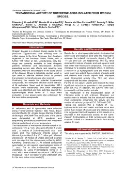
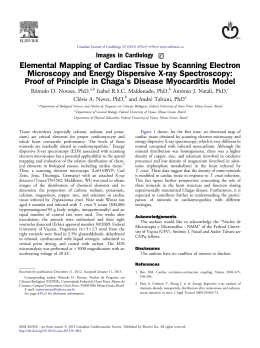
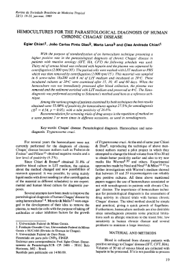
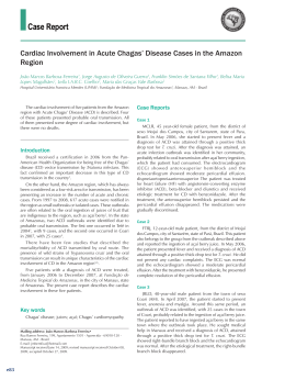
Mem Inst Oswaldo Cruz, Rio de Janeiro, Vol. 100(2): 111-116, April 2005
111
Blood transfusion and iatrogenic risks in Mexico city.
Anti-Trypanosoma cruzi seroprevalence in 43,048 blood donors,
evaluation of parasitemia, and electrocardiogram findings in
seropositive
Nidia Hernández-Becerril, Ana Maria Mejía* , Martha Alicia Ballinas-Verdugo,
Verónica Garza-Murillo, Elsa Manilla-Toquero*, Ruth López***, Sergio Trevethan**,
Manuel Cardenas, Pedro Antonio Reyes, Kenji Hirayama****, Victor Manuel Monteón/+
Laboratorio Inmunoparasitología *Banco de Sangre **Consulta Externa, Instituto Nacional Cardiología I. Chávez, Juan
Badiano 1, Taplan, 14080 México DF ***Centro Investigación Enfermedades Tropicales, Campeche, México
****Institute of Tropical Medicine, Nagasaki University, Japan
Iatrogenous transmission of Trypanosoma cruzi by blood transfusion was suggested as a potential risk by
Pellegrino (1949). Seropositive blood donors in Mexico were first reported in 1978, however, limited information is
available due to small sampling, the use of heterogeneous serologic assays, and geographically limited studies. A
wide survey carried out in 18 out of the 32 states of Mexico, showed a national mean of 1.6% seropositive among
64,969 donors, ranging from 0.2 to 2.8%.
In the present study, we have screened 43,048 voluntary blood donors in a period of five years at the Instituto
Nacional de Cardiología I. Chávez, a concentration hospital located in Mexico city which serves mainly the
metropolitan area and accepts from all over the country. Standardized ELISA and IIF were used to identify seropositive individuals in addition to hemoculture, PCR and standard 12 lead ECG tests that were applied to a group of
seropositive patients (29/161). The result showed a seropositivity of 0.37% (161/43,048). From the group of
seropositive individuals 40% (12/29) were potential carriers of T. cruzi at the donation time and 5/29 had
subclinical ECG abnormalities. Parasitological tests performed in 70 erythrocyte and platelet fractions from seropositive units (70/161) showed negative results. Our findings strongly support T. cruzi screening in the transfusion
medicine practice and identify subclinical heart disease among seropositive blood donors.
Key words: Trypanosoma cruzi - Chagas disease - blood bank - seroprevalence - Mexico
Blood transfusion is the second most important mode
of transmission of Chagas disease in Latinoamerica (Pinto
& Brener 1984, Schmunis 1991). Nearly 800 cases of blood
borne infection with Trypanosoma cruzi caused by iatrogenic mechanism related to transfusion medicine practice
had been collected in South America and the risk is covered by normative regulations in many countries including Mexico. In addition, iatrogenic infection surveillance
was included by the international initiatives to control
Chagas disease in the Americas.
Almost thirty years after the first report predicting
transfusion linked to T. cruzi infection (Pellegrino 1949),
seropositive blood donors were recognized in Oaxaca,
Mexico (Goldsmith & Kagan 1978), and several studies
since then have shown a seropositive incidence varying
between 0.2 to 17% in different regions of the country
(Monteón et al. 1987, Ramos-Echevarria et al. 1993,
Rodríguez-Felix et al. 1995, Trujillo-Contreras et al. 1995,
Rangel et al. 1998, Guzmán-Bracho et al. 1998, Monteón-
Padilla et al 1999, Sánchez-Guillen et al. 2002). A wide survey was carried out in the national blood bank network,
which included 64,969 samples collected in 18 Mexican
states showed a seroprevalence varying between 0.2 and
2.8%, with a general mean of 1.6% (Guzman-Bracho et al.
1998).
In Mexico, Chagas serological screening in blood bank
units is not routinely done in all the endemic areas
(Schmunis 1999), and the potential transfusion transmission of parasites together with the clinical status of seropositive subjects have been poorly studied (MonteónPadilla et al. 1999).
We screened, in a period of five years (1999-2003), all
voluntary blood donors attended in the National Institute of Cardiology I. Chávez in Mexico city. Additionally,
seropositive individuals were investigated in order to identify circulating parasite and/or parasite DNA, ECG was
also performed to identify subclinical abnormalities in these
presumptively infected individulas.
MATERIALS AND METHODS
+Corresponding author. E-mail: [email protected]
Received 19 May 2004
Accepted 26 January 2005
Studied population - In a period of five years (19992003) we invited consecutive blood donors attending the
blood bank service at the Instituto Nacional Cardiología
“I. Chávez” in Mexico city, to participate in a survey which
included an epidemiological questionnaire and serological screening for anti-T.cruzi antibodies.
112
Seropositive blood donors in Mexico • Nidia H-Becerril et al.
Routinely, donated blood is leucoreduced, and separated in three fractions: plasma, erythrocytes, and platelets.
Serological tests for anti-T. cruzi antibodies - The
home-made ELISA test previously validated (Monteón et
al. 1995, Sánchez et al. 2001) was used as the screening
assay. In brief, polystyrene plates were coated with 10
µg /ml of T. cruzi protein extract (INC-1 Mexican strain,
isolated from chronic cardiomyopathy chagasic case) in
alkaline-buffered solution. Human serum was diluted at
1:200 and anti-human IgG-peroxidase conjugate (Zymed
laboratories) was used at 1:15,000. The reaction was revealed by the addition of O-phenilen-diamine and read at
490 nm.
Sera from 30 healthy cases individually tested were
used to establish the cut off. The mean OD plus 5 SD
classified all healthy individuals as negative.
Indirect immunofluorescence (IIF) was used as the
second test. All the ELISA-T .cruzi positive sera were
submitted to the IIF test. In brief, IIF was performed in the
T .cruzi epimastigotes phase, fixed on a microscope slide.
Human serum was diluted at 1:32 and the flourescein conjugated goat anti-human IgG was used at 1:100 dilution.
Positive and negative controls were always included, and
the slide was read under epiflourescence microscope.
Blood supply from blood donor reactive only in the
ELISA-T. cruzi screening test was discarded. A confirmative positive blood donor for T. cruzi infection should
have both ELISA and IIF tests positive.
Parasitologic studies of T. cruzi-infected human blood
- Seventy out of 161 serological T. cruzi positive blood
supplies, in particular platelet and erythrocyte fractions
were submitted to hemoculture and PCR.
A subgroup of 29 out of 161 seropositive donors that
accepted to participate in a second round were resubmitted for hemoculture, PCR and ECG tests.
(i) Hemoculture: we followed the technique previously
described (Luz et al. 1994) with minor modifications. From
platelet and erythrocyte fractions approximately 50 ml were
centrifuged at 3500 g for 15 min. Platelet sediment was
resuspended in 10 ml of liver infusion tryptose (LIT) media supplemented with 10% fetal calf serum. In the case
of the erythrocyte fraction, after centrifugation, approximately 10 ml of the upper layer was divided into four tubes
and equal amount of LIT medium was added. The tubes
were incubated at 28°C for four months. Monthly evaluation of cultures was performed by taking out an aliquot to
be observed under light microscope (250 ×).
In the subgroup of 29 seropositive blood donors 20 ml
of blood sample was obtained for hemoculture. In brief,
the blood sample was centrifuged as mention above, the
supernatant was discarded and the sediment washed
twice with LIT medium. The sediment was divided into
four tubes and resuspended with equal amount of LIT
medium.
(ii) PCR for T. cruzi detection - From platelet and erythrocyte fractions and whole blood as second sample from
seropositive subjects, 1 ml aliquot was used for DNA extraction as previously reported (Monteón-Padilla et al.
1999). Lysis buffer was added to each sample (SDS final
concentration 1% and proteinase K 0.2 mg/ml). The mixture was incubated for 3 h at 42°C and extracted twice
with phenol/cloroform. Subsequently, sodium acetate (final concentration 0.3M) and ethanol were added for DNA
precipitation. DNA pellet was re-suspended in 20 µl of
sterile water.
Amplification reaction was performed in 50 µl volume
containing 1 µl of DNA sample, 300 ng of each minicircle
specific primers KNS1 and KNS2 (Monteon-Padilla et al.
1999), 0.2 mM of each dNTPs, and 2.5 U of Taq polymerase (Invitrogen), Tris-HCl 10 mM, pH 8.8, KCl 50 mM,
MgCl 1.5 mM. The reaction was subjected to 35 cycles:
94°C for 1 min, 56°C for 45 s, and 72°C for 1 min. The
amplified product was submitted to a 2% agarose gel electrophoresis and visualized after ethidium bromide staining under UV expusure. Positive and negative controls
were always included.
(iii) ECG: all blood donor seropositive volunteers that
accepted to participate in this second phase of the protocol (29 out of 161 seropositive), a standard 12 leads ECG
was recorded at a paper speed of 25 mm/s with a portable
Fukuda Denshi Model FX-2111 electrocardiograph
(Fokuda Co.). The tracing was blind analyzed independently by two cardiologists. The criteria for the ECG interpretation were those utilized by the Department of Electrocardiography and Electrophysiology of the Instituto
Nacional de Cardiología. Special attention was given to
the presence of arythmias, A-V heart block, bundle branch
blocks and ventricular hypertrophy and repolarization
abnormalities with changes in the ventricular gradient.
Ethical consideration - This work was done in accordance to “ Reglamento de la Ley General de Salud en Materia de Investigación” which stated forearm venipuncture as a minimal risk procedure and requires oral consent
when illiterate participants are invited to a clinical or epidemiological study. A written informed consent letter was
used. The Ethics Committee of the Institute (Instituto
Nacional Cardiología “ I. Chávez”) approved the study
protocol.
The subgroup of 29 seropositive individuals were delivered for medical attention and follow-up study.
Statistical analysis - The descriptive section consisted
only in determination of the mean, standard deviation and
percentage.
RESULTS
In a period of five years (1999-2003) 43,048 blood donors were serological screened. When a positive serum
was identified this was confirmed at the CNTS (Reference
Center) for HIV, HVB, and HVC, since T. cruzi serology is
not mandatory for urban areas such as Mexico city. All
positive cases were re-tested and confirmed by using a
second validated test in our Institution (Monteón et al.
1995, Sánchez et al. 2001). In Table I we can observe that
seroprevalence for anti-T. cruzi antibodies, by at least
two different tests, showed similar values along the different years that were analyzed with a significant increase
in the year of 2003 with a 0.6% seroprevalence, obtaining
113
Mem Inst Oswaldo Cruz, Rio de Janeiro, Vol. 100(2), April 2005
a 0.37% of seroprevalence in the total population. Similar
results were observed for HVC showing seroprevalence
between 0.16% to 0.44% for the period analyzed and 0.28%
in the total population. In the cases of HIV and HVB a
higher seroprevalence was detected in 2002 and 2000 respectively, with a global seroprevalence of 0.15% for HIV
and 0.07% for HVB.
Out of the 161 T. cruzi positive confirmed cases, only
in a group of 29 of them were able to carry out ECG exam,
hemoculture and PCR. We found in this group a median
age of 32 ± 10 years old with predominance of male over
female (72.4% male) (Table II).
Those 29 individuals came from 12 different states of
the 32 states of Mexico, and the geographic localization
is pointed out in Fig. 1 and Table II. From the central part
of Mexico there were 20 donors out of 29 (68.9%), from
costal Golf of Mexico 2 (6.9%) and from costal Pacific
Ocean 7 (24.1%).
Fifty nine percent (25,398/43,048) of blood donors
screened were born and raised in the urban area of Mexico
city, and 37% (15,928/43,048) in the Central part of Mexico
and only 2.6% (1,119/43,048) in the costal Pacific Ocean
and 1.4% (603/43,048) in the costal Golf of Mexico. This
data mean that 96% of blood donors seen in our Institu-
TABLE I
Seroprevalence in blood donors at the Instituto Nacional
Cardiología “I. Chávez” in a period of five years (1999-2003)
Year
T. cruzi
(%)
HVC
(%)
HIV
(%)
HVB
(%)
1999
n = 7673
25a
(0.32)
22
(0.28)
15
(0.19)
4
(0.05)
2000
n = 8756
28
(0.31)
24
(0.27)
10
(0.11)
9
(0.1)
2001
n = 8932
20
(0.22)
22
(0.24)
6
(0.06)
5
(0.05)
2002
n = 8230
30
(0.36)
37
(0.44)
31
(0.37)
7
(0.08)
2003
n = 9457
58
(0.6)
16
(0.16)
4
(0.04)
9
(0.09)
Total 43,048
161
(0.37)
121
(0.28)
66
(0.15)
34
(0.07)
a: number of positive blood donors confirmed by at least two
different tests. In the case of Trypanosoma cruzi by ELISA and
IIF, for HIV, HVB and HVC by ELISA and Western blot and
RIBA in exceptional cases (genome amplification).
TABLE II
Demographic, electrocardiographic and parasitological findings in Trypanosoma cruzi seropositive blood donors
Case
1
2
3
4
5
6
7
8
9
10
11
12
13
14
15
16
17
18
19
20
21
22
23
24
25
26
27
28
29
n = 29
Age/Sex
Place of birth
Place of residence
ECG
PCR
Hemoculture
20/M
26/F
22/M
47/M
44/M
26/F
27/F
19/M
43/M
25/F
54/M
21/M
43/F
25/M
28/M
36/M
22/F
43/M
30/M
42/M
40/M
35/M
20/F
59/M
34/M
26/F
39/M
35/M
23/M
Puebla
DF
Hidalgo
Veracruz
Hidalgo
Mexico State
DF
Hidalgo
Puebla
DF
Oaxaca
Mexico State
Oaxaca
DF
Guanajuato
Guanajuato
Michoacan
Puebla
Hidalgo
Oaxaca
Sinaloa
Oaxaca
Guerrero
Hidalgo
Morelos
DF
Veracruz
Tlaxcala
Guanajuato
Puebla
DF
Hidalgo
Veracruz
Hidalgo
Mexico State
DF
DF
Puebla
DF
Oaxaca
Mexico State
DF
DF
Guanajuato
Guanajuato
Michoacan
DF
Mexico State
Veracruz
Sinaloa
Oaxaca
DF
DF
Morelos
DF
Veracruz
DF
Guanajuato
Normal
Normal
LBBB
Normal
Normal
Normal
Normal
RBBB
Normal
Normal
Normal
Normal
Normal
Normal
Normal
Normal
VE
Normal
Normal
Normal
Normal
Normal
Short P-R
Normal
RBBB, VE
Normal
AVB
Normal
Normal
Positive
Positive
Negative
Positive
Negative
Negative
Positive
Negative
Negative
Positive
Negative
Positive
Negative
Negative
Positive
Positive
Positive
Negative
Positive
Negative
Positive
Negative
Negative
Negative
Positive
Negative
Negative
Negative
Negative
Negative
Negative
Negative
Negative
Negative
Negative
Negative
Negative
Negative
Negative
Negative
Negative
Negative
Negative
Positive
Negative
Negative
Negative
Positive
Negative
Negative
Negative
Negative
Negative
Positive
Negative
Negative
Negative
Negative
5/29
(17%)
12/29
(41%)
3/29
(10%)
Mean = 32 ±
10 years old
LBBB: left bundle-branch blockage; RBBB: rigth bundle-branch blockage; VE: ventricular extrasystole
114
Seropositive blood donors in Mexico • Nidia H-Becerril et al.
donors represents subclincal chagasic subjects. Three of
them came from the central part of Mexico (Hidalgo and
Morelos State), one from Veracruz State (costal Golf of
Mexico) and one more from Michoacan State (costal Pacific Ocean). The ECG findings were LBBB (left bundle
branch blockage) in one subject, RBBB (right bundle
branch blockage) in two, VE (ventricular extra systole) in
two and one AVB (auricular-ventricular blockage) in one
of them (Table II).
DISCUSSION
Fig. 1: geographic localization of seropositive blood donors. Dark
zone indicate the place of birth of the seropositive subjects.
tion came from the central part of Mexico, including Mexico
city (data not shown).
T. cruzi DNA was detected in 12 out of 29 subgroup of
seropositive blood donors (41%) by PCR (Fig. 2, Table II)
and positive hemoculture in only 3 out 29 (10%). Although,
hemoculture and PCR were also carried out in 70 out of
161 red blood and platelet fractions from seropositive
units, none gave positive results for T. cruzi parasites.
This negative result may be explained by the fact that
leukocyte rich fraction is eliminated throughout the
fractioning process and must likely eliminates inclusive
parasites.
Abnormal ECG compatible with Chagas disease was
found in 5 out of 29 (17.2%), that means seropositive blood
Fig. 2: representative PCR products. Lines - 1 and 15 blanks (without DNA); 2: negative control (DNA from a seronegative individual); 14: positive control (Trypanosoma cruzi DNA); 3 to 6 and
9-10: negative blood donors; 7, 11, 12, and 13: positive blood
donors; 8 and 16: molecular markers (100 bp ladder).
This study was carried out in an urban blood bank
center located in Mexico city that is ruled out of triatomines
contamination, but migrant people from endemic areas may
disseminate this antropozoonosis, mainly related to blood
transfusions.
Mexico city is an ideal model for this condition mentioned above due to the influx of permanent migrants who
have steadily increased the city’s population from the late
1930s to present.
In the present work we screened 43,048 blood donors,
being this, the first study in Mexico with such large sampling in a single blood bank unit. This size sample provided us confidence on the obtained results. The
seroprevalence observed for T. cruzi was 0.37%, being
higher than the values obtained for HIV and HVB, and
only comparable to HVC (0.28%) on which routine serological testing was performed (Table I).
Although, minor differences in seroprevalence for each
period analyzed was observed that may indicate that similar unchanged risk factors predominated in the period studied. A marked increase was observed for Chagas disease
in 2003 (0.6%), as well as observed for HVC and HIV in
2002 (0.44 and 0.37% respectively), this finding may be
linked to increasing poverty in the country side that obligate the people to move from endemic chagasic regions
to a large cities as Mexico city, having as a consequence
crowd and high risk for viral transmission infections.
The seroprevalence of 0.37% was similar to that previously reported seven years ago for the same blood bank
center in a smaller sampling (Monteón-Padilla et al. 1999)
and almost half of that reported in a sentinel survey conducted in 1994 which reported 1% seroprevalence in Mexico city in 2300 blood donors (Guzmán-Bracho et al. 1998).
This discrepancy may be attributed to methodological
procedures, namely the two subjective tests employed in
that work: Indirect hemaglutination (IHA) and (IIF). Since,
in a previous study carried out by these two laboratories
they observed a Kappa index close to 0.8 (Monteón et al.
1995).
In this context we can suggest that seroprevalence for
T. cruzi in Mexico city can be established between 0.3 up
to 1%.
For the blood donors screened at the Instituto Nacional Cardiología I. Chávez , 59% were born and raised in
Mexico city, and the remaining 41% in the country side.
We identified, in a group of 29 seropositive T. cruzi blood
donors (Table II) that 96% of them came from the central
part of Mexico bordering Mexico city, a distance that was
no more than 500 km away or 8 h travel. In this group 15
Mem Inst Oswaldo Cruz, Rio de Janeiro, Vol. 100(2), April 2005
out of 29 seropositive (51%) were born in this central part
of Mexico an area considered of low endemicity, including 5 (17%) that were born and raised in Mexico city. Only
9 out of 29 seropositive (31%) came from well known endemic areas located in the Pacific coast and Mexico Gulf.
Fortuneless we could not collect information about
the total number of donors originated from each state in
order to provide relative prevalences, however, for Mexico
city we registered 0.1% of relative seroprevalence (27/
23,398), for central part of Mexico excluding Mexico city
was 0.5% (82/15,884), for Pacific coast 3% (38/1235) and
for Mexico Gulf 0.55% (14/2531). This data indicate that
areas considered of low prevalence for T. cruzi infection
as the central part of Mexico including Mexico City may
represent a serious potentially risk in blood banking practice. In this sense our data support previously reported
findings (Goldsmith & Kagan 1978, Monteón et al. 1987,
Trujillo-Contreras et al. 1993, Rodríguez-Felix et al. 1995,
Rangel et al. 1998, Guzmán-Bracho et al. 1998, SanchézGuillen et al. 2002).
Considering all the data, these studies encourage the
need to search anti-T. cruzi antibodies in blood donors,
even in urban locations in Mexico.
Current strategies to prevent transfusion-associated
Chagas disease include the identification of T. cruzi-infected blood donors by questionnaire, serological tests,
and in areas of high endemicity, the treatment of collected
blood with gentian violet (Appleman et al. 1993, Carvalho
et al. 1993, Ramírez et al. 1995). Recently, white cell-reduction filters have been evaluated in artificially T. cruzi infected blood to remove the infectious agent showing that
the efficiency depends on the concentration of parasites
(Moraes-Souza et al. 1995).
Circulating parasites or parasite DNA in blood seropositive donors were achieved in 12 out of 29 seropositive
subjects (41%) by PCR and in 3 out of 29 (10%) by
hemoculture. These findings confirm that some seropositive subjects are potentially active transmitters of T. cruzi
infection at the moment of blood donation. Accordingly
to this data we can suggest that T. cruzi infection risk by
seropositive blood donors should be at least between 10
up to 40%, but positive leukoreduced supplies may represent a reduced risk of transmission of T. cruzi parasites, as was seen in the seventy red blood and platelet
fractions from seropositive units tested, since hemoculture
and PCR tests gave negative results, however, we consider it is necessary to carry out experimental test in order
to verify this observation.
Seroepidemiological surveys carried out in subjects
living in high and low endemic regions for T. cruzi infection in Mexico have showed that electrocardiographic alteration is present in 18 up to 22% of seropositive subjects (Rangel-Flores et al. 2001, Sosa-Jurado et al. 2003).
In this work we found that 5 out of the 29 seropositive
volunteer (17%) blood donors were subclinic subjects
presenting ECG alterations compatible with Chagas disease. The main ECG alterations were right bundle-branch
blockage, left bundle-branch blockage and ventricular
extrasystole. We excluded for this purpose one subject
with short P-R.
115
In conclusion we could observe that 17% of antiT.cruzi seropositive blood donors represents subclinic
chagasic subjects with rhythm or conduction defects;
furthermore at least 40% of them are potential carriers of
T. cruzi parasites at the moment of blood donation, but
use of leukoreduction process may be useful to prevent
T. cruzi transmission and finally blood banking practice
in central part of Mexico considering of low prevalence
for T. cruzi human infection, may represent a potential
risk factor for spreading of the desease.
ACKNOWLEDGMENTS
To Silvia Monteón for carefully reading of the manuscript
and Graciela Luque Kiel for administrative support.
REFERENCES
Appleman MD, Shulman IA, Saxena S, Kirchoff LV 1993. Use
of a questionnarie to identify potential blood donors at risk
for infecction with Trypanosoma cruzi. Transfusión 33: 6164.
Carvalho MR, Krieger MA, Almeida A 1993. Chagas disease
diagnosis: evaluation of several tests in blood bank screening. Transfusion 33: 830-834.
Goldsmith R, Kagan I 1978. El potencial de la transmisión en la
enfermedad de Chagas en el estado de Oaxaca. Gac Med
Mex 4: 439-444.
Guzmán-Bracho C, García-García L, Floriani-Verdugo J,
Guerrero-Martínez S, Torres-Cosme M, Ramírez-Melgar
C, Velasco-Castrejón O 1998. Riesgo de transmisión de Trypanosoma cruzi por transfusión de sangre en México. Rev
Panam Salud Pública 4: 94-98.
Luz ZMP, Couthinho MG, Cancado R, Krettli A 1994. Hemocultura: técnica sensível na detecção do Trypanosoma cruzi
em pacientes chagásicos na fase crônica da doença de Chagas.
Rev Soc Bras Med Trop 27: 143-148.
Monteón VM, Guzmán-Bracho C, Floriani-Verdugo J, RamosEcheverria A, Velasco-Castrejón O, Reyes PA 1995.
Diagnóstico serológico de la enfermedad de Chagas:
autosuficiencia y concordancia interlaboratorios. Sal Pub
(Mex) 37: 232-235.
Monteón-Padilla VM, Hernández-Becerril N, Guzmán-Bracho
C, Rosales-Encina JL, Reyes-López PA 1999. American
trypanosomiasis (Chagas’ disease) and blood banking in
Mexico city: seroprevalence and its potential transfusional
transmission risk. Arch Med Res 30: 393-398.
Monteón VM, Linares TC, Amador GF 1987. Anticuerpos
séricos a Trypanosoma cruzi en donadores de sangre en la
Ciudad de México. Bioquímica (Mex) 7: 6-9.
Moraes-Souza H, Bardossy L, MacPherson DW, Blajchman
MA 1995. Prevention of transfusion-asociated chagas disease: efficacy of withe cell-reduction filters in removing
Trypanosoma cruzi from infected blood. Transfusion 35:
723-726.
Pellegrino J 1949. Transmissão da doença de Chagas pela
transfusão de sangue. Primeiras comprovações serológicas
em doadores e em candidatos a doadores de sangue. Rev
Bras Med 6: 297-301
Pinto DJC, Brener S 1984. Chagas’ disease and blood transfusion. Mem Inst Oswaldo Cruz 79: 139-147
116
Seropositive blood donors in Mexico • Nidia H-Becerril et al.
Ramírez LE Lages-Silva E, Pianetti GM, Rabelo RMC, Bordin
JO, Moraes-Souza H 1995. Prevention of transfusion-associated Chagas’ disease by sterilization of Trypanosoma
cruzi-infected blood with gentian violet, ascorbic acid, and
light. Transfusion 35: 226-230.
Ramos-Echeverria A, Monteón-Padilla V, Reyes PA 1993.
Detección de anticuerpos contra Trypanosoma cruzi en
donadores de sangre. Sal Publica (México) 35: 56-54.
Rangel H, Gatica R, Ramos C 1998. Detection of antibodies
against Trypanosma cruzi in donors from a blood bank in
Cuernavaca, Morelos, Mexico. Arch Med Res 29: 79-82.
Rangel-Flores H, Sánchez B, Mendoza-Duarte J, Barnabé C,
Breniére FS, Ramos C, Espinoza B 2001. Serologic and
parasitologic demostration of Trypanosoma cruzi infections
in an urban area of central Mexico: correlation with electrocardiographic alterations. Am J Trop Med Hyg 65: 887895.
Rodríguez-Felix ME, Zavala-Velazquez J, Barrera-Pérez MA,
Guzmán-Marin E, Ramírez-Sierra MJ, Alvarez-Moguel R
1995. Riesgo de transmision de la enfermedad de Chagas
por donantes de sangre. Rev Biomed 6: 70-75.
Sánchez B, Monteón V, Reyes PA, Espinoza B 2001. Standardization of micro-enzyme-linked immunusorbent assay
(ELISA) and western blot for detection of Trypanosoma
cruzi antibodies using extracts from Mexican strains as antigen. Arch Med Res 32: 382-388.
Sánchez-Guillen MC Velásquez-Rojas M, Martínez-Mungía J,
Salgado-Rosas H, Torres-Rasgado E, Rosas-Ramírez MI,
Pérez-Fuentes R 2002. High prevalence anti-Trypanosoma
cruzi antibodies, among blood donors in the state of Puebla,
a non-endemic area of Mexico. Mem Inst Oswaldo Cruz 97:
947-952.
Schmunis G 1991. Trypanosoma cruzi, the etiologic agent of
Chagas’ disease: status in the blood supply in endemic and
nonedemic countries. Transfusion 31: 547-551.
Schmunis GA. 1999. Risk of Chagas disease through transfusions in the Americas. Medicina (B Aires) 59: 125-134.
Sosa-Jurado F, Mazariego-Aranda M, Hernández-Becerril N,
Garza-Murillo V, Cárdenas M, Reyes PA, Hirayama K,
Monteón VM 2003. Electrocardiographic findings in mexican
chagasic subjects living in high and low endemic regions of
Trypanosoma cruzi infection. Mem Inst Owaldo Cruz 98:
605-610.
Trujillo-Contreras F, Lozano-Kasten F, Soto-Gutiérrez M,
Hernández-Gutiérrez R 1993. Prevalencia de infección a
Trypanosoma cruzi en donadores de sangre en el estado de
Jalisco Mexico. Rev Soc Bras Med Trop 26: 89-92.
Download