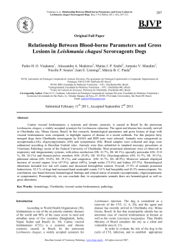
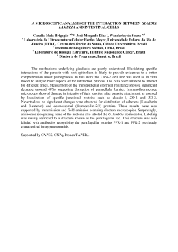
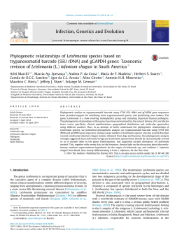
DNA Research Advance Access published July 15, 2013
DNA RESEARCH pp. 1–15, (2013)
doi:10.1093/dnares/dst031
The Genome Sequence of Leishmania (Leishmania) amazonensis:
Functional Annotation and Extended Analysis of Gene Models
FERNANDO Real1,†, RAMON OLIVEIRA Vidal2,†, MARCELO FALSARELLA Carazzolle2,3,†, JORGE MAURÍCIO COSTA Mondego4,
GUSTAVO GILSON LACERDA Costa3, ROBERTO HIROCHI Herai5, MARTIN Würtele6, LUCAS MIGUEL de Carvalho2,
RENATA CARMONA e Ferreira1, RENATO ARRUDA Mortara1, CLARA LUCIA Barbiéri1, PIOTR Mieczkowski7, JOSÉ FRANCO da
Silveira1, MARCELO RIBEIRO DA SILVA Briones1, GONC ALO AMARANTE GUIMARÃES Pereira2, and DIANA Bahia1,8, *
*To whom correspondence should be addressed: Tel. þ55 11 5576-4532. Fax. þ55 11 5571-1095.
E-mail: [email protected]
Edited by Naotake Ogasawara
(Received 24 January 2013; accepted 17 June 2013)
Abstract
We present the sequencing and annotation of the Leishmania (Leishmania) amazonensis genome, an etiological agent of human cutaneous leishmaniasis in the Amazon region of Brazil. L. (L.) amazonensis shares
features with Leishmania (L.) mexicana but also exhibits unique characteristics regarding geographical distribution and clinical manifestations of cutaneous lesions (e.g. borderline disseminated cutaneous leishmaniasis). Predicted genes were scored for orthologous gene families and conserved domains in comparison with
other human pathogenic Leishmania spp. Carboxypeptidase, aminotransferase, and 30 -nucleotidase genes
and ATPase, thioredoxin, and chaperone-related domains were represented more abundantly in L. (L.) amazonensis and L. (L.) mexicana species. Phylogenetic analysis revealed that these two species share groups of
amastin surface proteins unique to the genus that could be related to specific features of disease outcomes
and host cell interactions. Additionally, we describe a hypothetical hybrid interactome of potentially
secreted L. (L.) amazonensis proteins and host proteins under the assumption that parasite factors mimic
their mammalian counterparts. The model predicts an interaction between an L. (L.) amazonensis heatshock protein and mammalian Toll-like receptor 9, which is implicated in important immune responses
such as cytokine and nitric oxide production. The analysis presented here represents valuable information
for future studies of leishmaniasis pathogenicity and treatment.
Key words: genome; Leishmania amazonensis; interactome; amastin; heat-shock protein
1.
Introduction
Leishmaniases are neglected infectious diseases
caused by parasites belonging to the Trypanosomatidae
†
The authors agree that the first three authors should be regarded
as joint first authors.
family and the Leishmania genus. Leishmaniases are
prevalent in tropical countries; 12 million people are
affected by these diseases worldwide with 350 million
people at risk of infection and an estimated yearly incidence of 2 million cases.1 Leishmania spp. are digenetic
parasites that develop as promastigotes in the gut of phlebotominae sandflies and as intracellular amastigotes in
# The Author 2013. Published by Oxford University Press on behalf of Kazusa DNA Research Institute.
This is an Open Access article distributed under the terms of the Creative Commons Attribution License (http://creativecommons.org/licenses/by-nc/
3.0/), which permits non-commercial use, distribution, and reproduction in any medium, provided the original work is properly cited. For commercial
re-use, please contact [email protected].
Downloaded from http://dnaresearch.oxfordjournals.org/ at Universidade Federal de Minas Gerais on August 12, 2013
Departamento de Microbiologia, Imunologia e Parasitologia, Escola Paulista de Medicina, Universidade Federal de São
Paulo 2 EPM/UNIFESP, Rua Botucatu 862, 6o andar, 04023-062 São Paulo, Brazil1; Laboratório Nacional de
Biociências, LNBio/CNPEM, Campinas, Brazil2; Laboratório de Genômica e Expressão, LGE/UNICAMP, Campinas,
Brazil3; Centro de Pesquisa e Desenvolvimento de Recursos Genéticos Vegetais, Instituto Agronômico de Campinas – IAC,
Campinas, Brazil4; Department of Pediatrics, School of Medicine, University of California, San Diego, CA, USA5;
Departamento de Ciência e Tecnologia, Universidade Federal de São Paulo 2 UNIFESP, São José dos Campos, Brazil6;
Department of Genetics, School of Medicine, University of North Carolina, Chapel Hill, NC, USA7 and Departamento de
Biologia Geral, Instituto de Ciências Biológicas, Universidade Federal de Minas Gerais-ICB/UFMG, Minas Gerais, Brazil8
Page 2 of 15
F. Real et al.
Leishmania is regulated mainly at the post-transcriptional
level by RNA stability, rather than by promoters.13
Genes are organized into polycistronic transcriptional
units, and protein-encoding genes are co-transcribed
by RNA polymerase II. Precursor mRNAs subsequently
are trans-spliced and polyadenylated.14 – 16
The number of chromosomes has been established
for several Leishmania species.17 – 21 The molecular
karyotypes of Old World Leishmania species [L. (L.) infantum, L. (L.) donovani, L. (L.) major, and L. (S.) tarentolae] each comprise 36 chromosomes,17 whereas the
New World species, L. (V.) braziliensis, and L. (L.) mexicana have 35 and 34 chromosomes, respectively, due
to fusion events involving 2– 4 chromosomes.18,19
The genomes of two Old World Leishmania species,
L. (L.) major Friedlin, and L. (L.) infantum JPCM5, and
one New World species, L. (V.) braziliensis M2904,
have been sequenced and annotated.19,22 Recently,
the genomes of L. (L.) mexicana, 16 clinical isolates of
L. (L.) donovani, and the lizard parasite, L. (S.) tarentolae,
were sequenced and assembled using high-throughput
DNA sequencing technologies.20,23,24 Despite evolutionary divergence within the Leishmania genus,
Leishmania comparative genomics suggests a high
degree of synteny.19,20,23,24 Leishmania spp. from the
Leishmania and Viannia subgenera exhibit highly conserved gene sequences with remarkably few genes or
paralog groups that are unique to any given species.
However, L. (S.) tarentolae lacks genes associated with
the intracellular life stages of human pathogenic
Leishmania spp.20 On the other hand, the L. (V.) braziliensis genome includes features that are lacking in the
genomes of Old World Leishmania spp., such as transposable elements and RNA interference (RNAi)
machinery.19,25,26
To obtain a broader understanding of the pathogenesis of leishmaniasis, we sequenced the genome of the
New World species, L. (L.) amazonensis. Using a comparative bioinformatics approach with other available
Leishmania genomes, we searched for conserved
domains and orthologous gene families among predicted proteins of L. (L.) amazonensis. In addition, we
inferred the phylogeny of the surface glycoprotein,
amastin, and generated a hybrid protein interactome
to identify potential interactions between L. (L.) amazonensis secreted proteins and mammalian host factors.
2.
Materials and methods
All the procedures employed in this study, except for
phylogenetic analyses, are summarized in the workflow
presented in Fig. 2. The workflow was divided into
genome assembly and annotation steps, and functional
and extended analyses of gene models.
Downloaded from http://dnaresearch.oxfordjournals.org/ at Universidade Federal de Minas Gerais on August 12, 2013
the macrophages of vertebrate hosts. The Lainson and
Shaw classification2 subdivides the Leishmania genus
into two subgenera based on the localization of promastigotes in the insect alimentary tract. The subgenus
Leishmania comprises species limited to the midgut and
foregut of the sand fly, whereas the subgenus Viannia
includes species that develop a prolonged phase in the
hindgut with later migration of flagellates to the
midgut and foregut of the vector’s alimentary tract.
More recently, a third subgenus has been included in
Leishmania classification, the subgenus Sauroleishmania,
which comprises species that exclusively parasitize
lizards.3 A brief classification of Leishmania subgenera
and species associated with the diverse array of leishmaniasis clinical manifestations (cutaneous, mucocutaneous,
and visceral forms) is provided in Fig. 1A.
Leishmaniases are primarily zoonotic diseases, and a
variety of mammals acts as reservoirs of Leishmania
species. Specifically, rodents, edentates, and marsupials
typically harbor cutaneous leishmaniasis, whereas wild
canines and domestic dogs are the main reservoirs of
zoonotic visceral leishmaniases. In human hosts,
disease outcomes are determined by a combination of
parasitic properties (dermotropic versus viscerotropic
species) and host factors, such as genetic variability
and immune responses to infection.4,5 Among the
causative species of cutaneous leishmaniases in Brazil,
recent data indicate that 8% are attributed to
Leishmania (L.) amazonensis.6 This species can cause
simple and diffuse forms of cutaneous leishmaniasis
(DCL) and was implicated recently in borderline disseminated cutaneous leishmaniasis, an intermediate
form of disease.7
Infections with Leishmania species belonging to the
Leishmania (L.) mexicana complex involve the dermal infiltration of macrophages that harbor parasites in large
parasitophorous vacuoles (PVs). Most Leishmania
species including Leishmania (L.) major, Leishmania
(L.) donovani, and Leishmania (V.) braziliensis lodge
intracellularly within small membrane-bound PVs
that typically contain a single parasite and undergo
fission as the amastigotes divide.8 In contrast, amastigotes of L. (L.) amazonensis and L. (L.) mexicana are
housed in large numbers within spacious PVs (Fig. 1B)
that fuse together.9 These enlarged PVs may subvert
host cell defenses by facilitating conditions of relatively
diluted hydrolytic enzymes.10,11 The mechanistic basis
of spacious PV development remains unknown and
likely is triggered by unidentified parasitic factors produced by L. (L.) amazonensis and other species from
the L. (L.) mexicana complex, such as L. (L.) mexicana
and Leishmania (L.) pifanoi.12
During the past decade, several reports have
attempted to elucidate the factors used by Leishmania
to interact with its vertebrate host and establish an infection. Like other kinetoplastids, gene expression in
The Genome Sequence of L. (L.) amazonensis
Page 3 of 15
2.1. Genomic DNA extraction
Leishmania (L.) amazonensis (MHOM/BR/71973/
M2269 strain) was extracted from the cutaneous
lesions of a patient from Cafezal city, in the state of
Pará, Brazil, in 1973. Since then, this strain has been
maintained in the laboratory by inoculating hamsters
and mice and by axenic culture. Parasites were cultivated in M199 culture medium supplemented with
10% fetal bovine serum. Genomic DNA was extracted
from 109 promastigotes after the parasites were incubated in lysis buffer [50 mM Tris – HCl ( pH 8.0),
62.5 mM EDTA ( pH 9.0), 2.5 M LiCl, 4% Triton X-100]
at 378C for 5 min. DNA was purified using phenol –
chloroform extraction (1:1 v/v) and ethanol precipitation. The resulting pellets were resuspended in
50 ml of 10 mM TE [Tris – HCl ( pH 8.0), 1 mM EDTA]
containing 0.6 mg/ml of RNase A (Life Technologies
Corporation, USA), and were incubated at 378C for
30 min. Genomic DNA was precipitated with 2.5 v of
100% ethanol and 0.3 M sodium acetate, centrifuged
at 15 700g at 48C for 15 min, and resuspended in
DNase-free water.
2.2. Genome sequencing and assembly
Leishmania (L.) amazonensis DNA sequences were
obtained using the whole-genome shotgun strategy27
on a combination of 454 GS-FLX Titanium (Roche)
and Solexa (Illumina, Inc.) instruments from the
University of North Carolina (UNC, USA) sequencing
facility. The GS-FLX sequencer generated single-end
fragment reads (454 reads) with a mean length of
315 bp. The Illumina Genome Analyzer generated
76-bp paired-end fragment reads (Solexa reads) with
an average insert size of 400 bp. Using an Perl script
developed in-house, Illumina reads were filtered out if
the average Phred quality score was lower than 20. For
454 sequences, reads comprising fewer than 100 bp
or more than 500 bp (5% of each side of the normal
distribution of read sizes) and reads with more than 1
unknown nucleotide (N) also were filtered out.
The Solexa reads were assembled into longer scaffolds
using the Velvet 0.7.56 de novo assembler28 with a
k-mer parameter of 43. This value was calculated
using the Velvet Optimizer script (Victorian Bioinformatics Consortium, Monash University, Australia),
Downloaded from http://dnaresearch.oxfordjournals.org/ at Universidade Federal de Minas Gerais on August 12, 2013
Figure 1. Overview of the L. (L.) mexicana complex. (A) Classification of the Leishmania genus, subgenus and species complex (adapted from the
WHO reports and Bates, 2007). Leishmania (L.) amazonensis and L. (L.) mexicana belong to the L. (L.) mexicana complex, subgenus Leishmania,
and are causative agents of New World cutaneous leishmaniasis in which diffuse or disseminated lesions are hallmarks. The genomes of the
species marked in red were employed in the present comparative analyses [*L. (S.) tarentolae was employed only in the amastin phylogenetic
study]. (B) Large parasitophorous vacuoles (PVs) of L. (L.) amazonensis. Phase contrast microscopy image (left) of a bone marrow-derived
macrophage containing a spacious PV (asterisk) lined with rounded amastigotes. Bar ¼ 10 mm. Field-emission scanning electron
micrograph (right) of an amastigote-hosting macrophage. The fractured sample indicated that amastigote forms (in red) were
contained in a spacious PV. Bar ¼ 5 mm.
Page 4 of 15
F. Real et al.
which tested a range of k-mers from 31 to 69. Newbler
software29 was then used to assemble the 454 reads.
The N50 scaffold and contig lengths of the L. (L.) amazonensis assembly were 22 275 and 17 272 bp, respectively. Solexa scaffolds and 454 contigs were combined
by the Zorro assembler (Laboratório de Genômica e
Expressão, UNICAMP, Brasil, http://lge.ibi.unicamp.br/
zorro/downloads/Zorro2.2/DOC) to create the final
hybrid genome assembly. The hybrid assembly was performed by combining the 4441 Illumina scaffolds and
the 27 856 contigs from the 454 reads. Briefly, the
Zorro pipeline consisted of (i) masking repeat regions
in the contigs, (ii) detecting overlaps, (iii) unmasking
repeat regions, and (iv) assembling hybrid contigs.
In Phase 1, the repeat regions were determined by
counting the occurrences of k-mers in the 454 reads.
The assembler then masked k-mers in the contigs
that occurred at high frequency. The absence of
repeats enabled accurate overlap detection in Phase 2.
The contigs were then unmasked in Phase 3, and the
correct hybrid assembly was obtained in Phase 4 by
merging all overlapping contigs into hybrid contigs.
Overlap detection and consensus generation were
performed using the Minimus package.30 The hybrid
contigs were ordered and oriented with the Bambus
program31 using paired-end information and manual
verification, which yielded 2627 scaffolds.
2.3. Gene identification and annotation
2.3.1. Alignment of Leishmania spp. proteins with
the L. (L.) amazonensis genome Predicted
proteins for L. (V.) braziliensis (8153 proteins), L. (L.)
infantum (8154 proteins), L. (L.) major (8298 proteins), and L. (L.) mexicana (8007 proteins) were
mapped onto the L. (L.) amazonensis genome sequence
using the Exonerate program (v. 2.2.0),32 which performs genomic searches and spliced alignments in a
single run.
2.3.2. Ab initio prediction of gene models Glimmer,
v. 3.02 33 and Genemark.hmm, v. 3.3 34 programs were
used to perform ab initio gene predictions. The Glimmer
long-orfs program was trained on non-overlapping
open reading frames (ORFs) exceeding 200 bp in
length. Genemark.hmm was executed in self-training
mode by considering ORFs of at least 200 bp in length.
2.3.3. Combined gene models The above resources were used to automatically create L. (L.) amazonensis gene models using EVidenceModeler (EVM,
v. r03062010) software.35 For individual outputs
generated by Exonerate, Glimmer and Genemark.
hmm were given values reflecting our data confidence
to define gene structure. We considered the following
confidence values: Glimmer ¼ 3, Genemark ¼ 3,
Downloaded from http://dnaresearch.oxfordjournals.org/ at Universidade Federal de Minas Gerais on August 12, 2013
Figure 2. Bioinformatics analysis workflow used in the present study. Sequenced reads from the L. (L.) amazonensis genome were assembled
into 2627 scaffolds and 8100 genes were predicted using comparative and ab initio prediction tools. The functional analysis of these
predicted genes included: (i) AutoFACT functional annotation, which revealed that 45% of the predicted genes were unclassified or with
unassigned function; (ii) screening for orthologous families of genes among Leishmania spp. (OrthoMCL); and (iii) screening for
information about conserved protein domains deposited in CDD and PFAM databases. Expanded or exclusive orthologous proteins, or
those conserved domains detected in the L. (L.) amazonensis genome were selected for interactome analysis with mammalian host
proteins. This selection involved screening for possibly secreted proteins (using SecretomeP and TargetP) that also were orthologous to
immune function-related proteins in humans and mice.
The Genome Sequence of L. (L.) amazonensis
nucleotide-to-protein match ¼ 6. The gene model was
considered reliable only if it was predicted by all ab initio
software or if it had similarity with one of the compared
species. A final set of predicted gene models was then
selected and manually inspected. Incomplete genes or
genes overlapping gap regions were inspected with
input from blastx alignments against protein databases
and scaffold edges.
2.3.4.
2.3.5. Orthologous gene analysis A data set composed of all the Leishmania spp. gene models was
created and compared all-against-all using blastp
(e-value cutoff of 1 1025). The results were submitted to OrthoMCL (v. 1.4) software,41 which clustered
the proteins into orthologous and paralogous families.
We applied the default software parameters, including
an inflation index of 1.5. The inflation index regulates
the cluster tightness (granularity) associated with sensitivity and selectivity.42 Clusters of proteins that presented bidirectional similarities between at least two
Leishmania species were considered orthologs.
2.3.6. Pseudogene identification Leishmania and
Trypanosoma spp. proteins were aligned against the
L. (L.) amazonensis genome using blastx with an e-value
threshold of 1 1025. The coordinates of the first hit
alignment for all proteins were converted to GFF file
format using an in-house Perl script. The BEDTools
package43 was used to identify L. (L.) amazonensis
regions exceeding 200 bp that showed similarity with
Leishmania or Trypanosoma spp. proteins without overlapping with the gene model predictions. These regions
were compared against the NCBI non-redundant database using the blastx program and manual annotation.
2.3.7. Calculation of the codon adaptation index The
CodonW v. 1.4.4 software (http://bioweb.pasteur.fr/
seqanal/interfaces/codonw.html) was employed to
calculate the codon usage indices of each L. (L.) amazonensis predicted gene. The codon adaptation index
(CAI) estimates the extent of bias toward codons
known to be preferred in highly expressed genes.44
This index ranges from 0 to 1.0 with higher values indicating stronger codon usage bias and a higher expression level. The frequency of codon usage in highly
expressed genes defines the relative fitness values for
each synonymous codon. These values were calculated
from the relative synonymous codon usage rather than
from the raw codon usage and therefore were essentially independent of amino acid composition. Because
fitness values are highly species specific, we first identified a set of highly expressed genes in L. (L.) amazonensis.
This set was input into the calculation of the CAI. The
effective number of codons (Nc) also was used to quantify the codon usage bias of each gene. The Nc ranges
from 20 for a gene with extreme bias (using only 1
codon per amino acid) to 61 for a gene with no bias
(using synonymous codons equally).45,46 Sequences
for which Nc values are less than 30 were considered
highly expressed, whereas sequences with Nc values
exceeding 55 were considered poorly expressed genes.
2.4. Phylogeny of amastin surface proteins
A phylogeny of Leishmania amastin proteins was built
from a set of L. (L.) amazonensis amastins predicted by
our assembled genome and from all predicted
amastin proteins in the T. cruzi, T. brucei, L. (L.) major,
L. (L.) infantum, L. (V.) braziliensis, L. (L.) mexicana, and
L. (S.) tarentolae genomes. These amastin sequences
[except L. (L.) amazonensis amastins] were extracted
from TriTrypDB47 (accessed 6/28/2012) by searching
for ‘amastin’ or ‘amastin-like’ entries.
The 181 amastin/amastin-like protein sequences
encoded by L. (L.) braziliensis, L. (L.) infantum, L. (L.)
major, L. (L.) mexicana, and L. (S.) tarentolae and 24
amastin/amastin-like protein sequences from L. (L.)
amazonensis were aligned using Geneious software (v.
5.6.3),48 in which an embedded MUSCLE software
was applied.49 The alignment was performed using
default parameters. The phylogenetic tree was inferred
by Bayesian methods using MrBayes v. 3.1.250 with tree
parameter optimization during the generations. A
Bayesian tree was inferred based on 1 107 generations with a burn-in value of 75 000. Data were saved
every 100 generations and were run in 4 chains
Downloaded from http://dnaresearch.oxfordjournals.org/ at Universidade Federal de Minas Gerais on August 12, 2013
Automatic annotation and analysis of protein
domains The automatic annotation software, AutoFACT (v. 3.4),36 was used for functional
annotation of gene models. AutoFACTenables transitive
annotation based on sequence similarity searches
in several databases. We used the blastp algorithm
(e-value 1 10 – 5) to model gene alignments against
the following protein databases: non-redundant proteins (NCBI RefSeq database, downloaded 8/6/2010),
Swiss-Prot (only manually curated proteins, downloaded 4/15/2010),37 UniRef90, and UniRef100
(UniProt databases of clustered protein families, downloaded 4/15/2010), CDD (conserved protein
domains, downloaded 4/15/2010),38 PFAM ( protein
domains, downloaded 4/15/2010),39 and KEGG
(metabolic pathways, downloaded 4/15/2010).40
We set AutoFACT to consider the following order of importance for annotation: UniRef100, UniRef90, KEGG,
non-redundant proteins, and CDD. Data from CDD –
PFAM analyses were extracted from AutoFACT and
were used for comparative analyses of CDD – PFAM
protein domains among L. (L.) amazonensis, L. (V.) braziliensis, L. (L.) infantum, L. (L.) major, and L. (L.) mexicana.
These data also were evaluated using reverse PSI-BLAST
(RPS-BLAST, e-value cutoff of 1 1025).
Page 5 of 15
Page 6 of 15
F. Real et al.
during 2 runs. The Whelan and Goldman (WAG) substitution matrix was used for the protein alignment.51
2.5.
3.
Results and discussion
Annotation of Leishmania (L.) amazonensis gene
models
Approximately 37 million 76-bp paired-end reads
(average insert size, 400 bp) were assembled into
3.1.
Downloaded from http://dnaresearch.oxfordjournals.org/ at Universidade Federal de Minas Gerais on August 12, 2013
Interactome of mammalian proteins and
Leishmania secreted proteins
From the CDD and OrthoMCL analyses, protein families that were found to be expanded in L. (L.) amazonensis
[i.e. more gene/domain copies when compared with L.
(V.) braziliensis, L. (L.) infantum, and L. (L.) major] were
also screened for orthologous in human protein databases. To identify human proteins that are orthologous
to L. (L.) amazonensis proteins, we performed a blastp
search (e-value cutoff of 1 10210) against the
human protein databases using the parasite’s expanded
proteins as query. These selected gene models were then
analyzed using TargetP 1.1 and SecretomeP 2.0 prediction tools.52,53 Our objective was to identify proteins
that could be secreted to the extracellular compartment
or exposed for interaction with host cell components.
We considered as putative secreted proteins those gene
products for which TargetP returned a signal peptide
value exceeding 0.8 and for which other values, such as
the mitochondrial targeting peptide and the chloroplast
transit peptide, were below 0.2. L. (L.) amazonensis
proteins implicated in non-classical secretion or ectodomain shedding were predicted using SecretomeP v. 2.0
with the recommended threshold of 0.5.
Considering that amastigote is the developmental
form which maintains a durable relationship with the
mammalian host cell, an additional step was included
in the screening. Using the screened gene models
above mentioned, we performed a blastp analysis
(e-value cutoff of 1 1025) against L. (L.) mexicana proteins expressed by amastigotes and identified in proteomic databases.54 L. (L.) amazonensis gene models that
attend to these four criteria were considered for hybrid
interactomes: (i) expanded or exclusive in L. (L.) amazonensis; (ii) orthologous to human proteins; (iii) possibly
secreted (start codon was manually verified); and
(iv) possibly expressed by amastigotes.
Some of the screened gene products could allow
for interactions and interferences with native mammalian interactomes. Interactome networks for parasitesecreted proteins and for human and mouse proteins
were created using IPA software (Ingenuity Systems,
Redwood City, CA, USA) which was configured to build
interactomes considering only those proteins expressed
by cells of the immune system.
4411 scaffolds (coverage, 94) using Velvet de novo
assembler software. The 454 reads (179 112 reads;
average read length, 312 bp) were assembled using
Newbler software into 27 856 contigs (coverage,
2. The final assembly was performed using Zorro,
which combined scaffolds from Solexa and contigs
from 454 sequencing to generate 2627 scaffolds
(1000 bp in length) that specified an (L.) amazonensis
genome size of 29.6 Mb.
Ab initio gene prediction was conducted using
Glimmer and Genemark.hmm programs, which gave
8032 and 11 641 gene models (gene lengths 150
nt), respectively. Comparative gene predictions consisted of proteins from L. (V.) braziliensis, L. (L.) infantum,
L. (L.) mexicana, and L. (L.) major aligned against the
final L. (L.) amazonensis genome assembly. The final
set of 8168 gene models was created using a combination of ab initio and comparative gene model analyses,
EVM software to identify consensus gene structures
supported by these two approaches, and manual
annotation. Incomplete genes and genes overlapping
gap regions were discarded from further analyses.
Following manual annotation, 8100 gene models
remained with an average length of 1793 bp; this is
consistent with other sequenced Leishmania species.
These data are summarized and compared with other
Leishmania genomes in Table 1.
The final set of gene models were annotated automatically by comparing them against protein databases
(blastp) and summarizing with AutoFACT software. The
results indicated that 55% of the gene models showed
high sequence similarity (e-value 1 10210) to functionally annotated proteins, whereas 42% of the
models were similar to unassigned proteins (i.e. proteins with no functional annotation). The remaining
3% yielded no hits with any databases and were considered unclassified (Fig. 2).
In agreement with previous reports,19,20,22 – 24 our
genome sequence analysis indicated that more than
90% of the 8100 L. (L.) amazonensis genes are shared
with other human pathogenic Leishmania spp. with
little variation in orthologous gene content. Despite
varying clinical manifestations and features of lesions,
Leishmania spp. harbor a conserved genomic core
encoding functions ranging from fundamental biological processes to complex host – parasite interaction
networks.
We performed a comparative annotation of tRNAs
from the L. (L.) amazonensis, L. (L.) infantum, L. (V.)
braziliensis, and L. (L.) major genomes. Using
tRNAscan-SE software,55 the annotation yielded very
similar numbers among the studied Leishmania
species (Supplementary data, Table S1). Given the
number of tRNAs for each codon in L. (L.) amazonensis,
we calculated the CAI for all annotated gene models
(Supplementary data, Table S2). The index measures
The Genome Sequence of L. (L.) amazonensis
Page 7 of 15
Table 1. Summary of the information obtained from the genome sequences of Leishmania spp.
Contigs
L. (L.)
amazonensis
3199
L. (L.)
mexicana
35
L. (L.)
major
36
L. (L.)
infantum
37
L. (V.)
braziliensis
1041
L. (L.)
donovani
2154
L. (S.)
tarentolae
N/A
Genome length (Mb)
29.6
32.1
32.8
32.1
33.7
32.4
30.4
chromosomes
34*
34
36
36
35
36
36
Number of predicted
genes
8100
8007
8298
8216
8153
8252
8201
Gene density
(genes/Mb)
273
256
260
252
228
254
270
58.5
60.5
59.7
59.3
57.8
.60
57.2
61
61.23
62.5
62.45
60.38
61
58.4
References
Current
study
Rogers
et al.24
Ivens
et al.22
Peacock
et al.19
Peacock
et al.19
Downing
et al.23
Raymond
et al.20
The number of L. (L.) amazonensis chromosomes (*) was inferred by mapping against L. (L.) mexicana chromosomes using the
software SSAHA2 with all the L. (L.) amazonensis reads generated by Illumina.
N/A, not available.
the codon usage bias presented by highly expressed
genes and can be comparatively employed to score
native genes (higher CAI, more adapted to the pattern
of codon usage) and possibly foreign or transferred
genes (lower CAI, less adapted to the pattern of
codon usage).56 Ranging from 0 (non-expressing
gene, likely pseudogene) to 1 (highly expressed gene),
we found that L. (L.) amazonensis predicted genes
have a mean CAI value of 0.49 and median of 0.48
(Supplementary data, Fig. S1A). These data will be
useful for future analyses of recent events of horizontal
gene transfer in Leishmania.
Additionally, we scored regions in the L. (L.) amazonensis genome that showed similarities with predicted
genes in other Leishmania and trypanosomatid
species but were not identified as ORFs due to premature stop codons or frame shifts. This approach
yielded 36 genomic regions corresponding to potential
pseudogenes (Supplementary data, Table S3). One
L. (L.) amazonensis pseudogene identified in our analysis was a fragment of argonaute 1 (AGO1), which is
involved in the RNAi machinery. In the genome of
L. (L.) amazonensis, we did not detect the known trypanosomatid argonaute and dicer variants, AGO1, DCL1
or DCL2. In addition, proteins containing two RNAse
III domains (characteristic of dicer) or PAZ and
Piwi domains (characteristic of argonaute) were not
detected in this analysis. Our results suggest that RNAi
pathways are absent in L. (L.) amazonensis, corroborating the hypothesis that RNAi via dicer and argonaute
has been lost from the Leishmania subgenus following
its divergence from the Viannia subgenus.26
Leishmania generally is considered to be a diploid
organism because it carriestwo copies of most of its homologous chromosomes.19,22,24,57,58 However, there is increasing evidence suggesting that aneuploidy can occur
in Leishmania species.23,24,58,59 Chromosome copy
numbers can vary considerably among strains and
species from different geographic regions, even among
recent isolates.23,24 We mapped the L. (L.) amazonensis
reads generated by Illumina against the L. (L.) mexicana
chromosomes using the software SSAHA2.60 The
median of coverage along each chromosome indicated
a probable extra copy of chromosomes 7 and 26 and 3
extra copies of chromosome 30 in L. (L.) amazonensis
(Supplementary data, Fig. S1B). In L. (L.) amazonensis,
the exact number of chromosomes has not been
defined. Preliminary studies using pulsed field gel electrophoresis have reported that the L. (L.) amazonensis karyotype consists of 25 chromosomal bands ranging in size
from 0.2 to 2.2 Mb.61 Some bands exhibited variable ethidium bromide staining intensities possibly due to co-migration of chromosomes of similar sizes. Further studies
will be needed to define the number of chromosomes
and ploidy in L. (L.) amazonensis.
3.2. Functional analysis of gene models
In our functional analysis of gene models, we focused
on the common factors, rather than the species-specific
factors, predicted in L. (L.) amazonensis and L. (L.) mexicana genomes and not predicted in the genomes of the
other species. We chose one genome data set for each
disease outcome to compare with L. (L.) amazonensis
and L. (L.) mexicana. Specifically, we chose leishmaniasis
causative agents representative of cutaneous [L. (L.)
major], mucocutaneous [L. (V.) braziliensis], and visceral
[L. (L.) infantum] infections.
We searched for L. (L.) amazonensis genes that
could be expanded or contracted in terms of gene or
domain copies compared with the other species, particularly L. (L.) mexicana. We present a discussion of
Downloaded from http://dnaresearch.oxfordjournals.org/ at Universidade Federal de Minas Gerais on August 12, 2013
G þ C content (%)
CDS G þ C content (%)
Page 8 of 15
F. Real et al.
some of these expanded genes/domains that could participate in: (i) parasite tropism in host organisms via
adhesion molecules or amastin surface proteins; (ii)
the development of large PVs by lipid synthesis; and
(iii) intracellular establishment by enzymes related to
nutritional acquisition and resistance to host intracellular defenses, such as oxidative burst.
3.2.1.
3.2.2.
Expanded and contracted conserved
domains Another comparison between
Leishmania genomes was based on the identification
of predicted protein domains in CDD – PFAM databanks.
The result of CDD –PFAM analysis, included in the
AutoFACT annotation, was retrieved and manually evaluated. In total, 2509 protein domains were identified;
2186 of these domains were detected in at least
one L. (L.) amazonensis protein (Supplementary data,
Table S5). Most (1881) of the identified domains
were shared by other Leishmania spp.; 20 domains
were found exclusive to L. (L.) mexicana complex
[L. (L.) amazonensis, and L. (L.) mexicana], and 26
domains were unique to L. (L.) amazonensis (Fig. 3B,
Supplementary data, Table S5). The expanded and contracted protein domains in L. (L.) amazonensis were
evaluated by the same criteria applied in OrthoMCL
(i.e. variations in gene copies when compared with
other Leishmania genomes). Among the most prevalent
domains were the heat-shock protein (HSP)70 chaperone (CDD: 143803) and the vacuolar protein sortingassociated protein MRS6 (CDD: 34648). The following
protein domains were detected only in L. (L.) amazonensis and L. (L.) mexicana: thioredoxin domain (CDD:
32932), tat-binding protein 1 (CDD: 148614), sterile
alpha motif (SAM) domain of bicaudal C homolog
protein 1 (BCC1, CDD: 188919), hydrolase (CDD:
188206), and ATPase (CDD: 190944).
As examples of contracted protein domains identified
in L. (L.) amazonensis, we highlight: amastins (CDD:
140228), UDP-GlcNAc-dependent glycosyltransferase
(CDD: 140237), leishmanolysin peptidase M8 (CDD:
189994), cathepsin (CDD: 185513), tryparedoxin
peroxidase (CDD: 140280), non-long terminal repeat
reverse transcriptases (RTs-nLTR, CDD: 73156), rim
ABC transporters (CDD: 185513), adenylyl/guanylyl
cyclase (CDD: 128359), and paraflagellar rod protein
(CDD: 140353).
Downloaded from http://dnaresearch.oxfordjournals.org/ at Universidade Federal de Minas Gerais on August 12, 2013
Expanded and contracted orthologous gene
families The OrthoMCL software was
applied to genome data sets from L. (L.) amazonensis,
L. (L.) mexicana, L. (L.) infantum, L. (V.) braziliensis, and
L. (L.) major to identify Leishmania orthologous gene
families. We identified 7826 orthologous gene families
[7488 orthologous gene families containing 1 L. (L.)
amazonensis protein] with 468 families comprising at
least 7 members. Most (6784) of the orthologous
gene families were shared by other Leishmania spp.
Eight families were found only in L. (L.) amazonensis
and L. (L.) mexicana and 23 families were unique to L.
(L.) amazonensis (Fig. 3A, Supplementary data, Table
S4). The five largest families identified across human
pathogenic Leishmania spp. using OrthoMCL also were
identified in L. (L.) amazonensis: dyneins (Family 1),
glycoprotein GP63 (Family 3, leishmanolysins),
histone H4 (Family 4), ABC transporters (Family 8),
and amastin proteins (Family 14; Supplementary
data, Table S4). We identified several families associated
with 2-fold or more gene copies in L. (L.) amazonensis
than in any of the other human pathogenic species.
These families include aminotransferases (family
256), 60S ribosomal protein L37 (family 216), and
hypothetical proteins (families 323, 5508, and
7732). Among the families with at least 2-fold fewer
gene copies in L. (L.) amazonensis we highlighted the
GP63 gene family. The list of contracted gene families
requires further investigation because some of them
could be present in unassembled regions.
Comparative genome analyses of L. (L.) amazonensis
and L. (L.) mexicana indicated an expansion in the gene
family encoding a class-IV branched-chain amino acid
aminotransferase (OrthoMCL family 256) that consisted of 270–415 amino acid residues and shared few
regions of sequence similarity.62 Branched-chain aminotransferases catalyze the synthesis of leucine, isoleucine,
and valine, and may be used to fulfill the parasite’s nutritional requirements. They also may be involved in parasite sterol and prenol lipid synthesis because leucine is
the main isoprenoid precursor for L. (L.) mexicana promastigotes and amastigotes.63,64
A 30 -nucleotidase/nuclease (OrthoMCL family
7761) gene was predicted as being exclusive to the L.
(L.) amazonensis and L. (L.) mexicana genomes. This
gene encodes an enzyme responsible for nucleic acid
hydrolysis that was found to be dramatically upregulated on the cell surface of the trypanosomatid,
Chritidia luciliae, under purine starvation conditions.65
An ecto-30 -nucleotidase/nuclease was detected experimentally in L. (L.) amazonensis; this component has important implications for parasite nutrition, adhesion to
host cells, and infectivity.66
Substantial differences in the gene copy numbers
between certain Leishmania spp. may account for the
observed phenotypic variability in terms of pathogenesis
and virulence. Several genome features could contribute to quantitative variation in gene copies among
Leishmania spp. The expansion and contraction of genes
in tandem arrays could result in up- or down-regulation
of gene expression associated with copy-number variation. In addition, extensive variation in aneuploidy frequencies within parasite populations has been reported
for several Leishmania spp. and for different Leishmania
isolates within the same species.23,24,59
The Genome Sequence of L. (L.) amazonensis
Page 9 of 15
The present study detected a thioredoxin domain
unique to L. (L.) amazonensis and L. (L.) mexicana
(NCBI accession COG3118). Thioredoxin functions as
a hydrogen donor or disulfide reductase and is involved
in the response to oxidative stress and in protein
folding.67 Reactive oxygen species can be scavenged directly by thioredoxin or by thioredoxin-related dehydrogenases. Parasitic thiol and dithiol proteins may buffer
the redox environment of PVs; this could account for
the resistance of L. (L.) amazonensis to nitric oxide
(NO) production in interferon (IFN)-g-activated
macrophages.68 – 70
3.3. Extended gene model analysis
3.3.1. Amastin phylogeny suggests specialized
amastins in the Leishmania (L.) mexicana
complex Previous Leishmania phylogenetic
analyses, based on comparisons of isoenzymes, DNA
sequences, and HSP profiles among species agreed
with the adopted Linnean classification and with complexes proposed by Lainson and Shaw in 1987.4,71,72
The phylogenies from these studies indicated that
L. (L.) amazonensis has an evolutionary proximity to
L. (L.) mexicana, a finding that was interpreted as these
species comprising a monophyletic clade. These
studies also indicate that parasites responsible for cutaneous/mucocutaneous lesions could be as different
and divergent from one another as they are from the
parasites that cause visceral leishmaniasis. For instance,
the phylogenetic distance between the cutaneousassociated species, L. (L.) amazonensis and L. (L.) major,
is similar to the distance between L. (L.) amazonensis/
L. (L.) major and L. (L.) donovani, which causes visceral
leishmaniasis. Therefore, the same disease outcomes
in Leishmania mammalian hosts can result from a
variety of evasive strategies and factors distinctively featured by Leishmania spp.
Amastin belongs to a multi-gene family in Leishmania
that encodes small surface proteins of 200 amino
acids. Several members of the amastin gene family
are dispersed throughout the genomes of all
Leishmania species and exhibit various expression patterns.73 Phylogenetic analysis of trypanosomatid amastins defined four subfamilies of amastin (a, b, g, and d)
with distinct genomic organizations and expression patterns during the cell cycles of T. cruzi and
Leishmania spp.74 d-amastins comprise the largest and
most diverse amastin subfamily. In T. cruzi, d-amastin
expression was associated with parasite infectivity to
host cells.75 In Leishmania, the amastin N-terminal signature peptides are among the most immunogenic of
all leishmanial surface antigens in mice76 and generate
strong immune responses in humans with visceral
leishmaniasis.77 DNA microarray analyses have implicated amastin in the intracellular survival of the
parasite.78 Amastin gene expression was detected predominantly in amastigotes of several L. (L.) donovani
strains isolated from patients with visceral and postkala-azar dermal leishmaniasis.79 The roles of amastin
genes in parasite homeostasis and growth inside
acidic PVs also have been addressed.73,80 As transmembrane proteins, amastins could contribute to
proton or ion trafficking across the membrane to
adjust cytoplasmic pH under the harsh conditions of
Downloaded from http://dnaresearch.oxfordjournals.org/ at Universidade Federal de Minas Gerais on August 12, 2013
Figure 3. Diagrammatic representation of (A) species-specific orthologous gene families (OrthoMCL analysis) and (B) conserved domains
(CDD – PFAM analysis). A core of 6784 orthologous families and 1881 domains was conserved in all studied Leishmania species [L. (L.)
amazonensis, L. (L.) mexicana, L. (L.) major, L. (L.) infantum, and L. (V.) braziliensis]. We detected 8 orthologous families and 20 conserved
domains that were exclusive to L. (L.) mexicana complex. A complete list of orthologous families and conserved domains is presented in
Supplementary data, Table S4 and S5, respectively.
Page 10 of 15
F. Real et al.
of amastins (Fig. 4, red branches). These amastins could
play a role in the unusual housing of these parasites
within spacious PVs of infected macrophages.
Secreted Leishmania HSPs could interfere with
native host interactomes One of the most
striking features of the L. (L.) mexicana complex is the
development of giant PVs in infected macrophages
that harbor amastigotes. We speculate that the formation of large PVs may be related to factors secreted by
the parasite, the subversion of host native vesicular
trafficking, and potentially the production/incorporation of parasitic components into PV membranes. L.
(L.) amazonensis amastigotes interact with the internal
membranes of PVs via their posterior poles.8 The posterior pole behaves like an adhesion site between the
parasite and PV membranes, although no adhesion
factors or junction components have been identified
to date. De Souza Leão et al.82 suggested that in L.
(L.) amazonensis-infected macrophages, the internalization and degradation of major histocompatibility
complex (MHC) class II molecules by amastigotes
occur through their posterior poles. This degradation
could be performed by secreted components inserted
into the PV membrane. The posterior pole also may be
interpreted as part of a parasitic secretory pathway in
which secreted proteins directly encounter the host
cell cytosol, bypassing the acidic milieu of the PV.
Once in the host cell cytosol, secreted factors
may be transferred to the host cell nucleus and/or
plasma membrane, affecting gene expression, cellular
functions and metabolic processes. However, the classically described site for parasite exocytosis and
endocytosis is the anterior pole where the flagellar
pocket is located. The flagellar pocket faces the
lumen of the PV, and most secreted Leishmania proteins, regardless of their association with exosomes,
are expected to reach the acidic ( pH 4.5 – 5.0) PV
milieu from there.83
We hypothesized that Leishmania secreted factors
could mimic mammalian factors, thus perturbing
native host protein interactions. To identify possible
interactions between parasitic and mammalian host
factors, we constructed hybrid protein interaction networks in which human and mouse databases were
compared against the list of proteins that are potentially
secreted by L. (L.) amazonensis amastigotes. Our CDD –
PFAM and OrthoMCL analyses identified nine conserved domains and three orthologous gene families
that were exclusive to or expanded in L. (L.) amazonensis
and/or L. (L.) mexicana and are also orthologous to
human proteins (Supplementary data, Table S6). The
CDD 143803, an HSP 70 domain, exists as six copies
in L. (L.) amazonensis, four copies in L. (L.) mexicana,
and three copies in the other species’ genomes. We considered this as an expanded number of HSP70 domains
3.3.2.
Downloaded from http://dnaresearch.oxfordjournals.org/ at Universidade Federal de Minas Gerais on August 12, 2013
the phagolysosome. As a surface epitope, amastin may
be recognized by opsonizing host IgG antibodies and
could promote parasite uptake by host macrophages
(via Fc receptors) and subsequent release of interleukin
(IL)-10.81 We speculate that amastins could be involved
in certain peculiar characteristics of L. (L.) amazonensis,
such as its propensity to induce DCL, its development
inside spacious PVs, and its resistance to the highly
oxidative phagolysosomal environment in host cells.
The TriTrypDB provides a set of annotated
amastin and amastin-like surface proteins in the
Leishmania and Trypanosoma genomes. Our search of
the TriTrypDB yielded 181 annotated genes encoding
amastin or amastin-like proteins in L. (L.) braziliensis,
L. (L.) infantum, L. (L.) major, L. (L.) mexicana, and
L. (S.) tarentolae. The L. (L.) amazonensis genome presented in this study identified 12 orthologous groups
annotated as amastin or amastin-like proteins (families
14, 19, 20, 3539, 3935, 5852, 6119, 6120, 6543,
7556, 7771, and 7778). All families corresponded to
1 representative gene model, except family 19 (3
gene models were associated with this orthologous
group). Thus, our OrthoMCL analysis predicted 14
amastin/amastin-like proteins in the L. (L.) amazonensis
genome. All of these proteins also were identified in the
CDD – PFAM analysis. The CDD –PFAM list also identified gene models containing amastin domains that
were not identified in the OrthoMCL analysis. We gathered all gene models identified as amastin/amastin-like
proteins (OrthoMCL) or containing amastin domains
(CDD –PFAM) and built a list of 24 L. (L.) amazonensis
predicted amastin/amastin-like surface proteins to
perform our phylogenetic analysis.
Alignment of the Leishmania-annotated amastins
with 24 amastin/amastin-like proteins identified in
the L. (L.) amazonensis genome (OrthoMCL and CDD –
PFAM combined scoring) allowed us to build an
amastin phylogenetic tree (Fig. 4). By placing the phylogenetic root halfway between the two most divergent/
distant amastins (midpoint rooting) we identified
clades composed of species-related amastins. Early
branching clades could represent a class of amastin
surface proteins conserved in Leishmania prior to its
radiation (Fig. 4, blue branches). These Leishmania
pre-speciation amastins are gathered in a, b, and g subfamily clades.74 The presence of species-specific clades
of d-amastins at terminal tree branches (Fig. 4, red,
green, yellow, and purple branches) suggests that
several amastin surface proteins appeared because of
environmental selective pressures or pathogen speciation. At least in part, this could be associated with the
diverse leishmaniasis outcomes of different Leishmania species. It is possible to identify amastin subfamilies unique to L. (L.) major, L. (V.) braziliensis, and L. (L.)
infantum, and three groups in which L. (L.) amazonensis
and L. (L.) mexicana amastins represent a distinct clade
The Genome Sequence of L. (L.) amazonensis
Page 11 of 15
in the L. (L.) mexicana complex. The six gene models
in which these domains were identified (A42670,
A6630, A68920, A73510, A30200, and A45910)
were submitted to the TargetP and SecretomeP
servers and gene models A30200 and A45910 were
predicted for secretion. Additionally, A30200 and
A45910 were similar to two L. (L.) mexicana proteins
identified in a proteomic data analysis of L. (L.) mexicana
amastigotes54 (LmxM.28.2770 and LmxM.34.4710,
respectively; Supplementary data, Table S6). Although
these genes are not exclusive to the L. (L.) mexicana
complex, their similarity to two products from amastigote proteomic data is suggestive that, at least in L.
(L.) amazonensis and L. (L.) mexicana, these products
are expressed by the intracellular form of the parasite
and could be secreted within host cells.
Downloaded from http://dnaresearch.oxfordjournals.org/ at Universidade Federal de Minas Gerais on August 12, 2013
Figure 4. Bayesian consensus phylogeny of amastin surface proteins. The phylogram is represented by a consensus of 214 amastin sequences.
The root was inferred using midpoint rooting. WAG was used as the substitution matrix for the protein alignment. Posterior probabilities
exceeding 0.5 are shown in the branches. The tree topology suggests early branching of similar amastins shared by different species
(blue). These branches were classified as Leishmania pre-speciation amastins, composed by a, b, and g subfamily clades. We highlighted
the terminal taxa (late branching or apomorphic) of species-specific d-amastin clades of L. (L.) major (yellow), L. (V.) braziliensis (green),
and L. (L.) infantum ( purple). Complex-specific clades of L. (L.) amazonensis and L. (L.) mexicana amastin surface proteins are in red. The
scale of the generated tree (see 0.4 bar) represents the number of substitutions per sequence position. The classification of amastin
clades in subfamilies a, b, g, and d was based on the amastin phylogeny performed by Jackson et al. (2010).
Page 12 of 15
F. Real et al.
3.4. Conclusions
We present the genome of the protozoan L. (L.) amazonensis together with functional annotations and
extended analyses focused on host–parasite interactions.
We examined the genome sequences of L. (L.) amazonensis and L. (L.) mexicana for potentially expressed genes
at expanded copy numbers. Confirming that a few
Leishmania species-specific genes may exist despite striking conservation at the gene level, we report conserved
domains, orthologous gene families, and amastin
surface proteins unique to L. (L.) amazonensis and L. (L.)
Figure 5. Interactomes of potentially secreted L. (L.) amazonensis
[A30200 (A) and A45910 (B)] and mammalian immune cell
proteins. The secreted parasite gene products are represented
by red nodes in the interactome. The expression statuses of
these parasite proteins during the amastigote stage were
inferred using blastp with the proteomic database of L. (L.)
mexicana amastigotes. The secreted components of L. (L.)
amazonensis amastigotes share 28% identity and 94% coverage
(A30200, A) and 69% identity and 90% coverage (A45910, B)
with the mammalian HYOU1 and HSPA5 proteins, respectively.
Both secreted components could directly interact with TLR9. We
propose that orthologs of mammalian HYOU1 and HSPA5 are
secreted by L. (L.) amazonensis amastigotes, interfering with host
cell functions such as signaling and the production of NO and ILs.
Arrows represent direct interactions and dashed arrows represent
indirect interactions. The interactome was built using Ingenuity
software, considering only proteins expressed in human and
mouse immune cells and considering experimentally identified
protein–protein interactions.
mexicana. Additionally, we propose an innovative approach to interactome analysis that emphasizes the role
of parasite secreted proteins in host interaction networks.
4. Availability
The Leishmania (Leishmania) amazonensis Genome
Database is available at the URL http://www.lge.ibi.
unicamp.br/leishmania. This Whole Genome Shotgun
Downloaded from http://dnaresearch.oxfordjournals.org/ at Universidade Federal de Minas Gerais on August 12, 2013
The A30200 and A45910 gene models are candidates for the construction of hybrid interactomes,
given that they present a protein domain expanded in
both L. (L.) amazonensis and L. (L.) mexicana (CDD
143803), are predicted to be secreted and are similar
to proteins identified in the proteome of L. (L.) mexicana
amastigotes. The A30200 and A45910 gene models
present a considerable similarity with the mammalian
hypoxia up-regulated protein 1 (HYOU1) and the HSP
70 kDa protein 5 (HSPA5), respectively. We created
HYOU1 and HSPA5 interactome subsets to identify
host components that could be affected by the secretion of both A30200 and A45910 gene products.
This analysis led us to identify toll-like receptor (TLR)
9 and IL-6 as putative targets of the L. (L.) amazonensis
A30200 and A45910 gene products (Fig. 5A and B).
The HYOU1 and HSPA5 both directly interact with
TLR9, a receptor implicated in the recognition of CpG
DNA motifs and present in endolysosomal compartments where it is activated by proteolytic cleavage.84
TLR9 is preferentially expressed in the granulomas of
human cutaneous leishmaniasis caused by L. (V.) braziliensis,85 and TLR9-deficient mice are more susceptible
to L. (L.) major infection.86,87 Thus, TLR9 is implicated
in the immune response against Leishmania. The interaction between host TLR9 and the putative secreted L.
(L.) amazonensis HYOU1/HSPA5-mimic could block
TLR9 function and favor intracellular establishment of
the parasite. TLR9 also is implicated in the production
of NO via NO synthase 2, tumor necrosis factor, IL-6,
and IL-12B. The production of IL-6 is inhibited in dendritic cells differentiated from monocytes in the presence of L. (L.) amazonensis 88 and is present at low levels
in the sera of Chiclero’s ulcer patients infected for 3–8
months.89 Linares et al. 90 reported that in vitro infection
with L. (L.) amazonensis amastigotes decreases NO
production by macrophages stimulated with IFN-g plus
lipopolysaccharide. Thus, although hypothetical and
genome based, our proposed interactome can be used
to identify components implicated in the establishment
of Leishmania infection of mammalian host cells. Moreover, the interactome provides a model for studying
Leishmania-secreted proteins and their influence on
important effectors of the host cell immune response.
The Genome Sequence of L. (L.) amazonensis
project was deposited at DDBJ/EMBL/GenBank under the
accession APNT00000000 (SUBID SUB120161,
BioProject PRJNA173202). The version described in this
paper is the first version, APNT01000000. While revising
this manuscript, we realized that another Brazilian group
from Instituto Oswaldo Cruz (IOC)—Fiocruz, Rio de
Janeiro, is sequencing the genome of L. (L.) amazonensis.
10.
11.
12.
Supplementary Data: Supplementary Data are
available at www.dnaresearch.oxfordjournals.org.
13.
Funding
14.
The authors acknowledge the financial support of
Fundação de Amparo à Pesquisa do Estado de São
Paulo, FAPESP (Jovem Pesquisador 07/50551-2). F.R.
is recipient of a FAPESP post-doctorate fellowship
(10/19335-4). D.B., J.F.S., R.A.M., M.S.B., G.A.P. are recipients of a Conselho Nacional de Desenvolvimento
Cientı́fico e Tecnológico, CNPq, fellowship.
References
1. WHO. 2010, Control of the Leishmaniasis. WHO Technical
Report Series. WHO Press: Geneva.
2. Lainson, R. and Shaw, J.J. 1987, The leishmaniases in biology
and medicine. Evolution, classification and geographical
distribution.
3. Bates, P.A. 2007, Transmission of Leishmania metacyclic
promastigotes by phlebotomine sand flies, Int. J. Parasitol.,
37, 1097–106.
4. Dedet, J.P., Pratlong, F., Lanotte, G. and Ravel, C. 1999,
Cutaneous leishmaniasis. The parasite, Clin. Dermatol.,
17, 261– 8.
5. Murray, H.W., Berman, J.D., Davies, C.R. and Saravia, N.G.
2005, Advances in leishmaniasis, Lancet, 366, 1561– 77.
6. Camara Coelho, L.I., Paes, M., Guerra, J.A., et al. 2011,
Characterization of Leishmania spp. causing cutaneous
leishmaniasis in Manaus, Amazonas, Brazil, Parasitol.
Res., 108, 671– 7.
7. Silveira, F.T., Lainson, R. and Corbett, C.E. 2005, Further
observations on clinical, histopathological, and immunological features of borderline disseminated cutaneous
leishmaniasis caused by Leishmania (Leishmania) amazonensis, Mem Inst Oswaldo Cruz, 100, 525– 34.
8. Real, F. and Mortara, R.A. 2012, The diverse and dynamic
nature of Leishmania parasitophorous vacuoles studied
15.
16.
17.
18.
19.
20.
21.
22.
23.
by multidimensional imaging, PLoS Negl. Trop. Dis., 6,
e1518.
Real, F., Pouchelet, M. and Rabinovitch, M. 2008,
Leishmania (L.) amazonensis: fusion between parasitophorous vacuoles in infected bone-marrow derived
mouse macrophages, Exp. Parasitol., 119, 15– 23.
Alpuche-Aranda, C.M., Racoosin, E.L., Swanson, J.A. and
Miller, S.I. 1994, Salmonella stimulate macrophage
macropinocytosis and persist within spacious phagosomes, J. Exp. Med., 179, 601 –8.
Real, F., Mortara, R.A. and Rabinovitch, M. 2010, Fusion
between Leishmania amazonensis and Leishmania major
parasitophorous vacuoles: live imaging of coinfected
macrophages, PLoS Negl. Trop. Dis., 4, e905.
Ndjamen, B., Kang, B.H., Hatsuzawa, K. and Kima, P.E.
2010, Leishmania parasitophorous vacuoles interact continuously with the host cell’s endoplasmic reticulum;
parasitophorous vacuoles are hybrid compartments,
Cell Microbiol., 12, 1480– 94.
Clayton, C. and Shapira, M. 2007, Post-transcriptional
regulation of gene expression in trypanosomes and leishmanias, Mol. Biochem. Parasitol., 156, 93– 101.
Martinez-Calvillo, S., Yan, S., Nguyen, D., Fox, M., Stuart, K.
and Myler, P.J. 2003, Transcription of Leishmania major
Friedlin chromosome 1 initiates in both directions
within a single region, Mol. Cell, 11, 1291– 9.
Haile, S. and Papadopoulou, B. 2007, Developmental
regulation of gene expression in trypanosomatid parasitic protozoa, Curr. Opin. Microbiol., 10, 569–77.
Martinez-Calvillo, S., Vizuet-de-Rueda, J.C., FlorencioMartinez, L.E., Manning-Cela, R.G. and FigueroaAngulo, E.E. 2010, Gene expression in trypanosomatid
parasites, J. Biomed. Biotechnol., 2010, 525241.
Wincker, P., Ravel, C., Blaineau, C., et al. 1996, The
Leishmania genome comprises 36 chromosomes conserved across widely divergent human pathogenic
species, Nucleic Acids Res., 24, 1688– 94.
Britto, C., Ravel, C., Bastien, P., et al. 1998, Conserved
linkage groups associated with large-scale chromosomal
rearrangements between Old World and New World
Leishmania genomes, Gene, 222, 107– 17.
Peacock, C.S., Seeger, K., Harris, D., et al. 2007,
Comparative genomic analysis of three Leishmania
species that cause diverse human disease, Nat. Genet.,
39, 839– 47.
Raymond, F., Boisvert, S., Roy, G., et al. 2012, Genome sequencing of the lizard parasite Leishmania tarentolae
reveals loss of genes associated to the intracellular stage
of human pathogenic species, Nucleic Acids Res., 40,
1131– 47.
Rovai, L., Tripp, C., Stuart, K. and Simpson, L. 1992,
Recurrent polymorphisms in small chromosomes of
Leishmania tarentolae after nutrient stress or subcloning,
Mol. Biochem. Parasitol., 50, 115– 25.
Ivens, A.C., Peacock, C.S., Worthey, E.A., et al. 2005, The
genome of the kinetoplastid parasite, Leishmania major,
Science, 309, 436– 42.
Downing, T., Imamura, H., Decuypere, S., et al. 2011,
Whole genome sequencing of multiple Leishmania donovani clinical isolates provides insights into population
Downloaded from http://dnaresearch.oxfordjournals.org/ at Universidade Federal de Minas Gerais on August 12, 2013
Acknowledgements: F.R. and D.B. would like to
thank Dr Michel Rabinovitch for stimulating advice
and Dr Andrew Jackson for providing amastin
alignments. The authors also thank Dr Angela Kaysel
Cruz and Dr Colin Bowles for kindly revising the
manuscript and response to reviewers and BioMed
Proofreading (http://www.biomedproofreading.com)
for English editing services.
9.
Page 13 of 15
Page 14 of 15
24.
25.
26.
28.
29.
30.
31.
32.
33.
34.
35.
36.
37.
38.
39.
40.
41.
42.
structure and mechanisms of drug resistance, Genome
Res., 21, 2143– 56.
Rogers, M.B., Hilley, J.D., Dickens, N.J., et al. 2011,
Chromosome and gene copy number variation allow
major structural change between species and strains of
Leishmania, Genome Res., 21, 2129– 42.
Smith, D.F., Peacock, C.S. and Cruz, A.K. 2007,
Comparative genomics: from genotype to disease phenotype in the leishmaniases, Int. J. Parasitol., 37, 1173– 86.
Lye, L.F., Owens, K., Shi, H., et al. 2010, Retention and loss
of RNA interference pathways in trypanosomatid protozoans, PLoS Pathog., 6, e1001161.
Messing, J., Crea, R. and Seeburg, P.H. 1981, A system for
shotgun DNA sequencing, Nucleic Acids Res., 9, 309– 21.
Zerbino, D.R. and Birney, E. 2008, Velvet: algorithms for
de novo short read assembly using de Bruijn graphs,
Genome Res., 18, 821– 9.
Quinn, N.L., Levenkova, N., Chow, W., et al. 2008,
Assessing the feasibility of GS FLX Pyrosequencing for
sequencing the Atlantic salmon genome, BMC
Genomics, 9, 404.
Sommer, D.D., Delcher, A.L., Salzberg, S.L. and Pop, M.
2007, Minimus: a fast, lightweight genome assembler,
BMC Bioinformatics, 8, 64.
Pop, M., Kosack, D.S. and Salzberg, S.L. 2004, Hierarchical
scaffolding with Bambus, Genome Res., 14, 149– 59.
Slater, G.S. and Birney, E. 2005, Automated generation of
heuristics for biological sequence comparison, BMC
Bioinformatics, 6, 31.
Salzberg, S.L., Delcher, A.L., Kasif, S. and White, O. 1998,
Microbial gene identification using interpolated Markov
models, Nucleic Acids Res., 26, 544– 8.
Lomsadze, A., Ter-Hovhannisyan, V., Chernoff, Y.O. and
Borodovsky, M. 2005, Gene identification in novel eukaryotic genomes by self-training algorithm, Nucleic
Acids Res., 33, 6494 –506.
Haas, B.J., Salzberg, S.L., Zhu, W., et al. 2008, Automated
eukaryotic gene structure annotation using EVidence
Modeler and the program to assemble spliced alignments, Genome Biol., 9, R7.
Koski, L.B., Gray, M.W., Lang, B.F. and Burger, G. 2005,
AutoFACT: an automatic functional annotation and classification tool, BMC Bioinformatics, 6, 151.
Suzek, B.E., Huang, H., McGarvey, P., Mazumder, R. and
Wu, C.H. 2007, UniRef: comprehensive and non-redundant UniProt reference clusters, Bioinformatics, 23,
1282– 8.
Marchler-Bauer, A. and Bryant, S.H. 2004, CD-Search:
protein domain annotations on the fly, Nucleic Acids
Res., 32, W327– 31.
Bateman, A., Birney, E., Cerruti, L., et al. 2002, The Pfam
protein families database, Nucleic Acids Res., 30, 276– 80.
Kanehisa, M. and Goto, S. 2000, KEGG: kyoto encyclopedia of genes and genomes, Nucleic Acids Res., 28, 27– 30.
Chen, F., Mackey, A.J., Stoeckert, C.J. Jr and Roos, D.S. 2006,
OrthoMCL-DB: querying a comprehensive multi-species
collection of ortholog groups, Nucleic Acids Res., 34,
D363– 8.
Chen, F., Mackey, A.J., Vermunt, J.K. and Roos, D.S. 2007,
Assessing performance of orthology detection strategies
applied to eukaryotic genomes, PloS One, 2, e383.
43. Quinlan, A.R. and Hall, I.M. 2010, BEDTools: a flexible
suite of utilities for comparing genomic features,
Bioinformatics, 26, 841– 2.
44. Sharp, P.M. and Li, W.H. 1987, The codon adaptation
index—a measure of directional synonymous codon
usage bias, and its potential applications, Nucleic Acids
Res., 15, 1281– 95.
45. Sharp, P.M., Tuohy, T.M. and Mosurski, K.R. 1986, Codon
usage in yeast: cluster analysis clearly differentiates
highly and lowly expressed genes, Nucleic Acids Res., 14,
5125– 43.
46. Comeron, J.M. and Aguade, M. 1998, An evaluation of
measures of synonymous codon usage bias, J. Mol. Evol.,
47, 268– 74.
47. Aslett, M., Aurrecoechea, C., Berriman, M., et al. 2010,
TriTrypDB: a functional genomic resource for the
Trypanosomatidae, Nucleic Acids Res., 38, D457– 62.
48. Drummond, A.J., Ashton, B., Buxton, S., et al. 2011,
Geneious v5.6.3. http://www.geneious.com/ (June 2012,
date last accessed).
49. Edgar, R.C. 2004, MUSCLE: multiple sequence alignment
with high accuracy and high throughput, Nucleic Acids
Res., 32, 1792– 7.
50. Ronquist, F. and Huelsenbeck, J.P. 2003, MrBayes 3:
Bayesian phylogenetic inference under mixed models,
Bioinformatics, 19, 1572 –4.
51. Whelan, S. and Goldman, N. 2001, A general empirical
model of protein evolution derived from multiple
protein families using a maximum-likelihood approach,
Mol. Biol. Evol., 18, 691 –9.
52. Emanuelsson, O., Brunak, S., von Heijne, G. and Nielsen, H.
2007, Locating proteins in the cell using TargetP, SignalP
and related tools, Nat. Protoc., 2, 953– 71.
53. Bendtsen, J.D., Jensen, L.J., Blom, N., Von Heijne, G. and
Brunak, S. 2004, Feature-based prediction of nonclassical and leaderless protein secretion, Protein Eng
Des Sel: PEDS, 17, 349– 56.
54. Paape, D., Barrios-Llerena, M.E., Le Bihan, T., Mackay, L. and
Aebischer, T. 2010, Gel free analysis of the proteome
of intracellular Leishmania mexicana, Mol. Biochem.
Parasitol., 169, 108– 14.
55. Lowe, T.M. and Eddy, S.R. 1997, tRNAscan-SE: a program
for improved detection of transfer RNA genes in
genomic sequence, Nucleic Acids Res., 25, 955– 64.
56. Castillo-Ramirez, S., Vazquez-Castellanos, J.F., Gonzalez, V.
and Cevallos, M.A. 2009, Horizontal gene transfer and
diverse functional constrains within a common replication-partitioning system in Alphaproteobacteria: the
repABC operon, BMC Genomics, 10, 536.
57. Bastien, P., Blaineau, C. and Pages, M. 1992, Leishmania:
sex, lies and karyotype, Parasitol. Today, 8, 174– 7.
58. Mannaert, A., Downing, T., Imamura, H. and Dujardin, J.C.
2012, Adaptive mechanisms in pathogens: universal aneuploidy in Leishmania, Trends Parasitol., 28, 370–6.
59. Sterkers, Y., Lachaud, L., Bourgeois, N., Crobu, L., Bastien, P.
and Pages, M. 2012, Novel insights into genome plasticity
in Eukaryotes: mosaic aneuploidy in Leishmania, Mol.
Microbiol., 86, 15– 23.
60. Ning, Z., Cox, A.J. and Mullikin, J.C. 2001, SSAHA: a fast
search method for large DNA databases, Genome Res.,
11, 1725– 9.
Downloaded from http://dnaresearch.oxfordjournals.org/ at Universidade Federal de Minas Gerais on August 12, 2013
27.
F. Real et al.
The Genome Sequence of L. (L.) amazonensis
77.
78.
79.
80.
81.
82.
83.
84.
85.
86.
87.
88.
89.
90.
novel vaccine candidates against murine Leishmania
major infection, Vaccine, 24, 2602– 16.
Rafati, S., Hassani, N., Taslimi, Y., Movassagh, H.,
Rochette, A. and Papadopoulou, B. 2006, Amastin
peptide-binding antibodies as biomarkers of active
human visceral leishmaniasis, Clin. Vaccine Immunol.,
13, 1104– 10.
Salotra, P., Duncan, R.C., Singh, R., Subba Raju, B.V.,
Sreenivas, G. and Nakhasi, H.L. 2006, Upregulation of
surface proteins in Leishmania donovani isolated from
patients of post kala-azar dermal leishmaniasis,
Microbes Infect., 8, 637– 44.
Rochette, A., Raymond, F., Ubeda, J.M., et al. 2008,
Genome-wide gene expression profiling analysis of
Leishmania major and Leishmania infantum developmental stages reveals substantial differences between the two
species, BMC Genomics, 9, 255.
Azizi, H., Hassani, K., Taslimi, Y., Najafabadi, H.S.,
Papadopoulou, B. and Rafati, S. 2009, Searching for virulence factors in the non-pathogenic parasite to humans
Leishmania tarentolae, Parasitology, 136, 723– 35.
Naderer, T. and McConville, M.J. 2008, The Leishmaniamacrophage interaction: a metabolic perspective, Cell
Microbiol., 10, 301– 8.
De Souza Leao, S., Lang, T., Prina, E., Hellio, R. and
Antoine, J.C. 1995, Intracellular Leishmania amazonensis
amastigotes internalize and degrade MHC class II molecules of their host cells, J. Cell Sci., 108, 3219 –31.
Silverman, J.M., Chan, S.K., Robinson, D.P., et al. 2008,
Proteomic analysis of the secretome of Leishmania donovani, Genome Biol., 9, R35.
Mouchess, M.L., Arpaia, N., Souza, G., et al. 2011,
Transmembrane mutations in Toll-like receptor 9
bypass the requirement for ectodomain proteolysis and
induce fatal inflammation, Immunity, 35, 721 –32.
Tuon, F.F., Fernandes, E.R., Pagliari, C., Duarte, M.I. and
Amato, V.S. 2010, The expression of TLR9 in human cutaneous leishmaniasis is associated with granuloma,
Parasite Immunol., 32, 769– 72.
Abou Fakher, F.H., Rachinel, N., Klimczak, M., Louis, J. and
Doyen, N. 2009, TLR9-dependent activation of dendritic
cells by DNA from Leishmania major favors Th1 cell development and the resolution of lesions, J. Immunol., 182,
1386– 96.
Carvalho, L.P., Petritus, P.M., Trochtenberg, A.L., et al.
2012, Lymph node hypertrophy following Leishmania
major infection is dependent on TLR9, J. Immunol., 188,
1394– 401.
Favali, C., Tavares, N., Clarencio, J., Barral, A., BarralNetto, M. and Brodskyn, C. 2007, Leishmania amazonensis
infection impairs differentiation and function of human
dendritic cells, J. Leukoc. Biol., 82, 1401 –6.
Lezama-Davila, C.M. and Isaac-Marquez, A.P. 2006,
Systemic cytokine response in humans with chiclero’s
ulcers, Parasitol Res., 99, 546 –53.
Linares, E., Augusto, O., Barao, S.C. and Giorgio, S. 2000,
Leishmania amazonensis infection does not inhibit systemic nitric oxide levels elicited by lipopolysaccharide in
vivo, J. Parasitol., 86, 78– 82.
Downloaded from http://dnaresearch.oxfordjournals.org/ at Universidade Federal de Minas Gerais on August 12, 2013
61. Gentil, L.G., Lasakosvitsch, F., Silveira, J.F., Santos, M.R. and
Barbieri, C.L. 2007, Analysis and chromosomal mapping
of Leishmania (Leishmania) amazonensis amastigote
expressed sequence tags, Mem Inst Oswaldo Cruz, 102,
707 –11.
62. Hutson, S. 2001, Structure and function of branched
chain aminotransferases, Prog Nucleic Acid Res. Mol.
Biol., 70, 175– 206.
63. Ginger, M.L., Chance, M.L. and Goad, L.J. 1999,
Elucidation of carbon sources used for the biosynthesis
of fatty acids and sterols in the trypanosomatid
Leishmania mexicana, Biochem. J., 342, 397–405.
64. Arruda, D.C., D’Alexandri, F.L., Katzin, A.M. and Uliana, S.R.
2008, Leishmania amazonensis: biosynthesis of polyprenols of 9 isoprene units by amastigotes, Exp. Parasitol.,
118, 624– 8.
65. Neubert, T.A. and Gottlieb, M. 1990, An inducible 30 nucleotidase/nuclease from the trypanosomatid
Crithidia luciliae. Purification and characterization, J.
Biol. Chem., 265, 7236– 42.
66. Paletta-Silva, R., Vieira, D.P., Vieira-Bernardo, R., et al.
2011, Leishmania amazonensis: characterization of an
ecto-30 -nucleotidase activity and its possible role in virulence, Exp. Parasitol., 129, 277– 83.
67. Holmgren, A. and Lu, J. 2010, Thioredoxin and thioredoxin reductase: current research with special reference
to human disease, Biochem. Biophys. Res. Commun., 396,
120 –4.
68. Scott, P. and Sher, A. 1986, A spectrum in the susceptibility of leishmanial strains to intracellular killing by murine
macrophages, J. Immunol., 136, 1461 –6.
69. Krauth-Siegel, R.L. and Comini, M.A. 2008, Redox control
in trypanosomatids, parasitic protozoa with trypanothione-based thiol metabolism, Biochim. Biophys.
Acta, 1780, 1236– 48.
70. de Souza Carmo, E.V., Katz, S. and Barbieri, C.L. 2010,
Neutrophils reduce the parasite burden in Leishmania
(Leishmania) amazonensis-infected macrophages, PloS
One, 5, e13815.
71. Asato, Y., Oshiro, M., Myint, C.K., et al. 2009, Phylogenic
analysis of the genus Leishmania by cytochrome b gene
sequencing, Exp. Parasitol., 121, 352– 61.
72. Fraga, J., Montalvo, A.M., De Doncker, S., Dujardin, J.C. and
Van der Auwera, G. 2010, Phylogeny of Leishmania
species based on the heat-shock protein 70 gene, Infect.
Genet. Evol., 10, 238 –45.
73. Rochette, A., McNicoll, F., Girard, J., et al. 2005,
Characterization and developmental gene regulation of
a large gene family encoding amastin surface proteins
in Leishmania spp, Mol. Biochem. Parasitol., 140, 205– 20.
74. Jackson, A.P. 2010, The evolution of amastin surface glycoproteins in trypanosomatid parasites, Mol. Biol. Evol.,
27, 33– 45.
75. Cruz, M.C., Souza-Melo, N., da Silva, C.V., et al. 2012,
Trypanosoma cruzi: role of delta-Amastin on extracellular
amastigote cell invasion and differentiation, PloS One, 7,
e51804.
76. Stober, C.B., Lange, U.G., Roberts, M.T., et al. 2006, From
genome to vaccines for leishmaniasis: screening 100
Page 15 of 15
Baixar