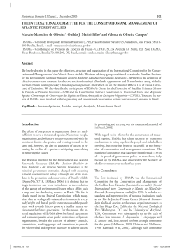
Sleep
Science
ORIGINAL ARTICLE
The presence of neuronal-specific nuclear
protein (NeuN) in the circadian timing system
of the capuchin monkey (Cebus apella)
A presença da proteína nuclear específica neuronal (NeuN) no sistema
de temporização circadiano do macaco capuchinho (Cebus apella)
Rayane Bartira Silva do Nascimento1, Janaína Siqueira Borda1, Rovena Clara Galvão Januário Engelberth1, Raysa Oliveira de
Medeiros1, Renata Frazão2, Luciana Pinato2, André Luiz Bezerra de Pontes1, Expedito Silva do Nascimento Jr2, Maria Inês
Nogueira2, Roelf Justino Cruz-Rizzolo3, Miriam Stela Maris de Oliveira Costa1, Jeferson de Souza Cavalcante1
ABSTRACT
Background and objective: The circadian timing system (CTS) is
composed of a group of specialized neuronal structures that establish a
temporal organization of physiological and behavioral processes within
precise patterns. The central components of this system are the suprachiasmatic nucleus of the hypothalamus (SCN) and the intergeniculate
leaflet of the thalamus (IGL). The objective of this study was to verify the
presence of the neuron-specific nuclear protein (NeuN) in the circadian
timing system of capuchin monkeys (Cebus apella) using immunohistochemical techniques. Methods: Capuchin monkeys (Cebus apella) were
anesthetized and transcardially perfused with 4% paraformaldehyde in
0.1 M phosphate buffer, and then their brains were removed and frozen. A
microtome was used to make 30 μm sections in the coronal plane. One of
the series was used for Nissl staining (thionin) to demarcate the cytoarchitecture, and the remainders of the sections were processed for immunohistochemical detection of NeuN (ABC protocol). Results: NeuN-positive
neurons were observed in the suprachiasmatic nucleus of the capuchin
monkey. The pregeniculate nucleus (PGN), a structure equivalent to the
ventral lateral geniculate nucleus (vLGN) and to the IGL in rodents, did
not have any NeuN-positive neurons. Conclusions: In this primate species, only the suprachiasmatic nucleus neurons of the central structures of
the circadian timing system express the NeuN protein.
Keywords: Circadian rhythm; Suprachiasmatic nucleus; Nuclear
proteins; Immunohistochemistry/methods; Geniculate bodies; Neurons; Animals; Cebus
RESUMO
Introdução e objetivo: O sistema de temporização circadiana (do inglês ������������������������������������������������������������������
circadian timing system, �����������������������������������������
CTS) é composto por um conjunto de estruturas neurais especializadas que estabelecem uma organização temporal
dos processos fisiológicos e comportamentos dentro de padrões precisos.
Seus componentes centrais são o núcleo supraquiasmático (SCN) do hipotálamo e o folheto intergeniculado (IGL) do tálamo. O objetivo deste
estudo foi verificar, através da técnica imunoistoquímica, a presença da
proteína nuclear neuronal específica (NeuN) no sistema de temporização
circadiana do macaco-prego (Cebus apella), um primata do Novo Mundo.
Métodos: Os animais foram previamente anestesiados e submetidos à
perfusão transcardíaca com solução salina heparinizada, seguida de solução
de paraformaldeído a 4% em tampão fosfato 0,1 M. Os encéfalos foram
removidos e submetidos à microtomia por congelação, obtendo-se secções
coronais de 30 μm. Secções de uma série foram submetidas ao método
de Nissl (Thionina) para demarcar a citoarquitetura e as outras secções
foram processadas por imunoistoquímica (protocolo ABC) a fim de revelar a presença de NeuN. Resultados: Neurônios NeuN positivos foram observados no núcleo supraquiasmático do macaco-prego. O núcleo
pré-geniculado (PGN), estrutura equivalente ao núcleo geniculado lateral
ventral (GLV) e ao IGL dos roedores, não apresenta neurônios NeuN positivos. Conclusão: Nas estruturas centrais do sistema de temporização circadiana, somente os neurônios do núcleo supraquiasmático nesta espécie
de primata expressam a proteína NeuN.
Descritores: Ritmo circadiano; Núcleo supraquiasmático; Proteínas nucleares; Imunoistoquímica/métodos; Corpos geniculados;
Neurônios; Animais; Cebus
INTRODUCTION
The generation and regulation of circadian rhythms in
mammals originate from a neural-specific system, the circadian timing system (CTS). This system utilizes a central
pacemaker, which includes inputs such as retinal afferents
that allow for the synchronization of environmental cycles
and outputs that lead to behavioral effectors (1). The supra-
Study carried out at Universidade Federal do Rio Grande do Norte – UFRN, Natal (RN), Brazil; Universidade de São Paulo – USP, São Paulo (SP), Brasil; Universidade
Estadual Paulista “Júlio de Mesquita Filho” – UNESP, Araçatuba (SP) Brazil.
1
Laboratório de Cronobiologia, Centro de Biociências, Universidade Federal do Rio Grande do Norte – UFRN, Natal (RN), Brazil.
2
Departamento de Anatomia da Universidade de São Paulo – USP, São Paulo (SP), Brazil.
3
Departamento de Ciências Básicas da Universidade Estadual Paulista “Júlio de Mesquita Filho” – UNESP, Araçatuba (SP) Brazil.
Corresponding author: Jeferson de Sousa Cavalcante – Centro de Biociências, Universidade Federal do Rio Grande do Norte – Caixa Postal 1511 – CEP 59078-970 –
Natal (RN), Brazil – Phone: +55 (84) 3215-3409 – Fax: +55 (84) 3211-9206 – E-mail: [email protected]
Received: January 8, 2010; Accepted: March 28, 2010
Sleep Sci. 2010;3(1):�����
����
–39
Nascimento RBS, Borda JS, Engelberth RCGJ, Medeiros RO, Frazão R, Pinato L, Pontes ALB, Nascimento Jr ES,
Nogueira MI, Cruz-Rizzolo RJ, Costa MSMO, Cavalcante JS
chiasmatic nucleus (SCN) and the intergeniculate leaflet
(IGL) are considered to be the central components of the
CTS (1). The SCN is a small region localized at the anterior
hypothalamus bilateral to the third ventricle and above the
optic chiasm (2). In the majority of mammalian species studied, the SCN is divided into two principal cell populations:
the ventrolateral SCN, which contains neurons that secrete
vasoactive intestinal polypeptide (VIP), and the dorsomedial SCN, which contains neurons that secrete vasopressin
(VP) (2,3). In addition to VIP and VP, the SCN also contains
a large number of other neuroactive substances that can act
as neurotransmitters or neuromodulators. The SCN contains
neuropeptide Y (NPY), serotonin (5-HT), glutamate (Glu),
bombesin (BBS), gastrin-releasing peptide (GRP), cholecystokinin (CCK), substance P (SP), angiotensin II, enkephalin (ENK), somatostatin (SS), tyrosine hydroxylase (TH),
and calcium binding proteins such as calbindin (CB) and
calretinin (CR) (2). Because the SCN has a crucial role in the
temporal organization of behavior, it is considered the central circadian pacemaker in mammals. SCN lesions result in
a loss of behavioral circadian, endocrine, and physiological
rhythms (4). Arrhythmic animals with SCN lesions recover
their circadian rhythms through fetal SCN transplant; however, these animals acquire the rhythm of the donor (5).
The intergeniculate leaflet (IGL), along with the ventral
lateral geniculate nucleus (vLGN) and the dorsolateral geniculate nucleus (dLGN), form the lateral geniculate complex of the thalamus in non-primate species (6). The IGL is a
thin layer of neurons containing NPY, interposed between
the vLGN and the dLGN, and its function is to modulate
the SCN (7). The organization of the lateral geniculate complex is different in primates in relation to rodents. Although
the primate dLGN corresponds to the rodent dLGN, the
primate pregeniculate nucleus (PGN), which is a wedgedshaped cellular grouping that dorsomedially encircles the
dLGN, is considered equivalent to the rodent vLGN (8,9).
NPY-immunoreactive neurons in the PGN have been found
in Rhesus monkeys (8), common marmosets (Callithrix jacchus)
(10)
, and Cebus monkeys (9). Thus, it can be assumed that the
PGN of primates also contains a rodent-equivalent IGL . A
new neuronal marker that has been utilized to identify nerve
cells is the neuronal-specific nuclear protein (NeuN), which
is expressed in both the nucleus and cytoplasm of the majority of vertebrate nervous system cells (11). This protein has
been utilized in studies on nervous system development (12),
in morphometric studies as a postmortem neuronal marker
of human cerebral tissue (13), in histopathology diagnosis as
a marker of neuronal differentiation in cerebral tumors, and
in studies of neurodegenerative disorders (14). Although this
protein has been identified in the majority of nerve cells,
there are some NeuN-negative neurons. In development,
Cajal-Retzius neurons of the first layer of the cerebral cortex,
the medullar inferior olivary nucleus, cerebellar Purkinje
cells, mitral cells of the olfactory bulb, retinal photoreceptors, and glial cells are all negative for NeuN (11,15).
Considering the importance of the CTS in mammals and
the quantity of neuroactive substances present in the central
components of this system, the objective of this study was
to identify NeuN-protein-immunoreactive neurons in the
central components of the CTS of the capuchin monkey (Cebus apella).
METHODS
For this study, we utilized two young adult capuchin monkeys (Cebus apella) obtained from the Júlio Mesquita Filho
Primate Center of Universidade Estadual Paulista, Araçatuba (SP), Brazil. The experimental procedures were in compliance with the guidelines for the care and use of mammals
in neuroscience and behavioral research. The capuchin monkeys were housed in individual cages under natural humidity, temperature, and lighting conditions and were fed with
a standard fruit and vegetable diet.
The animals were anesthetized with sodium thiopental (30
mg/kg, i.p.) and transcardially perfused with 800 mL of 0.9%
saline containing heparin (Hipolabor Laboratories, 5000 IU/
mL). The saline was followed by 1500 mL of 4% paraformaldehyde in 0.1 M acetate buffer (pH 6.5) and subsequently
with 1500 mL of 4% paraformaldehyde in borate buffer (pH
9.0). The brains were exposed and sliced into blocks utilizing stereotaxic equipment. The blocks were removed from
the cranium and placed in cryoprotectant solution composed
of 10% glycerol and 2% dimethylsulfoxide in 0.1 M borate
buffer (pH 9.0) at 4ºC. After three days, the blocks were transferred to a similar solution containing 20% glycerol for four
more days of cryoprotection. Next, the blocks were sliced into
30-μm coronal sections with a cryomicrotome and collected
in an anti-freezing solution. One of the series was used in
Nissl staining for the cytoarchitecture study, which utilized
thionin as a dye. Another series was pretreated for antigen
recovery utilizing 1% sodium borohydrate. After the pretreatment, the slices where treated immunohistochemically
with an anti-NeuN primary antibody (1:1000, Chemicon) in
0.4-% Triton X-100 and 2-% normal rabbit serum (1:50) at
room temperature for 24 hours. Next, slices were incubated
with secondary biotinylated antibody (1:200, Sigma) for two
hours. Next, the slices were incubated in avidin-biotin immunoperoxidase complex (Elite ABC kit, Vector Laboratories) for two hours and exposed to chromogenic 2.5% 3,3’–diaminobenzidine tetrahydrochloride (DAB) (Sigma, St Louis,
MO, USA) diluted in 0.1 M phosphate buffer (pH 7.4) for
15 minutes. Between incubations, the slices underwent a sequence of eight 5-minute washes each in 0.1 M phosphate
Sleep Sci. 2010;3(1):�����
����
–39
37
38
The presence of neuronal-specific nuclear protein (NeuN) in the circadian timing system
buffer (pH 7.4). Lastly, the slices were mounted on gelatinized
glass slides, intensified in osmium tetroxide, dehydrated in a
series of alcohols, cleared by xylene, and coverslipped for optic
microscopy analysis.
RESULTS
The SCN of Cebus apella is localized in the anterior hypothalamus dorsal to the optic chiasm and bilateral to the
third ventricle. The Nissl staining showed that the SCN
cells neighbored the wall of the third ventricle (Figure 1A).
This cellular group is triangular in the rostral level but becomes more rounded as it reaches the caudal levels. NeuN
immunoreactivity was observed in the three regions (rostral,
intermediate and caudal) of the SCN. The largest number
of NeuN-positive cells was observed in the intermediate region; however, the staining of these cells was pallid when
compared to the lateral hypothalamus, which presented robustly stained cells (Figure 1B).
oc: optic chiasm; 3v: third ventricle; dLGN: dorsal lateral geniculate
nucleus.
The scale bar is equivalent to 70 µm in A, C and D, and to 85 µm in B.
Figure 1: Photomicrographs of brain sections of Cebus apella showing the
components of the CTS in bright field. (A) SCN of Cebus apella stained
by Nissl staining. (B) Panel showing the immunoreactivity for NeuN in
the SCN of the Cebus apella monkey. (C) PGN of the Cebus apella monkey
stained by Nissl staining. (D) Panel showing the negative reaction for
immunoreactivity against NeuN in the PGN of the Cebus apella monkey.
The Nissl-stained sections demonstrated that the PGN
is localized dorsomedially to the dLGN (Figure 1C). It was
observed that the PGN cells in Cebus apella do not present
NeuN immunoreactivity (Figure 1D). However, we did
confirm that the immunohistochemical process was successSleep Sci. 2010;3(1):�����
����
–39
ful in dLGN neurons, thus representing a positive control
(Figure 1D).
DISCUSSION
The SCN is composed of oscillatory neurons that control a
series of effector systems, including those that regulate the
activity and rest cycles, body temperature, neuroendocrine
function, and psychomotor performance. Its role as the central pacemaker of the CTS is modulated by its inputs, which
are topographically organized within itself (16,17). The rodent
IGL or the PGN in primates, along with the SCN, are considered the central components of the CTS because they are
the first participants in the modulation of photic and nonphotic circadian responses (1).
The present study demonstrated that the SCN of Cebus Apella is elongated in the rostro-caudal axis. Neurons
strongly stained with thionin were localized in the anterior
hypothalamus above the optic chiasm and bilateral to the
third ventricle. Furthermore, the PGN had less intense thionin staining and was localized dorsomedially to the dLGN.
This result is in accordance with our previous studies in
Cebus monkeys (9,18). The SCN and PGN cells presented a
high level of homogeneity in the Nissl staining method.
Neuron-specific nuclear protein is considered to be a nuclear
regulatory molecule specific to the nervous system, and it
has been increasingly used in central nervous system studies
(11-13)
. The present study demonstrated that SCN neurons in
Cebus apella were immunoreactive for NeuN in the whole
rostro-caudal extension. The greatest number of NeuN-positive cells was found in the intermediate level of the SCN.
The more robustly stained cells were observed in the lateral
portion, the most pallid were present in the medial portion
of the SCN, but this result could be explained by immunoreactivity variability. A study with adult rats demonstrated
that NeuN expression in the SCN was low, particularly in
the dorsal region when compared to the neighboring hypothalamic neurons. However, detectable levels of NeuN
were observed in the ventral portion of the SCN when it
was co-localized with doublecortin, which is a microtubuleassociated protein that is also considered a marker for neurogenesis (19). The NeuN immunohistochemical labeling in
the PGN of Cebus apella was negative.
In summary, our study demonstrated that the localization
of the SCN and the PGN of Cebus apella is similar to other primate species. In addition, SCN neurons were immunoreactive
for NeuN, whereas the PGN did not exhibit labeled neurons.
Taking into consideration that some neurons of the nervous
system are NeuN negative (18,15), it still remains to be determined whether Cebus apella SCN contains specific subpopulations of neurons that are not immunoreactive for NeuN. The
data in this study are preliminary, but provide a perspective
Nascimento RBS, Borda JS, Engelberth RCGJ, Medeiros RO, Frazão R, Pinato L, Pontes ALB, Nascimento Jr ES,
Nogueira MI, Cruz-Rizzolo RJ, Costa MSMO, Cavalcante JS
for the comparative studies between primate and non-primate
species when it comes to understanding the role of NeuN as a
neuronal marker in this physiological system.
REFERENCES
1. Cavalcante JS, Nascimento Jr ES, Costa MSMO. Componentes
centrais do sistema de temporização circadiana: o núcleo supraquiasmático e o folheto intergeniculado. Neurociências 2006;3:
273-82.
2. Cassone VM, Speh JC, Card JP, Moore RY. Comparative anatomy
of the mammalian hypothalamic suprachiasmatic nucleus. J Biol
Rhythms 1988;3(1):71-91.
3. Smale L, Boverhof J. The suprachiasmatic nucleus and the intergeniculate leaflet of Arvicanthis niloticus, a diurnal murid rodent
from East Africa. J Comp Neurol 1999;403(2):190-208.
4. Van den Pol AN. The suprachiasmatic nucleus: morphology and
cytochemical substrates for cellular interaction. In: Klein DC,
Moore CRY, Reppert SM. Suprachiasmatic nucleus: the mind’s
clock. New York: Oxford University Press; 1991. p. 17-50.
5. Ralph MR, Foster RG, Davis FC, Menaker M. Transplanted
suprachiasmatic nucleus determines circadian period. Science
1990;247(4945):975-8.
6. Jones EG. The thalamus. 2ª ed. New York: Cambridge University
Press; 2007.
7. Hickey TL, Spear PD. Retinogeniculate projections in hooded and albino rats: an autoradiographic study. Exp Brain Res
1976;24(5):523-9.
8. Moore RY. The geniculohypothalamic tract in monkey and man.
Brain Res 1989;486(1):190-4.
9. Pinato L, Frazão R, Cruz-Rizzolo RJ, Cavalcante JS, Nogueira
MI. Immunocytochemical characterization of the pregeniculate
nucleus and distribution of retinal and neuropeptide Y terminals
in the suprachiasmatic nucleus of the Cebus monkey. J Chem
Neuroanat 2009;37(4):207-13.
10.Costa MS, Moreira LF, Alones V, Lu J, Santee UR, Cavalcante JS,
et al. Characterization of the circadian system in a Brazilian species of monkey (Callithrix jacchus): immunohistochemical analysis and retinal projections. Biol Rhythm Res 1998;29(5):510-20.
11.Mullen RJ, Buck CR, Smith AM. NeuN, a neuronal specific protein in vertebrates. Development 1992;116(1):201-11.
12.Preusser M, Laggner U, Haberler C, Heinzl H, Budka H, Hainfellner JA. Comparative analysis of NeuN immunoreactivity in
primary brain tumours: conclusions for rational use in diagnostic
histopathology. Histopathology 2006;48(4):438-44.
13.Gittins R, Harrison PJ. Neuronal density, size and shape in the
human anterior cingulated cortex: a comparison of Nissl and
NeuN staining. Brain Res Bull 2004;63(2):155-60.
14.Falke E, Nissanov J, Mitchell TW, Bennett DA, Trojanowski JQ,
Arnold SE. Subicular dendritic arborisation in Alzheimer’s disease correlates with neurofibrillary tangle density. Am J Pathol
2003;163(4):1615-21.
15.Kumar SS, Buckmaster PS. Neuron-specific nuclear antigen
NeuN is not detectable in gerbil subtantia nigra pars reticulata.
Brain Res 2007;1142:54-60.
16.Moga MM, Moore RY. Organization of neural inputs to the suprachiasmatic nucleus in the rat. J Comp Neurol 1997;389(3):
508-34.
17.Abrahamson EE, Moore RY. Suprachiasmatis nucleus in the
mouse: retinal innervation, intrinsic organization and efferent
projections. Brain Res 2001;916(1-2):172-91.
18.Pinato L, Allemandi W, Abe LK, Frazão R, Cruz-Rizzolo RJ,
Cavalcante JS, et al. A comparative study of cytoarchitecture and
serotonergic afferents in the suprachiasmatic nucleus of primates
(Cebus apella and Callithrix jacchus) and rats (Wistar and Lond
Evans strain). Brain Res 2007;1149:101-10.
19.Geoghegan D, Carter DA. A novel site of adult doublecortin expression: neuropeptide neurons within the suprachiasmatic nucleus circadian clock. BMC Neurosci [Internet]. 2008 [cited 2010
Mar 30];9:2. Available from: http://www.ncbi.nlm.nih.gov/pmc/
articles/PMC2253543/?tool=pubmed
Sleep Sci. 2010;3(1):�����
����
–39
39
Baixar