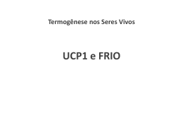
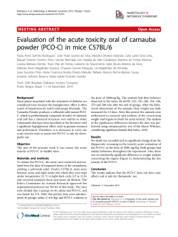
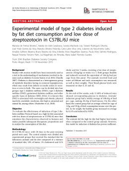
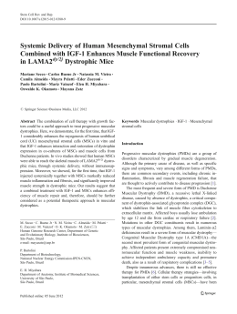
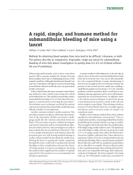
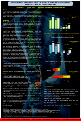
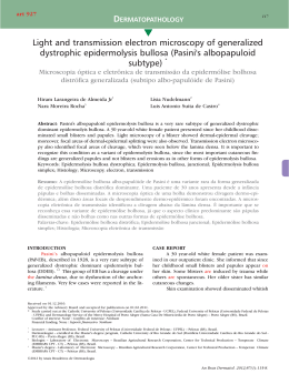
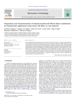
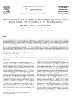
JNI-475261; No of Pages 6
Journal of Neuroimmunology xxx (2010) xxx–xxx
Contents lists available at ScienceDirect
Journal of Neuroimmunology
j o u r n a l h o m e p a g e : w w w. e l s ev i e r. c o m / l o c a t e / j n e u r o i m
Eicosapentaenoic acid decreases TNF-α and protects dystrophic muscles of mdx mice
from degeneration
Rafael Ventura Machado, Adriana Fogagnolo Mauricio, Ana Paula Tiemi Taniguti, Renato Ferretti,
Humberto Santo Neto, Maria Julia Marques ⁎
Departamento de Anatomia, Biologia Celular, Fisiologia e Biofísica, Instituto de Biologia, Universidade Estadual de Campinas, Campinas, São Paulo 13083-970, Brazil
a r t i c l e
i n f o
Article history:
Received 21 July 2010
Received in revised form 9 October 2010
Accepted 25 October 2010
Available online xxxx
Keywords:
EPA
Duchenne dystrophy
mdx mice
Muscle degeneration
Sarcolemma leakiness
TNF-α
a b s t r a c t
In dystrophin-deficient fibers of mdx mice and in Duchenne muscular dystrophy, inflammation and increased
production of tumor necrosis factor alpha (TNF-α) contribute to myonecrosis. We examined the effects of
eicosapentaenoic acid (EPA) on dystrophic muscle degeneration. Mdx mice (14 days old) received EPA for
16 days. The sternomastoid, diaphragm and biceps brachii muscles were removed. Control mdx mice received
vehicle. EPA decreased creatine kinase and myonecrosis and reduced the levels of TNF-α. These results
suggest that EPA plays a protective role in dystrophic muscle degeneration, possibly by reducing TNF-α, and
support further investigations of EPA as a potential therapy for dystrophinopathies.
© 2010 Elsevier B.V. All rights reserved.
1. Introduction
The absence of dystrophin in Duchenne muscular dystrophy
(DMD) and in the mdx mouse model of DMD is associated with
sarcolemma instability and elevated levels of calcium in muscle fibers,
factors that lead to myonecrosis. Muscle fiber damage activates an
inflammatory response that seems to play a central role in the
progression of myonecrosis (Tidball, 2005). Mast cells accumulate
rapidly in response to muscle injury, followed by neutrophils and
macrophages (Radley and Grounds, 2006). Progressive myonecrosis
ultimately results in the replacement of muscle fibers with fat and
fibrosis which, in turn, cause cardiorespiratory failure in dystrophic
patients (Engel et al., 1994).
The proinflammatory cytokine tumor necrosis factor alpha (TNF-α),
which is produced by inflammatory and muscle cells (Vassalli, 1992), is
of particular importance for dystrophic fiber necrosis. TNF-α is found to
be elevated in DMD and in mdx muscles (Kuru et al., 2003; Grounds
et al., 2008) and higher serum levels are observed in DMD patients
compared to healthy subjects (Saito et al., 2000). In addition, drug
therapy designed to reduce inflammatory cells (Hodgetts et al., 2006), to
block mast cell degranulation (Radley and Grounds, 2006) and to inhibit
TNF-α and TNF-signaling mechanisms (Waters et al., 2010) has been
shown to ameliorate dystrophy and demonstrates the role of inflammation and TNF-α in the progression of dystrophy.
The omega-3 fatty acid, eicosapentaenoic acid (EPA), has antiinflammatory properties and several clinical trials have reported
potential health benefits of omega-3 polyunsaturated fatty acids in
many diseases, including cardiovascular diseases (Harper et al., 2006),
epilepsy (Schlanger et al., 2002), inflammatory bowel disease (Calder,
2008), post-operative trauma (Roulet et al., 1997), exercise-trained
subjects (Bloomer et al., 2009), and cancer-associated cachexia (Babcock
et al., 2000). EPA has also been shown to inhibit the proinflammatory
transcription factor nuclear factor kappa B (NF-kB) (Babcock et al., 2000;
Singer et al., 2008), to reduce TNF-α production by macrophages
(Babcock et al., 2002) and to prevent the damaging effects of TNF-α
during skeletal muscle differentiation in vitro (Magee et al., 2008).
There is currently no effective therapy for DMD. Corticosteroids are
the main drugs of choice despite their side effects (Bonifati et al., 2000;
Moxley et al., 2005) and long-term treatment is necessary. Despite
advances in genetic and cell-based therapies (Tremblay et al., 2009;
Heemskerk et al., 2010), the search for new drugs or nutritional
interventions for DMD is relevant (Payne et al., 2006; Radley et al.,
2007). In view of the anti-inflammatory and anti-TNF-α properties of
EPA, we hypothesized that this agent would protect dystrophin-deficient
mdx muscle fibers against degeneration.
2. Materials and methods
2.1. Animals
⁎ Corresponding author. Tel.: + 55 19 3521 6395.
E-mail address: [email protected] (M.J. Marques).
Male and female mdx mice (C57BL/10-Dmdmdx/PasUnib) obtained
from a breeding colony maintained by our institutional animal care
0165-5728/$ – see front matter © 2010 Elsevier B.V. All rights reserved.
doi:10.1016/j.jneuroim.2010.10.032
Please cite this article as: Machado, R.V., et al., Eicosapentaenoic acid decreases TNF-α and protects dystrophic muscles of mdx mice from
degeneration, J. Neuroimmunol. (2010), doi:10.1016/j.jneuroim.2010.10.032
2
R.V. Machado et al. / Journal of Neuroimmunology xxx (2010) xxx–xxx
EPA-treated mdx mice (n = 15) received EPA (cis-5,8,11,14,17Eicosapentaenoic acid, 98.5% of EPA in composition, oil density
0.943 g/ml, Fluka/Sigma-Aldrich®, St. Louis, MO, USA) daily, by
gavage, at a dose of 300 mg/kg body weight (Matsumoto et al.,
2009) in 100% mineral oil (Nujol, liquid petrolatum for oral human
use, Mantecorp, SP, Brazil) for 16 days. Each mouse was weighed daily
so that the drug dosage could be adjusted accurately. The amount of
oil received was 0.01 ml/gavage. Control litter mdx mice (n = 15;
untreated) received an equivalent amount of mineral oil.
For each cross-section (4–5 sections for each muscle), the numbers of
central nucleated fibers and fibers with peripheral nuclei were
counted using a hand counter and expressed as the percentage of
the total number of fibers.
Areas with inflammatory cell infiltrate were characterized and
quantified as previously described (Marques et al., 2008). In short,
inflammatory cells were identified in hematoxilin–eosin sections
based on nucleus morphology and cell size, showing basophilic
nuclear staining and little cytoplasm. Areas containing inflammatory
cells densely packed were measured with the ImagePro-Express
software and were calculated as a percentage of the total muscle area
in each section studied (4–5 sections from each muscle) using the
microscope (Nikon Eclipse E400) fitted with a graduated eyepiece
micrometer at 200× magnification. Some sections were labeled with
antibody to F4/80 (Serotec; 1:250 dilution in 0.1 M PBS, pH 7.8, and
BSA 1%), a pan macrophage marker (Villalta et al., 2009), followed by
secondary antibody (CY-3; Jackson ImmunoResearch; 1:250 dilution
in 0.1 M PBS, pH 7.8, and BSA 1%). Control mounts for the primary
antibody were incubated with CY-3 anti-rat IgG in blocking solution
instead of the primary antibody. No stained structures were seen in
these controls. All the counting and measurements (EBD, central and
peripheral nucleated cells and inflammation area) were done by a
blinded observer.
2.3. Evans blue dye analysis
2.5. Analysis of creatine kinase
For visualization of muscle fiber leakiness/necrosis, treated and
untreated-mdx mice were injected with Evans blue dye (EBD;
Sigma®, St. Louis, MO, USA); (Matsuda et al., 1995). EBD was
dissolved in phosphate-buffered saline (PBS; 0.15 M NaCl, 10 mM
phosphate buffer, pH 7.4) and injected into the peritoneal cavity. The
animals (5 EPA-treated and 5 untreated) received an intraperitoneal
injection of 1% EBD in PBS at a dose of 100 μl per 10 g of body weight.
The mice were visually inspected for dye uptake. Discoloration of all
mice was observed within 50–60 min after intraperitoneal injection of
EBD, and successful injection of the dye was indicated by the blue
color of the ears and paws.
Twenty-four hours later, the mice were anesthetized with a
mixture of ketamine hydrochloride (130 mg/kg, Francotar, Virbac, São
Paulo, Brazil) and xylazine hydrochloride (6.8 mg/kg, 2% Virbaxyl,
Virbac, São Paulo, Brazil). The sternomastoid (STN), diaphragm (DIA),
and biceps braquii (BB) muscles were dissected out and snap frozen in
isopentane cooled in liquid nitrogen and stored at −80 °C. These
muscles were chosen because they are differently affected, with the
diaphragm being more severely impaired than other muscles in the
later stages of the disease (Stedman et al., 1991).
Cryostat cross-sections (7 μm thick) were incubated in ice-cold
acetone at −20 °C for 10 min, washed three times for 10 min with
PBS, and mounted in DABCO (mounting medium for fluorescence
microscopy; Sigma). EBD staining shows a bright red emission upon
fluorescence microscopy. Fiber counts of EBD-positive muscle fibers
were performed with a hand counter in all sections and photographed
under a Nikon fluorescence microscope connected to a Hamamatsu
video camera. The number of EBD-positive muscle fibers is expressed
as the percentage of the total number of muscle fibers counted in each
section (4–5 sections for each muscle).
For biochemical evaluation of muscle fiber damage, EPA-treated
(n= 8) and untreated (n= 8) mdx mice were anesthetized with a
mixture of ketamine hydrochloride (130 mg/kg, Francotar, Virbac, São
Paulo, Brazil) and xylazine hydrochloride (6.8 mg/kg, 2% Virbaxyl,
Virbac, São Paulo, Brazil). Blood samples (0.8 ml) were collected by
cardiac puncture. After incubation at room temperature for 1–2 h to
allow clotting, the samples were microcentrifuged at 936 g for 10 min,
and the supernatant (serum) was removed and used for analysis. The
creatine kinase (CK) assay was performed using a commercially
available kit (CK Cinético Crystal, Bioclin, Quibasa, Minas Gerais, Brazil)
and a Thermo Electron Corporation Genesys 20 spectrophotometer
(Krackeler Scientific, Albany, New York, USA). Values are reported as
international units (U/L).
facility were used in all experiments. Some C57BL/10 mice (C57BL/
10ScCr/PasUnib) were used for focused experiments. The mice were
housed according to institutional guidelines, with free access to food
and water. Pregnant females were isolated and monitored daily. The
date of birth was designated postnatal day 0. EPA treatment was
initiated on postnatal day 14 before the cycles of muscle degeneration–regeneration had started (Cullen and Jaros, 1988). Mice were
weaned at 4 weeks of age. The animal experiments described here
were done in accordance with the guidelines of the Brazilian College
for Animal Experimentation (COBEA; protocol # 2165-1) and the
guidelines set forth by our Institution.
2.2. Drug administration
2.4. Quantitative and morphometric analysis
Cryostat cross-sections of STN, DIA and BB from EPA-treated
(n = 7) and untreated (n = 7) were stained with hematoxilin–eosin
(HE). Slides were placed in a Nikon Eclipse E 400 microscope
connected to a personal computer and attached to a video camera
(Nikon Express Series; Tokyo, Japan). Non-overlapping images of the
entire cross-section were taken and tiled together using the
ImagePro-Express software (Media Cybernetic; Silver Spring, MD).
2.6. Western blot analysis
TNF-α was quantified by western blotting in control C57BL/10
mice (n = 8), and in EPA-treated (n = 8) and untreated (n = 8) mdx
mice. The method employed was described in Ferretti et al. (2009).
Briefly, muscles were lysed in assay lysis buffer (1% Triton, 10 mM
sodium pyrophosphate, 100 mM NaF, 10 μg/ml aprotinin, 1 mM PMSF,
and 0.25 mM Na3VO4), centrifuged and the soluble fraction was
resuspended in 50 μl Laemmli loading buffer (2% SDS, 20% glycerol,
0.04 mg/ml bromophenol blue, 0.12 M Tris–HCl, pH 6.8, and 0.28 M
ß-mercaptoethanol). An amount of 30 μg of aliquots from C57BL/10
and treated and untreated-mdx STN, DIA, and BB muscles was loaded
onto 8%–15% SDS-polyacrylamide gels. Proteins were transferred
from the gels to a nitrocellulose membrane using a submersion
electrotransfer apparatus (Bio-Rad Laboratories, Hercules, California,
USA). Membranes were incubated with the primary antibodies,
followed by peroxidase-conjugated secondary antibodies and by
development with SuperSignal West Pico Chemiluminescent Substrate
kit (Pierce Biotechnology, Rockford, Illinois, USA). To control for protein
loading, western blot transfer and nonspecific changes in protein levels,
the blots were stripped and re-probed for glyceraldehyde-3-phosphate
dehydrogenase (GAPDH). The luminescent signal from Western blot
bands was captured by a G:Box iChemi camera (Syngene, Cambridge,
UK) and band intensities were quantified using the analysis software
provided by the manufacturer (Gene Tools Version 4.01, Syngene,
Cambridge, UK).
Please cite this article as: Machado, R.V., et al., Eicosapentaenoic acid decreases TNF-α and protects dystrophic muscles of mdx mice from
degeneration, J. Neuroimmunol. (2010), doi:10.1016/j.jneuroim.2010.10.032
R.V. Machado et al. / Journal of Neuroimmunology xxx (2010) xxx–xxx
The following primary antibodies were used for Western blotting: 1)
TNF-α (rabbit anti-mouse polyclonal; Millipore, CA, USA), and 2)
GAPDH (rabbit polyclonal; Santa Cruz Biotechnology, Santa Cruz,
California, USA). The corresponding secondary antibody used for
Western blotting was peroxidase-labeled affinity-purified rabbit IgG
antibody (H+ L) (KPL, Gaithersburg, Maryland, USA).
2.7. Statistical analysis
All data are expressed as means ± standard deviation (SD).
Statistical analysis for direct comparison between means of two groups
was performed by the Student t-test and ANOVA was used for multiple
statistical comparisons between groups. P b 0.05 was considered
statistically significant.
3. Results
At the beginning of treatment (14 days of age), body mass was
comparable among the groups studied (6.8 ± 0.5 g in untreated-mdx
and 7.2 ± 0.7 g in EPA-mdx). There was a significant increase (about
45–50% compared to initial body mass) in body mass over time in both
untreated- (10.0± 1.2 g; p = 0.004 compared to initial body mass,
Student t-test) and EPA-treated (11.3± 2.0 g; p = 0.02 compared to
initial body mass, Student t-test) mdx. Thus, EPA treatment did not
interfere with the growth rate of young mdx mice.
3.1. Histopathology
The histological appearance of a muscle from normal C57BL/10 mice
and from EPA-treated and untreated-mdx mice is shown in Fig. 1. In
normal mice, muscle fibers with homogeneous diameter and peripheral
nuclei located directly below the sarcolemma were observed. In the mdx
mice, the inflammatory area was defined by areas containing
conspicuous inflammatory cells densely packed among regenerated
muscle fibers and muscle fibers under degeneration. Fully regenerated
muscle fibers, characterized by central nucleation and an apparent
normal diameter, predominated outside the inflammatory area. Groups
of F4/80-expressing cells were present in all untreated-mdx muscles
3
(shown only in BB) and were decreased in the EPA-treated mdx mice
(Fig. 1).
3.2. Analysis of myonecrosis: Evans blue dye, central nucleated fibers and
CK
EPA caused a significant decrease in Evans blue dye staining,
indicative of sarcolemma leakiness and myonecrosis, with a concomitant increase in fibers with peripheral nuclei, in all muscles studied
(Table 1). There was a reduction of central nucleated fibers in the
EPA-treated muscles, with a significant decreased seen only in STN
muscle (Table 1), possibly due to the higher levels of these fibers in the
untreated-STN in comparison to BB and DIA. CK levels were significantly
decreased by EPA compared to untreated-mdx mice (838 ± 347 U/L
in EPA-treated vs 1208 ± 376 U/L in untreated-mdx; p = 0.04, Student's
t-test).
3.3. Analysis of inflammation: inflammatory area and TNF-α levels
Quantitative data about the inflammatory area is shown in Fig. 2.
While untreated DIA presented the lowest inflammatory area (13%),
limb and STN muscles showed the highest values, with about 65% (BB)
to 100% (STN) increase in comparison to DIA. EPA decreased the
inflammatory area in all muscles studied (Fig. 2).
The levels of TNF-α in dystrophic STN, DIA and BB were
significantly higher in untreated-mdx muscles in comparison to
normal C57BL/10 mice (Fig. 3). The highest level of TNF-α was
observed in STN, followed by BB and DIA (Fig. 3). EPA led to a
significant reduction in the levels of TNF-α in all muscles (about 50%
reduction in STN and BB and 25% in DIA; Fig. 3).
4. Discussion
EPA is a 20-carbon omega (n)-3 polyunsaturated fatty acid with
anti-inflammatory properties, which is synthesized from ingested
alpha-linolenic acid or is consumed in fish and fish oil (Arterburn
et al., 2006). The present study demonstrated that treatment of mdx
mice with EPA resulted in a decrease of serum CK levels and Evans
blue dye-positive fibers and a concomitant increase in peripheral
Fig. 1. Histological appearance of control C57BL/10, untreated and EPA-treated mdx mice. Upper row: sternomastoid muscle stained with hematoxilin–eosin. Fiber with peripheral cell
nuclei (asterisk) in control and in EPA-treated muscles. In untreated-mdx, a representative inflammatory area is indicated by the outline, with necrotic fibers (arrow). In EPA-mdx, central
nucleated fiber (arrow). Bottom row: representative areas of biceps braquii muscle labeled with F4/80 antibody. Groups of F4/80-expressing cells are seen in untreated-mdx mice. Scale bar:
60 μm.
Please cite this article as: Machado, R.V., et al., Eicosapentaenoic acid decreases TNF-α and protects dystrophic muscles of mdx mice from
degeneration, J. Neuroimmunol. (2010), doi:10.1016/j.jneuroim.2010.10.032
4
R.V. Machado et al. / Journal of Neuroimmunology xxx (2010) xxx–xxx
Table 1
Effects of EPA treatment on the percentage of muscle fibers with peripheral cell nuclei (peripheral nuclei), centrally located nuclei (central nuclei), and fibers stained with Evans blue
dye (EBD).
STN
DIA
BB
Untreated
Treated
Untreated
Treated
Untreated
Treated
Total number of fibers
Peripheral nuclei (%)
Central nuclei (%)
EBD (%)
966 ± 52.27
882 ± 84.22
1250 ± 90.5
1320 ± 94.2
961 ± 88.6
1183 ± 109.35
50.0 ± 4.5
70.5 ± 5.5⁎p b 0.001
73.4 ± 6.6
86.7 ± 4.3⁎ p b 0.001
71.7 ± 8.3
83.3 ± 3.0⁎ p = 0.03
34.3 ± 2.3
25.2 ± 5.3⁎ p b 0.001
10.2 ± 3.5
8.0 ± 2.7
18.6 ± 7.3
13.8 ± 3.0
15.6 ± 2.6
4.2 ± 2.2⁎ p b 0.001
16.3 ± 7.0
5.2 ± 2.7⁎ p = 0.004
9.3 ± 3.6
4.2 ± 1.8⁎ p = 0.01
Values are expressed as the percentage (mean ± SD; n = 5 mice) of the total number of fibers of the sternomastoid (STN), diaphragm (DIA) and biceps brachii (BB) muscles.
⁎ Significantly different from untreated-mdx mice within the same muscle group (Student t-test).
nucleated fibers, evidence indicating a decline in myonecrosis. Taken
together, these results show that EPA treatment exerts a protective
effect on myofiber breakdown in limb (BB) and axial (STN and DIA)
muscles of young mdx mice.
In the present study, the dose of EPA used (300 mg/kg) seems to be
higher than might be expected to human patients, where a 1000 mg/day
of EPA is usually recommended. However, care must be taken to
extrapolate dose from mice to humans, especially if we consider
differences in metabolism, in the duration of treatment and in the
progression of dystrophy. In an experimental cachexia model in rats, it
was observed that EPA inhibited tumor growth and body weight loss in
a dose-dependent manner, with optimal effects seen at higher doses
(1.25–2.5 g/kg body weight) than that used here. Toxicity was observed
at a dose of 5 g/kg (Tisdale and Dhesi, 1990). In patients with advanced
pancreatic cancer, the oral intake of a nutritional supplement enriched
with 1.09 g of EPA, twice a day, attenuated the progression of cachexia
(Barber et al., 1999). Thus, considering that DMD patients may need a
long therapy, the lower recommended dose should be more appropriate, but clinical experiments are necessary to properly address this
question.
Inflammation is a hallmark of dystrophic muscles and strongly
contributes to the progression of the disease (Chen et al., 2000; Porter
Fig. 2. Quantitation of inflammatory area in sternomastoid (STN), diaphragm (DIA) and
biceps brachii (BB) muscles from untreated (n = 7) and EPA-treated (n = 7) mdx mice.
The inflammatory area is represented as a percentage of the total muscle area. *Means
significantly different from untreated-mdx. Student t-test. Error bars, SD.
et al., 2002). Although COX-2-derived eicosanoids do not seem to
contribute to the progression of dystrophic disease (Pierno et al.,
2007), the inhibitory action of EPA on arachidonic acid metabolism
through the 5-lipoxygenase and cyclooxygenase enzymatic pathways,
suppressing the production of n-6 eicosanoid inflammatory mediators
(James et al., 2000; Calder, 2008), may explain at least in part the
reduced muscle damage seen in dystrophic mdx muscle fibers.
In dystrophic muscles, mast cells rapidly accumulate once muscle
damage begins and macrophages are predominant between 12 h to 24 h
after the injury (Radley and Grounds, 2006). Distinct macrophages
populations may influence the course of mdx dystrophy (Villalta et al.,
2009) and both, mast cells and macrophages, produce proinflammatory
cytokines such as TNF-α. We observed F4/80-expressing cells in all
dystrophic muscles studied, at this early stage of dystrophy, further
supporting the primary roles of macrophages in mdx dystrophy (Villalta
et al., 2009; Wehling et al., 2001). It is well known that inhibition of
TNF-α production protects dystrophin-deficient mdx fibers against
muscle degeneration (Radley and Grounds, 2006; Waters et al., 2010). In
the present study, EPA reduced immunofluorescence labeling of
macrophages, reduced significantly TNF-α levels in all muscles studied
and ameliorated myonecrosis, findings indicating a role of macrophages
and TNF-α in the early stages of disease in muscles that are affected
differently by the lack of dystrophin. The present findings are in
agreement with previous studies showing that EPA is able to decrease
TNF-α levels by inhibiting its production by macrophages (Babcock
et al., 2002), and also to reduce serum TNF-α levels in exercised humans
(Bloomer et al., 2009).
The mechanisms whereby EPA exerts its anti-TNF effects are
poorly understood. One possibility is that EPA decreases transcription
of the TNF-α gene by reducing the translocation of NF-kB, a
proinflammatory transcription factor (Joussen et al., 2002), from the
cytoplasm to the nucleus (Lo et al., 1999; Singer et al., 2008).
Disturbances in NF-kB signaling have been described in mdx mice
(Acharyya et al., 2007) and in DMD (Monici et al., 2003). EPA may
prevent the damaging effects of TNF-α on muscle fibers as
demonstrated in in vitro studies on skeletal muscle differentiation
(Magee et al., 2008). Additionally, EPA may improve dystrophic
muscle regeneration, since it was demonstrated that this omega-3
fatty acid regulates myogenic regulatory factors and attenuates
muscle wasting by preventing muscle protein degradation (Castillero
et al., 2009).
In conclusion, the present study demonstrated that the administration of highly purified EPA to dystrophic mdx mice decreases
inflammation, myonecrosis and TNF-α levels in different dystrophindeficient skeletal muscles. EPA has been tested in several clinical trials
for different diseases and its effects in reducing the risk of cardiac death
are well documented (Nestel, 2000; Harper et al., 2006). Although
further studies are necessary to better understand the mechanisms
whereby EPA modulates immunoinflammatory pathways in dystrophic
muscle, the present results suggest that therapeutic strategies using EPA
as a supplemental or primary therapy may ameliorate dystrophic
muscle pathology.
Please cite this article as: Machado, R.V., et al., Eicosapentaenoic acid decreases TNF-α and protects dystrophic muscles of mdx mice from
degeneration, J. Neuroimmunol. (2010), doi:10.1016/j.jneuroim.2010.10.032
R.V. Machado et al. / Journal of Neuroimmunology xxx (2010) xxx–xxx
5
Fig. 3. Immunoblot analysis of tumor necrosis factor (TNF)-alpha levels in crude extracts of sternomastoid (STN), diaphragm (DIA), and biceps brachii (BB) muscles from control
C57BL/10 (n = 8), and from untreated (n = 8) and EPA-treated (n = 8) mdx mice. A. Western blot. B. Quantitation shows higher levels of TNF-α in dystrophic muscles compared to
normal mice (untreated × C57BL/10). EPA led to a significant reduction in TNF-α levels in all muscles. Levels of TNF-α expressed in arbitrary units normalized to the levels of GAPDH,
which was used as a loading control. Significantly different from EPA-mdx (a), from C57BL/10 (b) and from untreated-mdx (c), ANOVA. Error bars, SD.
Acknowledgments
This work was supported by Fundação de Amparo à Pesquisa do
Estado de São Paulo (FAPESP, grant 08/58491-1) and by Coordenação
de Aperfeiçoamento de Pessoal de Nível Superior (CAPES-PROEX). H.
S.N. and M.J.M. are recipients of fellowships from Conselho Nacional
de Pesquisas (CNPq; 301386/2007-2; 302006/2009-5). R. F. and A.P.T.
T were recipients of a CNPq fellowship and A. M. was the recipient of a
CAPES fellowship. We thank Mrs. Kerstin Markendof for English
revision of the manuscript.
References
Acharyya, S., Villalta, S.A., Bakkar, N., Bupha-Intr, T., Janssen, P.M., Carathers, M., Li, Z.W.,
Beg, A.A., Ghosh, S., Sahenk, Z., Weinstein, M., Gardner, K.L., Rafael-Fortney, J.A.,
Karin, M., Tidball, J.G., Baldwin, A.S., Guttridge, D.C., 2007. Interplay of IKK/NFkappa β signaling in macrophages and myofibers promotes muscle degeneration in
Duchenne muscular dystrophy. J. Clin. Investig. 117, 889–901.
Arterburn, L.M., Hall, E.B., Oken, H., 2006. Distribution, interconversion, and dose
response of n_3 fatty acids in humans. Am. J. Clin. Nutr. 83, 1467S–1476S.
Babcock, T.A., Helton, W.S., Espat, N.J., 2000. Eicosapentaenoic acid (EPA): an
antiinflammatory ω-3 fat with potential clinical applications. Nutrition 16,
1116–1118.
Babcock, T.A., Helton, W.S., Hong, D., Espat, N.J., 2002. Omega-3 fatty acid lipid emulsion
reduces LPS-stimulated macrophage TNF-α production. Surg. Inf. 3, 145–149.
Barber, M.D., Ross, J.A., Voss, A.C., Tisdale, M.J., Fearon, K.C.H., 1999. The effect of an oral
nutritional supplement enriched with fish oil on weight loss in patients with
pancreatic cancer. Br. J. Cancer 81, 80–86.
Bloomer, R.J., Larson, D.E., Fisher-Wellman, K.H., Galpin, A.J., Schilling, B.K., 2009. Effect
of eicosapentaenoic and docosahexaenoic acid on resting and exercise-induced
inflammatory and oxidative stress biomarkers: a randomized, placebo controlled,
cross-over study. Lipids Health Dis. 8, 36.
Bonifati, M.D., Ruzza, G., Bonometto, P., Berardinelli, A., Gorni, K., Orcesi, S., Lanzi, G.,
Angelini, C., 2000. A multicenter, double-blind randomized trial of deflazacort
versus prednisone in Duchenne muscular dystrophy. Muscle Nerve 23, 1344–1347.
Calder, P.C., 2008. Polyunsaturated fatty acids, inflammatory processes and inflammatory bowel diseases. Mol. Nutr. Food Res. 52, 885–897.
Castillero, E., Martín, A.L., López-Menduiña, M., Villanúa, M., López-Calderón, A., 2009.
Eicosapentaenoic acid attenuates arthritis-induced muscle wasting acting on
atrogin-1 and on myogenic regulatory factors. Am. J. Physiol. Regul. Integr. Comp.
Physiol. 297, R1322–R1331.
Chen, Y.W., Zhao, P., Borup, R., Hoffman, E.P., 2000. Expression profiling in the muscular
dystrophies: identification of novel aspects of molecular pathophysiology. J. Cell
Biol. 151, 1321–1336.
Cullen, M.J., Jaros, E., 1988. Ultrastructure of the skeletal muscle in the X chromosome-linked
dystrophic (mdx) mouse. Comparison with Duchenne muscular dystrophy. Acta
Neuropathol. 77, 69–81.
Engel, A.G., Yamamoto, M., Fischbeck, K.H., 1994. Dystrophinopathies. In: Engel, A.G.,
Franzini-Armstrong, C. (Eds.), Myology. McGraw-Hill Inc, New York, NY, pp. p.
1133–p. 1187.
Ferreti, R., Marques, M.J., Pertille, A., Santo Neto, H., 2009. Sarcoplasmic–endoplasmic–
reticulum calcium-ATPase and calsequestrin are overexpressed in spared intrinsic
laryngeal muscles of dystrophin-deficient mdx mice. Muscle Nerve 39, 609–615.
Grounds, M.D., Radley, H.G., Gebski, B.G., Bogoyevitch, M.A., Shavlakadze, T., 2008.
Implications of cross-talk between tumor necrosis factor and insulin-like growth
factor-1 signaling in skeletal muscle. Clin. Exp. Pharmacol. Physiol. 35, 846–851.
Harper, C.R., Edwards, M.J., DeFilipis, A.P., Jacobson, T.A., 2006. Flaxseed oil increases the
plasma concentrations of cardioprotective (n-3) fatty acids in humans. J. Nutr. 136,
83–87.
Heemskerk, H., Winter, C., van Kuik, P., Heuvelmans, N., Sabatelli, P., Rimessi, P.,
Braghetta, P., van Ommen, G.-J., de Kimpe, S., Ferlini, A., Aartsma-Rus, A., van
Deutekom, J.C.T., 2010. Preclinical PK and PD studies on 2′-O-methyl-phosphorothioate RNA antisense oligonucleotides in the mdx mouse model. Mol. Ther. 18,
1210–1217.
Hodgetts, S., Radley, H.G., Davies, M., Grounds, M.D., 2006. Reduced necrosis of
dystrophic muscle by depletion of host neutrophils, or blocking TNF alpha function
with Etanercept in mdx mice. Neuromuscul. Disor. 16, 591–602.
James, M.J., Gibson, R.A., Cleland, L.G., 2000. Dietary polyunsaturated fatty acids and
inflammatory mediator production. Am. J. Clin. Nutr. 71, 343S–348S.
Joussen, A.M., Poulaki, V., Mitsiades, N., Kirchhof, B., Koizumi, K., Dohmen, S., Adamis, A.
P., 2002. Nonsteroidal anti-inflammatory drugs prevent early diabetic retinopathy
via TNFalpha suppression. FASEB J. 16, 438–440.
Kuru, S., Inukai, A., Kato, T., Liang, Y., Kimura, S., Sobue, G., 2003. Expression of tumor
necrosis factor-α in regenerating muscle fibers in inflammatory and noninflammatory myopathies. Acta Neuropathol. 105, 217–224.
Lo, C.J., Chiu, K.C., Fu, M., Lo, R., Helton, S., 1999. Fish oil augments macrophage
cyclooxygenase II (COX-2) gene expression induced by endotoxin. J. Surg. Res. 86,
103–107.
Magee, P., Pearson, S., Allen, J., 2008. The omega-3 fatty acid, eicosapentaenoic acid
(EPA), prevents the damaging effects of tumor necrosis factor (TNF)-alpha during
murine skeletal muscle differentiation. Lipids Health Dis. 7, 24.
Marques, M.J., Machado, R., Minatel, E., Santo Neto, H., 2008. Disodium cromoglycate
protects dystrophin-deficient muscle fibers from leakiness. Muscle Nerve 37, 61–67.
Matsuda, R.A., Nishikawa, A., Tanaka, H., 1995. Vizualization of dystrophic muscle fibers
in mdx mouse by vital staining with Evans blue: evidence of apoptosis in
dystrophin-deficient muscle. J. Biochem. 118, 959–964.
Matsumoto, T., Nakayama, N., Ishida, K., Kobayashi, T., Kamata, K., 2009. Eicosapentaenoic acid improves imbalance between vasodilator and vasoconstrictor actions of
endothelium-derived factors in mesenteric arteries from rats at chronic stage of
Type 2 diabetes. J. Pharmacol. Exp. Ther. 329, 324–334.
Monici, M.C., Aguennouz, M., Mazzeo, A., Messina, C., Vita, G., 2003. Activation of
nuclear factor-kappaB in inflammatory myopathies and Duchenne muscular
dystrophy. Neurology 60, 993–997.
Please cite this article as: Machado, R.V., et al., Eicosapentaenoic acid decreases TNF-α and protects dystrophic muscles of mdx mice from
degeneration, J. Neuroimmunol. (2010), doi:10.1016/j.jneuroim.2010.10.032
6
R.V. Machado et al. / Journal of Neuroimmunology xxx (2010) xxx–xxx
Moxley, R.T., Ashwal, S., Pandya, S., Connolly, A., Florence, J., Mathews, K., Baumbach, L.,
McDonald, C., Sussman, M., Wade, C., 2005. Practice parameter: corticosteroid
treatment of Duchenne dystrophy: report of the Quality Standards Subcommittee
of the American Academy of Neurology and the Practice Committee of the Child
Neurology Society. Neurology 64, 13–20.
Nestel, P.J., 2000. Fish oil and cardiovascular disease: lipids and arterial function. Am. J.
Clin. Nutr. 71, 228S–231S.
Payne, E.T., Yasuda, N., Bourgeois, J.M., Devries, M.C., Rodriguez, M.C., Yousuf, J.,
Tarnopolsky, M.A., 2006. Nutritional therapy improves function and complements
corticosteroid intervention in mdx mice. Muscle Nerve 33, 66–77.
Pierno, S., Nico, B., Burdi, R., Liantonio, A., Didonna, M.P., Cippone, V., Fraysse, B.,
Rolland, J.F., Mangieri, D., Andreetta, F., Ferro, P., Camerino, C., Zallone, A.,
Confalonieri, P., De Luca, A., 2007. Role of tumor necrosis factor α, but not of
cyclo-oxygenase-2-derived eicosanoids, on functional and morphological indices
of dystrophic progression in mdx mice: a pharmacological approach. Neuropathol.
Appl. Neurobiol. 33, 344–359.
Porter, J.D., Khanna, S., Kaminski, H.J., Rao, J.S., Merrian, A.P., Richmonds, C.R., Leahy, P.,
Li, J., Guo, W., Andrade, F.H., 2002. A chronic inflammatory response dominates the
skeletal muscle molecular signature in dystrophin-deficient mdx mice. Hum. Mol.
Genet. 11, 263–272.
Radley, H.G., De Luca, A., Lynch, G.S., Grounds, M.D., 2007. Medicine in focus: Duchenne
muscular dystrophy: focus on pharmaceutical and nutritional interventions. Int. J.
Biochem. Cell Bio. 39, 469–477.
Radley, H.G., Grounds, M.D., 2006. Cromolyn administration (to block mast cell
degranulation) reduces necrosis of dystrophic muscle in mdx mice. Neurobiol. Dis.
23, 387–397.
Roulet, M., Frascarolo, P., Pilet, M., Chapuis, G., 1997. Effects of intravenously infused
fish oil on platelet fatty acid phospholipid composition and on platelet function in
postoperative trauma. J. Parenter. Enteral. Nutr. 21, 296–301.
Saito, K., Kobayashi, D., Komatsu, M., Yajima, T., Yagihashi, A., Ishikawa, Y., Minami, R.,
Watanabe, N., 2000. A sensitive assay of tumor necrosis factor α in sera from
Duchenne muscular dystrophy patients. Clin. Chem. 46, 1703–1704.
Schlanger, S., Shinitzky, M., Yam, D., 2002. Diet enriched with omega-3 fatty acids
alleviates convulsion symptoms in epilepsy patients. Epilepsia 43, 103–104.
Singer, P., Shapiro, H., Theilla, M., Anbar, R., Singer, J., Cohen, J., 2008. Anti-inflammatory
properties of omega-3 fatty acids in critical illness: novel mechanisms and
integrative perspective. Intensive Care Med 34, 1580–1592.
Stedman, H.H., Sweeney, H.L., Shrager, J.B., Maguirre, H.C., Panettieri, R.A., Petrof, B.,
Narusawa, M., Leferovich, J.M., Sladky, J.T., Kelly, A.M., 1991. The mdx mouse
diaphragm reproduces the degenerative changes of Duchenne muscular dystrophy.
Nature 352, 536–539.
Tidball, J.G., 2005. Inflammatory processes in muscle injury and repair. Am. J. Physiol.
Regul. Integr. Comp. Physiol. 288, R345–R353.
Tisdale, M., Dhesi, J.K., 1990. Inhibition of weight loss by Omega-3 fatty acids in an
experimental cachexia model. Cancer Res. 50, 5022–5026.
Tremblay, J.P., Skuk, D., Palmieri, B., Rothstein, D.M., 2009. A case for immunosuppression
for myoblast transplantation in Duchenne muscular dystrophy. Mol. Ther. 17,
1122–1124.
Vassalli, P., 1992. The pathophysiology of tumor necrosis factors. Annu. Rev. Immunol.
10, 411–452.
Villalta, S.A., Nguyen, H.X., Dengo, B., Gotoh, T., Tidball, J.G., 2009. Shifts in macrophage
phenotypes and macrophage competition for the arginine metabolism affect the
severity of muscle pathology in muscular dystrophy. Human Mol. Gen. 18, 482–496.
Waters, F.J., Shavlakadze, T., McIldowie, M.J., Piggott, M.J., Grounds, M.D., 2010. Use of
pifithrin to inhibit p53-mediated signaling of TNF in dystrophic muscles of mdx
mice. Mol. Cell Biochem. 337, 119–131.
Wehling, M., Spencer, M.J., Tidball, J.G., 2001. A nitric oxide synthase transgene
ameliorates muscular dystrophy in mdx mice. J. Cell Biol. 155, 123–131.
Please cite this article as: Machado, R.V., et al., Eicosapentaenoic acid decreases TNF-α and protects dystrophic muscles of mdx mice from
degeneration, J. Neuroimmunol. (2010), doi:10.1016/j.jneuroim.2010.10.032
Baixar