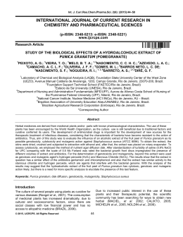
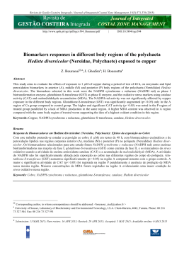
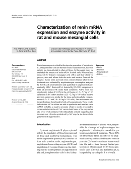
Journal of Medical and Biological Engineering, 31(5): 345-351
345
Direct and Indirect Effects of Ceramic Far Infrared Radiation
on the Hydrogen Peroxide-scavenging Capacity and on
Murine Macrophages under Oxidative Stress
Ting-Kai Leung1,†,*
Yung-Sheng Lin2,3,†
Huey-Fang Shang4
Sheng-Yi Hsiao3
Chi-Ming Lee1
Hsuan-Tang Chang1
Yen-Chou Chen4
Jo-Shui Chao1
1
Department of Radiology, Faculty of Medicine, Taipei Medical University and Hospital, Taipei 110, Taiwan, ROC
Department of Applied Cosmetology and Graduate Institute of Cosmetic Science, Hungkuang University, Taichung 433, Taiwan, ROC
3
Instrument Technology Research Center, National Applied Research Laboratories, Hsinchu 300, Taiwan, ROC
4
Graduate Institute of Medical Sciences, Taipei Medical University, Taipei 110, Taiwan, ROC
2
Received 16 Apr 2010; Accepted 17 Aug 2010; doi: 10.5405/jmbe.777
Abstract
Far infrared (FIR) rays are used for many therapeutic purposes, but the intracellular mechanisms of their beneficial
effects have not been entirely elucidated. The purposes of this study were thus to explore the effects of
ceramic-generated far infrared ray (cFIR) on RAW 264.7 cells by determining the scavenging activity of hydrogen
peroxide (H2O2), cell viability, and changes in cytochrome c levels and the NADP+/NADPH ratios. The results showed
that the H2O2-scavenging activity directly increased by 10.26% after FIR application. Additional FIR treatment resulted
in increased viability of murine macrophages with different concentrations of H2O2. cFIR significantly inhibited
intracellular peroxide levels and LPS-induced peroxide production by macrophages. The increased ratio of hypodiploid
cells elicited by H2O2 was significantly reduced by cFIR. The effects of cFIR on H2O2 toxicity were determined by
measuring intracellular changes in cytochrome c levels and the ratio of NADP+/NADPH, and results showed that cFIR
may block ROS-mediated cytotoxicity. In conclusion, data from this study suggest that cFIR may possess antiapoptotic
effects by reducing ROS production by macrophages. We also review past articles related to the effects of oxidative
stress from metabolically produced H2O2, and discuss possible beneficial effects of cFIR on living tissues.
Keywords: Ceramic-generated far infrared ray (cFIR), Hydrogen peroxide (H2O2) scavenging, Oxidative stress, Murine
macrophages, Cytochrome c, Hypodiploid cell
1. Introduction
Far infrared (FIR) rays are electromagnetic waves with
wavelengths within the infrared spectrum. FIR, especially at
8~14 µm, have many biological effects, including accelerated
wound healing via fibroblast proliferation, enhanced immunity
via leukocyte strengthening, and promotion of sleep [1-3].
Specific discoveries include a report by Shimokawa et al. [4]
which showed that FIR-treated water increased the number of
free tetrahedral water molecules or smaller-sized clusters. FIR
absorption causes the collapse of water clusters, and the energy
transfer may be converted into molecular vibrations. Also, by
† These authors contributed equally to this work
* Corresponding author: Ting-Kai Leung
Tel: +886-2-27372181 ext. 3148; Fax: +886-2-23780943
E-mail: [email protected]
measuring the scavenging activity against hydrogen peroxide
(H2O2), Jeon et al. [5] showed that the antioxidant effects of
rice hull extract are enhanced by thermal FIR irradiation. The
benefits of FIR were proven in their work by an experimental
model of H2O2-induced DNA damage in human lymphocytes,
in which the rice hull extract decreased DNA strand breakage.
H2O2 is a byproduct of normal oxygen metabolism in the
aerobic cells of animals and plants. All organisms possess
peroxidases or enzymes to break down low concentrations of
H2O2 into water and oxygen. However, the continuous
production of H2O2 contributes to increased concentration of
reactive oxygen species (ROS) within both the mitochondrial
matrix and cytosol. The rate of H2O2 production in
mitochondria is approximately 2% of the total oxygen uptake
under physiological conditions [6].
This potentially damages mitochondrial components and
initiates degradation. Therefore, the continuous generation of
H2O2 during aerobic metabolism is harmful, and acts as a
J. Med. Biol. Eng., Vol. 31 No. 5 2011
burden on living systems [7]. Superoxide and H2O2 are the
major primary sources of ROS, and play a role in certain
pathological processes, including neurodegeneration, aging,
and heart and lung toxicity. Superoxide anion is mainly formed
through one-electron reduction of O2 of the respiratory chain,
which is then catalyzed by superoxide dismutase to form H2O2.
A variety of inflammatory stimuli, such as
lipopolysaccharide (LPS), are thought to cause human diseases
through elevating ROS production. Such observations support
the notion that ROS play a critical role in causing certain
human diseases, and thus agents with the ability to block ROS
production have therapeutic potential and merit further
development. The changes in the intracellular ratio of
NADP+/NADPH reflect the adjustment of intracellular
chemical reduction, and thus are related to ROS-mediated
cytotoxicity [8].
On the other hand, the large amount of cytochrome c in
the mitochondrial intermembrane space should oxidize all the
superoxide produced by the respiratory chain and convert it
back to oxygen [9]. In the past, reduction of superoxide by
cytochrome c by superoxide was employed to measure
superoxide generation and oxidative stress [10].
In this study we investigated the effects of cFIR on
intracellular H2O2, cytochrome c, and the NADP+/NADPH
ratio, which are indices of the presence of antioxidants [9-12].
2. Materials and methods
2.1 FIR ceramic powder
The FIR-emitting ceramic powder was micro-sized
particles made from numerous ingredients consisting of mineral
oxides and mineral salts, such as aluminum oxide, which was
put together by the biomaterials laboratory of Taipei Medical
University, Taiwan. An scanning electron microscope (SEM)
image of the FIR ceramic powder used in this study is shown in
Fig. 1 FIR emissivity, the ratio of radiation energy irradiated
from the sample to an ideal black body as described by Plank’s
Law, was determined by a CI SR5000 infrared
spectroradiometer. The emissivity spectrum of the FIR ceramic
powder was in the wavelength range of 6 and 14 µm (Fig. 2)
[1,2].
Figure 1. SEM picture of far-infrared ceramic powder used in this
study.
1.00
Emissivity
Emissivity
346
0.80
0.60
0.40
0.20
0.00
3
4
5
6
7
8
9
10
11
12
13
14
Wavelength (um)
Figure 2. Emissivity spectrum of the far-infrared ceramic powder used
in this study.
Equal amounts (100 g) of FIR-emitting ceramic powder
were enclosed in different plastic bags (10 × 20 cm) and used
as the FIR irradiation source.
2.2 Direct scavenging of H2O2 with cFIR
An H2O2 solution (Sigma, St. Louis, MO, USA) with a
concentration of 1 M was prepared, and equal 9 mL amounts of
H2O2 solutions were added to the test tubes. The H2O2 solution
was categorized into two groups: control and FIR. For the FIR
group, test tubes were incubated at room temperature and
covered externally by plastic bags filled with 100 g of the FIR
ceramic powder for 3 h of irradiation. The control group was
treated similarly, with the exception of FIR treatment. After the
incubation period, the reagent (working concentration: 7.5 mM
phenol red and 5 mg/mL horseradish peroxidase, Sigma,
St. Louis, MD, USA) were added to each test tube. The mixture
was allowed to react for 10 min, and the absorbance was
observed at 550 nm with an enzyme-linked immunosorbent
assay (ELISA) reader (Gemini XPS Molecular Devices,
Sunnyvale, CA, USA), with a lower absorbance value
representing a higher H2O2-scavenging ability.
2.3 Cell preparation
RAW264.7 cells, a mouse macrophage cell line, were
obtained from the Bioresource Collection and Research Center
(BCRC). Cells were cultured in Dulbecco’s Modified Eagle
Medium (DMEM) supplemented with 2 mM glutamine,
antibiotics (100 U/mL penicillin A and 100 U/mL
streptomycin), and 10% heat-inactivated fetal bovine serum
(FBS; Gibco/BRL, Gaithersburg, MD, USA) and maintained in
a 37°C humidified incubator containing 5% CO2. Cells were
seeded until 80% confluent on the bottom of the dishes.
Equal amounts (100 g) of FIR-emitting ceramic powder or
a control (non-functional powder) were enclosed in different
plastic bag × 20 cm) as the FIR irradiation source. Total
cellular extracts were prepared according to our previous paper
[1,2], separated on 8%∼12% sodium dodecylsulfate
(SDS)-polyacrylamide minigels, and transferred to immobilon
polyvinylidene difluoride membranes (Millipore). Membranes
were incubated with 1% bovine serum albumin (BSA) and then
incubated with specific antibodies overnight at 4°C. The
expression of protein was detected by staining with nitroblue
Antioxidant Effect of Ceramic-generated FIR
tetrazolium (NBT) and 5-bromo-4-chloro-3-indolyl phosphate
(BCIP) (Sigma).
In order to determine steady-state intracellular H2O2
levels, RAW264.7 cells were trypsinized and resuspended in
ice-cold Hank’s balanced salt solution (HBSS) containing
0.2 mg/mL of soybean trypsin inhibitor. Cell suspensions were
preincubated for 5 min at 37°C and then treated with 100 mM
DMNQ at 37°C. Aliquots were taken at different times, mixed
with equal volumes of HBSS containing 10 mM DCFH-DA,
and further incubated for 5 min at 37°C. The cellular
fluorescence was then immediately determined by a flow
cytometric analysis.
2.4 Determination of extracellular
RAW264.7 cells without LPS
H2O2
production
of
RAW264.7 cells were incubated in a 5% CO2 incubator
for 24 and 48 h (1 and 2 days). After the cells had adhered to
the dish, we replaced the medium, and then treated them with
cFIR or control powder at 37ºC in a 5% CO2 incubator. We
used an NWLSSTM Hydrogen Peroxide Assay kit (Northwest
Life Science Specialties, LLC) to detect the extracellular H2O2.
The OD595 data represent the concentration of H2O2 within
RAW264.7 cells.
2.5 Determination of extracellular H2O2 production induced by
LPS in RAW264.7 cells
347
plastic bags which were inserted beneath the tissue culture
plates (Fig. 3).
H2O2
cFIR powder / Control powder bags
Figure 3. For cell culture, bags of ceramic-generated far-infrared
powder and control powder were inserted beneath discs
containing RAW264.7 cells to create FIR and control groups
of cells.
Cells were treated with the indicated compounds for a
further 24 h, and then were washed with PBS and stained with
3 µm propidium iodide (PI; Molecular Probes) for 30 min. The
fluorescence emitted from the PI-DNA complex was
quantitated after excitation of the fluorescent dye by FACScan
flow cytometry (Becton Dickinson).
2.8 Lactate dehydrogenase (LDH) activity release assay
RAW264.7 cells were incubated in a 5% CO2 incubator
for 24 h. After the cells had adhered to the dish, we replaced
the medium with one containing 1ug/mL lipopolysaccharide
(LPS; Sigma), then treated RAW264.7cells with cFIR or
control powder at 37°C in a 5% CO2 incubator for 1 and
2 days. We then detected the extracellular H2O2 with the
NWLSSTM Hydrogen Peroxide Assay kit. Data represent the
OD595 within RAW264.7 cells.
The percentage of LDH activity release was expressed as
the proportion of LDH released into the medium compared to
the total amount of LDH present in cells treated with lysis
buffer (Roche). LDH release concentrations of the designated
control and cFIR groups of H2O2 (400, 600, and
1000 µm)-treated cells were analyzed. After 6 h of incubation,
the activity was monitored as the oxidation of NADH at
530 nm with an LDH assay kit (Roche).
2.6 Cell viability of cFIR-treated RAW264.7 cells under H2O2mediated oxidative stress
2.9 Measurement of cytosolic cytochrome c levels
XTT was used as an indicator of cell viability as
determined by its mitochondrial-dependent reduction to
formazone. Cells were plated at a density of 4 × 10⁵ cells/well
into 24-well plates for 24 h, treated with different concentrations
of H2O2 (100, 250, 500, and 1000 µM), and followed by a
further 24 h of treatment. Cells were washed three times with
phosphate-buffered saline (PBS; Gibco), and XTT (1 mg/mL)
was added to the medium for 3 h, and the supernatant was then
collected. The absorbance was read at 450 nm with an ELISA
analyzer (Gemini XPS Molecular Devices, Sunnyvale, CA,
USA).
In order to identify cFIR’s effect on cytosolic
cytochrome c, RAW264.7 cells treated with FIR powder for 6 h
and other untreated cells were then harvested by centrifugation
at 3000 rpm for 5 min at 4°C. The cell pellets were washed once
with ice-cold PBS and resuspended in five volumes of 20 mM
HEPES-KOH (pH 7.5), 10 mM KCl, 1.5 mM MgCl2, 1 mM
EDTA, 1 mM EGTA, 1 mM DTT, 0.1 mM PMSF, and 250 mM
sucrose. Cells were homogenized and centrifuged at 1200 rpm
for 10 min at 4°C to separate them into supernatant and pellets.
The supernatant was then centrifuged at 12,000 rpm for 15 min
at 4°C, and the obtained supernatant was used to identify of
cytosolic cytochrome c by immunoblotting.
2.7 Assay of hypodiploid cell analysis
2.10 Measurement of NADP+/NADPH levels
RAW264.7 cells were seeded in six-well tissue culture
plates at a density of 4 × 10⁵ cells per well. After 24 h of
culturing, the medium was changed and various concentrations
of H2O2 (250 and 500 M) were added. For the FIR groups, the
enclosed FIR ceramic powder was distributed uniformly in
NADP+/NADPH levels were measured by determining the
rate of production of NADP+/NADPH, as previously described
[13]. NADP+ and NADPH were assayed spectrophotometrically
based on the measurement of the absorbance of the reduced
coenzyme at 340 nm.
J. Med. Biol. Eng., Vol. 31 No. 5 2011
2.11 Statistical analysis
After H2O2 degradation, the cell viability and intracellular
concentration of H2O2 were measured using a cell viability
assay (XTT). We also performed an analysis of hypodiploid
cells and an LDH release assay, and determined cytosolic
cytochrome c and NADP+/NADPH levels. The statistical
relationship between groups was determined using a t-test, with
p values of < 0.05 considered significant.
3. Results
3.1 Direct scavenging of H2O2 with cFIR
Figure 4 shows that the mean absorbance of the control
and FIR groups were 0.139 and 0.122, respectively (n = 25).
The extent to which H2O2 disappeared in the FIR group was
significantly larger than that of the control group, with a
decrease of 12.23% (p < 0.001). This result confirms that H2O2
can be directly scavenged by FIR treatment.
0.18
3.3 Extracellular H2O2 production of RAW264.7 cells with LPS
induction
The peroxide level induced by LPS was significantly
decreased by cFIR (p < 0.05) (Fig. 5).
3.4 Cell viability of cFIR-treated RAW264.7 cells under H2O2mediated oxidative stress
Additionally, cFIR possessed the ability to stimulate the
proliferation of RAW264.7 cells, according to the XTT assay
(Fig. 6). The percentage of cell proliferation differed
significantly when cells were treated with 250 µm H2O2
(p < 0.05).
140
Control
FIR
*
120
Cell proliferation (%)
348
100
80
60
40
Control
FIR
0.16
0.14
20
***
0
OD550
0.12
1000 uM
500 uM
250 uM
100 uM
H2O2 concentration
0.10
Figure 6. With different concentrations of H2O2 as the source of
oxidative toxicity, cFIR possesses the ability to stimulate the
proliferation of RAW264.7 cells against H2O2, according to
the XTT assay. *p < 0.05 indicates a significant difference
compared with the control group.
0.08
0.06
0.04
0.02
0.00
Control
FIR
3.5 Hypodiploid cell analysis and LDH activity release assay
Figure 4. Comparison of H2O2 content in direct H2O2 scavenging with
ceramic-generated far-infrared. ***p < 0.001 indicates a
significant difference compared with the control group.
3.2 Extracellular H2O2 production of RAW264.7 cells without
LPS induction
As shown in Fig. 5, significant decreases in the peroxide
level by cFIR were identified after both 1 and 2 days (p < 0.05).
0.5
Control
FIR
0.4
OD550
*
0.3
Table 1. The effect of cFIR on the apoptosis induced by H2O2 was
evaluated by flow cytometry via PI staining.
Percentage (%)
Cytotoxicity (%)
400 µm
600 µm
400 µm
600 µm
1000 µm
Control 30.5 ± 3.9 43.8 ± 4.7 135.2 ± 7.8 136.7 ± 9.8 131.9 ± 10.4
FIR
21.3 ± 2.5* 30.7 ± 3.8* 122.1 ± 7.3* 116.6 ± 3.7** 116.8 ± 10.4
*p < 0.05, **p < 0.01 indicate a significant difference compared with the control
group.
0.2
0.1
The effect of cFIR on apoptosis induced by H2O2 was
evaluated by flow cytometry with PI staining. As shown in
Table 1, the ratio of hypodiploid cells increased in H2O2-treated
cells, and this was significantly reduced by cFIR (p < 0.05).
The effect of cFIR on LDH release assays indicated a
significant difference between the control* and cFIR groups for
H2O2-treated cells (400 and 600 µm, p < 0.05); the cFIR group
showed a significant reduction in LDH release (Table 1).
*
*
0.0
1 Day without LPS 2 Days without LPS
1 Day with LPS
Figure 5. Significantly decreased in H2O2 by ceramic-generated
far-infrared irradiation with and without lipopolysaccharide
stimulation. *p < 0.05 indicates a significant difference
compared with the control group.
The cytoprotective effect of cFIR against ROS-induced
cell death was predicted. The morphology of cells was
observed under microscopy by demonstrating the apoptotic
features of cells, including irregular cytoplasmic membranes
and chromatin condensation. These characteristic findings of
hypodiploid cells after cFIR treatment were obviously lower
than those in the control group (Fig. 7).
Antioxidant Effect of Ceramic-generated FIR
349
2.5
Control
FIR
Relative Protein Amount
2.0
1.5
1.0
**
0.5
0.0
Control
FIR
FIR
Figure 9. Immunoblotting analysis revealed that the level of
cytochrome c in cFIR-irradiated groups of cells significantly
decreased. **p < 0.01 indicates a significant difference
compared with the control group.
3.7 NADP+/NADPH levels
The NADP+/NADPH ratio was increased in the cFIR
group (control group: 0.24 vs. cFIR group: 0.41). Through the
effects of cFIR, more NADPH was consumed and was
associated with an increase in NADP+, which reflects an
NADPH-reducing process.
4. Discussion
Figure 7. Effects of morphological changes induced by H2O2 on
RAW264.7 cells by observing the morphology of cells under
microscopy with a 200x power field. Cells were treated
(a) without and (b) with cFIR in the presence of H2O2, and
hypodiploid cells are indicated with white arrows.
3.6 Cytosolic cytochrome c levels
ROS reduction by cFIR was investigated. As shown in
Fig. 8, a significant decrease in H2O2 levels by cFIR was
identified (p < 0.05). The results of a flow cytometric analysis
showed that the intracellular peroxide level was reduced by
cFIR using DCHF-DA as a peroxide-sensitive fluorescent dye.
200
H2O2
H2O2 production
production
(fluorescent intensity)
(fluorescent
intensity)
Control
FIR
*
150
100
50
0
Control
2 Days
FIR
Figure 8. Flow cytometric analysis showing that levels of intracellular
H2O2 were reduced by ceramic-generated far infrared via a
flow cytometric analysis using DCHF-DA as a
peroxide-sensitive fluorescent dye. *p < 0.05 indicates a
significant difference compared with the control group.
The level of cytochrome c in the cFIR-irradiated groups of
cells was found to have increased significantly via an
immunoblotting analysis (p < 0.01; Fig. 9).
FIR can break hydrogen (H-O) bonds by exciting
‘stretching’ or ‘bending’ vibrations in water clusters [4], and
thus decreases the size of water clusters. Hydrogen bonds are a
factor that decreases the volatility of any liquid possessing
them. This is related to the reduction rate of H2O2, which exists
in a cluster form in general conditions, rather than as single
molecules. Therefore, the weakening of hydrogen bonds by
FIR may also explain why FIR accelerates H2O2 transformation
and releases H2O and O2 from H2O2 molecular clusters.
The H2O2-scavenging capacity elicited by FIR-emitting
ceramic materials may have beneficial biological effects. H2O2
is continually produced in oxidation-redox centers in both
animal and plant aerobic respiratory systems. Cumulative
increases in H2O2 and superoxide radicals can damage cells,
including proteins, lipids, and DNA, leading to proven
augmented mutation rates [14]. Reactive oxygen radicals of
H2O2 are an important factor in oxidative stress, and are related
to the pathogenesis of many important diseases in both animals
and plants [15-16].
FIR is the major heat-transmitting radiation at
wavelengths 3 µm to 1 mm, as defined by the CIE [17]. FIR,
especially in the range of 3~14 µm, has many biological
effects. Previous studies demonstrated that FIR has a wide
range of applications, including increasing microcirculation,
and its health-promoting properties are attracting more
attention [1-2,17,18]. However, the mechanisms underlying
these biological effects are still poorly understood. To our
knowledge, there were no previous reports investigating the
scavenging ability of FIR and its antitoxic effects toward H2O2.
In our experiment, we proved that H2O2 can be scavenged
directly by FIR treatment.
350
J. Med. Biol. Eng., Vol. 31 No. 5 2011
H2O2 is an oxygen species that causes the death of normal
human fibroblasts exposed to an external source of oxygen
radicals. Normal human cells may be damaged by
phagocyte-released oxygen radicals at the site of inflammation
and from sources other than phagocytic cells [19]. In
pathophysiological conditions, H2O2 is continuously generated,
and levels thus remain higher than normal [20]. Although
speculative, the concentrations of H2O2 used in this study may
be within the range that occur in some pathological states. H2O2
gradients form if the H2O2 production site and H2O2
degradation site are separated by membranes. Because H2O2 is
highly membrane-permeable, the equilibrium between extraand intracellular levels of H2O2 is reached extremely rapidly, or
within 1 s, such that the measurement of intracellular levels of
H2O2 also reflects extracellular levels [21]. Exposure of cells to
H2O2 leads to cell death by apoptosis and necrosis. Cell death
or cytotoxicity is classically evaluated by quantifying plasma
membrane damage.
In our experiment, we also used LDH to prove the
antioxidant ability of cFIR in circumstances of H2O2 toxicity.
LDH is a stable enzyme which is present in all cell types, and it
is rapidly released into the cell culture medium when the
plasma membranes are damaged. LDH is thus the most widely
used marker in cytotoxicity studies. Pathological or
aging-related overproduction of mitochondrial ROS may lead
to activation of apoptotic pathways [22]. Protection of cells
from these intracellular oxygen radicals appears to be due to
the presence of a variety of intracellular enzymes and naturally
occurring radical scavengers. Under normal conditions, these
protective mechanisms are adequate to prevent extensive
damage to vital cellular constituents. Excessive ROS cause the
degenerative diseases of aging, particularly cancer and
atherosclerosis, as consequence of oxidative damage [23-25].
We selected RAW264.7 murine macrophages as our target
cells because macrophages are vital for recognizing and
eliminating microbial pathogens, and the survival of
macrophages directly contributes to a host’s defense system.
Several previous studies showed that the virulence of some
bacteria is due to their ability to trigger the death of activated
macrophages by stimulating ROS production. Therefore,
investigating protective mechanisms and developing agents
with the ability to protect macrophages from ROS insults is an
important issue [26]. RAW264.7 macrophages play a
significant role in innate immunity and inflammation, and when
activated by pathogens and cytokines they produce large
amounts of H2O2 and ROS, exerting strong cytotoxicity against
microorganisms and many cells, including killing macrophages
themselves [27]. Therefore, increasing the survival rate of
macrophages engaged in defensive processes against pathogens
and cancer cells would enhance cell-mediated immunity [28].
Mitochondria are the main source of ROS in most aerobic
mammalian cells, and cytochrome c in the respiratory chain is
essential for maintaining a lower physiological H2O2
concentration in mitochondria [11]. Cytochrome c is thus an
ideal antioxidant that attacks superoxide and oxygen radicals.
In living cells, the generation of oxygen radicals and H2O2 in
the respiratory chain is a result of electron leakage, and the
levels of oxygen radicals and H2O2 are kept in a balanced state
between generation by the respiratory chain and elimination by
cytochrome c. Therefore, a lack of cytochrome c within the
respiratory chain causes higher levels of oxygen radicals and
associated H2O2 accumulation [29-31]. Our results
demonstrated that cFIR enhanced the antioxidant effect of
H2O2 by consuming more intracellular cytochrome c.
On the other hand, calmodulin (Cam) increases the rate at
which NADPH-derived electrons are transferred to enzyme
flavins, and also triggers electron transfer and the oxidation of
NADPH to NADP+. Meanwhile, the activity of cytochrome c
acting as a reductase promotes the activation of NO synthesis
from L-arginine [32].
This is the first study exploring the possibility that cFIR
may exhibit antioxidant characteristics in mammalian cells via
its effects on intracellular levels of H2O2, and the levels of
cytochrome c and NADP+/NADPH. We previously showed that
cFIR induces intracellular levels of Cam and NO in
RAW264.7 cells [2]. Summing up all of our data, we envision a
possible pathway through which cFIR might exert its
antioxidant effect (Figure 10). However, the in vitro
experiments described in this work are not a perfect model to
examine the influence of FIR-emitting ceramic materials on in
vivo H2O2 production. In the future, a more-precise
measurement method to detect tiny differences in the cellular
H2O2-scavenging capacity needs to be developed.
Figure 10. A possible pathway through which ceramic-generated far
infrared might achieve its antioxidant effects.
5. Conclusions
The present study first explores how to use a physical
method of cFIR irradiation that contributes to H2O2-scavenging
effects in cells. We correlated the results of cell viability,
hypodiploid cell analysis, LDH release assay, and the
cytochrome c and NADP+/NADPH levels of the RAW264.7
cell line (murine macrophages). Our results demonstrated that
the group of cells exposed to cFIR significantly differed from
the control group. It thus may be worth applying cFIR for its
antioxidant, anti-aging, immunity-boosting, and other related
health-promoting effects. However, to better elucidate the
precise effects of cFIR on living systems, it is important that
Antioxidant Effect of Ceramic-generated FIR
future research develops an in vivo experimental model. We
believe that these results justify further work to develop a more
mature explanation of the biomolecular mechanisms of cFIR
with regard to mammalian cells.
Acknowledgements
The authors gratefully acknowledge the support provided
to this study by Mr. Francis Chen (Franz Collection, Taipei,
Taiwan), Dr. Shawn Huang (Purigo Biotech, Taipei, Taiwan),
Mr. Li Chien Chiu (Hocheng, Taipei, Taiwan), Mr. Mike C.F.
Chen (All Star, Taipei, Taiwan), and Mr. Roy Y. H. Sun (Vital
7, Taipei, Taiwan).
References
[1]
T. K. Leung, C. M. Lee, M. Y. Lin, Y. S. Ho, C. S. Chen, C. H.
Wu and Y. S. Lin, “Far infrared ray irradiation induces
intracellular generation of nitric oxide in breast cancer cells,” J.
Med. Biol. Eng., 29: 15-18, 2009.
[2] T. K. Leung, Y. S. Lin, Y. C. Chen, H. F. Shang, Y. H. Lee, C. H.
Su and H. C. Liao, “Immunomodulatory effects of far infrared
ray irradiation via increasing calmodulin and nitric oxide
production in RAW 264.7 macrophages,” Biomed. Eng.-Appl.
Basis Commun., 21: 317-323, 2009.
[3] I. Shojiro and K. Morhihiro, “Biological activities caused by
far-infrared radiation,” Int. J. Biometeorol., 33: 145-150, 1989.
[4] S. Shimokawa, T. Yokono, T. Mizuno, H. Tamura, T. Erata and T.
Araiso, “Effect of far-infrared light irradiation on water as
observed by x-ray diffraction measurements,” Jpn. J. Appl. Phys.,
43: 545-547, 2004.
[5] K. I. Jeon, E. Park, H. R. Park, Y. J. Jeon, S. H. Cha and S. C.
Lee, “Antioxidant activity of far-infrared radiated rice hull
extracts on reactive oxygen species scavenging and oxidative
DNA damage in human lymphocytes,” J. Med. Food, 9: 42-48,
2006.
[6] B. Chance, H. Sies and A. Boveris, “Hydroperoxide metabolism
in mammalian organs,” Physiol. Rev., 59: 527-605, 1979.
[7] B. Vergauwen, M. Herbert and J. J. van Beeumen, “Hydrogen
peroxide scavenging is not a virulence determinant in the
pathogenesis of Haemophilus influenzae type b strain Eagan,”
BMC Microbiol., 6: 3, 2006.
[8] A. K. Agarwal and R. J. Auchus, “Minireview: cellular redox
state regulates hydroxysteroid dehydrogenase activity and
intracellular hormone potency,” Endocrinology, 146: 2531-2538,
2005.
[9] H. J. Forman and A. Azzi, “On the virtual existence of
superoxide anions in mitochondria: thoughts regarding its role in
pathophysiology,” FASEB J., 11: 374-375, 1997.
[10] M. O. Pereverzev, T. V. Vygodina, A. A. Konstantinov and V. P
Skulachev, “Cytochrome c, an ideal antioxidant,” Biochem. Soc.
Trans., 31: 1312-1315, 2003.
[11] Y. Zhao, Z. B. Wang and J. X. Xu, “Effect of cytochrome c on
the generation and elimination of O2 and H2O2 in mitochondria,”
J. Biol. Chem., 278: 2356-2360, 2003.
[12] S. H. Lee, S. O. Ha, H. J. Koh, K. Kim, S. M. Jeon, M. S. Choi,
O. S. Kwon and T. L. Huh, “Upregulation of cytosolic
NADP(+)-dependent isocitrate dehydrogenase by hyperglycemia
protects renal cells against oxidative stress,” Mol. Cells, 29:
203-208, 2010.
351
[13] Z. Zhang, J. Yu and R. C. Stanton, “A method for determination
of pyridine nucleotides using a single extract,” Anal. Biochem.,
285: 163-167, 2000.
[14] L. C. Seaver and J. A. Imlay, “Are respiratory enzymes the
primary sources of intracellular hydrogen peroxide?” J. Biol.
Chem., 279: 48742-48750, 2004.
[15] B. Halliwell and J. M. C. Gutteridge, Free Radicals in Biology
and Medicine, New York: Oxford University Press, 1999.
[16] T. Finkel and N. J. Holbrook, “Oxidants, oxidative stress and the
biology of aging,” Nature, 408: 239-247, 2000.
[17] Commission Internationale de L’Eclairage (CIE): International
Lighting Vocabulary, Vienna, 1987.
[18] A. Maurel, C. Hernandez, O. Kunduzova, G. Bompart, C.
Cambon, A. Parini and B. France’s, “Age-dependent increase in
hydrogen peroxide production by cardiac monoamine oxidase A
in rats,” Am. J. Physiol. Heart Circ. Physiol., 284: H1460-H1467,
2003.
[19] R. H. Simon, C. H. Scoggin and D. Patterson, “Hydrogen
peroxide causes the fatal injury to human fibroblasts exposed to
oxygen radicals,” J. Biol. Chem., 256: 7181-7186, 1981.
[20] P. A. Hyslop, Z. Zhang, D. V. Pearson and L. A. Phebus,
“Measurement of striatal H2O2 by microdialysis following global
forebrain ischemia and reperfusion in the rat: correlation with the
cytotoxic potential of H2O2 in vitro,” Brain Res., 671: 181-186,
1995.
[21] F. Antunes and E. Cadenas, “Estimation of H2O2 gradients across
biomembranes,” FEBS Lett., 475: 121-126, 2000.
[22] Y. H. Wei and H. C. Lee, “Oxidative stress, mitochondrial DNA
mutation, and impairment of antioxidant enzymes in aging,” Exp.
Biol. Med., 227: 671-682, 2002.
[23] M. Y. Lin and C. L. Yen, “Product-scavenging ability of yogurt
organisms, reactive oxygen species and lipid peroxidation,” J.
Dairy Sci., 82: 1629-1634, 1999.
[24] R. Kah, A. Kampkötter, W. Wätjen and Y. Chovolou,
“Antioxidant enzymes and apoptosis,” Drug Metab. Rev., 36:
747-762, 2004.
[25] R. I. Salganik, “The benefits and hazards of antioxidants:
controlling apoptosis and other protective mechanisms in cancer
patients and the human population,” J. Am. Coll. Nutr., 20:
464S-472S, 2001.
[26] H. Y. Lin, S. C. Shen, C. W. Lin, L. Y. Yang and Y. C. Chen,
“Baicalein inhibition of hydrogen peroxide-induced apoptosis via
ROS-dependent heme oxygenase 1 gene expression,” Acta
Biochim. Biophys., 1773: 1073-1086, 2007.
[27] Y. Yoshioka, T. Kitao, T. Kishino, A. Yamamuro and S. Maeda,
“Nitric oxide protects macrophages from hydrogen
peroxide-induced apoptosis by inducing the formation of
catalase,” J. Immunol., 176: 4675-4681, 2006.
[28] M. E. Gonzalez-Mejia and A. I. Doseff, “Regulation of
monocytes and macrophages cell fate,” Front. Biosci., 14:
2413-2431, 2009.
[29] X. Liu, C. N. Kim, J. J. R. Yang and X. Wang, “Induction of the
apoptotic program in cell-free extracts: requirement for dATP and
cytochrome c,” Cell, 86: 147-157, 1996.
[30] H. M. Abu-Soud, L. L. Yoho and D. J. Stuehr, “Calmodulin
controls neuronal nitric-oxide synthase by a dual mechanism.
Activation of intra- and interdomain electron transfer,” J. Biol.
Chem., 269: 32047-32050, 1994.
[31] Z. B. Wang, M. L. Y. Zhao and J. X. Xu, “Cytochrome c is a
hydrogen peroxide scavenger in mitochondria,” Protein Pept.
Lett., 10: 247-253, 2003.
[32] W. K. Alderton, C. E. Cooper and R. G. Knowles, “[31] Nitric
oxide synthases: structure, function and inhibition,” Biochem. J.,
357: 593-615, 2001.
352
J. Med. Biol. Eng., Vol. 31 No. 5 2011
Baixar