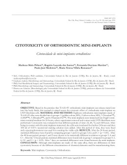
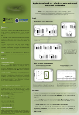
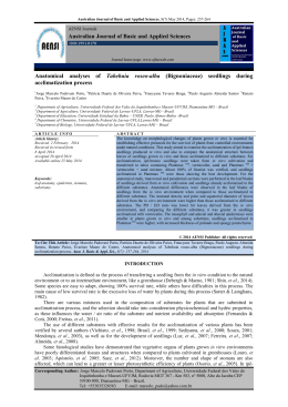
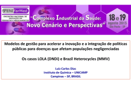
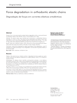
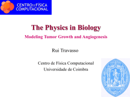
ORIGINAL ARTICLE Evaluation of immunologic profile in patients with nickel sensitivity due to use of fixed orthodontic appliances Marcelo Marigo, PhD,a Darcy Flávio Nouer, PhD,b Marisa Cristina Santos Genelhu, MS,a Luiz Cosme Cotta Malaquias, PhD,a Virgı́nia Ramos Pizziolo, MS,a Alexandre Sylvio Vieira Costa, PhD,c Olindo Assis MartinsFilho, PhD,d and Lucia Fraga Alves-Oliveira, PhDa Governador Valadares, São Paulo, and Belo Horizonte, Brazil The aim of this study was to develop a new approach to testing the impact of nickel antigen on in vitro cell-proliferation assay, to identify adverse reactions to casting alloys among orthodontic patients. Cellproliferation assay in vitro was used as the basic methodology to assess the influence of such variables as source of nickel antigen, type of serum used to supplement the culture medium, and number of cells in the culture. We selected 35 orthodontic patients who were classified as nickel sensitive and non–nickel sensitive, based on their clinical records. Our results showed that hexahydrated nickel sulfate at 10 g/mL, 10% of autologous sera, and 2 ⫻ 105 cells was the best condition for inducing the most marked nickel proliferation response in vitro. This optimized method was able to distinguish nickel-sensitive from non–nickel-sensitive dental patients and also to discriminate those with positive skin tests. Our data suggest that continuous exposure to nickel casting alloys might lead to oral tolerance mechanisms that modulate nickel sensitivity, as evidenced by the lower cell proliferation index in patients undergoing orthodontic treatment over 24 months. Finally, our findings demonstrated a known nickel-induced type 2 immune response and a marked lack of type 1 immunity (interferon ␥) as the hallmarks of nickel-sensitive patients. Further studies are needed to clarify the major cell phenotype associated with this type 2 immune response and the lack of type 1 immunity observed in nickel-sensitive people. (Am J Orthod Dentofacial Orthop 2003;123:000-00) (Am J Orthod Dentofacial Orthop 2003;124:46-52) M any objects contain nickel and can cause nickel sensitivity, and many people are in continuous contact with items that contain nickel.1 Nickel-containing objects are common in dentistry, especially in dental implants and compounds used in crowns, bridgework, and orthodontic appliances. Nickel-titanium alloys are widely used in orthodontic treatment, and all these devices can induce skin and mucosal reactions, leading to tissue inflammation.2-4 An immune response induced by nickel appliances is generally called contact dermatitis and, from a Núcleo de Pesquisa em Imunologia, Faculdade de Ciências da Saúde, Universidade Vale do Rio Doce, Governador Valadares, Minas Gerais, Brazil. b Faculdade de Odontologia de Piracicaba, UNICAMP, São Paulo, Brazil. c Faculdade de Ciências Agrárias, Universidade Vale do Rio Doce, Governador Valadares, Minas Gerais, Brazil. d Centro de Pesquisas René Rachou, Fundaçáo Oswaldo Cruz, Belo Horizonte, Minas Gerais, Brazil. Supported in part by Fundação de Amparo à Pesquisas do Estado de Minas Gerais, Brazil. Reprint requests to: Dr Marcelo Marigo, Rua João Pinheiro, 610 Centro, Governador Valadares, MG CEP 35020-270, Brazil; e-mail, [email protected]. Submitted, July 2002; revised and accepted, October 2002. Copyright © 2003 by the American Association of Orthodontists. 0889-5406/2003/$30.00 ⫹ 0 doi:10.1016/S0889-5406(03)00239-7 46 the immunologic standpoint, is considered type IV hypersensitivity. In this context, nickel binding to endogenous macromolecules can stimulate macrophages and cytotoxic cells, up-regulating the expression of adhesion molecules.5-7 It has also being reported that low-dose exposure can alter the metabolism of human monocytes.8 Additionally, nickel induces T lymphocytes to produce several cytokines, including interferon IFN-␥, interleukin IL-2, IL-5, and IL-10, and stimulates cellular proliferation.9 The pattern and level of cytokines secreted are critical to triggering differential immune responses, which can lead to different degrees of tissue damage. The temperature, microorganisms, enzymatic compounds, and ions normally present in the oral cavity particularly favor the breakdown and release of metallic elements from dental casting alloys.10 Contact dermatitis generally appears clinically as eczema. Reactions of the mucous membranes, such as stomatitis, also occur, and gum hyperplasias, cheilitis, labial desquamation, and multiform erythemas are frequently noted. Nickel hypersensitivity has increased in the last 10 years, and it is estimated that 15% to 30% of American Journal of Orthodontics and Dentofacial Orthopedics Volume 124, Number 1 Europeans and Americans, in a proportion of 1 to 8, men to women, show symptoms.11 A prevalence of 7% to 28% of the general population has been also reported.12,13 Nickel sensitivity has been diagnosed through biocompatibility testing,14,15 including cutaneous sensitivity tests,13 and also through clinical observations and family history. However, major problems regarding specificity of the cutaneous tests are frequently reported. Therefore, a great challenges in the diagnosis of nickel sensitivity has been to discover and develop alternative in vitro assays to improve specificity and minimize false-positive results. Lymphocyte stimulation assays with peripheral blood mononuclear cells (PBMC) and different concentrations of nickel salts have contributed to advances and show promising results.16 In one study, lymphocytes from all suspected nickel-sensitive patients showed a significantly greater response than did those of healthy controls.17 Lymphocyte transformation, as measured by increased deoxyribonucleic acid (DNA) synthesis, seems to be an important tool for investigating the problems arising from false-positive or false-negative patch tests.18 The purpose of this research was to develop a new approach to testing the effect of nickel antigen on lymphoproliferative response in vitro, by using PBMC from patients with orthodontic appliances. Using this strategy, we evaluated whether lymphoproliferative response is a useful method to discriminate nickelsensitive from non–nickel-sensitive dental patients. We also studied patients wearing orthodontic appliances (either with or without clinical symptoms) to determine the influence of certain variables on in vitro lymphoproliferation assay, including source of antigen, type of serum used to supplement the medium, and the number of cells used. MATERIAL AND METHODS Thirty-five patients, aged 10 to 21 years (mean 14.3), all undergoing orthodontic treatment with fixed appliances, were selected based on their clinical health records and classified into 2 groups. The first group comprised 26 patients (8 males, 18 females, aged 10 to 21 years) with clinical manifestations of contact dermatitis, including angular cheilitis, labial desquamation, or gum hyperplasia resulting from metallic compounds, especially earrings, bracelets, and other jewelry. The second group comprised 9 patients (3 males, 6 females, aged 11 to 21 years) who did not have clinical signs or histories of nickel sensitivity. All patients underwent cutaneous nickel sensitivity tests, and those selected for the control group had negative results. We could not use all 26 patients for all tests Marigo et al 47 owing to the limited quantity of PBMC we were able to extract from their blood samples. Informed consent was obtained from all subjects and their parents. The studies were approved by the Ethical Committee of Universidade Estadual de Campinas and Universidade Vale do Rio Doce. The cutaneous sensitivity test was performed as described by Carvalho.19 Briefly, a 5% nickel sulfate and petroleum jelly substrate (nickel sulfate 5%, Alergofar, Rio de Janeiro, Brazil) was kept in direct contact with the skin of the forearm for 48 to 72 hours. The results were classified as positive or negative, based on the reaction. Nickel sources used as stimulating agents for the cellular proliferation assays in vitro were obtained from solutions containing nickel from in vitro corrosion of orthodontic appliances (nickel extract) and from hexahydrated nickel sulfate solutions (NiSO4.6H2O, Sigma Chemical, St. Louis, Mo). The nickel extracts were obtained from 4 different brands of orthodontic appliances. Alloys were kept in a sterile 0.85% saline solution at 37°C for 7, 30, and 60 days. After centrifugation to remove the particles produced during the corrosion process, the solutions were sterilized by filtration (0.2 m, Filter Millex-HA Millipore Products Division, Bedford, Mass). Then the dosage of nickel in this solution was measured with an atomic absorption spectrophotometer (Hitachi Z-8200, Tokyo, Japan). The extracts thus obtained were diluted in culture media (RPMI-1640, Gibco BRL, Grand Island, NY) containing a 3% solution of antibiotic-antimycotic (stock solution: 10,000 IU penicillin, 10,000 IU streptomycin/mL, and 25 g amphotericin B/mL, Sigma Chemical) and 1.6% L-glutamine (stock solution: 200 mmol/L, Gibco), labeled incomplete RPMI. Extracts containing nickel at concentrations of 1.25, 2.5, and 5 g/mL were used in the cellular proliferation assays. The spectrophotometer readings of saline extract from the 4 appliances (A, B, C, and D) showed similar levels of nickel after 60 days of spontaneous corrosion. No significant differences on the yield of nickel between the 4 different brands were observed (A, 40.23 g/mL; B, 39.58 g/mL; C, 40.03g/mL, and D, 40.23 g/ mL). Solution A was used in cell culture. The nickel sulfate (NiSO4.6H2O, Sigma) was also diluted in the above-described incomplete RPMI medium and used at concentrations of 2.5, 5, and 10 g/mL in the cellular proliferation assays. In vitro cellular proliferation assays were performed with the procedure described by Gazzinelli et al.20 In 96-well, flat-bottom, tissue-culture plates, 2 different concentrations of PBMC (2.0 ⫻ 105 and 3.0 ⫻ 105 cells per well) were incubated with 100 L of 2 different 48 Marigo et al culture mediums (RPMI supplemented with 5% heatinactivated AB⫹ human serum and with 5% autologous serum) with several concentrations of the antigenic preparations, including nickel extract (1.25, 2.5, and 5 g/mL) and nickel sulfate (2.5, 5, and 10 g/mL). Triplicate cultures were incubated at 37°C (Queue Systems, Parkersburg, WVa), 95% humidity, 5% CO2 for 6 days. Six hours before harvesting the cells, 1.0 Ci of tritiated thymidine (3H) was added to each well. After this stage, the cells were collected on glass fiber paper (Whatman, Clifton, NJ) with an automatic cell harvester (Skatron Instruments, Sterling, Va). The incorporated radioactivity was determined with a Beckman LS 100 C scintillator (Beckman, Scientific Instruments Division, Irvine, Calif). The results obtained from the proliferation assays were expressed in counts per minute. The cellular stimulation index was calculated by taking the average value of the triplicate series of the stimulated cultures (E) divided by the average values from the triplicate series of controls (C). We chose E/C values greater than 2.0 to indicate significant enhanced PBMC proliferation, based on values described in previous studies.17,21-23 The analysis of cytokines, IFN-␥, and IL-5 in the supernatants from nickel-stimulated and control cultures were assayed with enzyme-linked immunosorbent assays, as described by Lunde et al.24 Briefly, a 1 ⫻ 106 PBMC sample from each patient undergoing orthodontic treatment was stimulated with nickel sulfate (10 g/mL) for 24 hours in medium containing autologous sera. Controls of nonstimulated cells were tested. For cytokine analysis in the supernatant, initially, 60 L per well of anti-IL-5 monoclonal antibodies (5 g/mL; DNAX, Palo Alto, Calif), and 60 L per well of anti-IFN-␥ (2 g/mL; R&D Systems, Minneapolis, Minn) were added to the 96-well plates (Imunolon 2, Dynatech Laboratories, Alexandria, Va) for 12 hours at room temperature. After washing, the plates were blocked with phosphate-buffered saline (PBS) solution supplemented with 1% of bovine serum albumin, 5% of sucrose, and 0.05% of sodium azide. After blockage, 50 L of culture supernatants were added to the test wells. Blank and standard wells were prepared with 50 L of PBS, recombinant IFN-␥ (25-0.78 ng/mL; R&D Systems), and recombinant IL-5 (50-1.56 ng/mL; DNAX), respectively. The plates were then incubated for 2 hours at room temperature. After washing with PBS 0.05% of Tween 20 (polyoxyethylene sorbitan monolaurate, Sigma), 60 L per well of biotinylated anti-IFN-␥ polyclonal antibody (0.3 g/mL; R&D Systems) and antiIL-5 (0.3 g/mL; DNAX) were added and the plates incubated for 1 hour at room temperature. After incubation, the plates were washed with PBS 0.05% of American Journal of Orthodontics and Dentofacial Orthopedics July 2003 Fig 1. Effect of antigen source on in vitro lymphoproliferation assay. Results are expressed as mean cell proliferation index (E/C) ⫾ standard error. E/C ⬎ 2.0 was adopted as arbitrary cutoff to signify significant cell proliferation index. Tween 20 and 100 L of peroxidase-conjugated streptavidin (1 g/mL, Sigma) and reincubated for 20 minutes at room temperature. After this step, 60 L of the substrate (2,2 azino-bis 3 ethylbenz-thiazolidine-6sulfonic acid, Sigma, 1 mg/mL) were added to all wells. After color development, the reaction was stopped by adding 50 L of 1 M sulfuric acid. The optical density was read with an automatic reader (Benchmark Microplate Reader, Bio-Rad Laboratories, Hercules, Calif) with a 405-nm filter. Data analysis was performed by one-way analysis of variance followed by Student t tests. Significance was set at P ⬍ .05. RESULTS To improve specificity of nickel sensitivity diagnosis, we tested the effect of nickel antigen source on the in vitro lymphoproliferative response by using PBMC from 22 patients wearing orthodontic appliances. Using the basic blastogenesis method described by Gazzinelli et al,20 we tested 3 different concentrations of nickel extract and nickel sulfate (Fig 1). Data analysis demonstrated that exposure of PBMC to the nickel extract did not lead to significant proliferation (E/C ⬍ 2.0). Concentrations of 2.5 and 5.0 g/mL of nickel sulfate did not give significant results. On the other hand, a concentration of 10 g/mL of nickel sulfate significantly stimulated cell proliferation (E/C ⬎ 2.0), measured by 3H-thymidine incorporation. Therefore, the antigen source chosen for further study was nickel sulfate. The responses of PBMC from 21 subjects to nickel sulfate 10 g/mL was evaluated in the presence of autologous and AB⫹ type sera with 2 ⫻ 105 and 3 ⫻ 105 cells per well (Fig 2). The in vitro responses of 2 ⫻ American Journal of Orthodontics and Dentofacial Orthopedics Volume 124, Number 1 Fig 2. Influence of serum-type supplement and number of cells on in vitro lymphoproliferation assay. Results are expressed as mean cell proliferation index (E/C) ⫾ standard error. E/C ⬎ 2.0 was adopted as arbitrary cutoff to signify significant cell proliferation index. Fig 3. Comparative analysis of cell proliferation index between nickel sensitive and non–nickel-sensitive patients. Results are expressed as mean cell proliferation index (E/C) ⫾ standard error. E/C ⬎ 2.0 was adopted as arbitrary cutoff to signify significant cell proliferation index. 105 PBMC in the presence of autologous serum differed significantly (E/C ⬎ 2.0) from 3 ⫻ 105 cells in autologous serum and AB⫹ serum. Additional comparative studies were performed with 2 ⫻ 105 PBMC in the presence of autologous serum to derive E/C values. The PBMC responses of 21 subjects, 12 nickelsensitive and 9 non–nickel-sensitive, were measured after in vitro stimulation with nickel sulfate in medium containing autologous serum (Fig 3). The nickel-sensitive group had significantly higher cell proliferation indexes than did the non–nickel-sensitive group. No differences were observed when nickel-sensitive patients were subdivided into groups based on morbidity of oral disease (data not shown). It has been proposed that long-term exposure to nickel-containing devices can induce immunologic tol- Marigo et al 49 Fig 4. Down-regulation of nickel sulfate–induced lymphoproliferation in vitro associated with exposure time to orthodontic appliances. Results are expressed as mean cell proliferation index (E/C) ⫾ standard error. E/C ⬎ 2.0 was adopted as arbitrary cutoff to signify significant cell proliferation index. erance.25 To investigate this hypothesis, PBMC from 21 dental patients with different times of exposure to orthodontic appliances were submitted to the lymphoproliferation protocol described above. Evidence of cell proliferation index inhibition due to long-term exposure to orthodontic appliances was found when patients were divided into 2 groups based on the time of exposure to nickel-containing devices (Fig 4). Patients with less than 24 months of exposure (n ⫽ 10) showed significant cell proliferation indexes (E/C ⬎ 2.0) in contrast to those with more than 24 months of oral nickel exposure (n ⫽ 11). To validate the use of cell proliferation assay in vitro in clinical trials, we conducted a parallel study of PBMC nickel sulfate-induced proliferation in vitro with skin tests (patch test) for nickel sensitivity. Our findings demonstrated that a positive skin test is associated with a higher proliferation index, leading to significant results compared with patients with a negative patch test (Fig 5). In our system, the variables of sex and allergic reaction were also evaluated, but they had no impact on cell proliferation index (data not shown). To further investigate the impact of in vitro nickel stimulation on PBMC from 19 subjects (13 nickelsensitive and 6 non–nickel-sensitive), IFN-␥ and IL-5 were measured after 24 hours of stimulation with nickel sulfate in medium containing autologous sera (Fig 6). Our data demonstrated a significant difference in IL-5 production between nickel-stimulated and nonstimulated cultures from nickel-sensitive patients. This difference was not detected in control cultures. On the other hand, nickel stimuli significantly inhibit in vitro IFN-␥ production by PBMC from nickel-sensitive patients but not non–nickel-sensitive subjects (Fig 6). 50 Marigo et al Fig 5. Parallel study of in vivo and in vitro cellular immune response to nickel. Results are expressed as mean cell proliferation index (E/C) ⫾ standard error. E/C ⬎ 2.0 was adopted as arbitrary cutoff to signify significant cell proliferation index. DISCUSSION Induction of nickel hypersensitivity has been studied extensively.13,26-34 Orthodontic appliance therapy could enhance the liberation of nickel directly into the oral cavity, and therefore it is a potential source of antigenic stimulation for the human immune system. Orthodontic treatment is available to many more people. In general, the appliances and archwires used in treatment are composed of metal alloys that are more than 55% nickel, representing a potential source of this heavy metal in the buccal cavity.11 The biodegradation of orthodontic appliances has been studied by various authors, and the allergic response to nickel-containing dental alloys has been reported in several publications.12,35,36 Because nickel can sensitize certain people and cause severe allergic reactions, the safe use of these alloys is still under study. The assay most commonly reported to evaluate cellular immune response in vitro is the measure of lymphoproliferative activity of PBMC in the presence of specific antigenic materials of interest. From the dental care standpoint, it has been demonstrated that lymphocytes from nickel-sensitive dental patients showed significantly greater responses compared with those of controls.17 Several methods are available to assess cell proliferation in vitro, including protocols with whole blood and purified PBMC.20 Despite its potential use for evaluating immunologic status, the in vitro lymphocyte transformation test is not standardized for nickel sensitivity studies. Questions regarding nickel source and concentration for antigenic stimulation are still topics of research.21,22 In attempting to evaluate whether the lymphoproliferative response in vitro could be a useful method for discriminating American Journal of Orthodontics and Dentofacial Orthopedics July 2003 nickel-sensitive from non–nickel-sensitive patients, we performed a parallel study of lymphocyte transformation in vitro with different concentrations of 2 distinct sources of nickel: nickel released from orthodontic appliances and nickel sulfate. Our data demonstrated that nickel sulfate at 10 g/mL was the best solution to induce lymphocyte transformation in vitro. We observed that nickel extract in 3 different concentrations (1.25, 2.5, and 5.0 g/mL) could not induce significant lymphocyte proliferation, and high doses of nickel extract were cytotoxic for PBMC cultures in vitro (data not shown). These data agree with those reported by Silvennoinen-Kassinen,37 showing that higher concentrations of nickel can induce lymphocyte death in vitro. Moreover, we found that optimization of lymphoproliferation assay in vitro can be achieved by substituting autologous serum for AB⫹ and by using fewer cells per culture (2.0 ⫻ 105 cells per well). We believe that several factors, such as the cytokines’ microenvironment in cultures with autologous sera, were responsible for the differences observed. By using this improved methodology, it was possible to discriminate nickelsensitive from non–nickel-sensitive patients (Fig 3). Our study also found a correlation between nickelinduced proliferation in vitro and nickel cutaneous sensitivity test results (Fig 5). Because skin tests have been routinely used not just to evaluate nickel hypersensitivity, but also for diagnostic purposes,26,27,29,32,34 lymphocyte activation and proliferation measured by the DNA synthesis test is a method for diagnosing nickel sensitivity that does not expose patients to the hazards of patch testing. A question that remained without a clear answer is whether long-term intraoral exposure to nickel from orthodontic appliances might result in a higher incidence of allergic reactions or lead to oral tolerance. In this study, we found that patients who had been undergoing orthodontic treatment for more than 24 months showed lower nickel-induced cell proliferation indexes than those with less than 24 months of exposure (Fig 4). These data suggest that the longer the treatment continues, the lower the nickel-induced PBMC proliferation index; this in turn suggests that mechanisms of oral tolerance might develop in this context. Immunologic tolerance to nickel was described by Vreebur et al25 in 1984, when oral administration of nickel induced partial tolerance in guinea pigs with a splint fixed to their teeth or receiving nickel in their food. According to these authors, this state of partial tolerance could contribute to reducing the incidence of allergic reactions in patients undergoing orthodontic treatment in which nickel alloys are used. In agreement with this hypothesis, it has also been reported that Marigo et al 51 American Journal of Orthodontics and Dentofacial Orthopedics Volume 124, Number 1 Fig 6. Effect of nickel stimuli on cytokine profile. Results are expressed as cytokine concentration in the cell culture supernatant (ng/mL) for control cultures (white bars) and nickel-stimulated cultures (black bars) ⫾ standard error. Statistical analysis was performed by analysis of variance followed by Student t test. Significance was considered when P ⬍ .05. Differences are marked by distinct letter. treatment with nickel-containing appliances before sensitization to nickel (ear piercing) can lead to reduced frequency of nickel hypersensitivity.30 Tolerance induction might be a possible benefit of using intraorally placed alloys.3 We have also begun to investigate the role of type 1 and type 2 immune responses in the pathogenesis of nickel sensitivity. It seems that nickel stimuli can elicit IL-5 production in nickel-sensitive patients. There was a significant difference between their cytokine levels and those observed in the control cultures. Moreover, we showed that our optimized method for lymphocyte transformation in vitro is a useful tool for identifying the lack of type 1 (IFN-␥) immune response in nickelsensitive compared with non–nickel-sensitive patients (Fig 6). Further studies are needed to elucidate the major cell phenotype associated with the type 2 immune response, as well as the lack of type 1 immunity observed in nickel-sensitive people. Moreover, phenotypic studies will help define the target cells for nickel-specific activation that could lead to immunologic interventions in dental patients having orthodontic appliances. CONCLUSIONS ● Optimization of lymphoproliferation assay in vitro was achieved by using fewer cells per culture (2.0 ⫻ 105cells per well), substituting autologous for AB⫹ sera, and stimulating cells with 10 g/mL of nickel sulfate. With this ideal culture, it was possible to assess the nickel-induced cell proliferation index and thus distinguish nickel-sensitive from non–nickelsensitive patients. ● ● ● This optimized methodology for assessing cellular immune status could enable diagnostic testing of nickel sensitivity without exposing patients to the hazards of patch testing. The exposure to nickel alloy castings for more than 24 months resulted in lower cell proliferation indexes, suggesting that the development of oral tolerance mechanisms might play a role in modulating the cellular response to nickel. A known nickel-induced type 2 immune response and a marked lack of type 1 immunity (IFN-␥) were the hallmarks of nickel-sensitive patients. We thank Marlucy Rodrigues Lima, Maria de Fátima da Silva, and Lilia Cardoso Moreira for technical assistance. REFERENCES 1. Jacobsen N, Hensten-Pettersen A. Occupational health problems and adverse patient reactions in orthodontics. Eur J Orthod 1989;3:254-64. 2. Hensten-Pettersen A. Skin and mucosal reactions associated with dental materials. Eur J Oral Sci 1998;106:707-12. 3. Hensten-Pettersen A. Casting alloys: side-effects. Adv Dent Res 1992;6:38-43. 4. Wataha JC, O’Dell NL, Singh BB, Ghazi M, Whitford GM, Lockwood PE. Relating nickel-induced tissue inflammation to nickel release in vivo. J Biomed Mater Res 2001;58:537-44. 5. Wataha JC, Hanks CT, Sun Z. Effect of cell line on in vitro metal ion cytotoxicity. Dent Mater 1994;3:156-61. 6. Wataha JC, Lockwood PE, Marek M, Ghazi M. Ability of Ni-containing biomedical alloys to activate monocytes and endothelial cells in vitro. J Biomed Mater Res 1999;45:251-7. 7. Wataha JC, Sun ZL, Hanks CT, Fang DN. Effect of Ni ions on expression of intercellular adhesion molecule 1 by endothelial cells. J Biomed Mater Res 1997;36:145-51. 52 Marigo et al 8. Wataha JC, Lockwood PE, Schedle A, Noda M, Bouillaguet S. Ag, Cu, Hg and Ni ions alter the metabolism of human monocytes during extended low-dose exposures. J Oral Rehabil 2002;29:133-9. 9. Fernandez-Botran R, Sanders VM, Mosmann TR, Vittetta ES. Lymphokine-mediated regulation of the proliferative response of clones of T helper I and T helper 2 cells. J Exp Med 1988;168: 543-58. 10. Wataha JC, Lockwood PE, Nelson SK. Initial versus subsequent release of elements from dental casting alloys. J Oral Rehabil 1999;26:798-803. 11. Greppi AL, Smith DC, Woodside DG. Nickel hypersensitivity reactions in orthodontic patients: a literature review. Univ Tor Dent J 1981;3:11-4. 12. Bass JK, Fine H, Cisneros GJ. Nickel hypersensitivity the orthodontic patient. Am J Orthod Dentofacial Orthop 1993;103: 280-5. 13. Menné T, Brandup F, Thestrup-Pedersen K, Veien NK, Andersen JR, Yding F, et al. Patch test reactivity to nickel alloys. Contact Dermatitis 1987;16:255-9. 14. Wataha JC, Hanks CT. Biocompatibility testing—what can we anticipate? Trans Acad Dent Mater 1997:109-20. 15. Wataha JC. Biocompatibility of dental casting alloys: a review. J Prosthet Dent 2000;83:223-34. 16. Wataha JC, Lockwood PE, Schedle A. Effect of silver, copper, mercury, and nickel ions on cellular proliferation during extended, low-dose exposures. J Biomed Mater Res 2000;52:360-4. 17. Al-Tawil NG, Marcusson JA, Moller E. Lymphocyte transformation test in patients with nickel sensitivity: an aid to diagnosis. Acta Derm Venereol 1981;61:511-5. 18. Malvish AE, Strong M. Lymphocyte proliferation. In: Rose NR, Friedman H, Fahey JL, editors. Manual of clinical laboratory immunology. Washington DC: American Society for Microbiology; 1986. 274-81. 19. Carvalho LP. Eczemas por contato. In: Negreiros B, Ungier C, editors. Alergologia clı́nica. Rio de Janeiro: Atheneu; 1995. 350-61. 20. Gazzinelli G, Katz N, Rocha RS, Colley DG. Immune responses during human Schistosomiasis mansoni.VIII. Differential in vitro cellular responsiveness to adult worm and schistosomular tegumental preparations. Am J Trop Med Hyg 1983;32:326-33. 21. Everness KM, Gawkrodger DJ, Botham PA, Hunter JA. The discrimination between nickel-sensitive and non-nickel-sensitive subjects by an in vitro lymphocyte transformation test. Br J Dermatol 1990;122:293-8. American Journal of Orthodontics and Dentofacial Orthopedics July 2003 22. Grimstóttir MR, Hensten-Pettersen A, Kullmann A. Proliferation of nickel-sensitive human lymphocytes by corrosion products of orthodontic appliances. Biomaterial 1994;15:1157-60. 23. Hutchinson HF, Raffle J, Macleod TM. The specificity of lymphocyte transformation in vitro by salts in nickel sensitive subjects. J Invest Dermatol 1972;58:362-5. 24. Lunde MN, Ottesen EA, Cheever AW. Serological differences between acute and chronic Schistosomiasis mansoni detected by enzyme-linked immunosorbent assay Elisa. Am J Trop Med Hyg 1979;28:87-91. 25. Vreebur KJJ, et al. Induction of immunological tolerance by oral administration of nickel and chromium. J Dent Res 1984;62: 124-8. 26. Diógenes MJN, de Morais RM, Carvalho FF, Veras OB, Meireles TE. Nickel contact dermatitis in Fortaleza, Ceará, Brazil: 1993-1994. Rev Inst Med Trop Sao Paulo 1997;39:291-2. 27. Fisher AA, Shapiro A. Allergic eczematous contact dermatitis due to metallic nickel. JAMA 1956;161:717-21. 28. Janson GRP, Dainesi EA, Concolaro A, Woodside DG, de Freitas MR. Nickel hypersensitivity reaction before, during, and after orthodontic therapy. Am J Orthod Dentofacial Orthop 1998;113:655-60. 29. Jones TK, Hansen CA, Singer MT, Kessler HP. Dental implications of nickel hypersensitivity. J Prosth Dent 1986;56:507-9. 30. Kerosuo H, Kullaa A, Kerosuo E, Kanerva L, Hensten-Pettersen A. Nickel allergy in adolescents in relation to orthodontic treatment and piercing of ears. Am J Orthod Dentofacial Orthop 1996;109:148-54. 31. Menezes LM, Souza FL, Rolognese AM, Chevitaresc O. Reação alŕgica em paciente ortodôntico: um caso clı́nico. Ortodontia Gaúcha 1997;1:51-6. 32. Peltonen L. Nickel sensitivity in the general population. Contact Dermatitis 1979;5:27-32. 33. Starkjeer L, Menné T. Nickel allergy and orthodontic treatment. Eur J Orthod 1990;12:284-9. 34. Veien NK, Hattel T, Justesen O, Norholm A. Contact dermatitis in children. Contact Dermatitis 1982;8:373-5. 35. Feasby WH, Ecclestone ER, Grainger RM. Nickel sensitivity in dental patients. Pediatr Dent 1988;10:127-9. 36. Miranda FJ, Duncanson MG. The allergenic potential of metals in dental alloys. Natl Dental Assoc J 1985;42:25-38. 37. Silvennoinen-Kassinen S. Lymphocyte transformation in nickel allergy: amplification of T-lymphocyte responses to nickel sulphate by macrophages in vitro. Scand J Immunol 1980;12:61-5.
Baixar