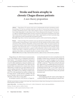
International Journal of Cardiology 149 (2011) 310–314
Contents lists available at ScienceDirect
International Journal of Cardiology
j o u r n a l h o m e p a g e : w w w. e l s ev i e r. c o m / l o c a t e / i j c a r d
Biodistribution of bone marrow mononuclear cells in chronic chagasic
cardiomyopathy after intracoronary injection
Lea Mirian Barbosa da Fonseca a,d,⁎, Sérgio S. Xavier a,b,e, Paulo Henrique Rosado de Castro a,d,
Ronaldo S.L. Lima a,e, Bianca Gutfilen a,d, Regina C.S. Goldenberg c,f, Angelo Maiolino a,g,
Claudia L.R. Chagas a,d, Roberto C. Pedrosa a,e, Antonio Carlos Campos de Carvalho c,f
a
Hospital Universitário Clementino Fraga Filho, Universidade Federal do Rio de Janeiro, Rio de Janeiro, Brazil
Instituto Evandro Chagas, Fundação Oswaldo Cruz (FIOCRUZ), Rio de Janeiro, Brazil
c
Instituto de Biofísica Carlos Chagas Filho, Universidade Federal do Rio de Janeiro, Rio de Janeiro, Brazil
d
Department of Radiology, Hospital Universitário Clementino Fraga Filho, Universidade Federal do Rio de Janeiro, Rio de Janeiro, Brazil
e
Department of Cardiology, Hospital Universitário Clementino Fraga Filho, Universidade Federal do Rio de Janeiro, Rio de Janeiro, Brazil
f
Laboratório de Cardiologia Celular e Molecular, Instituto de Biofísica Carlos Chagas Filho, Universidade Federal do Rio de Janeiro, Rio de Janeiro, Brazil
g
Department of Hematology, Hospital Universitário Clementino Fraga Filho, Universidade Federal do Rio de Janeiro, Rio de Janeiro, Brazil
b
a r t i c l e
i n f o
Article history:
Received 23 February 2009
Received in revised form 6 October 2009
Accepted 6 February 2010
Available online 2 March 2010
Keywords:
Homing
Cell therapy
Intracoronary infusion
Myocardial Scintigraphy
Chagasic cardiomyopathy
a b s t r a c t
Background: Animal and human clinical studies have indicated that bone marrow (BM) mononuclear cell
(MNC) therapy for Chagasic Cardiomyopathy (ChC) is feasible, safe and potentially efficacious. Nevertheless,
little is known about the retention of these cells after intracoronary (IC) infusion.
Methods: Our study investigated the homing of technetium-99m (99mTc) labeled BM MNCs and compared it
to thallium-201 (201Tl) myocardial perfusion images using the standard 17-segment model. Six patients with
congestive heart failure of chagasic etiology were included.
Results: Scintigraphic images revealed an uptake of 5.4% ± 1.7, 4.3% ± 1.5 and 2.3% ± 0.6 of the total infused
radioactivity in the heart after 1, 3 and 24 h, respectively. The remaining activity was distributed mainly to
the liver and spleen. Of 102 segments analyzed, homing took place in 36%. Segments with perfusion had
greater homing (58.6%) than those with decreased or no perfusion (6.8%), p b 0.0001. There was no
correlation between the number of injected cells and the number of segments with homing for each patient
(r = − 0.172, p = 0.774).
Conclusions: These results indicate that 99mTc-BM MNCs delivered by IC injection homed to the chagasic
myocardium. However, cell biodistribution was heterogeneous and limited, being strongly associated with
the myocardial perfusion pattern at rest. These initial data suggest that the IC route may present limitations
in chagasic patients and that alternative routes of cell administration may be necessary.
© 2010 Elsevier Ireland Ltd. All rights reserved.
1. Introduction
Chronic Chagas disease is a consequence of infection by the
protozoan parasite Trypanosoma cruzi and is endemic in certain
regions of Latin America. Recent estimates from the World Health
Organization indicate that 18 million persons are chronically infected
and 200.000 new cases occur each year [1]. Chronic cardiomyopathy
is the most important and severe manifestation of human Chagas
disease, affecting approximately 30% of infected patients [2,3].
Patients with end-stage Chagas disease have long been recognized
⁎ Corresponding author. Hospital Universitário Clementino Fraga Filho, Departamento
de Radiologia, subsolo, Universidade Federal do Rio de Janeiro, Ilha do Fundão, 21949-913,
Rio de Janeiro, Brazil. Tel./fax: +55 21 2562 2339.
E-mail address: [email protected] (L.M. Barbosa da Fonseca).
0167-5273/$ – see front matter © 2010 Elsevier Ireland Ltd. All rights reserved.
doi:10.1016/j.ijcard.2010.02.008
to have a low survival rate and presently there are no effective
therapies other than heart transplantation [2]. In this setting, the use
of cell therapies may offer an alternative for these patients.
Experimental models in mice with ChC have suggested BM MNC
therapy has beneficial effects [4,5] and human clinical studies have
indicated that BM MNC therapy is feasible, safe and potentially
efficacious [6]. Nevertheless, little is known about the homing,
retention and mechanisms of action of these cells. In ChC, myocardial
perfusion defects, fixed or reversible, have been described and
attributed to microvascular disease and fibrosis [7–10]. These
perfusion abnormalities could affect the biodistribution of BM MNCs
after IC injection, limiting their homing and their potential benefits.
Therefore, using labeled cells we investigated the biodistribution
pattern of BM MNCs after IC injection in chagasic patients, and
compared it to 201Tl myocardial perfusion scintigraphy.
L.M. Barbosa da Fonseca et al. / International Journal of Cardiology 149 (2011) 310–314
2. Materials and methods
2.1. Patients
Six patients with congestive heart failure of chagasic etiology that remained in New York
Heart Association class III despite optimized treatment were included in this study. The
general characteristics of the patients are described in Table 1. They had two positive antitrypanosoma cruzi serological reactions by three distinct techniques (indirect hemaglutination, indirect immunofluorescence and enzyme linked immunosorbent assay). Uni and
bidimensional Doppler echocardiographic analysis were performed in all patients prior to
cell therapy. Echocardiographic views were obtained as recommended by the American
Society of Echocardiography (ASE) [11]. Left ventricular systolic function was evaluated by
the ejection fraction, estimated by Simpson's method [11]. The coronariography was normal
in all patients. The study was conducted according to the guidelines of the local ethics
committee and of the National Research Ethics Committee, and informed consent was
obtained from all patients.
2.2. Cell harvesting, labeling and injection
Cells were obtained by marrow aspiration of the posterior iliac crest (50–80 ml) and
processed by Ficoll density centrifugation at 400 ×g for 30 min (Ficoll 1.077, 1:2,
Amersham Biosciences). After washing, cells were resuspended in 20 ml of saline solution
with 5% autologous serum. Five ml (25%) of this solution was used for labeling with 99mTc
as previously described [12–18]. In short, a saline solution of SnCl2 was added to the cell
suspension in 0.9% NaCl and the mixture incubated at room temperature for 10 min. Then,
45 mCi 99mTc were added and the incubation continued for another 10 min. After
centrifugation (500×g for 5 min), the supernatant was removed and the cells were
washed again in saline solution. The pellet was resuspended in saline solution.
Labeling efficiency was always N90%. Cell-bound activity ranged from 148 to
444 MBq (4–12 mCi). 99mTc-labeled cells demonstrated viability N 95% as assessed by
the trypan blue exclusion test. Cells labeled as described above were added back to the
total mononuclear cell suspension (final volume of 20 ml) and slowly injected into the
left (50%), right (25%) and left circumflex (25%) arteries, via an angioplasty catheter.
2.3. Cell characterization
A small aliquot of the mononuclear cells was used for immunophenotipic
characterization of the injected cell population. Flow cytometry analysis was
performed on a BD FacsCanto (BD Biosciences, USA) using antibodies (CD34, CD45,
CD73, CD90, CD105, CD133 — all from BD Biosciences) that allowed identification of
hematopoietic stem cells (HSCs), mesenchymal stem cells (MSCs) and endothelial
progenitor cells (EPCs). The injected mononuclear fraction contained a mean of 1.56%
(range 0.9 to 3.30) HSCs (Ishage Protocol), 0.01% (range 0.003 to 0.011) MSCs and
0.01% EPCs (range 0.003 to 0.013).
311
used to compare the proportion of cell homing in segments with and without perfusion.
The significance level adopted was 5%. After evaluating the pattern of distribution by
the Kolmogorov–Smirnov Test, the correlation between the number of segments with
homing and the total number of injected cells, as well as the number of CD34+, MSC
and EPC cell populations, was analyzed by Pearson's product-moment correlation
coefficient.
3. Results
The general characteristics of the patients, as well as the number of cells injected in
each patient are shown in Table 1. Scintigraphic images revealed an uptake of 5.4% ± 1.7,
4.3%± 1.5 and 2.3%± 0.6 of the total infused radioactivity in the heart after 1, 3 and 24 h,
respectively. The remaining activity was distributed mainly to the liver, spleen, kidneys
and bladder in all time points. Intestinal uptake was observed 24 h after IC infusion. Fig. 1
illustrates whole body planar views of a representative patient. 201Tl SPECT and 99mTc-BM
MNC SPECT images allowed us to compare perfusion and cell homing in all patients. Long-,
vertical- and short-axis views of the myocardium revealed a decreased uptake of 201Tl in
inferior, inferolateral and apical walls in all patients, corresponding to the areas of
hypokinesis on the echocardiograms. 99mTc-BM MNC SPECT indicated a very heterogeneous uptake, but for all patients there was lack of or decreased uptake of 99mTc-BM MNC
in walls with perfusion defects. Fig. 2 shows this comparison for two representative
patients. Fig. 3 illustrates Polar maps for 201Tl and 99mTc-BM MNC of a representative
patient.
Furthermore, we performed a segmental analysis in all patients comparing perfusion
and cell homing. In the 201Tl study perfusion defects were identified in 44 (43%) of 102
segments analyzed. The perfusion pattern varied according to the myocardial segment
(Fig. 4A). Taken together, the analysis of all 6 patients indicated that perfusion defects were
more frequent in segments 4 (basal inferior), 5 (basal inferolateral), 10 (mid inferior), 15
(apical inferior) and 17 (apical). The study of cardiac biodistribution of the 99mTc BM MNC
showed cell homing in only 37 (36%) of 102 segments analyzed. Cell homing varied
according to the myocardial segment (Fig. 4B). Lack of homing was more frequent in
segments 1 (basal anterior), 4 (basal inferior), 5 (basal inferolateral), 10 (mid inferior), 15
(apical inferior).
The analysis of the myocardial perfusion pattern and cell homing for each patient is
shown in Fig. 5. For each patient the number of segments with or without perfusion and
with or without cell homing was quantified and plotted in Fig. 5A and B, respectively. There
was a strong association between perfusion and cell homing. Of 58 segments with
perfusion, homing took place in 34 (58.6%) Of 44 segments without perfusion, homing
took place in only 3 (6.8%), p b 0.0001 as seen in Fig. 6. The analysis of interobserver
variability showed a kappa of 0.529 (p = 0.000) for perfusion and 0.455 (p = 0.000) for cell
homing. Also, the correlation coefficient between the number of injected cells and the
2.4. Myocardial imaging
Myocardial 201Tl and 99mTc-BM MNC images were acquired with a dual-head
gamma camera (Millennium MG GE Medical Systems, Milwaukee, WI). 201Tl
myocardial perfusion Single Photon Emission Computed Tomography (SPECT) was
performed at rest prior to IC cell therapy [19]. 99mTc-BM MNC planar and SPECT images
were obtained 1, 3 and 24 h after IC infusion in all patients. Uptake was defined as the
percentage of myocardium-originated number of counts, compared to the total number
of counts of radiolabeled cells [19]. Short-axis, horizontal long-axis and vertical longaxis views for 201Tl and 99mTc-BM MNC SPECT were obtained for each patient [20,21].
Perfusion and cell homing data were also transferred to polar maps of 17 segments for
each patient [20,21]. The image analysis was performed by 2 independent observers,
without knowledge of clinical data.
2.5. Statistical analysis
Interobserver agreement to define the segmental biodistribution of labeled cells
and perfusion with 201Tl was evaluated using the Kappa test. The chi-square test was
Table 1
Patients characteristics.
Patient
Age
(years)
Gender
NYHA
Class
LVEF at baseline
(%)
Number of injected cells
(×108)
1
2
3
4
5
6
53
56
62
59
49
60
M
F
M
M
M
M
III
III
III
III
III
III
27
29
32
23
28
24
5
9.5
9.6
1
3.6
2
Abbreviations: M = male; F = female; LVEF=Left Ventricular Ejection Fraction by
Echocardiogram.
Fig. 1. Anterior views of whole body scintigraphy of a representative patient (Patient 1)
carried out 1 (A), 3 (B) and 24 h (C) after cell infusion in the coronary arteries show the
biodistribution of BM MNCs labeled with 99mTc. Black, no uptake; blue-red-yellow-white,
increasing uptake.
312
L.M. Barbosa da Fonseca et al. / International Journal of Cardiology 149 (2011) 310–314
Fig. 2. Comparison of 201Tl SPECT demonstrating perfusion and 99mTc-BM MNC SPECT demonstrating homing of two representative patients (A — patient 2, B — patient 3) carried out
1 hour after cell infusion in the coronary arteries. SA=Short Axis; VLA=Vertical Long Axis; HLA=Horizontal Long Axis. Black, no uptake; blue-red-yellow-white, increasing uptake.
number of segments with homing was low or even negative (r = −0.172), with a
nonsignificant p value (p = 0.774), as seen in Fig. 7. Likewise, correlation coefficients
between the number of HSCs (CD34+), MSCs (CD34−/CD45−/CD73+/CD90+/CD105+) or
EPCs (CD34+/CD45−/CD133+) and the number of segments with homing did not reach
statistical significance.
4. Discussion
Experimental models involving the injection of bone marrow
derived cells in cardiac diseases have focused mainly in ischemic
models and demonstrated an appreciable improvement in myocardial
function that has recently been attributed to the cardioprotective or
proangiogenic effects of secreted paracrine factors [22,23]. In humans,
the available evidence suggests that therapy with BM MNCs seems safe
and associated with modest improvements in anatomic and physiologic
parameters in patients with both acute myocardial infarction and
chronic ischemic heart disease, above and beyond conventional therapy
[24–30].
Chagasic cardiomyopathy is not a regional disease of the heart as
are the ischemic syndromes. It affects the heart globally and it is
characterized by an intense inflammatory infiltrate and fibrosis. In
ChC, experimental models have been developed in mice and BM
MNCs from control or Trypanosoma cruzi chronically infected mice
have been used significantly decreasing inflammation and fibrosis for
as long as 6 months after cell transplantation [4]. Another study using
magnetic resonance imaging indicated BM MNC therapy resulted in a
Fig. 3. Display, on a circumferential polar plot, of the 17 myocardial segments — 1: basal anterior; 2: basal anteroseptal; 3: basal inferoseptal; 4: basal inferior; 5: basal inferolateral; 6: basal
anterolateral; 7:mid anterior; 8: mid anteroseptal; 9: mid inferoseptal; 10: mid inferior; 11: mid inferolateral; 12: mid anterolateral; 13: apical anterior; 14: apical septal; 15: apical inferior;
16: apical lateral; 17: apex (A). Polar maps of 201Tl SPECT (B) and 99mTc-BM MNC SPECT (C) from a representative patient (Patient 5). Black, no uptake; blue-red-yellow-white, increasing
uptake.
L.M. Barbosa da Fonseca et al. / International Journal of Cardiology 149 (2011) 310–314
313
Fig. 4. Analysis of myocardial perfusion (A) and of cell homing (B) for each of the 17 myocardial segments.
Fig. 5. Analysis of the myocardial perfusion pattern (A) and analysis of cellular homing
(B) of each patient.
reported [6]. Nonetheless, many questions remain unanswered about
the homing, retention and possible mechanisms by which cell therapy
may act in this disease [31].
In the present study we begin to address some of these questions by
labeling cells with 99mTc to track them and investigate their homing and
short time retention in the chagasic myocardium. Over the past fifteen
years, we have developed and used a simple technique for the labeling of
mononuclear leukocytes with 99mTc. It has been used for visualization of
infection, osteomyelitis, graft rejection and fever of unknown origin
[11–14]. When compared to techniques such as 99mTc-hexamethyl
propylene-amine oxime (99mTc-HMPAO) labeling, there are advantages
such as smaller amount of blood required, shorter performing time and
lower cost [12–15]. Based on this experience, we have developed a new
and simple technique for labeling BM MNCs and BM MSCs with 99mTc
[16–18]. The 6-hour half-life of 99mTc allows the monitoring of cell
distribution for approximately 24 h, which is an important advantage
over the half-life of 110 min of 18F-fluorodeoxyglycose (FDG). In
comparison with indium-111 oxine, another commonly used radiopharmaceutical, 99mTc results in better image resolution and a lower
radiation burden to the patient [12–15,32].
A case report had previously indicated cardiac homing of 99mTcHMPAO labeled BM MNCs 2 h after their injection in one patient with ChC
[33]. However, since 99mTc HMPAO-labeled cells were administered only
into the anterior descending artery [33], this could have accounted for a
Fig. 6. Cell homing according to the myocardial perfusion pattern of all segments of the
6 patients. There was a strong association between perfusion and cell homing.
Fig. 7. Correlation between the number of injected cells and the number of segments
with homing analyzed by Pearson's product-moment correlation coefficient.
significant reduction of the right ventricular dilatation typically
observed in the chagasic mouse model [5]. In humans, a phase I
clinical trial demonstrated that BM MNC IC cell injection in patients
with ChC is feasible, safe and potentially efficacious, as recently
314
L.M. Barbosa da Fonseca et al. / International Journal of Cardiology 149 (2011) 310–314
limitation in the homing of BM MNCs. In our protocol, the labeled cells
were injected into the three main arteries, in accordance to clinical trials
involving IC BM MNC injection for ChC that don't evaluate cell homing
[6,34]. In addition to the quantification of labeled cells, an analysis using
the 17-segment model allowed a more adequate comparison between
cell homing and myocardial perfusion.
In our study, cell homing was detectable in the heart up to 24 h after IC
delivery, which is the longest possible time interval to track cells labeled
by 99mTc. However, myocardial homing was found to be heterogeneous
and limited. All patients presented extensive perfusion defects mainly in
inferior, inferolateral and apical myocardial walls, where cell homing was
absent or reduced, despite adequate injection in the corresponding
coronary arteries. These perfusion deficits are consistent with previous
reports on ChC and have been attributed to the presence of fibrosis and
microvascular disease, already demonstrated in nuclear medicine,
echocardiographic and anatomic pathology studies [7–10]. It is possible
that these perfusion abnormalities may restrict cell homing, and this
could represent a limitation to IC administration of BM MNCs in chagasic
patients with greater perfusion defects, as the ones in this study. On the
other hand, chagasic patients with less perfusion defects may have greater
homing to the myocardium after IC injection. As for patients with more
extensive perfusion defects, it is possible that other approaches may be
necessary, such as intramyocardial (IM) injections. There are no studies
comparing different techniques in ChC, but recent reports in experimental
myocardial infarction models with peripheral blood MNCs [35], BM MSCs
[36] and BM MNCs [37] have suggested that IM route may achieve higher
myocardial retention rates when compared to IC infusion.
Our study provides for the first time quantification of BM MNC
retention for up to 24 h after IC injection and indicates that cell homing
may be decreased in myocardial regions with perfusion deficits in the
setting of Chronic Chagasic Cardiomyopathy. These initial data suggest
that the IC route may present limitations in chagasic patients and that
alternative routes of cell administration may be necessary. Nevertheless,
further experimental and clinical studies are warranted and, to this end,
the field of nuclear cardiology may play an important role in the
evaluation of parameters such as cell tracking, cell biodistribution and
its correlation to myocardial perfusion/viability in Chagas disease.
Acknowledgements
This work was supported by grants from the Research Support
Foundation of the State of Rio de Janeiro (to BG and ACCC) and the
Brazilian National Council of Technological and Scientific Development
(to ACCC).
The authors of this manuscript have certified that they comply
with the Principles of Ethical Publishing in the International Journal of
Cardiology [38].
References
[1] Control of Chagas' disease: second report of the WHO expert committee. Technical
Report Series 905. Geneva, Switzerland: World Health Organization; 2002.
[2] Rocha MO, Teixeira MM, Ribeiro AL. An update on the management of Chagas
cardiomyopathy. Expert Rev Anti Infect Ther 2007;5:727–43.
[3] Bilate AM, Cunha-Neto E. Chagas disease cardiomyopathy: current concepts of an
old disease. Rev Inst Med Trop Sao Paulo 2008;50:67–74.
[4] Soares MBP, Lima RS, Rocha LL, et al. Transplanted bone marrow cells repair heart
tissue and reduce myocarditis in chronic chagasic mice. Am J Pathol 2004;164:
441–7.
[5] Goldenberg RC, Jelicks LA, Fortes FS, et al. Bone marrow cell therapy ameliorates and
reverses chagasic cardiomyopathy in a mouse model. J Infect Dis 2008;197:544–7.
[6] Vilas-Boas F, Feitosa GS, Soares MB, et al. Early results of bone marrow cell
transplantation to the myocardium of patients with heart failure due to Chagas
disease. Arq Bras Cardiol 2006;87:3–9.
[7] Rossi MA, Ramos SG. Coronary microvascular changes in Chagas' disease. Arq Bras
Cardiol 1996;66:169–72.
[8] Simoes MV, Pintya AO, Bromberg-Marin G, et al. Relation of regional sympathetic
denervation and myocardial perfusion disturbance to wall motion impairment in
Chagas' cardiomyopathy. Am J Cardiol 2000;86:975–81.
[9] Marin-Neto JA, Cunha-Neto E, Maciel BC, Simoes MV. Pathogenesis of chronic
Chagas heart disease. Circulation 2007;115:1109–23.
[10] Acquatella H. Echocardiography in Chagas heart disease. Circulation 2007;115:
1124–31.
[11] Gottdiener JS, Bednarz J, Devereaux R, et al. American Society of Echocardiography. American Society of Echocardiography recommendations for use of
echocardiography in clinical trials. J Am Soc Echocardiogr 2004;17:1086–119.
[12] Gutfilen B, Pellini MP, de Roure, et al. 99mTc labeling white blood cells with a
simple technique: clinical application. Ann Nucl Med 1994;8:85–9.
[13] Gutfilen B, Rossini A, Martins FP, da Fonseca LM. Tc-99m-leukocytes—is it an
intracellular labeling? J Clin Lab Immunol 1999;51:1–7.
[14] Souza SA, Fonseca LM, Gonçalves RT, et al. Diagnosis of renal allograft rejection
and acute tubular necrosis by 99mTc-mononuclear leukocyte imaging. Transplant
Proc 2004;36:2997–3001.
[15] Gutfilen B, Lopes de Souza SA, Martins FP, et al. Use of 99mTc-mononuclear
leukocyte scintigraphy in nosocomial fever. Acta Radiol 2006;47:699–704.
[16] Carvalho AB, Quintanilha LF, Dias JV, et al. Bone marrow multipotent mesenchymal stromal cells do not reduce fibrosis or improve function in a rat model of
severe chronic liver injury. Stem Cells 2008;26:1307–14.
[17] Quintanilha LF, Mannheimer EG, Carvalho AB, et al. Bone marrow cell transplant does
not prevent or reverse murine liver cirrhosis. Cell Transplant 2008;17:943–53.
[18] Barbosa da Fonseca LM, Battistella V, de Freitas GR, et al. Early tissue distribution of
bone marrow mononuclear cells after intra-arterial delivery in a patient with
chronic stroke. Circulation 2009;120:539–41.
[19] Hansen CL, Goldstein RA, Akinboboye OO, et al. Myocardial perfusion and
function: single photon emission computed tomography. J Nucl Cardiol 2007;14:
e39–60.
[20] Goussetis E, Manginas A, Koutelou M, et al. Intracoronary infusion of CD133+ and
CD133-CD34+ selected autologous bone marrow progenitor cells in patients with
chronic ischemic cardiomyopathy: cell isolation, adherence to the infarcted area,
and body distribution. Stem Cells 2006;24:2279–83.
[21] Cerqueira MD, Weissman NJ, Dilsizian V, et al. Standardized myocardial
segmentation and nomenclature for tomographic imaging of the heart. A
Statement for Healthcare Professionals from the Cardiac Imaging Committee of
the Council on Clinical Cardiology of the American Heart Association. Circulation
2002;105:539–42.
[22] Smart N, Riley PR. The stem cell movement. Circ Res 2008;102:1155–68.
[23] Passier R, van Laake LW, Mummery CL. Stem-cell-based therapy and lessons from
the heart. Nature 2008;453:322–9.
[24] Perin EC, Dohmann HF, Borojevic R, et al. Transendocardial, autologous bone
marrow cell transplantation for severe, chronic ischemic heart failure. Circulation
2003;107:2294–302.
[25] Perin EC, Dohmann HF, Borojevic R, et al. Improved exercise capacity and ischemia
6 and 12 months after transendocardial injection of autologous bone marrow
mononuclear cells for ischemic cardiomyopathy. Circulation 2004;110:213–8.
[26] Schachinger V, Erbs S, Elsasser A, et al. Intracoronary bone marrow-derived
progenitor cells in acute myocardial infarction. N Engl J Med 2006;355:1210–21.
[27] Lunde K, Solheim S, Aakhus S, et al. Exercise capacity and quality of life after
intracoronary injection of autologous mononuclear bone marrow cells in acute
myocardial infarction: results from the Autologous Stem cell Transplantation in
Acute Myocardial Infarction (ASTAMI) randomized controlled trial. Am Heart J
2007;154(710):e1–8.
[28] Abdel-Latif A, Bolli R, Tleyjeh IM, et al. Adult bone marrow-derived cells for cardiac
repair: a systematic review and meta-analysis. Arch Intern Med 2007;167:989–97.
[29] Lipinski MJ, Biondi-Zoccai GG, Abbate A, et al. Impact of intracoronary cell therapy on
left ventricular function in the setting of acute myocardial infarction: a collaborative
systematic review and meta-analysis of controlled clinical trials. J Am Coll Cardiol
2007;50:1761–7.
[30] Martin-Rendon E, Brunskill SJ, Hyde CJ, Stanworth SJ, Mathur A, Watt SM.
Autologous bone marrow stem cells to treat acute myocardial infarction: a
systematic review. Eur Heart J 2008;29:1807–18.
[31] Soares MB, Garcia S, Campos de Carvalho AC, et al. Cellular therapy in Chagas'
disease: potential applications in patients with chronic cardiomyopathy. Regen
Med 2007;2:257–64.
[32] Peters AM. A brief history of cell labelling. Q J Nucl Med Mol Imaging 2005;49:304–7.
[33] Jacob JL, Salis FV, Ruiz MA, Greco OT. Labeled stem cells transplantation to the
myocardium of a patient with Chagas' disease. Arq Bras Cardiol 2007;89:e10–1.
[34] Tura BR, Martino HF, Gowdak LH, et al. Multicenter randomized trial of cell
therapy in cardiopathies—MiHeart Study. Trials 2007;8:2.
[35] Hou D, Youssef EA, Brinton TJ, et al. Radiolabeled cell distribution after
intramyocardial, intracoronary, and interstitial retrograde coronary venous
delivery: implications for current clinical trials. Circulation 2005;112:I150–6.
[36] Perin EC, Silva GV, Assad JA, et al. Comparison of intracoronary and transendocardial delivery of allogeneic mesenchymal cells in a canine model of acute
myocardial infarction. J Mol Cell Cardiol 2008;44:486–95.
[37] George JC, Goldberg J, Joseph M, et al. Transvenous intramyocardial cellular
delivery increases retention in comparison to intracoronary delivery in a porcine
model of acute myocardial infarction. J Interv Cardiol 2008;21:424–31.
[38] Coats AJ. Ethical authorship and publishing. Int J Cardiol 2009;131:149–50.
Baixar