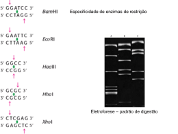
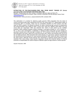
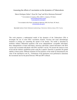
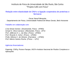
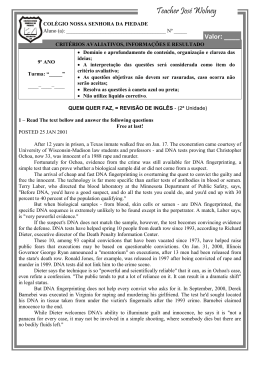
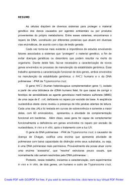
Brazilian Journal of Genetics Online version ISSN 16784502 Braz. J. Genet. vol. 20 no. 4 Ribeirão Preto Dec. 1997 Services on Demand Article Article in xml format http://dx.doi.org/10.1590/S010084551997000400030 SHORT COMMUNICATION How to cite this article Hb Köln [2298(FG5) valmet] identified by DNA analysis in a Brazilian family Silvia R.P. Miranda1, Silvana F. Fonseca 2, Maria S. Figueiredo 2, Myoko Yamamoto 2, Helena Z.W. Grotto1, Sara T.O. Saad 1 and Fernando F. Costa1 1 Departamento de Clínica Médica and Departamento de Patologia Clínica, Faculdade de Ciências Médicas, Universidade Estadual de Campinas, Caixa Postal 6111, 13083970 Campinas, SP, Brasil. Phone: (019) 2397866; Fax: (019) 2393114. Email: [email protected]. Send correspondence to F.F.C. 2 Departamento de Medicina, Universidade Federal de São Paulo, São Paulo, SP, Brasil. Article references Curriculum ScienTI Automatic translation Send this article by email Indicators Cited by SciELO Access statistics Related links Share More More Permalink ABSTRACT Hb Köln was identified by DNA analysis in a Brazilian patient. A fouryear old Brazilian female, with jaundice since birth, presented an abnormal band, between A2 and S, in hemoglobin electrophoresis on a cellulose acetate membrane, and a band with electrophoretic migration similar to Hb C on agar gel. Thermic instability and isopropanol precipitation tests were positive. Heinz bodies were observed in the patient s peripheral blood. Sequencing of the three exons of the globin gene detected a transition from G to A in the first position of codon 98. This alteration does not create or abolish any known restriction site. In this case, confirmation of the mutation was accomplished by allelespecific oligonucleotide hybridization, which is a simple and fast identification method when the clinical data and hematological and electrophoretic patterns are suggestive of Hb Köln. INTRODUCTION Hemoglobin (Hb) Köln, in which the 98 (FG5) valine residue is replaced by methionine, is the most frequent unstable hemoglobin; however, it presents very few features that allow its distinction from other unstable Hb without performing structural protein analysis (Carrell et al., 1966; Miller et al., 1971; Pedersen et al., 1973). Mild anemia, jaundice and persistent excretion of dark urine characterize the clinical manifestations of the Hb Köln (Fairbanks et al., 1969; Lolskin and Miller, 1976). Splenectomy diminishes the anemia and improves the clinical course of the disease (Scott et al, 1960; Vaughan et al., 1967). The relation between the amino acid substitution (valmet) and the instability of this Hb suggests that the valine residue is in contact with the heme in the normal chain. The substitution of valine for methionine (position FG5) changes the steric configuration of the globin and either alters the enzymatic reduction of iron or results in an increased conversion of metahemoglobin into denatured hemoglobin (Harley and Mauer, 1961). When Hb Köln is in the oxygenated conformation, the molecule is saturated with heme groups, and it is stable. The transition to the deoxygenated state results in the loss of heme groups, instability, precipitation and formation of Heinz bodies, which are cytoplasmic degradation products (denatured globin and hemichromes) attached to the cell membrane (Eisenger et al., 1985). To our knowledge, three cases of Hb Köln were described in Brazil, and in all of them the identification was presumptive or carried out by protein analysis (Naoum, 1993a,b). We describe a fouryear old Brazilian female patient with Hb Köln identified by DNA analysis. MATERIAL AND METHODS The patient was a fouryear old white female, probably of Portuguese descent, seen at the Hematology Clinic of Universidade Estadual de Campinas and Universidade Federal de São Paulo. She presented jaundice since birth. The family included two other members with anemia, her father who was splenectomized 20 years ago, and her aunt who presented splenomegaly. The red blood cell (RBC) count, Hb concentration, and mean cell volume (MCV) were determined electronically (Coulter Counter S.Sr model T890). HbA2 values were measured spectrophotometrically after elution from cellulose acetate strips, following electrophoresis (MarengoRowe, 1965), and the Hb F value was determined by alkali denaturation (Pembrey et al., 1972). Thermic instability and isopropanol precipitation tests were performed by standard procedures (Dacie and Lewis, 1984). Genomic DNA was obtained from peripheral leukocytes using standard methods (Sambrook et al., 1989). A double strand DNA Cycle Sequencing System (GibcoBRL, Gaithersburg, MD, USA) was used for sequence analysis according to the manufacturer s instructions. The primers used for amplification and sequencing of the three exons of the globin gene are described in Table I and Figure 1. Table I Primer sequences used for amplification and sequencing of the three exons of the b globin gene. Primers Sequence 5 3 Direction Position 1 GCCAAGGACAGGTACGGCTGTCATC S 140 2 TTTGCTTCTGACACAACTGT S +2 3 AGACAGAGAAGACTCTTG S +198 4 CTCATGGCAAGAAAGTGCTC S +348 5 CCCTTCTTCCTATGACATGAACTTAACCAT A +521 6 AATCCAGCTACCATTCTGC S +1214 7 TCCAGATGCTCAAGGCCCTTC A +1556 S Sense; A antisense; Position position in relation to the Cap site. The allelespecific oligonucleotide hybridization was performed using DNA fixed in nylon membranes and hybridized with the normal (5 CTCAGGATCCACGTGCCGC3") and with the mutant (5 CTCAGGATCCATGTGCCGC o 3") radioactive labeled sequences. The membranes were incubated for 2 h at 48 C in the prehybridization solution (5x SSPE, 5x Denhardt s, 0.5% SDS), followed by the addition of the denatured probes. The hybridization was conducted for 16 h at 48oC and was followed by two washes with 2x SSPE, 0.1% SDS at room temperature (5 min and 10 min, respectively), and one wash with 5x SSPE, 0.15% SDS at 62oC for 5 min. RESULTS The hematological parameters showed mild hemolytic anemia in the patient and the abnormal hemoglobin in all family members studied (Table II). Table II Hematological and hemoglobin data of the patient and two relatives. Hb (g/dl) Ht (%) RBC (x1012/l) MCV (fl) MCH (pg) HbF (%) HbA2 (%) Hb Elect X(%) 1 10.3 37 4.0 103 32.0 2.8 4.7 7.6 2 14.9 48 4.5 104 33.0 1.7 2.2 10.4 3 12.0 41 3.9 104 30.5 1.6 2.2 6.5 1 Patient; 2 father; 3 aunt; Hb = hemoglobin; Ht = hematocrit; RBC = red blood cells; MCV = medium corpuscular volume; MCH = medium corpuscular hemoglobin; Hb F = fetal hemoglobin; Hb Elect. = hemoglobin electrophoresis; X = abnormal Hb. The variant was initially detected as an abnormal band, which migrated between A2 and S, in electrophoresis on a cellulose acetate membrane. The migration pattern was similar to Hb C on agar gel (sodium citrate buffer, pH 6.1). Thermic instability and isopropanol precipitation tests were positive. The three exons of the globin gene were sequenced. A 367bp fragment of exon 2 (amplified with primers 3 and 5; Figure 1) was sequenced with an internal primer (primer 4; Figure 1). There were two bands at the first position of the codon 98 (Figure 2), at the same height (G and A), clearly showing the presence of two nucleotides in that region. The finding represents a transition G ® A, and also indicates that besides the codon GTG, which codifies for valine, the patient has the codon ATG, which codifies for methionine, indicative of Hb Köln. The result of the oligonucleotide allelespecific hybridization assay demonstrated that the patient is heterozygous for Hb Köln (Figure 3). DISCUSSION Characterization of unstable hemoglobins by protein analysis is sometimes difficult. However, it is facilitated by DNA analysis, specially in the case of hyperunstable chain variants. The use of direct sequencing from PCR amplified fragments, without the cloning steps, simplifies and speeds up the process. The establishment of an oligonucleotide allelespecific hybridization protocol, since the alteration present in Hb Köln (GTG ® ATG) does not create or abolish any known restriction site, allows confirmation of this mutation, and also offers a simple and fast identification method when the clinical data and hematological and electro phoretic patterns are suggestive of Hb Köln. The case described in this paper represents another example of Hb Köln in the Brazilian population and the first one in which the mutation was demonstrated by DNA analysis. ACKNOWLEDGMENTS This work was supported in part by grants from CNPq, Hemocentro (UNICAMP) and FAPESP (Brazil). Publication supported by FAPESP. RESUMO Hb Köln resulta da substituição do aminoácido valina por metionina na posição 98 da cadeia da hemoglobina. Embora seja a mais freqüente das hemoglobinas instáveis, possui poucas características que permitem distinguíla sem a realização de análise estrutural da molécula. Com o objetivo de desenvolver um procedimento alternativo à análise protéica, este trabalho descreve a identificação da Hb Köln através de análise de DNA. Uma paciente branca, sexo feminino, 4 anos de idade, com icterícia desde o nascimento, foi analisada, apresentando uma banda anômala entre A2 e S em eletroforese em acetato de celulose, e migração eletroforética semelhante a Hb C em gel de agar. Os testes de instabilidade térmica e de precipitação pelo isopropanol foram positivos e foram observados corpúsculos de Heinz no sangue periférico. Os três exons do gene da globina foram seqüenciados e uma transição de G ® A foi identificada na primeira posição do codon 98. Essa alteração não cria ou abole nenhum sítio conhecido de enzima de restrição. Desta forma, a confirmação da mutação foi levada a efeito através de hibridização com oligonucleotídeos aleloespecíficos, o que permitiu o estabelecimento de um método simples e rápido de identificação de novos casos quando os dados clínicos e o padrão hematológico e eletroforético sugerem Hb Köln. REFERENCES Carrell, R.W., Lehmann, H. and Hutchison, H.E. (1966). Haemoglobin Köln ( 98 ValineMethionine): an unstable protein causing inclusion body anaemia. Nature 210: 915916. [ Links ] Dacie, J.V. and Lewis, S.M. (1984). Practical Haematology. Churchill Livingstone, Edinburg, New York. [ Links ] Eisenger, J., Flores, J., Tyson, J.A. and Shohet, S.B. (1985). Fluorescent cytoplasm and Heinz bodies of hemoglobin Köln erythrocytes: evidence for intracellular heme catabolism. Blood 65: 886893. [ Links ] Fairbanks, V.F., Opfell, R.W. and Burgert Jr., E.O. (1969). Three families with unstable hemoglobinopathies (Köln, Olmsted and Santa Ana) causing hemolytic anemia with inclusion bodies and pigmenturia. Am. J. Med. 46: 344359. [ Links ] Harley, J.D. and Mauer, A.M. (1961). Studies on the formation of Heinz bodies. II. The nature and significance of Heinz bodies. Blood 17: 418433. [ Links ] Lolskin, G.B. and Miller, D.R. (1976). Heme synthesis in hereditary hemolytic anemias: decrease of aminolevulinic synthetase in hemoglobin Köln disease. Pediat. Res. 10: 702706. [ Links ] MarengoRowe, A.J. (1965). Rapid electrophoresis and quantitation of haemoglobin on cellulose acetate. J. Clin. Pathol. 18: 790792. [ Links ] Miller, D.R., Weed, R.I., Stamatoyannopoulos, G. and Yoshida, A. (1971). Hemoglobin Köln disease occurring as a fresh mutation: erythrocyte metabolism and survival. Blood 38: 715729. [ Links ] Naoum, P.C., Domingos, C.R.B., Moreira, H.W. and Alvares Fo., F. (1993a). Hb Köln or 22 98 (FG5) ValMet in Brazil. 5th International Conference on Thalassemias and the Haemoglobinopathies. Nicosia, Cyprus. [ Links ] Naoum, P.C., Domingos, C.R.B., Moreira, H.W. and Alvares Fo., F. (1993b). Hb Köln or 22 98 (FG5) ValMet in Brazil. Simpósio Nacional de Hemoglobinopatias. São José do Rio Preto, SP, Brasil. [ Links ] Pedersen, P.R., McCurdy, P.R., Wrightstone, R.N., Wilson, J.B., Smith, L.L. and Huisman, T.H.J. (1973). Hemoglobin Köln in a black: pre and postsplenectomy red cell survival (DF32P and 51Cr) and the pathogenesis of hemoglobin instability. Blood 42: 771781. [ Links ] Pembrey, M.E., MacWade, P. and Weatherall, D.J. (1972). Reliable routine stimulation of small amounts of foetal hemoglobin by alkali denaturation. J. Clin. Pathol. 25: 738740. [ Links ] Sambrook, J., Fritsch, E.F. and Maniatis, T. (1989). Molecular Cloning. Cold Spring Harbor Laboratories, New York. [ Links ] Scott, J.L., Haut, A., Cartwright, G.E. and Wintrobe, M.M. (1960). Congenital hemolytic disease associated with red cell inclusion bodies, abnormal pigment metabolism and electrophoretic hemoglobin abnormality. Blood 16: 12391252. [ Links ] Vaughan Jones, R., Grimes, A.J., Carrel, R.W. and Lehman, H. (1967). Köln haemoglobinopathy: further data and a comparison with other hereditary Heinz bodies anaemias. Brit. J. Haematol. 13: 394408. [ Links ] (Received December 17, 1996) Figure 1 Diagram of the three exons of the b globin gene and primer positions (see Table I). The arrow indicates the position of the mutation codon 98, exon 2. Figure 2 Direct genomic sequencing of the globin gene exon 2 of the patient. The PCR fragment was amplified with the primers 3 (sense) and 5 (antisense). The sequencing reaction was conducted using primer 4 (see Figure 1). The asterisk (*) represents position +455 of the globin gene, relative to the Cap site, and indicates two bands at the same height (G A). This transition at codon 98 leads to substitution of the amino acid valine (GTG) for a methionine (ATG), which is described as Hb Köln. Figure 3 Allelespecific oligonucleotide hybridization. Genomic DNA from a normal control and the patient were amplified using primers 1 (sense) and 5 (antisense, see Figure 1). Upon fixation on nylon membranes, the PCR products were hybridized with probes corresponding to the normal (columns 1 to 6) and mutant (columns 7 to 12) alleles (see Material and Methods). B2 and B8 normal control DNA; C3 and C9 patient DNA. A positive signal is observed for the normal control DNA only in the membrane hybridized with the normal probe (columns 1 to 6). In the patient sample a positive signal is seen in both membranes, confirming the heterozygous nature of the mutation. Rua Capitão Adelmio Norberto da Silva, 736 14025670 Ribeirão Preto SP Brazil Tel. / Fax: +55 16 621.8540, 620.1251, 620.1253 [email protected]
Baixar