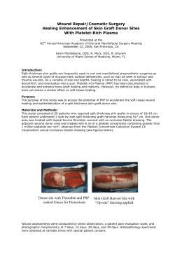
Learning
to Bask questions
A
C
By Dean Ripa , Ronaldo & Rodrigo C. G. de Souza
To E.G.M., who paid the Price, Cecília Haddad who opened the door, Polly Matzinger who brought
the Light and to Serendipity, our invisible guide.
"Computers are useless. They can only give you answers."
Pablo Picasso
Contacts:
[email protected]
[email protected]
[email protected]
INTRODUCTION
The road that leads to what we call today "modern snakebite treatment" is littered with myths, folklore and well
intended treatments created more out of desperation and compassion than reason and knowledge. Myths and
folklore tend to fade away with the scientific advance, although modern research in areas such as ethno-biology
are learning to separate wheat from chaff, examining folk knowledge with both an open mind and critical thinking.
At some point in history, each treatment was believed to be effective and beneficial to the patient, until it had to
face the crossroads the evolution of science always creates as new evidence gathers: prove itself or clear the way
for new approaches. This was the fate of the tourniquet, suction of the venom with the mouth, making cuts across
the wounded area and many others. Each of these methods, faced its crossroads and all took the wrong turn into
oblivion.
As an ever changing endeavor, in Science, even oblivion is relative. From time to time, these ancient techniques
are revisited and sometimes given new applications, such as the use of ice in Loxosceles spider envenomings.
Today, the slow gathering of in loco observations and new theoretical insights suggest that a common treatment
taught in all medical schools and common practice in virtually all emergency hospitals might be facing its
crossroads: the invasive management of wounds created by snake and spider envenomings.
A growing body of evidence seems to suggest that necrosis in snake and spider envenomings, specially when the
necrosis is long lasting, is the result of the body's own defensive system attacking and destroying itself as a
reaction to damage directly caused by toxins.
The body reacts to damage by activating healing and cleanup systems along with the immune system. It seems
that this first attempt to clean and heal is conservative or, in other words, it is not in itself damaging. If this first step
doesn't work, the second step will be to bring in the immune system's "big guns": the Delayed Type Hypersensitivity
response (Tumor Necrosis Factor, Gamma Interferon, activated macrophages, oxygen radicals, etc.).
In theory, this response could be also activated by aggressive wound management, resulting in a double whammy
attack to the body, which may result in a catastrophic necrosis in which the immune system will destroy large
amounts of tissue along with whatever pathogen it is fighting against. Since infection is such a common
intercurrence in these cases, this auto-immune reaction may easily be misinterpreted as an infection when none
exists, or may be overshadowed by a real infection.
Everyone has seen a movie about one of those clear cut police cases in which the perpetrator conveniently leaves
his DNA and fingerprints all over the crime scene. At the precinct, the suspect is identified and his rap sheet is
several yards long so, all that is left to do is lock him up and have a beer, felling pretty good about the day.
Sometimes, though, the crimes don't stop and the detectives wonder if their suspect is the right guy. Fingerprints
are rechecked and new DNA tests are done. Same results. Yes, he is the guy, so he must have a furtive, stealthy
partner. Then the real story, which in general is much more complex, begins...
As far as we know, med schools all over the world, teach the treatment of snake and spider bites in the similar
fashion: it is just a question of identifying the venom (from a very short list) and neutralizing it with the proper antivenom. Of course each case will be different according to many factors like age, weight, time since the bite, etc.,
thus demanding different amounts of clinical care. But, despite a few cases in our experience which didn't fit the
pattern, this was our mindset until a most unusual case of Loxoscelism came into our lives.
THE CASE
Background information: E.G.M.; male; 39 years; obese; 8 year old kidney transplant (the organ was removed
from a cadaver) and on immunosuppressants since (Sirolimus and Mycophenolate); gout (treated with Allopurinol);
also taking Prednisone, Verapamil and Furosemide.
TIMELINE
12/13/05
12:30 11 year old daughter said: "Dad was really itching during lunch", which means that the spider bite
happened some 4 to 6 hours before, i.e. 6:30 - 8:30.
14:00 Girlfriend, a nurse, sees the wound for the first time: "a big red round area (4 inches wide) with purple
spots in the center".
15:30 Pain increases, taken to a clinic, MD misdiagnosed as contact dermatitis; treated with Promethazine
(anti-staminic) IM and pain reliever; sent home around 16:30.
17:30 Severe, agonizing pain. Transfer to the regional toxicology reference center (just 35 miles away) had to
be aborted due to unbearable pain. Stayed overnight in a hospital in a small town on the way where the
case was again misdiagnosed as bacterial and treated with Benzathine Penicillin (1.200.000 units, IM)
and morphine.
12/14/05
09:00 Transfer to the toxicology reference center.
11:05 Arrived at the hospital's triage center: immediately diagnosed as loxoscelism and received 10 IV veils of
specific anti-venom. Started proper pain relief treatment and corticotherapy. Oliguria. Without antirejection drugs for over 30 hours.
12:50 Admission into observation ward.
19:40 First exams: GLU 103; BUN 86; CREAT 4.3; TP 5.3; GLOB 3.2; ALB 2.1; MG 1.3; AST 41; ALT 32;
DBIL 0.4; IBIL 0.5; TBIL 0.9; CK 159; AMY 62; NA 136; K 6.1; CL 106; WBC 20.0 (BAND 25; SEG 57;
EOSI 2; MONO 1; LIMPH 15); RBC 4.27; HGB 11.1; HCT 32.9; MCV 77; MCH 25.9; MCHC 33.6; RDW
15.8; PLT 175; MPV 8.4; PCT 0.148; PDW 14.3; TP 16.8s: 68.5/INR 1.27; TTPA 33.6s; FIBRIN 672;
12/15/05
02:30 Minor debridement of blisters in area of epitheliolisis in the antero-medial face of the right thigh.
04:15 Transferred to ICU. Decision to keep Sirolimus and Mycophenolate suspended until stabilization;
-x- First major debridement: "patient under venous sedation; extensive skin and subcutaneous necrosis on
right thigh; debridement; banding; no intercurrences".
12/17/05
-x- Vascular surgeon consultation: “patient presenting expanding necrotic areas after debridement on right
thigh; femural pulse: ok; distal pulse: ok; viable looking thigh musculature; conclusion: right lower limb
with pervious vessels; consider anti-coagulation therapy”.
17:00 Second major debridement: "patient under general anesthesia; extensive necrosis of the right thigh;
resection of tensor faciae late, gracilis and sartorius muscles; posterior area; skin and subcutaneous of
the lateral and posterior faces of right leg; silver sulfadiazine; compressive banding; no intercurrences".
12/21/05
-x- Third major debridement: “debridement of devitalized tissue of right lower limb; adhesive dressing with
silver sulfadiazine”.
12/22/05
09:00 Plastic surgeon: ”extensive dressing of anterior, medial and posterior areas of right thigh with exposed,
viable musculature; observed epidermic lysis on the posterior area of the leg; general impression: viable
limb to this date; schedule surgical debridement in 3 days”.
19:00 Clinical condition starts to deteriorate.
12/23/05
13:05 Severe worsening of clinical condition: hemodynamic instability despite the progressive increase of
vasoactive amines; No significant acidosis or hypercalcemia; BUN 354; CREAT 7.3 ; K 5.6;
13:10 C.A. in asystole; successful CPR after 12 minutes; atropine 2mg; adrenaline 4mg; narrow QRS
tachycardia observed on the monitor.
13:25 Second C.A. in V-Fib; amiodarone 300mg; unsuccessful sequential defribilation leading to irreversible
asystole.
13:55 Patient declared dead.
For complete blood work and biochemistry, see Table 1 and Table 2, respectively.
His entire stay in the ICU (12/16 – 12/23) can be resumed in a few points:
• A continuous struggle to restore kidney function and to keep electrolyte and acid-base balance under control.
• A fight against a possible, although unlikely infection (the wound had no smell or secretions). Two negative
blood cultures for bacteria and fungi, eliminated this hypothesis.
•
•
•
•
A vascular surgeon found no obstruction to blood flow in his entire right leg.
Three major debridemants.
Frequent, minor debridements ("scrape-offs") and clean-up jobs.
Although anti-scar protocols were followed, necrosis kept on progressing until the end.
Local poison control center captured two spiders in his room, including one inside a pair of pants and several in the
backyard. Up until this date, no report on the identification of the spiders has been released.
PICTURES
Pictures 1 - 6
Picture 4: Notice the well defined upper border while the lower border is fuzzy and fast expanding.
Courtesy of Drs Cecilia Haddad , Solange Magalhães & Joana Valente, MDs, Intoxication Control Center at João XXIII HPS ER-BH/MG-Brazil
Picture 7
After second major debridement on 12/17
(Notice the swollen knuckles)
Pictures 8 - 10
Cleanup and redressing on 12/22
Notice the blackened, necrotic areas which had to be removed almost daily.
Courtesy of Drs Cecilia Haddad , Solange Magalhães & Joana Valente, MDs, Intoxication Control Center at João XXIII HPS ER-BH/MG-Brazil
TABLE 1 – BLOOD WORK
Day - Hour
(dec. 2005)
13 - 17:30
14 - 19:40
15 - 03:30
15 - 09:30
16 - 09:20
16 - 16:00
17 - 09:50
18 - 09:501
18 - 20:05
19 - 10:25
20 - 09:40
21 - 09:40
21 - 22:002
WBC
(x103/mm3)
18.2
20.0
31.4
33.7
32.4
36.2
30.3
24.6
24.5
24.0
21.5
21.3
20.8
Band Segm Eosi Mono
(%)
(%)
(%)
(%)
19
67
--3
25
57
2
1
26
58
1
1
10
81
1
3
12
79
1
2
11
69
5
5
4
88
1
2
15
72
0
2
4
80
3
4
10
66
1
2
9
73
--2
--------12
70
1
2
(1) 18 - 09:50: MIEL 1; META 0;
(2) 21 - 22:00: MIEL 3; META 2;
Limph
(%)
11
15
14
5
6
10
5
10
9
21
16
--10
RBC
HGB HCT MCV
3
(x106/mm3) (g/dL) (%) (µ
µm )
4.18
10.7 33.3 79.7
4.27
11.1 32.9
77
3.63
9.7 28.1
77
3.95
10.2 30.4
77
2.85
7.3 21.7
76
2.80
7.0 21.4
76
2.45
6.0 18.6
76
2.31
6.1 18.5
80
2.37
6.4 19.3
81
2.60
7.2 21.2
82
2.90
7.9 23.6
82
2.68
7.9 23.2
87
2.63
7.7 22.5
86
MCH
(pg)
25.7
25.9
26.8
25.7
25.5
25.1
24.6
26.5
27.0
27.5
27.4
29.5
29.1
MCHC
(g/dL)
32.2
33.6
34.7
33.5
33.6
32.8
32.5
33.1
33.2
33.8
33.6
34.1
33.9
RDW
(%)
18.3
15.8
16.1
15.8
16.3
16.6
17.1
16.5
17.4
16.5
17.3
17.7
18.5
PLT
MPV PCT
3
(x103/mm3) (µ
(%)
µm )
------175
8.4
0.148
163
8.1
0.132
148
8.4
0.125
91
9.2
0.084
90
9.1
0.082
94
9.2
0.087
87
9.2
0.080
91
8.6
0.078
113
8.1
0.091
110
8.4
0.093
177
8.4 0.149
226
8.7
0.196
PDW
(%)
--14.3
13.8
14.5
16.8
17.8
18.5
18.3
16.3
15.3
17.0
18.3
18.8
TABLE 2 – BIOCHEMISTRY
Day / Hour
(dec. 2005)
13 - 17:30
14 - 19:40
15 - 03:30
15 - 09:30
15 - 17:40
15 - 21:10
16 - 09:10
16 - 17:00
16 - 20:45
17 - 09:45
17 - 20:15
18 - 10:20
18 - 20:05
19 - 10:00
19 - 21:30
20 - 08:50
20 - 20:45
21 - 09:20
21 - 21:05
GLU
(mg/dL)
97
103
160
232
191
171
197
138
143
110
112
134
136
113
134
118
144
114
118
BUN
(mg/dL)
--86
97
94
--81
90
----114
--186
--255
--330
222
268
---
CREAT
(mg/dL)
2.8
4.3
5.3
4.5
--4.2
4.2
----4.8
--5.9
--6.6
--6.6
4.5
5.0
---
12/14-09:30 - AMY 62;
12/15-09:30 - ALP 102; PHOSPH 8.6;
12/15-17:40 - CA 6.0;
12/19-10:00 - Uric Acid 12.2 mg/dL;
TP
GLOB
(g/dL) (g/dL)
----5.3
3.2
----4.8
2.9
--------------------4.1
2.8
------------4.3
2.9
----4.5
3.1
-------------
ALB
(g/dL)
--2.1
--1.9
----------1.3
------1.4
--1.4
-------
MG
(mg/dL)
--1.3
1.7
1.5
--1.6
1.8
--1.9
2.2
--2.6
2.6
2.8
2.8
2.8
2.4
2.5
---
AST
(U/L)
13
41
--81
----------62
------42
--36
-------
ALT
(U/L)
6
32
--78
----------74
------44
--40
-------
DBIL
(mg/dL)
--0.4
--0.5
----------0.3
-------------------
IBIL
(mg/dL)
--0.5
--0.3
----------0.3
-------------------
TBIL
(mg/dL)
--0.9
--0.8
----------0.6
-------------------
CK
(U/L)
--159
154
185
--323
---7
588
----491
----279
--190
-------
Na
K
Cl
(mmol/L) (mmol/L) (mmol/L)
------136
6.1
106
133
6.0
105
136
5.3
105
136
5.1
--136
5.2
105
135
4.7
103
------136
4.0
105
136
5.2
107
137
5.6
108
140
5.2
109
138
5.5
109
140
5.3
109
140
5.4
108
140
4.9
107
139
4.3
104
137
4.3
102
138
4.7
104
THE PROBLEM
Trying to find na explanation for this complex case and to answer the nagging question: “Why did the necrosis
kept on progressing, even after neutralization by specific anti-venom?”, at first we looked for the usual
suspects: bacterial infection and vascular problems:
• Bacterial infection can be discarded by two reasons: (1) the wound didn’t have any particular smell and the
typical secretions were absent. (2) Two negative blood cultures for bacteria and fungi.
• Examination by a vascular surgeon revealed that the blood flow was preserved (femural and distal pulses) and
the thigh musculature appeared viable, concluding “right lower limb with pervious vessels”.
[3]
Without a “partner in crime”, the venom alone could not do this kind of damage , so we have to look somewhere
else for clues.
A THEORY
The clues came from Dr. Polly Matzinger’s new insights on how the Immune System might work, the so called
“Danger Model”[1,2]. Under this new paradigm, things started to make sense. At least we had a new suspect for the
venom’s stealthy partner: the patient’s own Immune System (IS).
The basic idea of the Danger Model is deceivingly simple: instead of being able to tell “self” from “non-self” apart,
the IS simply detects signs of danger coming from the tissues and responds accordingly. The tissues assume an
active role in their own survival: by emitting “Danger Signals” (DS), they establish a “conversation” with the IS. As
Dr. Matzinger puts it, "Because cells dying by normal programmed processes are usually scavenged before they
desintegrate,whereas cells that die necrotically release their contents, any intracelular product could potentially be
a danger signal when released. Inducible alarm signals could include any substance made, or modified by
distressed or injured cells.The important feature is that danger/alarm signals should not be sent by healthy cells or
by cells undergoing normal physiologycal deaths"[1]. There are a number of danger signals, but they are basically
associated with “bad cell death”, i.e. death of cells which is not the result of a natural process of the body
(apoptosis).
In the case we presented, we can identify at least four sources of danger signals: (1) a double attack from the
direct action of the venom itself: its vasoconstrictive effect and cell membrane disruption via sphingomyelinase D;
(2) the effect of the suspension of the immunosuppressants might have had on his transplanted kidney (from a
cadaver); (3) Gout patient: urate crystals acting as adjuvants; (4) the aggressive manipulation of necrotic tissue
(more on this below).
Taking into account so many possible DS sources, it seems reasonable to ask if maybe, the IS skipped its natural
conservative tendency and launched a Delayed Type Hypersensitivity (DTH) response, with all that comes along
(Tumor Necrosis Factor, Gamma Interferon, macrophages, oxygen radicals, etc). Another possible interpretation is
that the IS launched it’s first, gentler response and, since the DS’s kept on coming, it assumed it was ineffective
and brought the “big guns” (DTH) into the battlefield. So, maybe, what we have seen is just the response from the
IS to what it perceived as a series of continuous aggressions in the wound site.
One might ask if the necrotic tissue should be removed at all. The answer is clear: yes. How and when are the real
questions. The rationale behind removing dead tissue is not to give pathogens a growth medium in which to
reproduce, which could be a real problem with the "little monsters" found in hospitals these days. In general terms,
we are avoiding to turn into systemic what is local (gangrene, septicemia, etc.). The problems begin when the limits
between healthy and necrotic tissue are taken for granted: "Remove until it bleeds" seems to be a common motto
among surgeons treating snake and spider bites.
One of the authors (Dean) has a lot to say regarding his own experiences: "Whenever the doctor cut away the
dead material from the bite site, it seemed an equal portion of dead material then appeared in its place,
surrounding the area - and then this too had to be removed, a vicious cycle. When I managed to keep the dead
material ("gray matter") intact (with great care not to disturb it), this did not happen and the necrosis remained static
(these are small superficial wounds managed at home).
This recently happened here with a rattlesnake bite, and the doctors kept cleaning the man's arm up until there was
eventually nothing left but a stump at the elbow. What would have happened if they had never touched it? Same
thing happened to my friend Brian, with his Bothrops bite... now his finger looks like a pretzel.
They wanted to cut the dead tissue out of my index finger with my Bothrops bite, but I wouldn't let them, because I
had just seen the same thing in Brian's bite, and knew what would happen. Today I have perfectly normal finger his is disfigured.
Here is what I think happens: the venom is hemorrhagic; venom necrosis is a case of venom exploding the cells.
But it cannot explode them except into areas containing less pressure than the cell’s own internal pressure. This is
why, say, blebs form on the outside of the skin and not inside. Cut away dead material and new hemorrhage will
appear there... relieve pressure and more hemorrhage will occur. It seems that the area of dried liased blood
cells acts as a barrier between the necrosis and the healthy tissue (a concept I call "Pressure Barrier" PB). Remove the barrier and the necrosis will spread. It may seem perfectly harmless to remove tissue that is
already dead, but what happens is that a puncture is made in the cellular dam and the whole wall falls down"
Generally speaking, we noticed some kind of action-reaction taking place in the debridement site, but could not
figure out what it could be until we considered the role of the Immune System in snake and spider bites. A brief
search on the literature revealed that this trail has been explored before:
[3]
Dr. Gary W. Tamkin says: “The Brown Recluse Spider (BRS) bite is significant primarily in its ability to cause
major tissue necrosis. The primary dermonecrotic factor of the venom is phospholipase sphingomyelinase D
(2)
(3,4,5)
that acts by disrupting cell membranes . A variety of other enzymes are present in the venom of the BRS
.
The patient's immune response to the foreign antigens found in the venom may be responsible for the
resultant tissue necrosis(6,7,8). The fact that the quantity of cytotoxic proteins in venom are inadequate to
account for the degree of necrosis observed., and that reduced PMN leukocyte migration results in
decreased necrosis supports the role of immunity in the necrotic process(9,10). Altering the body’s immune
response to the BRS's venom is the focus of many investigational treatment modalities.”
Dr. Matzinger has been kind enough to help us, patiently clarifying our doubts and teaching us what should be
obvious: before you rush for answers, make sure you have clear questions in your mind.
From Ambroise Paré to Halstead we've learned the importance of gentleness with tissues. From Dr. Matzinger on
we started considering the immune response as a dialogue between tissues. Could aggressive wound
management be taken as some sort of cursing amidst this dialogue, calling for an equally aggressive response? In
this case, does the DTH work as a backup in a situation where the IS has to speak “a little louder” to be heard?
Dr. Matzinger also accounts that Dr. Mel Cohn once said: "...anyone who is willing to stick his neck out and be clear
about his thoughts will advance the field. Because if he is clear, someone can plan an experiment to test those
thoughts, and will therefore find out things about nature that they would not find otherwise. It really doesn't matter
if the original thinker is right or wrong, as long as he's clear.”
With a clear question in our minds: “Does the debridement of wounds cause liberation of alarm signals such
as HMGB1, hyalluron breakdown byproducts, etc. in the systemic circulation?”, sticking our neck out, we
drafted a study to test this hypothesis.
TESTING THE THEORY
Study Proposal: Experiment #1 on Venom Induced Necrosis
Objectives:
1) To find out whether the active debridement wounds causes liberation of alarm signals (HMGB1, hyalluron
breakdown byproducts, etc.) in the systemic circulation.
2) To compare the biochemistry and clinical evolution of wounds treated under different approaches: no treatment,
aggressive wound management, state of the art wound debridement, debridement with maggots.
Materials and Methods:
PHASE I: Determination of some of the parameters for PHASE II
Estimated duration: 1 week.
Five genetically homogenic hogs in the 20 kg range will undergo a first trial with the following objectives:
1) Determination of baseline values for all parameters to be monitored.
2) Determination of the optimum amount of venom to be subcutaneously injected (SCI) in the rest of the group so
as to cause no more than a 20 cm (8 inches) necrosis in the site of injection on the limb. We suggest the use of
venom of the Bothrops Jararacussu which statistically, is the species which causes more damage in humans in
Brazil. Based on personal experience of mild Bothrops discharges, we suggest an initial value of 0,2 ml, with
gradual increments of 0,2 ml: on day one, the first animal receives 0,2 ml, the second receives 0,4 ml and so on
up to 0,8 ml which we believe is more than enough to achieve the desired results. Observation/monitoring for
the following 2 or 3 days should be enough to determine the optimum value.
3) Since most of the maggots’ contact with the venom area will be through their digestive system we expect they’ll
do just fine, but we must be sure and evaluate some unknown factors regarding the behavior of maggots in
venom induced necrotic wounds: (I) Will the maggots eat this flesh at all? (II) If so, will they survive for the
necessary period? (III) Will they be as effective as if they were in a “normal" necrotic wound? In the hog which
had the optimum amount of venom, place the maggots in the wound, careffuly dressing it. Evaluate the results
daily for the period necessary to address questions I,II and III.
4) The site of inoculation should be in the outer part of the limb, midway along the femural axis line and was
chosen with the following concerns in mind: (1) permit easy observation without restraining (2) permit a
progressive necrosis up to 20 cm in diameter (3) closeness to important anatomic features such as medium
caliber blood vessels since we're trying to evaluate the release (if any) of danger signals into systemic
circulation. In this first phase, this initial inoculation site would be fine tuned and procedures to ensure consistent
localization in Phase II, would be developed.
PHASE II: Study the effects of various techniques in necrotic wound management
Estimated duration: 3-4 weeks
Group I: 12 genetically homogenic hogs in the 20 kg range, are divided into 5 subgroups of 2:
All animals should have blood samples collected at least twice a day, along with temperature, heart rate, blood
pressure and other basic physiologic data.
Group I-A) Control group; no venom injection.
Group I-B) SCI; no treatment at all.
Group I-C) SCI; conservative wound management: ice, antisepsis, antibiotics, debridement only if signs of
compartment syndrome or, if necessary, after the necrosis stops progressing.
Group I-D) SCI; aggressive wound management: daily debridements to remove dead tissue, antisepsis,
antibiotics.
Group I-E) SCI; state of art surgical debridement: extremely gentle surgical procedure using dermatologic
surgery tools, respecting the PB, i.e. preserving the natural boundary between necrotic and healthy
tissue.
Group I-F) SCI; maggot debridement, preferably by a specialist. (depending on results of PHASE I).
Using genetically homogenic hogs can be a double edged sword: if on one hand it makes the comparison between
the groups more meaningful, on the other, the particular lineage used may not be representative of the population.
If this point cannot be properly clarified, maybe we should create a second group using homogenic hogs from a
completely different lineage from those used in Group I or, even, a genetically diverse group.
Group II would be divided and treated in the same manner as Group I.
CONCLUSION
The risk of an auto-immune reaction resulting in a catastrophic necrosis overtaking an entire extremity (resembling
heparin-induced thrombocytopenia) seems to increase with invasive wound management and correlates with the
aggressiveness and extension of the intervention. Since infection is such a common intercurrence in these cases,
the auto-immune reaction may easily be misinterpreted as an infection when none exists, or may be or
overshadowed by a real infection. Especially when, even after proper toxin neutralization with specific anti-venom,
the necrosis persists, it seems reasonable to hypothesize that much of the necrosis observed in snake and spider
envenomings, is the result of the body's own defense system attacking and destroying itself as a reaction not only
to the damage caused by toxins but also by the breaching of the pressure barrier, a concept which we also submit
to those interested in solving the enigma.
Without the means to conduct such study, we can’t offer any conclusions. We send this question in a bottle in the
hope someone reads it and, using our text as a first draft, conduct some sort of experiment which may shed some
light on the question "Does the debridement of wounds cause liberation of alarm signals such as HMGB1, hyalluron
breakdown byproducts, etc. in the systemic circulation?", which is the critical first step in the revaluation of current
practices in the treatment of severe necrotic snake and spider envenomings.
REFERENCES
[1] The Danger Model: A Renewed Sense of Self
Polly Matzinger
Science 12 April 2002 296: 301-305
Supplementary Material: http://www.sciencemag.org/cgi/content/full/296/5566/301/DC1
[2] The real function of the immune system or Tolerance and the four D’s (danger, death, destruction and distress)
Polly Matzinger
http://cmmg.biosci.wayne.edu/asg/polly.html
[3] Emergency Management of Brown Recluse Spider Bites: A Review
Gary W. Tamkin, MD
http://www.highway60.com/mark/brs/tamkin.txt
Download