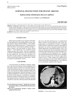
Silva et al; Spinal Cord Extradural Abscess Caused by Arcanobacterium pyogenes
in a Lamb in the Northeast Region of Brazil. Braz J Vet Pathol, 2012, 5(2), 40 - 41
40
Letter to the Editor
Spinal Cord Extradural Abscess Caused by Arcanobacterium
pyogenes in a Lamb in the Northeast Region of Brazil
Reanne M.M. Silva1, Filipe R.B. Carvalho2, Robson Bahia
Cerqueira2, Juliana T.S.A. Macêdo1, Pedro M.O. Pedroso1
1
Laboratório de Patologia Veterinária, Universidade Federal do Recôncavo da Bahia (UFRB),
Cruz das Almas – BA, 2Laboratório de Doenças Infecciosas, UFRB, Cruz das Almas, BA
* Corresponding Author: Pedro M.O. Pedroso, Rua Rui Barbosa 710, Campus Universitário,
UFRB, Cruz das Almas – BA, 44380-000, Brazil. E-mail: [email protected]
Submitted June 13th 2012, Accepted July 28th 2012
Dear Editor,
Abscesses of the central nervous system occur mainly in
young animals, usually below one year of age. In the
spinal cord, where they are seldom observed (1), they can
be located either within the nervous tissue or in the
meninges or osseous structures, compressing the spinal
cord (3, 5, 6).
A one-month-old male lamb of the mixed breed Santa Inês
was presented with neurological signs characterized by
motor incoordination, posterior paralysis and a dog-sitting
position (Fig. 1).
Figure 1 – Sheep in dog-sitting position.
When in movement the lamb frequently fell.
According to the attending veterinarian, the lamb had been
castrated with an emasculatome in the first days of life and
the navel showed difficulty of healing. Due to the poor
clinical condition the lamb was euthanized. At necropsy
an epidural abscess was observed (Fig. 2), measuring
4x3x3 cm, between the lumbar vertebrae 1 and 2 (L1-L2)
compressing the left side of the spinal cord. Fragments of
several organs were collected and fixed in 10% buffered
formalin, processed routinely for histology and stained
with hematoxylin and eosin. Additionally, abscess material
was collected for bacteriological examination. In cross
section of the lumbar spinal cord (Fig. 2, inset), in the
white matter, marked Wallerian degeneration was
observed, characterized by the presence of axonal
spheroids, phagocytosis of myelin debris by macrophages
in digestion chambers and moderate astrogliosis in the
lateral and ventral funiculi. Gray matter neurons had
moderate chromatolysis, characterized by swelling and
central pale cytoplasm and dispersal of the Nissl substance
and moderate astrocytosis and swelling endothelial cells.
Arcanobacterium pyogenes was cultured on blood agar
after incubation at 370C for 24 hours.
Arcanobacterium pyogenes has not been previously
reported as an agent linked to abscesses in ovine and
caprine species in the Northeast Region of Brazil (2, 4). In
this region Corynebacterium pseudotuberculosis is the
main etiological agent of abscesses in the CNS of sheep
and goats, where caseous lymphadenitis is very common,
especially in lambs. Abscesses may be a consequence of
castration wounds or umbilical infection (2, 4) as observed
in this case. Ataxia of the pelvic limbs, without lesions on
the thoracic limbs, indicates thoracic-lumbar lesion. In the
case of severe lesions in the lumbar region the animal can
adopt a dog-sitting position and move himself around
using the thoracic limbs only. Lambs with a cervical spinal
Brazilian Journal of Veterinary Pathology. www.bjvp.org.br . All rights reserved 2007.
Silva et al; Spinal Cord Extradural Abscess Caused by Arcanobacterium pyogenes
in a Lamb in the Northeast Region of Brazil. Braz J Vet Pathol, 2012, 5(2), 40 - 41
cord lesion are unable to maintain sternal recumbency and
have paresis of all four limbs (2). A. pyogenes is an
important opportunistic pathogen, responsible for
suppurative infections in a variety of domestic animals.
The formation of abscesses in the central nervous system
caused by A. pyogenes in sheep in the Northeast Region of
Brazil should be considered.
Figure 2 – Burst extradural abscess (arrow) between lumbar
vertebrae 1 and 2. Inset: Wallerian degeneration and axonal
spheroids within the white matter.
References
1.
2.
3.
4.
5.
6.
GRANT MAXIE, M, YOUSSEF, S. Nervous system.
GRANT MAXIE, M. Ed. Jubb, Kennedy and
Palmer Pathology of Domestic Animals. 5 ed.
Philadelphia: Saunders Elsevier, 2007. 1, 281-455.
GUEDES, K.M.R., RIET-CORREA, F., DANTAS,
A.F.M., SIMÕES, S.V.D., MIRANDA NETO, E.G.,
NOBRE, V.M.T., MEDEIROS, R.M.T. Doenças do
sistema nervoso central em caprinos e ovinos no
semi-árido. Pesq. Vet. Bras., 2007, 27, 29-38.
RADOSTITS, E.M., GAY, C.C., BLOOD, D.D.,
HINCHCLIFF, K.W. Veterinary Medicine. A
textbook of the diseases of cattle, sheep, goats, pigs
and horses. 10 ed. London: Saunders, 2000. 1881p.
SANTA ROSA, J., SANTA ROSA, M.G.
Inflamações supuradas e granulomatosas no sistema
nervoso de caprinos. Ciên. Vet. Trop., 1999, 2, 108114.
SCOTT, P.R., PENNY, C.D., MURRAY, L.D. A
field study of eight ovine vertebral body abscess
cases. N. Z. Vet. J., 1991, 39, 105-107.
SUMMERS, B.A., CUMMINGS, J.F., LAHUNTA,
A. Inflammatory diseases of the central nervous
system. In: Veterinary Neuropathology. 1 ed. St.
Louis: Mosby, 1995, 95-162.
Brazilian Journal of Veterinary Pathology. www.bjvp.org.br . All rights reserved 2007.
41
Baixar