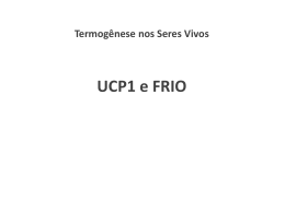
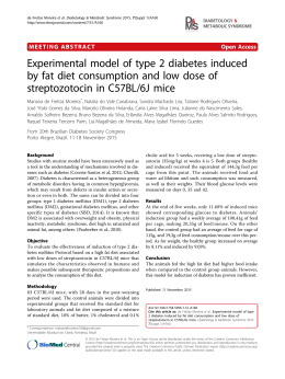
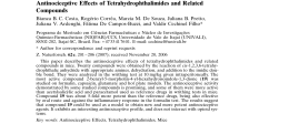
Brazilian Journal of Medical and Biological Research (2006) 39: 253-261
Th1 response in H. heilmannii infection
ISSN 0100-879X
253
The role of IFN-γγ and IL-4 in gastric
mucosa inflammation associated with
Helicobacter heilmannii type 1 infection
1Laboratório
de Pesquisa em Bacteriologia, 2Departamento de Patologia,
Faculdade de Medicina, 3Departamento de Bioquímica e Imunologia,
4Departamento de Microbiologia, Instituto de Ciências Biológicas,
Universidade Federal de Minas Gerais, Belo Horizonte, MG, Brasil
5Cento de Pesquisa Renê Rachou, Fundação Osvaldo Cruz, Belo Horizonte,
MG, Brasil
S.M.S. Cinque1, G.A. Rocha1,
R. Correa-Oliveira5,
T.F. Soares1, S.B. Moura4,
A.M.C. Rocha1,
A.M.M.F. Nogueira2,
M.M.D.A. Cabral2,
L.Q. Vieira3,
O.A. Martins-Filho5
and D.M.M. Queiroz1
Abstract
Correspondence
D.M.M. Queiroz
Laboratório de Pesquisa em
Bacteriologia
Faculdade de Medicina, UFMG
Av. Alfredo Balena, 190, Sala 4026
30130-100 Belo Horizonte, MG
Brasil
Fax: +55-31-3274-2767
E-mail: [email protected]
Research supported by CNPq, CAPES,
FIOCRUZ, and FAPEMIG.
Received April 26, 2005
Accepted September 30, 2005
Although Helicobacter heilmannii infection is less common than H.
pylori infection in humans, it is considered to be of medical importance because of its association with gastritis, gastric ulcer, carcinoma,
and mucosa-associated lymphoid tissue lymphoma of the stomach.
However, there have been no studies evaluating the role of the Th cell
response in H. heilmannii gastric infection. We evaluated the participation of pro-inflammatory and anti-inflammatory cytokines, IFN-γ
and IL-4, in H. heilmannii gastric infection in genetically IFN-γ- or IL4-deficient mice. The serum IFN-γ and IL-4 concentrations were
determined by ELISA. The gastric polymorphonuclear infiltrate was
higher (P = 0.007) in H. heilmannii-positive than in H. heilmanniinegative wild-type (WT) C57BL/6 mice, whereas no significant inflammation was demonstrable in the stomach of H. heilmannii-positive IFN-γ -/- C57BL/6 mice. The degree of gastric inflammatory cells,
especially in oxyntic mucosa, was also higher (P = 0.007) in infected
IL-4-/- than in WT BALB/c mice. Serum IFN-γ levels were significantly higher in IL-4-/- than in WT BALB/c mice, independently of H.
heilmannii-positive or -negative status. Although no difference in
serum IFN-γ levels was seen between H. heilmannii-positive (11.3 ±
3.07 pg/mL, mean ± SD) and -negative (11.07 ± 3.5 pg/mL) WT
BALB/c mice, in the group of IL-4-/- animals, the serum concentration
of IFN-γ was significantly higher in the infected ones (38.16 ± 10.5 pg/
mL, P = 0.04). In contrast, serum IL-4 levels were significantly
decreased in H. heilmannii-positive (N = 10) WT BALB/c animals
compared to the negative (N = 10) animals. In conclusion, H. heilmannii
infection induces a predominantly Th1 immune response, with IFN-γ
playing a central role in gastric inflammation.
Key words
•
•
•
•
Helicobacter heilmannii
Gastric mucosa
inflammation
IFN-γ -/- mice
IL-4-/- mice
Braz J Med Biol Res 39(2) 2006
254
S.M.S. Cinque et al.
Introduction
Helicobacter pylori infects the gastric
mucosa of half the world’s population and
the infection is considered to be an important factor in the pathogenesis of peptic ulcer
(1,2), distal gastric carcinoma (3) and mucosa-associated lymphoid tissue lymphoma
(4). The reason why most infected subjects
remain asymptomatic whereas only a few
develop the related diseases is controversial.
Besides environmental and bacterial virulence factors, differences in the host genetics may influence the type of tissue inflammatory reaction and subsequently the outcome of infection.
Following contact with antigen-presenting cells, T cells can develop into either Th1
or Th2 cells. Th1 cells produce the proinflammatory cytokines interferon-γ (IFNγ) and tumor necrosis factor-α that activate
macrophages. Th2 clones synthesize IL-4
and IL-5, with consequent maturation of
plasma cells and, ultimately, the production
of immunoglobulins. Th1 and Th2 cells also
cross-regulate each other, whereas IFN-γ
down-regulates the production of Th2 cytokines and IL-4 can regulate the Th1 pathway
(5). Animal models such as H. pylori and H.
felis infection in knockout mice deficient for
specific cytokines have been used to clarify
the immune response in Helicobacter infection (6-9). Data have shown that, in contrast
to extracellular organisms such as helminths
and many bacteria, the immune response to
H. pylori is predominantly Th1 (4,10,11).
In addition to H. pylori, another gastric
spiral bacterium, H. heilmannii, has been
demonstrated in the gastric mucosa of human beings (12-14). Although H. heilmannii
infection is rare in humans, it is considered
to be of medical importance because of its
association with gastritis, gastric ulcer, carcinoma, and mucosa-associated lymphoid
tissue lymphoma (15-18). In contrast to H.
pylori infection, H. heilmannii more frequently colonizes the antral gastric mucosa.
Braz J Med Biol Res 39(2) 2006
Furthermore, the gastritis scores, based on
the degree of polymorphonuclear (PMN)
and mononuclear (MN) cells, are lower in
the gastric mucosa of H. heilmannii-positive
patients than in the mucosa of H. pyloripositive patients (14,16). The reason for these
differences may be associated with the host’s
response to distinct Helicobacter species that
may have different strategies for colonization and persistence in the gastric mucosa.
Studies that evaluate cell-mediated responses
in H. heilmannii infection would be of great
value to explain why H. heilmannii induces
a more modulated inflammatory response in
the host.
H. heilmannii infection is considered to
be a zoonosis and swine seem to be the most
important reservoir of H. heilmannii for humans (19). In the pig, the infection is associated not only with gastritis, but also with
pre-ulcerous lesions and ulcer of the pars
esophagea (20,21). Although the bacterium
has not been cultured in vitro, oral inoculation of mice and rats with H. heilmanniipositive mucus of pig stomach results in
100% gastric infection that also leads to
chronic gastritis (22-24). Based on comparative 16S rDNA data, the sequence of the
spiral porcine bacterium was shown to be
identical to that of H. heilmannii type 1
(21,25).
Since, to the best of our knowledge, there
are no studies evaluating the role of Th
immune response in the H. heilmannii gastric infection, we investigated the participation of pro-inflammatory and anti-inflammatory cytokines, IFN-γ and IL-4, in the
gastric infection by H. heilmannii type 1 in
mice genetically deficient for IFN-γ or IL-4.
Material and Methods
Animals
Groups of 10 Helicobacter-free female
mice (7-8 weeks old) were studied: wild type
(WT) and knockout (-/-) for IFN-γ C57BL/6
255
Th1 response in H. heilmannii infection
mice, whose genetic background is prone to
a predominantly Th1 response, and WT and
knockout for IL-4 BALB/c mice whose genetic background is prone to a predominantly Th2 response (26,27).
The WT C57BL/6 and BALB/c mice
were kindly provided by Oswaldo Cruz Institute (Rio de Janeiro, RJ, Brazil). IFN-γ -/C57BL/6 mice were obtained from the Laboratory of Gnotobiology, Department of Biochemistry and Immunology, ICB/UFMG,
Brazil, bred from pairs purchased from
Jackson Laboratories (B6.129S7-ifngtm1Ts,
ME/USA). IL-4-/- BALB/c mice were obtained from breeding pairs kindly provided
by Dr. Nancy Noben-Trauth (NIH, Bethesda,
MD, USA). The animals were bred in
microisolators and kept under specific pathogen-free conditions according to the international guidelines for laboratory animal care.
Helicobacter heilmannii inoculum
Since H. heilmannii is a non-cultivable
bacterium, mucus from a pig stomach had to
be used as an inoculum of the microorganism. The stomach of a pig naturally infected
with H. heilmannii and the stomach of a pig
not infected with the bacterium were used.
Mucus was obtained from scrapings of the
whole area of the antral region of the H.
heilmannii-positive or -negative stomach.
Mucus positive for H. heilmannii showed
a rapidly positive result for the urease test,
presented many tightly spiraled bacteria in
carbolfuchsin-stained smears and was PCR
positive for H. heilmannii type 1 (24). Bacterial viability was assayed by checking motility by phase contrast microscopy. Mucus
negative for H. heilmannii had a negative
urease test, did not reveal any spiral bacteria
in the smears and was PCR negative for the
genus Helicobacter (28).
One part of the mucus was vigorously
homogenized in three parts of 0.85% (w/v)
saline in a vortex blender and then used for
inoculation.
Experimental design
All animal procedures were carried out
in accordance with the National Institute of
Health Guide for the Care and Use of Laboratory Animals.
Groups of 10 mice of each strain (WT
and IFN-γ-/- C57BL/6, WT and IL-4-/BALB/c mice) were inoculated orally with
0.5 mL of the H. heilmannii-positive mucus
homogenate, corresponding to an inoculum
of approximately 105 bacteria/mL. Four other
groups, also consisting of 10 mice of each
strain, were inoculated orally with 0.5 mL of
the H. heilmannii-negative mucus homogenate. All H. heilmannii-positive and -negative mice were inoculated on the same day
with the gastric mucus homogenate from the
same H. heilmannii-positive and -negative
pig stomach, respectively. The animals were
kept in separate cages and had free access to
an autoclaved pelleted diet and sterile water
during the experimental period.
The animals were sacrificed 4 weeks after inoculation under ether anesthesia and
blood was obtained by heart section. Blood
samples were centrifuged and the sera stored
at -4ºC. Fragments of the antral and oxyntic
gastric mucosa of each animal were obtained for microbiological and histological
studies.
Microbiology
The specimens were inserted into 2%
Christensen’s urea agar for the urease test
and examined within 24 h to detect preformed urease. For carbolfuchsin-stained
smears, specimens were smeared onto a glass
slide, heat fixed, stained with 40% carbolfuchsin for 5 min and examined under an oil
immersion lens for the presence of tightly
spiraled bacteria (29). Bacterial viability was
determined by observing motility by phase
contrast microscopy of fresh gastric mucus
preparations.
Specimens for culture were plated onto
Braz J Med Biol Res 39(2) 2006
256
S.M.S. Cinque et al.
Belo Horizonte medium (30) and Skirrow’s
medium (30) and incubated at 37ºC under
micro-aerobic conditions obtained by a gas
generation system (Anaerocult C, Merck,
Darmstadt, Germany) for up to 10 days.
Colonies with the macroscopic aspect of
Helicobacter were tested for catalase, cytochrome-oxidase and urease and then Gram
stained.
DNA from mucosal fragments was extracted with a QIAamp DNA minikit (Qiagen
GmbH, Hilden, Germany) and 16S rRNA
gene was amplified by nested PCR using
primer sets specific for Helicobacter genus
according to Oliveira et al. (28). For the
detection of H. heilmannii type 1 species,
16S rRNA cistrons were amplified with primers chosen on the basis of the 16S rRNA
gene sequence of H. heilmannii type 1 as
described by Duval-Araujo et al. (24).
ommendations. The concentrations of IL-4
and IFN-γ are reported as means ± SD for 10
mice in each group.
Statistical analysis
The results of inflammatory cell infiltration in the gastric mucosa are reported as
median percent values and analyzed by the
two-tailed Mann-Whitney U-test. The mean
values of IFN-γ and IL-4 concentrations were
compared by the Student t-test. Correlations
were evaluated by Pearson’s correlation coefficient (r). The level of significance was
set at P ≤ 0.05 (32).
Results and Discussion
This is the first report on the role of the
Th response in H. heilmannii gastric infection.
Histology
Microbiological study
Histological sections of the antral and
oxyntic mucosa were stained with hematoxylin-eosin and examined by two pathologists who were unaware of their origin. The
levels of MN and PMN cells in the antral and
oxyntic mucosa were scored as absent (0),
mild (1), moderate (2), or marked (3), according to the modified Sydney system for
human gastritis classification (31).
Since H. heilmannii is a non-cultivable
organism, histological sections were also
stained with carbolfuchsin (29) and used for
the evaluation of bacterial density. Ten adjacent areas of the mucosa were evaluated by
counting the number of bacteria inside the
glands.
Serum IL-4 and IFN-γγ concentrations
The serum concentrations of IFN-γ (sensitivity <5 pg/mL) and IL-4 (sensitivity <1
pg/mL) were measured using a commercial
ELISA (Biosource International, Camarillo,
CA, USA) according to manufacturer recBraz J Med Biol Res 39(2) 2006
Four weeks after inoculation, all mice
infected with H. heilmannii-positive mucus
showed tightly spiraled bacteria in carbolfuchsin-stained smears and in the histological sections, and also presented a positive
urease test and were positive for H.
heilmannii type 1 PCR. All tests carried out
in all mice inoculated with H. heilmanniinegative mucus were negative.
No other Helicobacter species was detected by PCR and no spiraled bacterium
grew in the medium used, regardless of
whether the mucus from the source animal
was H. heilmannii-positive or -negative.
The gastric sections of H. heilmanniipositive mice, irrespective of the animal
strain, showed so many tightly spiraled bacteria that clusters of them were seen within
every lumen of the glands. Even though this
prevented the counting of individual bacteria, there was no apparent difference in bacterial density among groups. The urease tests
applied to the gastric mucosa of all infected
257
Th1 response in H. heilmannii infection
mouse strains also indicated the high degree
of bacterial colonization by giving positive
results within the first hour in all animals.
Since the degree of H. heilmannii colonization was similar in WT and IFN-γ-/C57BL/6 mice, no protective effect against
H. heilmannii infection may be attributed to
IFN-γ. Similar results were observed for H.
pylori-infected WT and IFN-γ-/- C57BL/6
mice (9). The present results agree with the
conclusions of Mohammadi et al. (6) who
demonstrated that IFN-γ neutralization with
anti-IFN-γ antibodies had no effect on the
magnitude of H. felis colonization.
The gastric mucosa of IL-4-/- mice was
as heavily colonized as the gastric mucosa of
WT BALB/c mice, suggesting that IL-4 does
not modulate H. heilmannii load. Similar
results were obtained by Smythies et al. (9)
in H. pylori-infected IL-4-/- mice. Conversely, Mohammadi et al. (33) showed that,
in H. felis infection, the number of bacteria is
greater in IL-4-/- than in WT C57BL/6 mice.
In addition, Zavros et al. (34) showed that
treatment with IL-4 reduced the H. felis load
in a Th1 mouse model of Helicobacter infection. These differences may be due to
differences between both the Helicobacter
species and the host studied. Therefore, IL-4
may play a role in the host defense against H.
felis colonization (33), but not against H.
pylori (9) or H. heilmannii.
Histological study
The results of the histological evaluation
are shown in Table 1.
WT C57BL/6 and BALB/c mice
The degree of PMN infiltrate in the antral
mucosa was higher in H. heilmannii-positive than -negative WT C57BL/6 mice (P =
0.007), but no difference in MN levels was
observed (P = 0.27). Conversely, no difference was observed in the degree of MN (P =
0.58) or PMN cell infiltration (P = 0.11) in
the antral mucosa of H. heilmannii-positive
and -negative WT BALB/c. When the oxyntic mucosa was studied, the levels of inflammatory cells were higher in H. heilmanniipositive than in H. heilmannii-negative WT
C57BL/6 [MN (P = 0.005) and PMN (P =
0.007)] and WT BALB/c [MN (P = 0.01)]
mice. When WT H. heilmannii-positive
C57BL/6 and BALB/c mice were compared,
Table 1. Gastric inflammation scores in Helicobacter heilmannii-negative and -positive wild-type and knockout C57BL/6 and BALB/c mice.
Antral mucosa
Hh-negative
Oxyntic mucosa
Hh-positive
Hh-negative
Hh-positive
MN
PMN
MN
PMN
MN
PMN
MN
PMN
C57BL/6
WT
IFN-γ-/-
0 (0-1)
0 (0-0)
0 (0-0)a
0 (0-1)
1 (0-1)l
0 (0-0)i,l
1 (0-2)a,m
0 (0-0)m
0.5 (0-1)b
0 (0-1)
0 (0-1)c
0 (0-1)
2 (1-2)b,n
0 (0-1)k,n
2 (1-3)c,e,o
0 (0-1)j,o
BALB/c
WT
IL-4-/-
0 (0-1)
0 (0-1)f
0 (0-1)
0 (0-0)
0.5 (0-1)
1 (0-1)f,i
0 (0-1)p
1 (0-1)p
0 (0-1)d
0 (0-1)g,h
0 (0-1)
0 (0-0)
1.5 (1-3)d
2 (0-2)h,k
0 (0-1)e,q
1 (0-1)g,j,q
Data are reported as median (range) of the score of inflammatory cell infiltration in the gastric mucosa (0 =
absent, 1 = mild, 2 = moderate, and 3 = marked). Hh = H. heilmannii; MN = mononuclear cells; PMN =
polymorphonuclear cells; WT = wild type; -/- = knockout; IFN-γ = interferon-γ; IL-4 = interleukin-4. Equal
superscript letters mean that differences between results were significant (P ≤ 0.05, two-tailed Mann-Whitney
U-test).
Braz J Med Biol Res 39(2) 2006
258
S.M.S. Cinque et al.
the levels of PMN in the oxyntic mucosa
were significantly higher in C57BL/6 animals (P = 0.001). No other differences were
demonstrable [PMN (P = 0.21) and MN (P =
0.58) in the antral mucosa and MN (P =
0.52) in the oxyntic mucosa]. These data
confirm our demonstration that the degree of
the gastric inflammatory infiltration was
higher in H. heilmannii-positive C57BL/6
than in BALB/c mice (35) and provide more
in-depth information since we discriminated
between PMN and MN cells. Also, in agreement with literature data, the levels of inflammatory cells in H. heilmannii-infected
mice were not marked, ranging from absent
(0) to moderate (2), as demonstrated in human gastric infection by H. heilmannii (16).
Knockout C57BL/6 and BALB/c mice
In the group of IFN-γ-/- C57BL/6 mice,
no difference was observed between H.
heilmannii-positive and -negative animals
[MN (P = 1.0) and PMN (P = 0.36) in the
antral mucosa; MN (P = 0.91) and PMN (P =
0.82) in the oxyntic mucosa].
In contrast to the results obtained for WT
BALB/c mice, in H. heilmannii-positive IL4-/- animals, the degree of MN infiltration in
the antral mucosa (P = 0.05) and of PMN
infiltration in the oxyntic mucosa (P = 0.001)
was significantly higher in H. heilmanniipositive than in the -negative animals. In
contrast, similarly to WT BALB/c mice, the
degree of PMN infiltration in the antrum did
not differ between H. heilmannii-positive
and -negative IL-4-/- mice (P = 0.4) and the
degree of MN cell infiltration in the oxyntic
mucosa was higher in the IL-4 -/- H.
heilmannii-positive than in the IL-4-/- H.
heilmannii-negative group (P = 0.002).
When H. heilmannii-positive IL-4-/- and
IFN-γ-/- mice were compared, the scores of
MN infiltration (P = 0.03) in the antral mucosa and the scores of PMN (P = 0.05) and
MN (P = 0.001) infiltration in the oxyntic
mucosa were significantly higher in BALB/
Braz J Med Biol Res 39(2) 2006
c than in C57BL/6 mice, whereas the degree
of PMN infiltration in the antral mucosa was
not significantly different (P = 0.39).
Helicobacter heilmannii-positive knockout
versus WT animals
When H. heilmannii-positive WT and
knockout mice were compared, differences
were observed between IFN-γ-/- and WT
C57BL/6 [MN (P = 0.02) and PMN (P =
0.007) in the antrum; MN (P = 0.002) and
PMN (P = 0.01) in the oxyntic mucosa].
In the group of H. heilmannii-positive
BALB/c mice, the degree of PMN infiltration in the antral (P = 0.03) and oxyntic
mucosa (P = 0.007), but not of MN infiltration in the antral (P = 0.86) and oxyntic
mucosa (P = 0.48) was significantly higher
in the IL-4-/- than in the WT group.
In agreement with the studies of D’Elios
et al. (10) in H. pylori-positive patients and
Mohammadi et al. (6) in H. felis-infected
mice, our results also support the pathogenic
role of Th1 cytokines and, in particular, of
IFN-γ in H. heilmannii-induced gastritis.
First, the gastritis was more intense and not
restricted to the corpus in H. heilmanniipositive WT C57BL/6 mice, which are prone
to a predominantly Th1 response, than in H.
heilmannii-positive WT BALB/c mice,
which tend to exhibit Th2 polarization. Second, Zavros et al. (34) observed a significant
increase of MN cells in the gastric mucosa of
mice after 7 days of IFN-γ infusion. Third, in
agreement with the data of Sawai et al. (8)
and Smythies et al. (9) who did not observe
gastric inflammation in H. pylori-infected
IFN-γ-/- mice and those of Mohammadi et
al. (6) who observed a significant reduction
of the gastric inflammation in H. felis mice
after anti-IFN treatment, in the present study,
no gastric inflammation was seen in H.
heilmannii-positive IFN-γ-/- C57BL/6 mice.
Therefore, as observed for H. pylori and H.
felis infection, IFN-γ seems to contribute to
the severity of the gastritis associated with
259
Th1 response in H. heilmannii infection
H. heilmannii infection.
There are conflicting results in the literature regarding the role of IL-4 in mucosal
inflammation. Chen et al. (7), while evaluating H. pylori-infected IL-4-/- and IL-4 transgenic (that produce higher levels of IL-4)
C57BL/6J mice, did not observe a difference
in the degree of gastric inflammation between these groups and concluded that IL-4
does not play a central role in the protection
against gastric inflammation. Conversely,
Zavros et al. (34) showed that IL-4 administration prevented the inflammatory response
of the gastric mucosa of a Th1 background
mice infected with H. felis. In addition,
Smythies et al. (9) demonstrated a higher
gastric inflammation in H. pylori-infected
IL-4-/- than in WT mice, which was supposed to be due to an up-regulation of IFNγ in the absence of IL-4. In agreement with
Smythies et al. (9), we observed that IL-4-/animals partially switched to a Th1 phenotype. The gastric inflammation scores were
higher in infected IL-4-/- than in WT BALB/
c mice.
Thus, it seems that, in Helicobacter infection, the severity of gastric inflammation
depends on IFN-γ and some protective effect may be attributed to IL-4.
even in BALB/c mice, H. heilmannii infection elicits a Th1 response since the serum
levels of IL-4 were significantly decreased
in infected animals.
Serum IFN-γ levels were undetectable in
IFN-γ-/- C57BL/6 mice. Similar IFN-γ levels were observed in H. heilmannii-positive
(11.03 ± 3.07 pg/mL) and H. heilmanniinegative (11.07 ± 3.5 pg/mL) WT BALB/c
mice (P = 0.98), but IFN-γ levels were higher
in the H. heilmannii-positive (38.16 ± 10.5
pg/mL) than -negative (23.9 ± 10.6 pg/mL)
WT C57BL/6 group (P = 0.04; Figure 2),
confirming the predominant Th1 profile of
the C57BL/6 mouse strain.
In contrast to the results obtained for WT
BALB/c mice, the serum levels of IFN-γ
were significantly higher in H. heilmanniipositive (34.14 ± 8.7 pg/mL) than -negative
(21.2 ± 9.6 pg/mL) IL-4-/- BALB/c mice (P
= 0.02).
When WT and IL-4-/- BALB/c mice were
compared, the serum IFN-γ levels were significantly higher in both the H. heilmanniipositive (P = 0.000) and -negative (P = 0.05)
IL-4-/- groups than in the H. heilmannii-posi-
Serum IL-4 and IFN-γγ concentration
Irrespective of the infection, the levels of
IL-4 were nearly undetectable in the IL-4-/groups. Although the serum levels of IL-4
were very low in the other groups, difference
was observed between H. heilmannii-positive (0.36 ± 0.57 pg/mL) and -negative (1.20
± 0.59 pg/mL) WT BALB/c mice (P = 0.03),
as well as between H. heilmannii-negative
WT BALB/c and WT C57BL/6 (0.0 ± 0.2
pg/mL) animals (P = 0.001). No other significant association was observed [H.
heilmannii-positive vs -negative IFN-γ -/C57BL/6 mice (P = 0.14) and H. heilmanniinegative WT vs IFN-γ -/- C57BL/6 mice (P =
0.26)] (Figure 1). These results show that
Figure 1. Serum IL-4 levels in Helicobacter heilmannii-negative and -positive wild-type and
knockout BALB/c and C57BL/6 mice. Hh = H. heilmanni i ; WT = wild type; -/- = knockout;
IFN = interferon; IL-4 = interleukin-4. P values are shown only when the differences were
significant (Student t-test).
Braz J Med Biol Res 39(2) 2006
260
S.M.S. Cinque et al.
tive and -negative WT BALB/c groups. No
differences between H. heilmannii-negative
IL-4-/- and H. heilmannii-negative WT C57BL/
6 mice (P = 0.67) nor between H. heilmanniipositive IL-4-/- and H. heilmannii-positive WT
C57BL/6 mice (P = 0.44) were seen. These
results show that H. heilmannii infection leads
to an increase of the Th1 cytokine IFN-γ.
The serum concentrations of IFN-γ positively correlated with the degree of MN (r =
0.48, P = 0.001) and PMN infiltration in the
oxyntic mucosa (r = 0.46, P = 0.01). A weak
significant inverse correlation was detected
between the levels of IL-4 and the degree of
MN infiltration in the oxyntic mucosa (r =
-0.27, P = 0.05). These results again show
the relevance of IFN-γ in inducing gastric
inflammation especially in the corpus mucosa.
In conclusion, the results of the present
study demonstrated that the inflammatory
response of mice to H. heilmannii infection
is similar to that seen in mice infected with
other Helicobacter species, with IFN-γ playing a pivotal role in gastric inflammation.
We also demonstrated that the host background is a critical factor for the severity of
the gastritis induced by H. heilmannii that is
the basis for the development of severe diseases associated with Helicobacter infection.
Acknowledgments
Figure 2. Serum IFN-γ levels in Helicobacter heilmannii-negative and -positive wild-type and
knockout BALB/c and C57BL/6 mice. Hh = H. heilmannii; WT = wild type; -/- = knockout; IFN
= interferon; IL-4 = interleukin-4. P values are shown only when the differences were
significant (Student t-test).
The authors thank Dr. Luiz Vicente Rizzo,
Department of Biochemistry and Immunology, USP, São Paulo, SP, Brazil, for providing IL-4-/- mice.
References
1. Logan RPH & Walker MM (2001). ABC of the upper gastrointestinal
tract: Epidemiology and diagnosis of Helicobacter pylori infection.
British Medical Journal, 323: 920-922.
2. Cover RL & Blaser MJ (1995). Helicobacter pylori: A bacterial cause
of gastritis, peptic ulcer disease, and gastric cancer. ASM News, 61:
21-26.
3. Parsonnet J, Friedman GD, Vandersteen DP et al. (1991). Helicobacter pylori infection and the risk of gastric carcinoma. New England Journal of Medicine, 325: 1127-1131.
4. Isaacson PG (1994). Gastric lymphoma and Helicobacter pylori.
New England Journal of Medicine, 330: 1310-1311.
5. Lee A (2000). Elimination of Helicobacter pylori is dependent on a
Th2 response. In: Hunt RH & Tytgat GNJ (Editors), Helicobacter
pylori. Basic Mechanisms to Clinical Cure. Kluwer Academic Publishers, Dordrecht, The Netherlands.
6. Mohammadi M, Czinn S, Redline R et al. (1996). Helicobacter-
Braz J Med Biol Res 39(2) 2006
7.
8.
9.
10.
specific cell-mediated immune responses display a predominant
Th1 phenotype and promote a delayed-type hypersensitivity response in the stomachs of mice. Journal of Immunology, 156: 47294738.
Chen W, Shu D & Chadwick VS (1999). Helicobacter pylori infection
in interleukin-4-deficient and transgenic mice. Scandinavian Journal
of Gastroenterology, 34: 987-992.
Sawai N, Kita M, Kodama T et al. (1999). Role of gamma interferon
in Helicobacter pylori-induced gastric inflammatory responses in a
mouse model. Infection and Immunity, 67: 279-285.
Smythies LE, Waites KB, Lindsey JR et al. (2000). Helicobacter
pylori-induced mucosal inflammation is Th1 mediated and exacerbated in IL-4, but not IFN-γ, gene deficient mice. Journal of Immunology, 165: 1022-1029.
D’Elios MM, Manghetti M, De Carli M et al. (1997). T helper 1
effector cells specific for Helicobacter pylori in the gastric antrum of
261
Th1 response in H. heilmannii infection
11.
12.
13.
14.
15.
16.
17.
18.
19.
20.
21.
22.
23.
patients with peptic ulcer disease. Journal of Immunology, 158: 962967.
Bamford KB, Fan X, Crowe SE et al. (1998). Lymphocytes in the
human gastric mucosa during Helicobacter pylori have a T helper
cell 1 phenotype. Gastroenterology, 114: 482-492.
Queiroz DMM, Cabral MMDA, Nogueira AMMF et al. (1990). Mixed
gastric infection by Gastrospirillum hominis and Helicobacter pylori.
Lancet, 336: 507-508.
Heilmann KL & Borchard F (1991). Gastritis due to spiral shaped
bacteria other than Helicobacter pylori: clinical, histological, and
ultrastructural findings. Gut, 32: 137-140.
Ierardi E, Monno RA, Gentile A et al. (2001). Helicobacter heilmannii
gastritis: a histological and immunohistochemical trait. Journal of
Clinical Pathology, 54: 774-777.
Morgner A, Bayerdorffer E, Meining A et al. (1995). H. heilmannii
and gastric cancer. Lancet, 346: 511-512.
Stolte M, Kroher G & Meining A (1997). A comparison of Helicobacter pylori and H. heilmannii gastritis. A matched control study
involving 404 patients. Scandinavian Journal of Gastroenterology,
32: 28-33.
Debongnie JC, Donnay M, Mairesse J et al. (1998). Gastric ulcers
and Helicobacter heilmannii. European Journal of Gastroenterology
and Hepatology, 10: 251-254.
Morgner A, Lehn N, Andersen CT et al. (2000). Helicobacter
heilmannii-associated primary gastric low-grade MALT lymphoma:
complete remission after curing the infection. Gastroenterology,
118: 821-828.
Meining A, Kroher G & Stolte M (1998). Animal reservoirs in the
transmission of Helicobacter heilmannii. Results of a questionnairebased study. Scandinavian Journal of Gastroenterology, 33: 795798.
Queiroz DMM, Rocha GA, Mendes EN et al. (1990). Spiral microorganism in the stomach of pigs. Veterinary Microbiology, 24: 199204.
Queiroz DMM, Rocha GA, Mendes EM et al. (1996). Association
between Helicobacter and gastric ulcer disease of the pars
esophagea in swine. Gastroenterology, 111: 19-27.
Moura SB, Queiroz DMM, Mendes EN et al. (1993). The inflammatory response of the gastric mucosa of mice experimentally infected
with Gastrospirillum suis. Journal of Medical Microbiology, 39: 6468.
Mendes EN, Queiroz DMM, Moura SB et al. (1998). Mouse inocula-
24.
25.
26.
27.
28.
29.
30.
31.
32.
33.
34.
35.
tion for the detection of non-cultivable gastric tightly spiraled bacteria. Brazilian Journal of Medical and Biological Research, 31: 373376.
Duval-Araujo I, Queiroz DMM, Magnago AGP et al. (2000). Increased gastric emptying induced by Helicobacter heilmannii type 1
infection in rats. Journal of Medical Microbiology, 49: 627-634.
Solnick JV, O’Rourke J, Lee A et al. (1993). An uncultured gastric
spiral organism is a newly identified Helicobacter in humans. Journal of Infectious Diseases, 168: 379-385.
Nedrud JG, Mohammadi M, Blanchard T et al. (1998). Th1/Th2
lymphocyte responses in Helicobacter infections. In: Hunt RH &
Tytgat GNJ (Editors), Helicobacter pylori - Basic Mechanisms to
Clinical Cure. Kluwer Academic Publishers, Dordrecht, The Netherlands, 101-109.
Houghton J & Wang TC (2005). Helicobacter pylori and gastric
cancer: a new paradigm for inflammation associated epithelial cancer. Gastroenterology, 128: 1567-1578.
Oliveira AG, Sanna MGP, Rocha GA et al. (2004). Helicobacter
species in the intestinal mucosa of patients with ulcerative colitis.
Journal of Clinical Microbiology, 42: 384-386.
Rocha GA, Queiroz DMM, Mendes EN et al. (1989). Simple carbolfuchsin staining for showing C. pylori and other bacteria in gastric
mucosa. Journal of Clinical Pathology, 42: 1004-1005.
Queiroz DMM, Mendes EN & Rocha GA (1987). Indicator medium
for isolation of Campylobacter pylori. Journal of Clinical Microbiology, 25: 2378-2379.
Dixon MF, Genta RM, Yardley JH et al. (1996). Classification and
grading of gastritis. The update Sydney system. American Journal
of Surgical Pathology, 10: 1161-1181.
Snedecor GW & Cochran WG (1989). Statistical Methods. 8th edn.
Iowa State University Press, Ames, IA, USA.
Mohammadi M, Nedrud J, Redline R et al. (1997). Murine CD4 T-cell
response to Helicobacter infection: Th1 cells enhance gastritis and
Th2 cells reduce bacterial load. Gastroenterology, 113: 1848-1857.
Zavros Y, Rathinavelu S, Kao JY et al. (2004). Treatment of Helicobacter gastritis with IL-4 requires somatostatin. Proceedings of the
National Academy of Sciences, USA, 100: 12944-12949.
Cinque SMS, Queiroz DMM, Rocha GA et al. (2002). Cellular immune responses in Helicobacter heilmannii infection. Evaluation of
the role of the host and the bacterium. Digestive Diseases and
Sciences, 47: 823-830.
Braz J Med Biol Res 39(2) 2006
Baixar