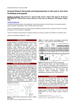
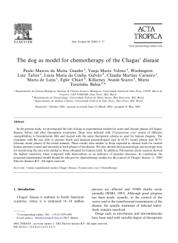
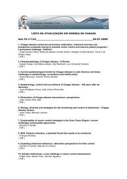
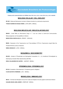
Revista da Sociedade Brasileira de Medicina Tropical
ARTIGO
36(4):441-447, jul-ago, 2003.
Treatment with benznidazole in association with immunosuppressive
drugs in mice chronically infected with Trypanosoma cruzi:
investigation into the possible development of neoplasias
Tratamento com benzonidazol em associação com drogas imunossupressoras em
camundongos crônicamente infectados com Trypanosoma cruzi: investigação
sobre a possibilidade de desenvolvimento de neoplasias
Sonia G. Andrade1, Igor Marcelo Oliveira Mesquita1 2, Jamile F. Jambeiro1 2,
Isis F. Magalhães Santos1and Renata Siqueira Portella1
Abstract Benznidazole is recommended in Brazil for the treatment of Trypanosoma cruzi infection in acute and
early chronic phases of Chagas’ disease. Observations by others have indicated a higher incidence of neoplasias
in immunosuppressed patients, presenting Chagas’ disease reactivation, submitted to treatment with benznidazole.
In the present study, we investigated whether there is a potentiation in the generation of lymphomas in chronically
infected mice, treated with immunosuppressive drugs and benznidazole. For this, 142 Swiss mice chronically
infected with the 21 SF strain of T. cruzi and 72 normal Swiss mice were used. Both infected and normal mice
were divided into experimental groups and submitted to one of the following treatment regimens: benznidazole
alone; immunosuppressive drugs (azathioprine, betamethasone and cyclosporin); a combination of
immunosuppressive drugs and benznidazole; and untreated controls. In the infected group treated with
benznidazole, one mouse developed a non-Hodgkin’s lymphoma. This finding has been interpreted as a spontaneous
tumor of mice. The study of the chronically infected mice treated with the combination of immunosuppressive
drugs and benznidazole demonstrated an absence of lymphomas or other neoplasias. These findings support
the indication of benznidazole, as the drug of choice, for immunosuppressed patients that develop a reactivation
of Chagas’ disease.
Key-words: Trypanosoma cruzi. Experimental infection. Chemotherapy. Benznidazole. Immunosuppressive drugs.
Non-Hodgkin’s lymphoma.
Resumo O benzonidazol é recomendado no Brasil para o tratamento da infecção pelo Trypanosoma cruzi na
fase aguda e na fase crônica precoce da doença de Chagas. Observações de outros autores mostraram uma
elevação na incidência de neoplasias em pacientes imunossuprimidos, com reativação da doença de Chagas,
submetidos a tratamento com o benzonidazol. No presente estudo foi investigado em camundongos cronicamente
infectados, tratados com drogas imunossupressoras e com benzonidazol se há uma potencialização de
desenvolvimento de linfomas por esta combinação. Para isso foram usados 142 camundongos suiços crônicamente
infectados pela cepa 21SF do T.cruzi e 72 camundongos suiços normais. Tanto os camundongos infectados
como os normais foram divididos em grupos experimentais submetidos respectivamente a diferentes esquemas
de tratamento: com benzonidazol apenas; com drogas imunossupressoras (azatioprina, betametasona e
ciclosporina); com drogas imunossupressoras mais benzonidazol; e controles não tratados. No grupo infectado
e tratado com benzonidazol, um camundongo desenvolveu um linfoma não-Hodgkin. Este achado foi interpretado
como um tumor espontâneo do camundongo. O estudo de camundongos crônicamente infectados e tratados
com uma combinação de drogas imunossupressoras e benzonidazol demonstrou ausência de linfomas ou de
outras neoplasias. Estes achados reforçam a indicação do tratamento com benzonidazol como droga de escolha
em pacientes imunossuprimidos que desenvolvem reativação da doença de Chagas.
Palavras-chaves: Trypanosoma cruzi. Infecção experimental. Quimioterapia. Benzonidazol. Drogas
imunossupressoras. Linfoma não Hodgkin.
1. Laboratório de Doença de Chagas Experimental do Centro de Pesquisas Gonçalo Moniz da Fundação Oswaldo Cruz, Salvador, BA..2. PIBIC da Fundação
Oswaldo Cruz e Conselho Nacional de Pesquisa, Salvador, BA.
Address to: Dra. Sonia G. Andrade. Centro de Pesquisas Gonçalo Moniz/FIOCRUZ. Rua Waldemar Falcão 121, 40295-001 Salvador, BA, Brasil.
Recebido para publicação em 17/9/2002
Aceito em 18/5/2003
441
Andrade SG et al
Although the transmission of Chagas’ disease in
endemic areas has been successfully controlled 12,
patients with chronic infection, and new acute cases,
due to different mechanisms of infection, necessitate
chemotherapy treatment. As recently reviewed by Coura
and Castro11, nitroimidazolyl benznidazole continues to
be the drug in clinical use in Brazil, with an effective
cure in early cases 3 and is recommended by the
Fundação Nacional de Saúde14 Reactivation of Chagas’
disease in immunosuppressed individuals, either due to
AIDS13 18 19 20 or to the use of immunosuppressive drugs
in transplant patients1 2 8 17 21, has been reported. In such
cases, chemotherapeutic treatment is mandatory and
benznidazole is the drug generally used. However, clinical
observations have shown an influence of treatment with
benznidazole associated to immunosuppression upon
the development of neoplasias, as described by Bocchi
et al 8. The latter treated patients with reactivation of
T. cruzi infection, after cardiac transplantation and the
use of immunosuppressive drugs. This fact has been
confirmed by Almeida 2 and Almeida et al 1. These
observations have implications, mainly related to the
replacement of benznidazole by allopurinol1 2, the efficacy
of which has not been established. Other authors18, on
the contrary, succeeded in treating the reactivation of
Chagas’ disease using benznidazole, without reference
to the development of neoplasias. In four heart-transplant
patients, Stolf et al21, detected 3 cases with reactivation
of Chagas’ disease. The treatment of these patients with
benznidazole was successful and a follow-up of 160 to
500 days showed reversion of the acute manifestations,
apparently with no detectable clinical or histological
sequelae.
Since experimental observations by Teixeira et
al23 24 regarding the presence of lymphomas in rabbits
under treatment with nitroarenes24, and, specifically,
benznidazole23 a controversy has been established.
Clinical observations by Cançado9, and a recent review
by Coura and Castro11 , did not indicate an increased
incidence of neoplasias in chagasic patients treated with
benznidazole. Experimentally, treatment of the chronic
phase of infection in mice with benznidazole, has never
resulted in the development of neoplasias, either of the
lymphoid organs or others, at least in our experience6 7.
With the objective of contributing to the clarification of
whether there is in fact a potentiation of mutagenesis
and lymphoma generation by the combined use of
immunosuppressive drugs and benznidazole, an
investigation was conducted with the experimental murine
model of chronic T. cruzi infection using azathioprine,
betamethasone and cyclosporin, in doses that could be
compared with those used in transplant patients5.
MATERIAL AND METHODS
Experimental animals. A group of 178 Swiss mice,
weighing 10 to 12g were experimentally infected with the
21 SF strain of T. cruzi classified as biodeme Type II4.
Inoculation consisted of 4x10³ trypomastigotes obtained
from infected mouse blood. From this infected group,
142 mice that survived up to 100 days of infection, were
divided into 4 groups of chronically infected mice as
detailed below. For treatment control, 80 normal Swiss
mice weighing 10 to 12g were maintained until 100 days.
Seventy two surviving mice were divided into 4 groups
of uninfected controls. All the experimental mice were
sacrificed at the end of treatment.
Parasitemia and cumulative mor tality: were
registered in the acute phase.
Experimental groups. Group 1: infected controls: 31
infected and untreated mice (Inf Co). Group 2: 31
infected mice treated with benznidazole (Inf Benz).
Group 3: 40 infected mice treated with immunosuppressive
combination: azathioprine (Imuran) + betamethasone
(Celestone) + cyclosporin (Sandimun) (Inf IM). Group 4:
40 infected mice treated with immunosuppressive drugs
followed by benznidazole (Inf IM Benz). Group 5: 25
uninfected control mice treated with immunosuppressive
drugs (Co IM). Group 6: 14 uninfected control mice treated
with benznidazole (Co Benz). Group 7: 14 uninfected
control mice treated with immunosuppressive drugs and
benznidazole (Co IM Benz). Group 8: 19 normal intact
controls (No Co).
Schedules of immunosuppressive treatment. The
surviving mice were in par t, treated with the
442
immunosuppressive drugs: azathioprine (Imuran),
betamethasone (celestone) and cyclosporin (Sandimun),
100 days after infection. Doses and administration of
the drugs were as follows: azathioprine, 10mg/kg b.w.
administered by gavage, 3 days a week for 4 weeks
followed by one weekly dose for 90 days.
Betamethasone, 2mg/kg b.w. administered
intraperitoneally, 3 days a week, during 4 weeks and
once a week during 90 days.
Cyclosporin, 30mg/kg b.w., 3 days a week, by
gavage, during 4 weeks, followed by a dose of 15mg/
kg b.w. once a week for 90 days.
Chemotherapy treatment with benznidazole.
Treatment with benznidazole (Rochagan) was initiated 4
weeks after beginning the immunosuppressive treatment.
The drug was administered by gavage at a dose of
100mg/kg b.w. day, for 5 days a week, during 90 days,
concomitantly with the immunosuppressive treatment.
Group 1 (Inf Co) received sterile saline by gavage
(1ml/day) during the same period of treatment.
Parasitaemia was checked before and after treatment.
Cumulative mortality was evaluated at the pre-treatment
phase until 100 days and during the treatment period for
all groups, until 280 days. Mice from the various groups
were evaluated according to the total number of
leukocytes and the number of lymphocytes in peripheral
blood, up to the end of the experiment.
White blood cell counts: blood collection was
performed during the sacrifice of the mice, by sectioning
Revista da Sociedade Brasileira de Medicina Tropical 36:441-447, jul-ago, 2003
the axillary plexus and collecting the blood with heparin
into plastic vials, six weeks after the beginning of
treatment (for treated animals) and at the same period
for untreated controls. White blood cell counts were
performed with an automatic Coulter count (T-890).
Statistical analysis: variance test was applied by using
the Kruskal-Wallis method for comparing the number of
leukocytes and lymphocytes in peripheral blood in normal
controls and chronically infected mice from the
experimental groups. Comparison was also performed for
leukocytes and lymphocytes in chronically infected mice
after immunosuppressive and or chemotherapy treatment.
For evaluation of the significance of spleen indices in the
several groups, the same test (variance test of Kruskal
Wallis) was performed. Significance was post-tested by
the Dunn’s multiple comparison test.
Serology: serum antibodies were examined by indirect
immunofluorescence (IIFT) using culture forms of T. cruzi
as antigens and rabbit-anti-T. cruzi monospecific serum as
primary antibody in dilutions of 1:10 until 1:640. As
secondary antibody, a goat anti-rabbit IgG conjugated to
fluorescein (Sigma) was used.
Histopathological study. Mice from the experimental
groups, were sacrificed under anesthesia with ether, 30
days after the end of treatment. The blood was collected for
white cell count and serological tests. Body weight and
spleen weight were registered for each animal, and the
relationship between spleen weight and body weight (spleen
index) was determined. The various organs were fixed in
Milloning formalin10 and paraffin embedded. Five micrometer
thick (5m) sections of the heart, skeletal muscle, liver, spleen
and lymph nodes were stained with Hematoxylin and Eosin.
RESULTS
Parasitemia. Figure 1 shows the characteristic
profile of the parasitemic curve of mice in the acute
phase of infection with T. cruzi strain, biodeme Type
II, with the highest peak at the 15 th day, and
negativation in the chronic phase.
Cumulative mortality: the mortality rate was 15.2%
in the pretreatment period, until 100 days post
inoculation (Figure 2). After 100 days of infection, the
indices of cumulative mortality varied according to
the experimental groups, as shown in Figure 3. The
No. of trypomastigotes per 50
microscope fields (x400)
70
60
50
40
30
20
10
0
0
9
11
15 17 21 23 25 29 31 35 37 39 43 53 67 79 100
Days of infection
Figure 1 - Parasitemia levels in mice infected with the 21SF strain of Trypanosoma
cruzi representing the acute phase and the chronic pre-treatment phase.
16
Cumulative mortality (%)
14
Infected
Not infected
12
10
8
6
4
2
0
1
10
20 30
40 50
60 70
80 90
98 100
Days of infection
Figure 2 - Cumulative mortality of mice infected with
Trypanosoma cruzi (21 SF strain) in the pre-treatment
phase, as compared with normal controls. The mortality
index was of 15,2% until 100 days post inoculation.
443
Andrade SG et al
60
Inf Co
)
Inf Benz
Inf IM
50
Inf IM B enz
Co IM
40
No Co
30
20
10
0
100 120 140 160 180 200 220 240 260 280
Days of infection
Figure 3 - Cumulative mortality of mice of different experimental groups: l-Inf Co; ¨ Inf
Benz; p-In IM; ¡-Inf IM Benz; ¢-Co IM ; r-No Co . The highest indices of mortality
occurred in the groups of mice , infected and uninfected controls that have been treated
with immunosuppressive drugs (Groups Inf IM, Inf IM Benz, Co IM), excluding group Co
IM Benz, with nule mortality (not included). Group 6 - Co Benz also showed nule mortality.
highest indices of mortality occurred in the groups of
mice, both infected and uninfected controls that were
treated with immunosuppressive drugs (Groups Inf IM,
In IM Benz, Co IM), excluding Group Co IM Benz,
with no mortality. Also the control group, treated with
Benznidazole (Co Benz) presented zero mortality.
Histopathological study. General data: the heart
of mice chronically infected with T. cruzi from Group 2
(In Benz) showed regression of the myocarditis, as
compared with the infected controls (Inf Co), showing
residual, focal inflammatory infiltrates limited to the
atria, and focal areas of fibrosis of the myocardium.
The other groups of infected mice: Inf Co, Inf IM, Inf
IM Benz, presented either mild (+) or moderate (++)
focal mononuclear infiltrates in the myocardium and
skeletal muscle, with slight variation of intensity.
In several mice from the groups infected and
treated with immunosuppressive drugs, pneumonia,
pyelonephritis and pericarditis were present, as well
as amyloid deposits in the spleen and liver.
Lymphoid tissue: in Group 2 (Inf Benz), out of 31 mice
chronically infected and treated with benznidazole, one
mouse presented neoplastic proliferation of lymphoid cells
of the spleen, histologically characterized as a malignant
non-Hodgkin’s lymphoma (Figure 4-A, B, C and D).
Infiltration with blastic lymphoid cells was present in the
subepicardium and pericardiac lymph nodes, in the
pulmonary tissue and in the sinusoidal and perivascular
spaces of the liver (Figure 4-D). In this same group, the
other animals showed either normal lymphoid organs or a
mild (+) to moderate (++) hyperplasia of the lymphoid
follicles of the spleen.
In the other infected groups, the spleen and lymph
nodes revealed a normal structure in most of the cases,
444
with mild (+) or moderate (++) hyperplasia of lymphoid
follicles.
Uninfected mice: in the animals of Group 5 (Co IM)
the heart showed mild mononuclear infiltration and small
focal areas of myocytolysis of heart. The spleen showed
normal structure or atrophy of the lymphoid follicles in
50% of the cases and mild hyperplasia of the lymphoid
follicles in the remaining 50%. In the latter cases,
amyloid deposits were present in the red pulp as well
as in the liver. In the mice of Groups Co Benz, Co IM
Benz and NoCo, no significant alterations were observed
in the various organs.
Relationship between spleen weight and body weight
(spleen index): Figure 5 shows the splenic indices for
the several experimental groups. Case #15 was not
included (Group 2-Inf Benz), that developed a lymphoma
and showed a significant increase in spleen weight (2.69g)
in comparison with the mean weight of the spleen of the
animals of the same group (0.39). Excluding this case,
no significant statistical difference was detected between
the spleen indices of the four infected groups and the
intact controls. However a significant difference was
observed between the spleen index of Group 1 (Inf Co)
as compared with Group 7 (Co IM Benz) in which a
significant decrease of spleen weight was detected
(p <0.05).
White blood cells count. Results of the evaluation
of the total number of leukocytes on infected and
uninfected mice, treated or untreated, are shown in
Figure 6. No significant statistical differences were
detected between the infected groups in comparison
with the intact controls. The mouse with a lymphoma
(Group 2- Inf Benz) presented a higher number of
Revista da Sociedade Brasileira de Medicina Tropical 36:441-447, jul-ago, 2003
Figure 4 - Histopathological aspects: non-Hodgkin’s lymphoma of the spleen: A – sections of
the spleen showing neoplastic proliferation of small lymphoid cells, with irregular nuclei, with
substitution of the follicular structure of the organ, H & E (400X); B – sections of a lymph node
with substitution of the follicular structure by blastic lymphoid cell proliferation, H & E (250 X).
C – Heart section, with the presence of lymphoid blastic cells in the subepicardium. H & E
(100X). D – Invasion of the liver tissue by the neoplastic lymphoid cells. H & E (200X).
Leucocyte count (x103)
50
40
30
20
10
0
1
2
3
4
5
6
7
8
groups
Figure 5 - Splenic indices of mice of the experimental groups
infected with Trypanosoma cruzi and uninfected controls: 1-Inf
Co; 2- Inf Benz ; 3 - In IM; 4 -Inf IM Benz; 5 - Co IM; 6 - Co Benz;
7 - Co IM Benz; 8 – No Co. Highest spleen index was observed
in Group 1 – In Co. No significant difference was detected by
statistical analysis between the four infected groups and the
normal controls (Group 8 – No Co). A significant difference was
observed between the spleen index of Group 1 (Inf Co) compared
with Group 7 (Co IM Benz) in which a significant decrease of
spleen weight was detected (p <0.05).
Figure 6 - Results of the evaluation of the total number of leukocytes
in the peripheral blood of mice representative of the several
experimental groups: 1-Inf Co; 2 - Inf Benz; 3 - In IM; 4 -Inf IM Benz;
5 - Co IM; 6 - Co Benz; 7 - Co IM Benz; 8 - No Co. No significant
statistical differences were detected between the infected groups
in comparison with the intact controls. One mouse (*) of Group 2 –
Inf Benz that presented a lymphoma of the spleen presented a
higher number of leukocytes. Significant decrease of the total
number of leukocytes (p <0.05) was observed in Group 7 (Co IM
Benz) as compared with Groups 3 (Inf IM) and 4 (Inf IM Benz).
445
Andrade SG et al
lymphocytes (47.7 x 10³ ) in comparison with the other
mice of the same group. A significant decrease in the
total number of leukocytes was seen in Group 7 (Co IM
Benz) as compared with Groups 3 (Inf IM) and 4 (Inf IM
Benz). The number of lymphocytes, showed slight
variations between the several groups, excluded the mice
with lymphoma (Case #15). A statistically significant
difference (P <0.05) (Figure 7), was detected between
Group 4 (Inf IM Benz) and Group 7 (Co IM Benz).
Serological test (IIFT): Table 1 shows the variation of
titers of the IIFT in the several groups. A decrease in the
titers occurred in Group 2 (Inf Benz) and varied from 1/40
to 1/640; the other infected groups maintained the
serological titers. All uninfected control mice were negative.
Table 1 - Variation of the titers of IIFT in the infected mice of
Groups 1 to 4.
Variation in titers*
Infected Groups
Minimum
Maximum
Group 1
1:320
1:1280
Group 2
1:40
1:640
Group 3
1:320
1:640
Group 4
1:40
1:320
*Uninfected controls: negative results in the 4 groups
Groups:1 - Inf Co; 2 - Inf Benz; 3 - Inf Im; 4 – Inf IM Benz
Lym phocyte count (x103)
30
20
10
0
1
2
3
4
5
6
7
8
groups
Figure 7 - Evaluation of the number of lymphocytes in the experimental
groups: 1-Inf Co; 2- Inf Benz; 3 - In IM; 4 -Inf IM Benz; 5 - Co IM; 6 - Co
Benz; 7 - Co IM Benz; 8 – No Co. The number of lymphocytes, showed
slight variations between the groups. One mouse (*) of group 2 (Inf Benz)
with lymphoma, presented a higher number of lymphocytes. A statistically
significant difference (P <0.05) was detected between Group 4 (Inf IM
Benz) and Group 7 (Co IM Benz).
DISCUSSION
The results indicate that the association of
benznidazole with immunosuppressive drugs did not
contribute to the appearance of lymphomas or other
neoplasias in mice chronically infected with T. cruzi.
In a previous study5 done with Swiss mice chronically
infected with different strains of T. cruzi, and submitted
to immunosuppressive treatment, but not treated with
benznidazole, none developed neoplastic proliferation
of lymphoid tissues. In the control group of normal
mice, the immunosuppressive treatment combined
with benznidazole caused a significant decrease in
the number of leukocytes and lymphocytes in peripheral
blood and of the spleen index that indicate a response
of the lymphoid organs to this type of treatment.
However, the infected mice submitted to the same
446
regimens, did not show this type of response. This
appears to indicate that the infected mice maintained
their leukocyte response, even under the influence of
immunosuppressive drugs or benznidazole treatment,
with mild to moderate hyperplasia of lymphoid follicles,
without proliferation of the cells of the germinal centers.
In the present study a non-Hodgkin’s lymphoma of
the spleen and lymph nodes was detected in one
mouse of the group treated exclusively with
benznidazole. The finding of one case of non-Hodgkin’s
lymphoma out of 31 chronically infected mice treated
with chemotherapy (3.2%), could not be considered a
priori as evidence of a direct action of the
nitroimidazolyl on its development. Spontaneous
lymphoma in mice has been registered not only in
Revista da Sociedade Brasileira de Medicina Tropical 36:441-447, jul-ago, 2003
some inbred strains most susceptible to the
development of such tumors as in outbred strains of
mice currently used in laboratories, as reported by
Furmanski and Rich 15. According to these authors
leukemia and other hematological malignancy in mice
are estimated to have a spontaneous incidence of 1
to 2%. Many highly inbred strains of mice can be
selected with a high incidence of these types of
neoplasias, for example the SLJ16 22. Considering the
Swiss mice, Furmanski and Rich 15, registered the
results of different authors with a variation from 1 to
22.8% in the incidence of spontaneous lymphomas.
The present investigation contributed to
demonstrating that the combined use of
immunosuppressive drugs with benznidazole, in
doses that mimic the treatment of transplant
patients, did not effectively determine the
appearance of lymphomas in Swiss outbred mice
strain. These have proven not to be refractory to
the spontaneous development of this type of tumor
as presently demonstrated. Our findings support
the indication of treatment with benznidazole
f o r immunosuppressed patients that develop
reactivation of T. cruzi infection.
REFERENCES
1. Almeida DR. Transplante cardíaco no tratamento da
miocardiopatia chagásica. (Resumo de Tese) Revista da
Sociedade Brasileira de Medicina Tropical 30:425-427, 1997.
meningoencephalitis in a woman immunodeficiency virus-positive
hemophiliac patient. American Journal of Tropical Medicine and
Hygiene 45:723-727, 1991.
2. Almeida DR, Carvalho AC, Branco JN, Pereira AP, Correa L,
Vian Buffolo E, Martinez EE. Chagas’ disease reactivation after
heart transplantation: efficacy of allopurinol treatment. Journal
of Heart Lung Transplantation 15:998-992, 1996.
14. Fragata Filho AA, Luquetti AO, Prata A, Rassi A, Gontijo ED,
Ferreira HO, Cançado JR, Coura JR, Andrade SG, Macedo V,
Amato Neto VA, Oliveira Jr W, Brener Z. Etiological treatment of
Chagas Disease (The National Health Foundation of Brazil).
Parasitology Today 13:127-128, 1997.
3. Andrade AL, Zicker F, de Oliveira RM, Almeida Silva S, Luquetti A,
Travassos LR, Almeida IC, de Andrade SS, de Andrade JG, Martelli
CM. Randomised trial of efficacy of Benznidazole in treatment of
early Trypanosoma cruzi infection. Lancet 348:1407-1413, 1996.
4. Andrade SG. Caracterização de cepas do Trypanosoma cruzi
isoladas no Reconcavo Bahiano. Revista Patologia Tropical
3:65-121, 1974.
5. Andrade SG, Carneiro Filho A, Maia de Souza AJ, Lima ES,
Andrade ZA. Influence of treatment with immunosuppressive drugs
in mice chronically infected with Trypanosoma cruzi. International
Journal of Experimental Pathology 78:391-399, 1997.
6. Andrade SG, Magalhães JB, Pontes AL. Evaluation of
chemotherapy with Benznidazole and Nifurtimox in mice infected
with Trypanosoma cruzi strains of different types. Bulletin of the
World Health Organization 63: 721-726, 1985.
7. Andrade SG, Magalhães JB, Pontes AL. Terapêutica da fase
crônica da infecção experimental pelo Trypanosoma cruzi com
Benzonidazol e Nifurtimox. Revista da Sociedade Brasileira de
Medicina Tropical 22:113-118, 1989.
8. Bocchi EA, Bellotti VIP, Kalil J, Higuchi ML, Fiorelli A, Stolf N, Jatene
A, Pilleggi F. Long-term follow up after heart transplantation in
Chagas’ disease. Transplantation Proceedings 25:1329-1330,1993.
15. Furmanski P, Rich MA. Neoplasms of the hematopoietic system.
In The Mouse in Biomedical Research. Vol.4, Eds. Academic
Press Inc, 1982.
16. Hiai H. Genetic predisposition to lymphomas in mice. Pathology
International 46: 707-718, 1996.
17. Libow LF, Beltrani VP, Silvers DN, Grossman ME. Post-cardiac
transplant reactivation of Chagas’ disease diagnosed by skin
biopsy. Cutis 48: 37-40, 1991.
18. Nishioka AS, Ferreira MS, Rocha A, Burgarelli MKN, Silva AM,
Duarte MIS, Schmitt FC. Reactivation of Chagas’ disease
successfully treated with Benznidazole in a patient with acquired
immunodeficiency syndrome. Memórias do Instituto Oswaldo
Cruz 88:493-496, 1993.
19. Rocha A, de Menezes AC, da Silva AM, Ferreira MS, Nishioka
SA, Burgarelli MK, Almeida E, Turcato Júnior G, Metze K, Lopes
ER. Pathology of patients with Chagas’ disease and acquired
immunodeficiency syndrome. American Journal of Tropical
Medicine and Hygiene 50:261-268, 1994.
20. Rosemberg S, Chaves CJ, Higuchi ML, Lopes MBS, Castro
LHM, Machado LR. Fatal meningoencephalitis caused by
reactivation of Trypanosoma cruzi infection in a patient with
AIDS. Neurology 42: 640-642, 1991.
9. Cançado JR. Tratamento específico In: Cançado JR, Chuster
M (eds) Cardiopatia Chagásica, 1a edição, Fundação Carlos
Chagas de Pesquisa Médica, p.327-355, 1985.
21. Stolf NA, Higushi L, Bocchi E, Bellotti G, Auler JO, Uip D, Amato Neto
V, Pileggi F, Jatene AD. Heart transplantation in patient with Chagas’
disease cardiomyopathy. Journal Heart Transplant 6:307-312, 1987.
10. Carson FL, Martin JH, Lyn JA. Formalin fixation for electron
microscopy a re-evaluation. American Journal of Clinical
Pathology 59:365-373,1973.
22. Tang JCO, Ho FCS, Chan ACL, Srivastava G. Clonality of
lymphomas at multiple sites in SJL mice. Laboratory Investigation
78:205-212, 1998.
11. Coura JR, Castro SL. A critical review on Chagas disease
chemotherapy. Memórias do Instituto Oswaldo Cruz 97:3-24,2002.
23. Teixeira ARL , Cordoba JC, Souto Maior I, Solórzano E. Chagas’
disease: lymphoma growth in rabbits treated with Benznidazole.
American Journal of Tropical Medicine and Hygiene 43:156158, 1998.
12. Dias JCP, Coura JR. Epidemiologia. In: Dias JCP, Coura JR
(eds) Clínica e Terapêutica da Doença de Chagas: Uma
Abordagem Prática para o Clínico Geral, FIOCRUZ, Rio de
Janeiro-p. 36-66, 1997.
13. Ferreira MS, Nishioka Sde A, Rocha A, Silva AM, Ferreira RG,
Oliver W, Tostes Junior S. Acute fatal Trypanosoma cruzi
24. Teixeira ARL, Silva R. Cunha Neto E, Santana JM, Rizzo LM.
Malignant non-Hodgkin’s lymphoma in Trypanosoma cruzi –
infected rabbits treated with nitroarenes. Journal of Comparative
Pathology 103:37-48, 1998.
447
Baixar