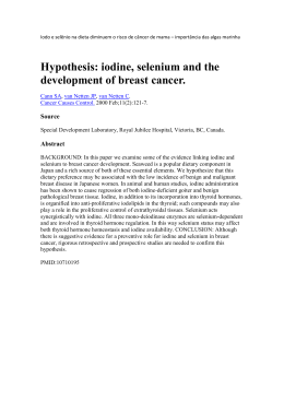
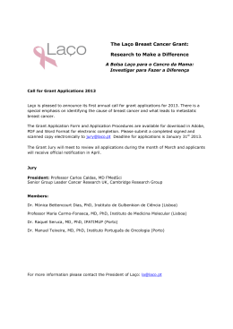
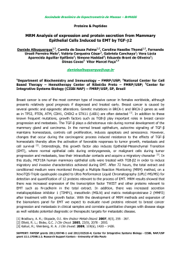
Proceedings of ENCIT 2010
Copyright © 2010 by ABCM
13th Brazilian Congress of Thermal Sciences and Engineering
December 05-10, 2010, Uberlandia, MG, Brazil
DOSE AND RISK EVALUATION IN DIGITAL MAMMOGRAPHY USING
COMPUTER MODELING
Samanda Cristine Arruda Correa, [email protected]
Centro Universitário Estadual da Zona Oeste (CCMAT/UEZO)
Avenida Manuel Caldeira de Alvarenga, 1203, 23070-200, Rio de Janeiro, RJ, Brasil.
Humberto de Oliveira Silva, [email protected]
Instituto de Física, Universidade Federal do Rio de Janeiro, RJ, Brasil.
Edmilson Monteiro de Souza, [email protected]
Centro Universitário Estadual da Zona Oeste (CAPI/UEZO)
Avenida Manuel Caldeira de Alvarenga, 1203, 23070-200, Rio de Janeiro, RJ, Brasil.
Programa de Engenharia Nuclear, COPPE, Universidade Federal do Rio de Janeiro (UFRJ)
Ilha do Fundão, Caixa Postal 68509, 21945-970, Rio de Janeiro, RJ, Brasil.
Centro Universitário Estadual da Zona Oeste (CAPI/UEZO)
Avenida Manuel Caldeira de Alvarenga, 1203, 23070-200, Rio de Janeiro, RJ, Brasil.
Ademir Xavier da Silva, [email protected]
Ricardo Tadeu Lopes, [email protected]
Sarah Braga Magalhães, [email protected]
Programa de Engenharia Nuclear, COPPE, Universidade Federal do Rio de Janeiro (UFRJ)
Ilha do Fundão, Caixa Postal 68509, 21945-970, Rio de Janeiro, RJ, Brasil.
Abstract. Digital mammography has been introduced in several countries in the last years. The new technology
requires new optimising methods considering for instance the increased possibility of changing the absorbed dose,
mainly in modern mammographic systems that allow the operator to choose the beam quality by varying the tube
voltage, and filter and target materials. In this work, the Monte Carlo code MCNPX is used in order toinvestigate how
the average glandular dose (AGD) vary with tube voltage (23-32 kV) and anode–filter combination (Mo-Mo,Mo-Rh
and Rh-Rh) in digital mammographic examinations. Furthermore, the risk of breast cancer incidence attributable to
mammography exams was estimated using the Biological Effects of Ionizing Radiations (BEIR) VII Committee Report.
The results show that the risk of breast cancer incidence in women younger than 30 years of age tends to decrease
significantly using Rh-Rh anode–filter combination and higher tube voltage. For women older than 50 years of age the
variation of tube voltage, and anode–filter combination did not influence the risk values considerably.
Keywords:digital mammography, MCNP, computer modeling, dose.
1. INTRODUCTION
Breast cancer is the second most common type of cancer among Brazilian women, with about 49,240 new cases
expected in Brazil in 2010 (INCA, 2009). According to the American Cancer Society, about 465,000 women die from
the disease annually worldwide (Lopez, 2009). However, the survival of breast cancer patients has been improving in
the last decades. Early diagnosis is one means to lower breast cancer mortality (American Cancer Society, 2003).
Mammography is the most accurate tool for early detection because in many cases it can detect abnormalities such
as masses, calcifications, and other suspicious anomalies up to two years before they are palpable (Shinde, 2003). In
addition, technological advances have resulted in large scale improvements in the performance of newer generation
mammography systems, such as digital mammography systems. Such systems have been proven to be more sensitive in
cancer detection, especially in younger women who tend to have breast tissue more dense (Lewin et al., 2001, Pisano et
al., 2005, Yang et al, 2006). Furthermore, modern mammography systems have a variety of anode–filter combinations
that allow the operator to modify the X-ray beam quality in order to improve the image quality.
Thanks to great performance of the mammography, a considerable number of women in different ages have
undergone the exam. In Brazil, mammograms are recommended for women over 50 years of age, and younger women
with a family history of breast cancer or evidence of a specific genetic change (BRCA1/BRCA2 mutation) (INCA,
2004). However, according to IBGE (2010), among Brazilian women who had undergone mammography, 7% had
done it between the ages of 25 and 29 years, 18.5% had done it between the ages of 30 and 39 years, 28.3% had done it
between the ages of 40 and 49 years and 46.1 % had done it after the age of 50 years.
Although the benefits of mammography for the early detection and treatment of breast cancer are very significant,
the carcinogenic risk associated with mammography and the absorbed radiation dose to the breast have given rise to
concern, especially in younger women who have higher risk of cancer induction by radiation (BEIR, 2005).
Furthermore, optimization is required and, with regard to patient exposure, this refers to the determination of the lowest
average glandular dose (AGD) that yields a sufficient level of clinical image quality. However, scientific efforts to
optimise the choice, in terms of finding the parameter settings which yield sufficient image quality at the lowest
13th Brazilian Congress of Thermal Sciences and Engineering
December 05-10, 2010, Uberlandia, MG, Brazil
Proceedings of ENCIT 2010
Copyright © 2010 by ABCM
possible cost in dose, are still rare (Bouzarjomehri, 2004) mainly for modern mammography systems such as digital
mammography systems.
The first essential step in optimising the patient dose is to make radiologists aware of their own performance in this
regard and how it relates to the risk of radiation damage to the patient. This is not a simple task. In routine radiological
procedures, it is not practical to conduct in vivo measurements of organ doses. The possible practical methods for
deriving the organ doses are measurements in a phantom, or Monte Carlo calculations that simulate the X-ray source
and patient. The latter is the better established approach, is well validated and has the important advantage of flexibility
(IAEA, 2007).
The purpose of this work is to use the Monte Carlo code MCNPX to investigate how the the average glandular dose
and the breast entrance skin dose vary with tube voltage (23-32 kV) and anode–filter combination (Mo-Mo, Mo-Rh and
Rh-Rh) for a modern digital mammography system. The risk of breast cancer incidence attributable to mammography
exams is also estimated for women in different ages, using the Biological Effects of Ionizing Radiations (BEIR) VII
Committee Report (BEIR, 2005).
2. SYSTEM MODELING
The computer code used in this study was MCNPX version 2.5 (Pelowitz, 2005). Although a number of Monte
Carlo codes can be used for dosimetry in diagnostic radiology, MCNP was chosen because it is widely used and has
already been validated in studies on radiation transport involving neutrons, photons, electrons, and more recently
protons, in medical physics and radiological protection. It is also a very versatile code, allowing users to specify
parameters such as source type and geometric configuration, to indicate general conditions of the system such as size,
shape, energy distribution and composition of the medium where radiation interaction takes place, and also to configure
the information-recording commands (tally).
In order to calculate the average glandular dose (AGD) the breast model proposed for Ullman et al. (2005) was
considered. This model of the breast consists of a homogeneous mixture of glandular and adipose tissue in the central
region surrounded by an adipose layer. In this work was adapted in the breast model a skin layer of 0.12 cm and the
tissue compositions are taken from ICRU 44 (1989). The breast is assumed to be in the shape of a cylinder with
semicircular cross-section. The density of glandular tissue was 1.02 g cm-3, for skin tissue 1.09 g.cm-3, and for adipose
tissue 0.95 g.cm-3. The breast dimensions are given in Table 1 and the breast model is shown in Figure 1.
Table 1. Breast thickness, breast radius and thickness of adipose and skin layers.
Thickness (cm)
5.0
Radius (cm)
11
Adipose layer (cm)
0.5
Skin layer (cm)
0.12
Figure 1. Breast model
The energy spectrum of X-rays used as an input parameter in the simulation of the radiation beams was obtained
with the software SRS 78 (Cranley et al., 1997) using added filtration of 30 μm molybdenum (Mo) or 25 μm rhodium
(Rh), molybdenum or rhodium target and tube voltage ranging from 25 to 32 kV. Three target/filter combinations were
considered: Mo/Mo, Mo/Rh and Rh/Rh. The energy spectrum was described in the MCNPX code using 0.5 energy bins.
The photons were emitted by a 0.003-centimetre-diameter plane circular source collimated into a cone of directions by
using the variance reduction technique called source biasing (Pelowitz, 2005). The distance from the source to the
posterior surface of the breast model was kept at 66 cm.
The photons emitted by the radiation source propagated through the air-filled medium before interacting with the
breast model. The average glandular dose and breast entrance skin dose were calculated through MCNPX’s F6
command.
Proceedings of ENCIT 2010
Copyright © 2010 by ABCM
13th Brazilian Congress of Thermal Sciences and Engineering
December 05-10, 2010, Uberlandia, MG, Brazil
The dose values were normalised by the value of entrance air kerma at the breast model. To estimate the entrance air
kerma a point detector (tally F5) was used. The value of fluence registered at point detector was converted to the air
kerma through the conversion factors provided by ICRP 51 of the International Commission on Radiological Protection
(ICRP, 1987).
In this study, every calculation was based on the generation of a number of histories large enough to guarantee a
relative statistical error smaller than 0.05.
2.1. Consideration of the entrance air kerma in digital mammography exams
In order to obtain the average glandular dose and the breast entrance skin dose in digital mammography exams,
entrance air kerma values in digital mammography procedures were measured. All measurements were obtained with a
Senographe DS (GE) mammography unit using tube voltage ranging from 25 to 32 kV and anode–filter combinations
of Mo-Mo, Mo-Rh and Rh-Rh. Exposure automatic control (EAC) was used in order to fix the radiation exposure in the
image detector during the measurements. The fixed radiation exposure value was the same used to obtain digital
mammograms of women with 5-cm-thick breast.
Each experimental entrance air kerma was measured with a mammographic ion chamber positioned on the
geometric breast phantom (Pina, 2006) with thickness of 5 cm, as shown in Figure 2. The distance from the source to
the posterior surface of the geometric breast phantom was kept at 66 cm.
Ion chamber
Breast phantom
Figure 2. Positioning of the mammographic ion chamber on the geometric breast phantom. In this study was used the
10x5-6M ion chamber coupled to a radiation monitor 9010 manufactured by Radcal
The measured entrance air kerma were multiplied by normalized values of the average glandular dose and breast
entrance skin dose calculated with MCNPX code.
2.2. Cancer risk estimation
The lifetime breast cancer incidence that is attributable to mammography was estimated by multiplying the average
glandular dose values with a risk coefficient for breast provided by the BEIR VII Committee Report (BEIR, 2005).
3. RESULTS
Figures 3 and 4 present the values of the average glandular dose and breast entrance skin dose as a function of the
voltage for different anode–filter combinations.
Proceedings of ENCIT 2010
Copyright © 2010 by ABCM
13th Brazilian Congress of Thermal Sciences and Engineering
December 05-10, 2010, Uberlandia, MG, Brazil
Figure 3. Breast entrance skin dose as a function of the voltage for different anode–filter combinations
Figure 4. Average glandular dose as a function of the voltage for different anode–filter combinations
The results show that it is possible to minimize the breast entrance skin and average glandular dose values by raising
the voltage applied to the X-ray tube. The reduction of the dose when the voltage is raised stems from the fact that the
penetrating power of the beam increases with the voltage.
It was also observed that the dose values are also dependent on the anode–filter combination. When the Mo/Mo
anode–filter combination is used, the doses are large. This can be explained by the number of low energy photons that
are included in Mo/Mo spectrum. These photons are mainly absorbed near the surface of the phantom, increasing dose.
In Figure 5 X-ray spectrums obtained with anode–filter combinations of Mo-Mo, Mo-Rh and Rh-Rh are shown.
13th Brazilian Congress of Thermal Sciences and Engineering
December 05-10, 2010, Uberlandia, MG, Brazil
Proceedings of ENCIT 2010
Copyright © 2010 by ABCM
7.00E+009
number photons
6.00E+009
MoMo
MoRh
RhRh
5.00E+009
4.00E+009
3.00E+009
2.00E+009
1.00E+009
0.00E+000
5
10
15
20
25
Energy (keV)
Figure 5. X-ray spectrums
Figures 6, 7, 8, 9 and 10 present the risk of breast cancer incidence for women in different ages exposed to a single
dose. For women under 40 years, the results show that although the risk of breast cancer incidence is large, it can be
significantly minimizes when Rh/Rh anode–filter combination is used. When comparing the risk of breast cancer
incidence for women of 20 years obtained with Mo/Mo and Rh/Rh anode–filter combinations, it is observed a
minimization of about 10 cases per million women when Rh/Rh anode–filter combination is used. It can be seen from
Figures 6, 7, 8, 9 and 10 that is also possible to minimize the risk of breast cancer incidence for younger women by
raising the voltage applied to the X-ray tube.
Figure 6. The lifetime attributable risk of breast cancer incidence for women of 20 years exposed to a single dose
Proceedings of ENCIT 2010
Copyright © 2010 by ABCM
13th Brazilian Congress of Thermal Sciences and Engineering
December 05-10, 2010, Uberlandia, MG, Brazil
Figure 7. The lifetime attributable risk of breast cancer incidence for women of 30 years exposed to a single dose
Figure 8. The lifetime attributable risk of breast cancer incidence for women of 40 years exposed to a single dose
Proceedings of ENCIT 2010
Copyright © 2010 by ABCM
13th Brazilian Congress of Thermal Sciences and Engineering
December 05-10, 2010, Uberlandia, MG, Brazil
Figure 9. The lifetime attributable risk of breast cancer incidence for women of 50 years exposed to a single dose
Figure 10. The lifetime attributable risk of breast cancer incidence for women of 60 years exposed to a single dose
It is important to highlight the fact that the risk of breast cancer incidence presented in this work is for a single
exposure. It increases with the number of exposures, and this is especially important when one considers that millions
of mammograms are performed annually. Therefore, it is desirable to minimize the patient dose.
The risk information given here should provide radiologists and their patients with reasonable estimations of the risk
of breast cancer incidence from radiation received during the mammography. By comparing the radiation risk
Proceedings of ENCIT 2010
Copyright © 2010 by ABCM
13th Brazilian Congress of Thermal Sciences and Engineering
December 05-10, 2010, Uberlandia, MG, Brazil
associated with a wide range of exposures in the mammography, radiologists can minimize the patient dose and
increase the net benefit from the diagnostic procedure.
4. CONCLUSIONS
The results obtained in this work suggest that different precautions need to be taken depending on age of patient. For
women under 40 years, the appropriate choice of anode–filter combination and of tube voltage can minimize
significantly the risk of breast cancer incidence in mammography exams. The dose values and risk of breast cancer
incidence tend to be lower when Rh/Rh anode–filter combination and higher voltages are used. In addition, the results
show that younger women should avoid the mammography and that the recommendation of exam for women over 50
years of age should be considered. Screening mammography with female patients under the age of 50 years should only
be recommended in particular circumstances, such as a family history of breast cancer or evidence of a specific genetic
change.
5. ACKNOWLEDGEMENTS
The authors wish to thank the financial support of the Conselho Nacional de Desenvolvimento Científico e
Tecnológico (CNPq), Coordenação de Aperfeiçoamento de Pessoal de Nível Superior (CAPES) and Fundação Carlos
Chagas Filho de Amparo a Pesquisa do Estado do Rio de Janeiro (FAPERJ), Brazil.
6. REFERENCES
American Cancer Society, “Cancer Facts and Figures 2003”, Atlanta, GA: American Cancer Society, 2003.
Bouzarjomehri, F. (2004) ‘Patient dose in routine X-ray examinations in Yazd state’, Iran. J. Radiat. Res., Vol. 1, No. 4,
pp.199–204.
Committee on the Biological Effects of Ionizing Radiations (BEIR Committee). “Health Risks from Exposure to Low
Levels of Ionizing Radiation: BEIR VII – Phase 2”, Washington, DC: National Academy Press, 2005.
Cranley, K., Gilmore, B.J., Fogarty, G.W.A., Desponds, L., “Catalogue of Diagnostic X-Ray Spectra and Other Data”.
Institute of Physics and Engineering in Medicine. Report 48, 1997.
IBGE. “Um Panorama da Saúde no Brasil: Acesso e utilização dos Sreviços, condições de Saúde e Fatores de Risco e
Proteção à Saúde 2008”, Pesquisa Nacional por Amostra de Domicílios (PNAD), Instituto Brasileiro de Geografia e
Estatística (IBGE), 2010.
INCA.”Controle do Cancer de Mama: Documento de Consenso”, Instituto Nacional do Câncer, Rio de Janeiro: INCA,
2004.
INCA. “Estimativa 2010: Incidência de Cancer no Brasil”. Instituto Nacional de Câncer, Rio de Janeiro: INCA, 2009.
International Atomic Energy Agency (2007) ‘Dosimetry in diagnostic radiology: an international code of practice’,
Technical Report No. 457, Vienna.
International Commission on Radiological Protection. Data for Use in Protection Against External Radiation. ICRP
Publication 51, Oxford: Pergamon Press, 1987.
International Commission on Radiation Units and Measurements. “Tissue Substitutes in Radiation Dosimetry and
Measurement”. ICRU Publication 44. Bethesda, MD, 1989.
Lewin J.M., Hendrick R.E., D'Orsi C.J., Isaacs P.K., Moss L.J., et al. “Comparison of full-field digital mammography
with screenfilm mammography for cancer detection: results of 4,945 paired examinations”. Radiology 218, 873–
880, 2001.
Lopez, M.E. “The Association of Religiosity and Use of Breast Cancer Screening among Older Women in Latin
American and the Caribbean”. Dissertation. University of North Texas Health Science Center, 2009.
Pelowitz, D.B. (2005) MCNPXTM User’s Manual, version 2.5.0. LA-CP-05-0369, Los Alamos National Laboratory.
Pina, D.R., Morceli. J., Duarte, S.B., et al. “Otimização de imagens mamográficas”. Radiol. Bras. 39, 351–354, 2006.
Pisano E.D., Gatsonis C., Hendrick E., Yaffe M., Baum J.K., Acharyya S., et al. “Digital Mammographic Imaging
Screening Trial (DMIST) Investigators Group. Diagnostic performance of digital versus film mammography for
breastcancer screening”. N Engl J Med. 353:1773-83, 2005.
Shinde, M. “Computer Aided Diagnosis in Digital Mammography: Classification of Mass and Normal Tissue”.
Dissertation. Engineering Univerty of South Florida, 2003.
Ullman, G., Sandborg, M., Dance, D.R., Yaffe, M., Carlsson, G.A., “A Search for Optimal X-ray Spectra in Iodine
Contrast Media Mammography”. Physics in Medicine and Biology 50, 3143-3152, 2005.
Yang, W.T., Lai, C.J., Whitman, G.J., et al.”Comparison of Full-Field Digital Mammography and Screen-Film
Mammography for Detection and Characterization of Simulated Small Masses”. American Journal of Roentgen
187(6), w576-w581, 2006.
Baixar