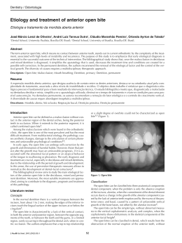
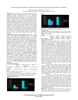
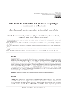
Anterior glenoid rim fracture
77
ORIGINAL ARTICLE
Anterior glenoid rim fracture: the value of helical CT with threedimensional reconstruction and electronic humeral disarticulation
Fratura da margem anterior da glenóide: o valor da tomografia computadorizada helicoidal
com reconstruções 3D e desarticulação eletrônica do úmero*
Heverton César de Oliveira1, Luís Cesar Paiva2, Lúcio Cesar Silva Righi3
ABSTRACT
Objectives: To show a new three-dimensional reconstruction
technique based on helical computed tomography images with
electronic humeral disarticulation in anterior glenoid rim fractures,
correlating the anatomic specimen with simulation of an anterior
glenoid rim fracture, as well as evaluating the extension of the
fracture, the bone fragment position and distance in relation to the
glenoid cavity in six patients. Methods: One scapula and one
humerus with no signs of fracture or congenital malformations
were placed in anatomical position using an adhesive tape after
simulating an anterior glenoid rim fracture made by an osteotome.
Helical CT imaging was acquired and three-dimensional
reconstructions were made based on these images, with and
without electronic humeral disarticulation. The bone fragment was
located, measured and its position in relation to the glenoid cavity
was assessed. Six patients with anterior glenoid rim fracture were
submitted to CT of the shoulder using the same parameters as
those applied to the anatomic specimen. Results: In the anatomic
specimen and in all six patients the bone fragment was clearly
demonstrated; bone fragment measurements in the anatomic
specimen and in three-dimensional reconstructions were
equivalent. The fragment was better demonstrated in the images
taken with electronic humeral disarticulation, particularly in the
frontal view of the glenoid cavity as observed in all six patients.
Conclusion: We concluded that our experiment with the anatomic
specimen and the study of six patients allow us to state that this
technique is safe and accurate to demonstrate the extension, size
and location of the bone fragment in anterior glenoid rim fractures,
and it provides essential elements for therapeutic planning.
Keywords: Fractures; Shoulder/injury; Humerus; Tomography,
spiral computed; Imaging, three dimensional
RESUMO
Objetivos: Mostrar nova técnica de reconstrução em 3D, a partir
de imagens obtidas por tomografia computadorizada espiral, com
desarticulação eletrônica do úmero, nas fraturas da margem
anterior da glenóide, correlacionando peça anatômica com
simulação de fratura da margem anterior da glenóide, além de
avaliar a extensão da fratura, a posição e a distância do fragmento
ósseo em relação à glenóide em seis pacientes. Métodos: Uma
escápula e um úmero sem sinais de fratura ou malformações
congênitas foram colocados em posição anatômica com o auxílio
de fita adesiva, após a simulação de fratura da margem anterior
da glenóide, realizada com um osteótomo, então foram obtidas
imagens tomográficas com técnica espiral a partir dessas imagens
foram realizadas reconstruções em 3D com e sem desarticulação
eletrônica do úmero. O fragmento ósseo foi localizado, mensurado
e estudada sua posição em relação à glenóide. Seis pacientes
com fratura da margem anterior da glenóide foram submetidos a
exame tomográfico do ombro com os mesmos parâmetros
utilizados na peça anatômica. Resultados: Na peça anatômica e
nos seis pacientes, o fragmento ósseo foi bem demonstrado; as
medidas do fragmento ósseo na peça anatômica e nas
reconstruções em 3D foram equivalentes, sendo melhor
demonstrado nas imagens com desarticulação eletrônica do
úmero, especialmente na visão frontal da glenóide, que também
ocorreu com os seis pacientes. Conclusão: Concluímos que o nosso
experimento com a peça anatômica e o estudo dos seis pacientes
nos permitem afirmar que essa técnica oferece segurança e
precisão para demonstrar a extensão, as dimensões e a localização
do fragmento ósseo nas fraturas da margem anterior da glenóide,
propiciando elementos fundamentais para o planejamento
terapêutico.
Descritores: Fraturas; Ombro/lesões; Úmero; Tomografia
computadorizada espiral; Imagem tridimensional
INTRODUCTION
The shoulder is the most mobile joint of the human
body and most subluxations occur in the glenohumeral
joint(1). In 96% of the cases, dislocation is caused by
direct trauma and 84% of the patients present anterior
* Study carried out in the Imaging Diagnosis Department, Escola Paulista de Medicina/Unifesp, São Paulo (SP) and Lúmen - Centro de Diagnósticos. São Bernardo do Campo (SP).
1
2
3
Professor, Imaging Diagnosis Department, Escola Paulista de Medicina/Unifesp; Director of Lúmen - Centro de Diagnósticos. São Bernardo do Campo (SP).
Radiologist, Lúmen - Centro de Diagnósticos. São Bernardo do Campo (SP).
Orthopedist, Hospital Ifor; Instructor of medical residency at Hospital Ifor. São Bernardo do Campo (SP).
Corresponding author: Heverton César de Oliveira - R. Américo Brasiliense, 628 - Centro - CEP 09715-021 - São Bernardo do Campo (SP), Brazil. Fax: 4331-6465 - Tel.: 4331-6464- e-mail: [email protected]
Received on September 1, 2003 – Accepted on November 8, 2003
einstein 2003; 1:77-83
78
Oliveira HC, Paiva LC, Righi LCS
subluxation due to fall with the arm in hyperxtension,
abduction and external rotation(2-3).
The anterior glenoid rim fractures occur in
consequence to traumatic glenohumeral dislocation,
by means of forces applied to the humeral head during
subluxation movement; it is defined as an acute fracture
and may result from bone erosions associated with
recurrent glenohumeral instability(4).
These fractures are very often difficult to diagnose
by conventional radiographic techniques, even using
special views(4-6).
Computed tomography imaging usually demonstrates
A
B
C
E
D
F
Figure 1. Anatomic specimen. (A) Scapula showing simulation of anterior glenoid rim fracture (arrow).
(B and C) 3D reconstruction images. Lateral and anterior views with electronic humeral disarticulation
showing the bone fragment and its relations with the glenoid rim (arrow). (D and E) 3D reconstruction
image with the humerus. In the lateral view (D), humeral head overlaps the glenoid cavity and hinders
visualization of the fracture, whereas in the anterior view (E), the bone fragment of the anterior
glenoid rim could be seen (arrow). (F) Cross-sectional CT images showing amputation of the anterior
glenoid rim and bone fragment (arrow).
einstein 2003; 1:77-83
Anterior glenoid rim fracture
fracture and bone fragment; however the overlapping
of humeral head and other elements, and no availability
of complete imaging of the glenohumeral joint, make
diagnosis, spatial location of the bone fragment and
surgical treatment planning difficult.
The number of publications with three-dimensional
reconstructions of the musculo-skeletal system based
on helical CT images has increased in the past years.
Nevertheless, we found only one study about anterior
glenoid rim fractures using this technique(5) and no
study with electronic humeral disarticulation exposing
the entire glenoid cavity for observation.
The main objective of this article is to demonstrate
a new three-dimensional reconstruction technique with
electronic humeral disarticulation in anterior glenoid rim
fractures. Thus, we correlated the anatomic specimen
with simulated anterior glenoid rim fracture and
assessed the extension of the fracture, as well as the
position and distance of the bone fragment in relation
to the glenoid cavity, in six patients.
METHODS
One scapula and one humerus with no signs of fracture
or congenital malformations were placed in anatomical
position using an adhesive tape after simulating an
anterior glenoid rim fracture made by an osteotome.
The bone fragment was fixed beside the anterior
glenoid rim with a radiotransparent gelly-like material.
Cross-sectional helical CT images were made using the
following parameters: contiguous 3 mm-thick slices,
pitch of 1, and 1.5-mm reconstructions using Philips
Tomoscan AV performance (120kV, 150 mA, 2250 UH
window width, 800 UH center), with 512 x 512 matrix,
and a 320-mm field of vision. After acquiring the images,
three-dimensional reconstructions were performed in a
Philips – Easy Vision workstation using specific software
with and without electronic humeral disarticulation.
The anatomic specimen and the images acquired
by three-dimensional reconstructions were correlated
in order to determine the location, size and position
of the bone fragment in relation to the glenoid cavity
(figures 1A to 1F).
Cross-sectional helical CT images of the shoulder
of six patients with anterior glenoid rim fracture and
three-dimensional reconstructions, with or without
electronic humeral disarticulation, were made using the
same parameters described.
All patients were male, age range 23-40 years, mean
age of 33 years. Five patients had recurrent anterior
glenohumeral instability symptoms due to previous
trauma and one patient had an acute direct trauma.
79
RESULTS
In the scapula submitted to simulation of glenoid rim
fracture, the bone fragment measured 16.7 x 4.2 mm in
the longitudinal and axial planes, respectively, and 9
mm when moved downwards in relation to the inferior
glenoid rim (figure 2). The measurements made in the
three-dimensional reconstruction images of the scapula
were 16.4 x 4.0 mm in the longitudinal and axial planes,
respectively, and 9.1 mm in relation to the inferior
glenoid rim (figures 2A and 2B).
The cross-sectional CT images of the six patients
demonstrated anterior glenoid rim fractures and bone
fragments (figures 3A, 4A, 5A), like in the threedimensional reconstructions, with no electronic
humeral disarticulation, particularly in the anterior view
(figures 3C, 4C, 5C). However, three-dimensional
reconstructions with electronic humeral disarticulation
showed anterior glenoid fracture in all views. The
frontal view of the glenoid cavity stood out (figures
3D, 3E, 4D, 4E, 5D, 5E) because the entire glenoid
rims are displayed, showing the bone fragment with no
overlapping image of the humerus, unlike other views
16,7 mm
16,4 mm
4,2 mm
9,0 mm
4,0 mm
A
9,1 mm
B
Figure 2. Three-dimensional reconstruction of the anatomic specimen
demonstrating the longitudinal and transversal diameters of bone fragment
(A), and its inferior dislocation in relation to the anterior glenoid rim (B).
einstein 2003; 1:77-83
80
Oliveira HC, Paiva LC, Righi LCS
B
A
C
D
E
Figure 3. A 26-year-old male patient injured in a road accident; anterior subluxation reduced two days before. (A)
Cross-sectional CT image of the right shoulder shows fracture in anterior glenoid rim and adjacent fragment (arrow).
(B) 3D reconstruction image of the right shoulder, lateral view with humerus. Humeral head overlaps the glenoid cavity
and hinders visualization of the fracture. (C) 3D reconstruction image of the right shoulder, anterior view with
humerus, showing bone fragment of the anterior glenoid rim (arrow). (D and E) 3D reconstruction image of the right
shoulder, lateral (D) and anterior views (E) after electronic humeral disarticulation, demonstrating anterior glenoid rim
fracture and bone fragment and their relations (arrow).
without electronic humeral disarticulation (figures 3B,
4B, 5B). The technique described clearly showed the
bone fragment in all patients, and enabled surgical
planning using a three-dimensional view of the fracture.
einstein 2003; 1:77-83
DISCUSSION
Thanks to technological developments, the imaging
diagnosis methods now demonstrate details of the
interior human body that were observed only during
Anterior glenoid rim fracture
surgeries or in anatomical studies. Therefore, some
diagnoses that could not be made without invasive
methods are routinely made and enable less aggressive,
more functional and more economic treatments.
The diagnosis of anterior glenoid rim fractures by
imaging methods is often difficult, and has the
disadvantage of not providing subsidies for therapeutic
planning (5) . Rockwood et al. mentioned eight
radiographic views used for the same purpose in several
centers(7), and showed how conventional radiology is
limited in these cases.
A
81
Thus, many authors use CT with or without intraarticular contrast as well as magnetic resonance
imaging(8-12) to diagnose anterior glenoid rim fractures
and plan their treatment; however, there are some
limitations, such as overlapping of structures and
difficulty to demonstrate the bone fragment position
and its relation with the glenoid cavity.
The literature on selective three-dimensional
reconstruction of anatomic structures using CT images
is scarce and the few studies published are mostly
related to calcaneal fractures(10).
B
C
D
E
Figure 4. A 26-year-old male patient with glenohumeral instability on the right joint and subluxation reduced
five days before. (A) Cross-sectional CT imaging of the right shoulder showing fracture in anterior glenoid rim
(arrow). (B) 3D reconstruction image of the right shoulder, lateral view with humerus. Humeral head overlaps
the glenoid cavity hinders visualization of the fracture. (C) 3D reconstruction image of the right shoulder,
anterior view with humerus shows fracture in anterior glenoid rim (arrow). (D and E) 3D reconstruction image
of the right shoulder, lateral (D) and anterior (E) views after electronic humeral disarticulation demonstrating
fracture in anterior glenoid rim and their relations (arrow).
einstein 2003; 1:77-83
82
Oliveira HC, Paiva LC, Righi LCS
A
B
C
D
E
Figure 5. A 30-year-old male patient with glenohumeral instability in the left shoulder and history of six
previous subluxations (the last subluxation occurred 15 days before the examination). (A) Cross-sectional
CT images of the left shoulder shows fracture in anterior glenoid rim (arrow). (B) 3D reconstruction image
of the left shoulder, lateral view with humerus. Humeral head overlapping the glenoid cavity and hindering
visualization of the fracture. (C) 3D reconstruction image of the left shoulder, anterior view with humerus,
shows slight irregularity on the anterior glenoid rim (arrow). (D) 3D reconstruction image of the left shoulder,
lateral view after electronic humeral disarticulation demonstrates fracture in anterior glenoid rim and their
relations (arrow). (E) 3D reconstruction image of the left shoulder, anterior view after electronic humeral
disarticulation showing slight irregularity on the anterior glenoid rim (arrow).
einstein 2003; 1:77-83
Anterior glenoid rim fracture
In these fractures, with no electronic calcaneal
disarticulation, Freund et al.(10) demonstrated that
three-dimensional reconstructions have the same
disadvantages of conventional radiology, primarily bone
overlapping, an obstacle that was easily overcome by
separating the bone through electronic disarticulation,
thus enabling observation of the fractured calcaneus
in all its views.
Stevens et al.(5) used three-dimensional reconstruction
of the anterior glenoid rim fracture and demonstrated
clear superiority of this technique in relation to
conventional radiology; however, these authors did not
carry out studies showing the glenoid cavity and bone
fragment. They compared the performance of crosssectional CT imaging with radiographic images and
suggested 3D reconstruction of CT images could
contribute to diagnosis of these fractures.
We believe it is fundamental to show the glenoid
cavity in frontal view in order to understand the
fracture; moreover, electronic humeral disarticulation
enables visualizing the entire joint with no overlapping
of the humeral head.
We concluded that our experiment with the
anatomic specimen and the study of six patients allow
us to state that this technique is safe and accurate to
demonstrate the extension, size and location of the
bone fragment in anterior glenoid rim fractures, and it
provides essential elements for therapeutic planning.
83
REFERENCES
1. Kazar B, Relovsky E. Prognosis of primary dislocation of the shoulder. Acta
Orthop Scand 1969;40:216-24.
2. Cave EF, Burke JF, Boyd RJ. Posterior dislocations. In: Cave EF. Trauma
management. Chicago: Year Book; 1974. p. 437.
3. Danzig L, Resnick D, Greenway G. Evaluation of unstable shoulders by computed
tomography. A preliminary study. Am J Sports Med 1982;10:138-41.
4. Bigliani LU, Newton PM, Steinmann SP, Connor PM, Mcllveen SJ. Glenoid rim
lesions associated with recurrent anterior dislocation of the shoulder. Am J
Sports Med 1998;26:41-5.
5. Stevens KJ, Preston BJ, Wallace WA, Kerslake RW. CT imaging and threedimensional reconstructions of shoulders with anterior glenoumeral instability.
Clin Anat 1999;12:326-36.
6. Resnick D. Physical injury: sports related abnormalities. In: Resnick D. editor.
Diagnosis of bone and joint disorders. 4th ed. Philadelphia: WB Saunders;
2002. p. 2783-933.
7. Rockwood CA, Wirth MA. Subluxations and dislocations about the glenoumeral
joint. In: Rockwood CA, Green DP, Bucholz RW, Heckman JD. editors. Rockwood
and Green’s fractures in adults. Philadelphia: Lippincott-Raven; 1996. p.
1193-255.
8. Seeger LL, Gold RH, Bassett LW. Shoulder instability: evaluation with MR
imaging. Radiology 1988;168:695-7.
9. Singson RD, Feldman F, Bigliani L. CT arthrographic patterns in recurrent
glenohumeral instability. AJR Am J Roentgenol 1987;149:749-53.
10. Freund M, Thomsen M, Hohendorf B, Zenker W, Heller M. Optimized
preoperative planning of calcaneal fractures using spiral computed tomography.
Eur Radiol 1999;9:901-6.
11. Sanders TG, Morrison WB, Miller MD. Imaging techniques for the evaluation
of glenoumeral instability. Am J Sports Med 2000;28:414-34.
12. Seltzer SE, Weissman BN. CT findings in normal and dislocating shoulders. J
Can Assoc Radiol 1985;36:41-6.
einstein 2003; 1:77-83
Baixar