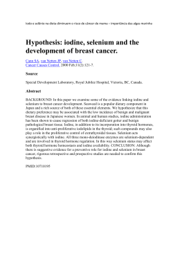
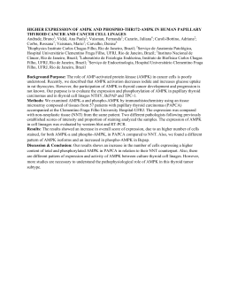
Int J Endocrinol Metab. 2014 October; 12(4): e15806.
DOI: 10.5812/ijem.15806
Case Report
Published online 2014 October 1.
Invasive Thyroid Angiosarcoma With a Favorable Outcome
1,*
1
1
2
Joana Couto ; Raquel G Martins ; Ana Paula Santos ; Joana Matos ; Isabel Torres
1
1Endocrinology Department, Portuguese Institute of Oncology FG, Porto, Portugal
2Pathology Department, Portuguese Institute of Oncology FG, Porto, Portugal
*Corresponding author: Joana Couto, Endocrinology Department, Portuguese Institute of Oncology FG, Porto, Portugal. Tel: + 351-969014901, Fax: + 351-225084001, E-mail: [email protected]
Received: October 30, 2013; Revised: May 31, 2014; Accepted: June 13, 2014
Introduction: Thyroid angiosarcoma is a malignant neoplasm, which usually shows local aggressive behavior and associated with a high
recurrence rate. It was originally described in the Alpine region and extremely rare in other parts of the world.
Case Presentation: An asymptomatic 61-year-old woman presented with a right lobe thyroid nodule with fine needle aspiration cytology
indicating “suspicious for malignancy” was reported in our study. Histological examination revealed a vascular 35mm neoplasm with
areas of necrosis. Immunohistochemistry staining had negative results for MNF, CAM5.2, CD34, thyroglobulin, and positive for CD31. The
findings described were compatible with angiosarcoma diagnosis. Besides, a 4mm papillary microcarcinoma was found in the left lobe.
Computerized tomography (CT) scan performed about two months after the operation, showed a right neck nodular lesion, conditioning
tracheal deviation. At our institution, the study performed was consistent with local recurrence (angiosarcoma). Tumor excision was
performed and invasion to larynx, trachea and esophagus was detected intraoperatively. Histopathologic examination confirmed tumor
recurrence and the patient was submitted to radiotherapy (60 Gy), completed four years ago. There is not, so far, any evidence of tumor
recurrence.
Conclusions: We described a rare case of a usually aggressive thyroid angiosarcoma in a patient living in a non-Alpine region, with an
unusual favorable outcome after the operation and radiotherapy.
Keywords:Angiosarcoma; Thyroid; Neoplasia
1. Introduction
Most thyroid tumors are primary and of epithelial origin, while tumors of mesenchymal origin, common in
other organs, rarely occur in this gland (1-3). Vascular
thyroid lesions include benign neoformation such as
hemangiomas, and malignant tumors, like angiosarcoma and angiosarcomatoid carcinoma. Angiosarcoma
is a rare soft tissue tumor, accounting for about 1-2 % of
all sarcomas, mainly occurring in the skin and head and
neck superficial soft tissues (1, 4, 5). Thyroid angiosarcoma is a rare malignant tumor of endothelial differentiation, mainly found in European mountainous regions
of the Alps (Switzerland, Austria and Northern Italy),
where it represents up to 16% of thyroid malignancies
(1, 5-8). Its high prevalence in those regions is thought
to be due to iodine deficiency associated goiter, because
most thyroid angiosarcomas occur in a multinodular
gland (6, 7). Although its etiology remains unknown, it
was suggested that endothelial proliferation after recurrent hemorrhage in nodules within a long-standing
multinodular goiter, can induce neoplastic transformation (1, 2). It is more common in females (ratio 9:3) and
mainly occurs between the fifth and eighth decades (the
median age of 65 years) (1, 6, 9). Generally, these tumors
present as a painless infiltrating mass with tendency to
increase its size (1), which can vary from 2.5 to 11cm, being usually unifocal (6, 10). Cytological characteristics of
this type of cancer have not been clearly defined yet and
are rarely described in the literature (5). Histological examination often reveals extensive areas of necrosis, hemorrhage with the presence of anastomosing channels
lined by endothelial cells. Endothelial differentiation is
evidenced by the expression of vascular markers such as
CD31 (the most sensitive and specific marker), CD34 and
factor VIII related antigen. In many cases, there may be
coexpression of cytokeratins. Immunonegativity for thyroglobulin supports its diagnosis (6-8). Some authors
believe that most thyroid angiosarcomas are in fact anaplastic thyroid carcinomas with angiomatoid features;
however, this distinction is considered academic, since
their prognosis and treatment are similar (1, 8, 11). Treatment of these tumors is conditioned by local aggressive
behavior and high recurrence rate, consisting of radical
surgery wherever feasible. Radiation therapy seems to
be effective in some patients and can be complemented
with chemotherapy using Adriamycin (1, 7). Recurrence
and development of metastasis are common, even after
complete resection (2) and the prognosis is poor, due to
local aggressive behavior. Presence of distant metastasis
Copyright © 2014, Research Institute For Endocrine Sciences and Iran Endocrine Society; Published by Kowsar. This is an open-access article distributed under the
terms of the Creative Commons Attribution-NonCommercial 4.0 International License (http://creativecommons.org/licenses/by-nc/4.0/) which permits copy and
redistribute the material just in noncommercial usages, provided the original work is properly cited.
Couto J et al.
and extracapsular tumor extension are frequently associated with a poor prognosis (1). Metastasis is commonly
found in local lymph nodes, lung and bone marrow (1, 12).
Mean 5-year survival rate is near 33% and most patients
die in less than six months, regardless of treatment (1, 6).
Tumor resection was performed and invasion to larynx,
trachea and esophagus was noticed intraoperatively. Histological examination revealed a 2.5 cm angiosarcoma,
with positive surgical margins and immunohistochemistry staining showed the same features described above,
including strong positivity result for CD31 with cytoplasmic and membranous pattern (Figure 2).
We present a 61-year-old-women with no relevant medical
history, born in northern Portugal, where she has always
lived. When a neck mass was noticed, neck ultrasound was
performed showing two solid thyroid nodules, one in the
right lobe with about 3 cm and another in the left lobe
with about 9 mm. There were no associated compression
symptoms. Fine needle aspiration (FNA) cytology was held
reporting “suspicious for malignancy". The patient underwent total thyroidectomy and histological examination
showed a 35 mm heterogeneous and hemorrhagic vascular neoplasm with dilated vessels, areas of necrosis, with
swollen endothelial cells and atypical mitoses between
thyroid follicles and cells. Immunohistochemistry staining showed negative results for cytokeratins MNF, CAM5.2,
CD34, thyroglobulin and positive finding for CD31. These
findings confirmed the diagnosis of angiosarcoma. A 4
mm papillary microcarcinoma was also identified in the
left lobe, without any poor prognosis criteria. Neck computerized tomography (CT) scan performed two months
after the operation showed a 35 mm nodular lesion in the
right surgical bed, with irregular margins, conditioning
tracheal deviation. The patient was referred to our institution. FNA of the nodule was held showing local recurrence
of the previously diagnosed malignancy. 18F-FDG PET-CT
identified a single neck hypermetabolic lesion compatible
with tumor recurrence (Figure 1).
Figure 2. Histological Examination Revealed a 2,.5 cm Angiosarcoma,
2. Case Presentation
With Positive Surgical Margins
A, Tumor cells with vesicular nuclei with prominent nucleoli and abundant eosinophilic cytoplasm (H&E staining); B, Strong positive result for
CD31 with cytoplasmic and membranous pattern (Immunohistochemistry staining).
It was decided to treat the patient with local radiotherapy (60Gy), completed four years ago. The patient was kept
under close surveillance at our institution, and about six
years after the initial diagnosis, there was no clinical, analytical or imaging (neck CT, magnetic resonance imaging) evidence of tumor recurrence.
3. Conclusions
Figure 1. 18F-FDG PET-CT Identified a Single Neck Hypermetabolic Lesion
2
We described a rare case of thyroid angiosarcoma associated with a papillary microcarcinoma on the contralateral
Int J Endocrinol Metab. 2014;12(4):e15806
Couto J et al.
lobe in a patient living outside the Alpine region. A recent
literature review by Kaur et al. (13) identified 48 cases of
thyroid angiosarcoma worldwide. Since then, as far as we
know, three more cases were published (14-16), and including one we described, there are a total of 52 patients with
thyroid angiosarcoma in the literature. Generally, there
was a favorable outcome despite initial local recurrence
with invasion to adjacent organs. We are unaware of what
might exactly influenced the patient’s prognosis, but we
believe that local radiotherapy may have an important
role in the treatment of these types of lesions. Data available is insufficient to suggest the most suitable approach
to this type of cases, but surgical treatment of head and
neck angiosarcoma followed by radiotherapy improved
patient’s survival (13). However, there are some reports of
patients treated with radiotherapy who did not respond to
that modality of treatment (5, 6, 17). We could not find any
similar case of such a long disease free period, after radiation treatment. In areas not associated with endemic multinodular goiter, cases with higher than average survival
have been reported (1), comparable to our patient’s case.
Good prognosis has been associated with absence of extra glandular extension (9), which was found in this case,
but not changing the final outcome. The low incidence of
these tumors in non-Alpine regions makes them difficult
to diagnose and treat. Being so, we believe that multidisciplinary collaboration is essential in addressing this and
other rare thyroid neoplasms.
Authors’ Contribution
Study concept and design: Couto, Torres. Acquisition,
Analysis and interpretation of data: Couto, Martins, Matos. Drafting of the manuscript: Couto. Critical revision
of the manuscript for important intellectual content:
Santos, Torres.
References
1.
Petronella P, Scorzelli M, Luise R, Iannaci G, Sapere P, Ferretti M,
et al. Primary thyroid angiosarcoma: an unusual localization.
Int J Endocrinol Metab. 2014;12(4):e15806
2.
3.
4.
5.
6.
7.
8.
9.
10.
11.
12.
13.
14.
15.
16.
17.
World J Surg Oncol. 2012;10:73.
Eusebi V. Angiosarcoma. In: DeLellis RA, Lloyd RV, Heitz PU, Eng C
editors. World health organisation classification of tumours Pathology and genetics tumours of endocrine organs.. Lyon: IARC Press;
2004. pp. 113–4.
Sniezek JC, Holtel M. Rare tumors of the thyroid gland. Otolaryngol Clin North Am. 2003;36(1):107–15.
Weiss SW, Goldblum JR. Malignant vascular tumors. In: Weiss SW,
Goldblum JR editors. Enzinger and Weiss's Soft Tissue Tumors.. St
Louis: Mosby; 2001. pp. 917–45.
Isa NM, James DT, Saw TH, Pennisi R, Gough I. Primary angiosarcoma of the thyroid gland with recurrence diagnosed by fine needle aspiration: a case report. Diagn Cytopathol. 2009;37(6):427–32.
Goh SG, Chuah KL, Goh HK, Chen YY. Two cases of epithelioid
angiosarcoma involving the thyroid and a brief review of nonAlpine epithelioid angiosarcoma of the thyroid. Arch Pathol Lab
Med. 2003;127(2):E70–3.
Kalitova P, Plzak J, Kodet R, Astl J. Angiosarcoma of the thyroid.
Eur Arch Otorhinolaryngol. 2009;266(6):903–5.
Ryska A, Ludvikova M, Szepe P, Boor A. Epithelioid haemangiosarcoma of the thyroid gland. Report of six cases from a non-Alpine
region. Histopathology. 2004;44(1):40–6.
Maiorana A, Collina G, Cesinaro AM, Fano RA, Eusebi V. Epithelioid angiosarcoma of the thyroid. Clinicopathological analysis
of seven cases from non-Alpine areas. Virchows Arch. 1996;429(23):131–7.
Zouaidia F, Jahid A, Elouazzani H, Laraki L, Bernoussi Z, Mansouri
F, et al. Angiosarcoma of the Thyroid Gland: A Case Report. J Med
Cases. 2010;1(1):29–31.
Kim NR, Ko YH, Sung CO. A case of coexistent angiosarcoma
and follicular carcinoma of the thyroid. J Korean Med Sci.
2003;18(6):908–13.
Yilmazlar T, Kirdak T, Adim S, Ozturk E, Yerci O. A case of hemangiosarcoma in thyroid with severe anemia due to bone marrow
metastasis. Endocr J. 2005;52(1):57–9.
Kaur A, Didolkar MS, Thomas A. Angiosarcoma of the thyroid: a case report with review of the literature. Endocr Pathol.
2013;24(3):156–61.
Innaro N, Succurro E, Tomaino G, Arturi F. Nonalpine thyroid
angiosarcoma in a patient with hashimoto thyroiditis. Case Rep
Oncol Med. 2013;2013:901246.
Kefeli M, Mete O. An Unusual Malignant Thyroid Nodule: Coexistence of Epithelioid Angiosarcoma and Follicular Variant Papillary Thyroid Carcinoma. Endocr Pathol. 2013.
Gouveia P, Silva C, Magalhaes F, Santos C, Guerreiro E, Santos
F, et al. Non-Alpine thyroid angiosarcoma. Int J Surg Case Rep.
2013;4(5):524–7.
Astl J, Duskova J, Limanova Z, Povysil C, Kuchynkova Z. Hemangiosarcoma of the Thyroid Gland. A case report. Neuro Endocrinol
Lett. 2000;21(3):213–6.
3
Baixar