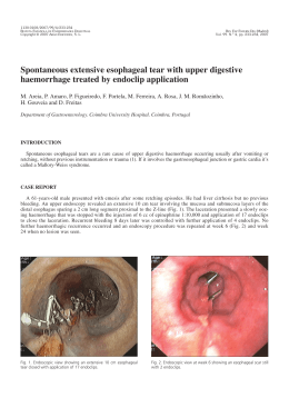
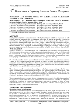
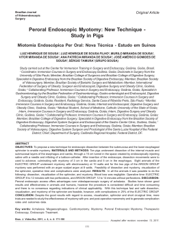
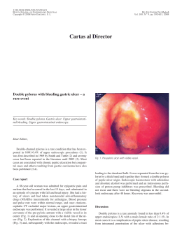
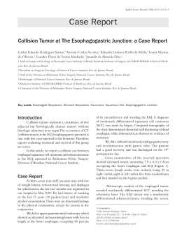
1130-0108/2010/102/5/341-342
REVISTA ESPAÑOLA DE ENFERMEDADES DIGESTIVAS
Copyright © 2010 ARÁN EDICIONES, S. L.
REV ESP ENFERM DIG (Madrid)
Vol. 102, N.° 5, pp. 341-342, 2010
Letters to the Editor
Glycogenic acanthosis of the esophagus: an
unusually endoscopic appearance
Key words: Glycogenic acanthosis. Endoscopic appearance.
Dear Editor,
Glycogenic acanthosis of the esophagus is a common benign
entity characterized by multifocal plaques of hyperplastic squamous epithelium with abundant intracellular glycogen deposits
(1). At esophagoscopy these lesions appear as slightly raised
grey-white plaques which are usually 2-10 mm in diameter and
may be confluent. Although they may involve any segment, the
literature suggests that are most common in the distal third (2).
Fig. 1. Upper endoscopy: pediculated polyp as large as 7 mm in the
middle esophagus.
Case report
A 78-year-old white male admitted to the Medicine Department with microcytic anaemia [haemoglobin: 8.8 g/dl (13-15);
mean corpuscular volume: 66 fl] and for a suspicion of lead
poisoning. He had a diffuse abdominal pain, nausea, anorexia,
asthenia and constipation. There were no symptoms of
esophageal reflux or dysphagia. He drank heavily (100 g of
wine per day) and smoked three packs of cigarettes a day for 30
years. Current medications included furosemide, perindopril
and metformin for hypertension and diabetes mellitus. On examination, he presented cutaneomucosal pallor, tenderness in
the lower quadrants and peripheral edema. An upper endoscopy
revealed a pale-pink, lobulated and pediculated polyp as large
as 7 mm in diameter in the middle esophagus (Fig. 1). A
polypectomy was performed and histology demonstrated
glycogenic acanthosis (Figs. 2 and 3). A grade B esophagitis in
Fig. 2. Low-power photomicrograph: squamous epithelium thickened
by groups of enlarged and clear cells located in the upper layers of the
epithelium.
342
LETTERS TO THE EDITOR
Fig. 3. Medium-power photomicrograph: enlarged squamous cells
with abundant clear cytoplasm.
the distal third and a linear area of red mucosa, 2 cm above the
esophago-gastric transition were also visualized. Biopsy of that
area revealed Barrett’s epithelium with no dysplasia. Acid-reducing drug was instituted. A total colonoscopy was performed
and revealed no changes. Abdominal ultrasonography showed
only hepatic steatosis. The blood lead level was 3,848 µg/L
(40-290) which confirmed the lead poisoning’s diagnosis.
Chelation therapy was started with symptomatic and laboratory
improvement.
The overall incidence in the general population is difficult to
determine (endoscopic series have ranged from 5-15%). Endoscopy shows whitish mucosal elevations usually less than
0.5 cm surrounding by normal mucosa. A few or multiple lesions may be found assuming sometimes a confluent conglomeration with a cobblestone mucosal pattern. Exceptionally,
nodularity is focal or there is diffuse fold thickening, such cases
endoscopy may be necessary to exclude some other possibilities, particularly superficial carcinoma (2). In contrast with literature, the endoscopic appearance of the current case has never been reported. Morphology and the underlying disease have
led to a resection because it’s important to recognize the nature
of the lesion. Pathologically, the squamous epithelium is focally thickened by groups of enlarged and clear cells located in the
upper layers of the epithelium which contain abundant cytoplasmic glycogen and appear clear because of routine histological processing removal (1,3).
In summary, glycogenic acanthosis seems to have no clinical significance and endoscopy should be used only when the
appearance is atypical and/or the clinical suspicion of
esophageal disease is high. Furthermore, this condition must be
differentiated from lesions of similar appearance and more important prognostic significance.
S. Lopes, P. Figueiredo, P. Amaro, P. Freire, S. Alves,
M. A. Cipriano, H. Gouveia, C. Sofia and M. Correia-Leitão
Department of Gastroenterology. Coimbra University
Hospital. Coimbra, Portugal
Discussion
Glycogenic acanthosis is a benign entity of unknown pathogenesis and etiology, perhaps representing a degenerative or reactive phenomenon involving the squamous esophageal epithelium, apparently age-related and progressive (1). Despite the
name, there is no association with abnormalities of glucose metabolism or with skin disorders, such as acanthosis nigricans.
There isn’t a clear relationship to smoking, alcohol, hot liquids
or acid reflux (3,4). In fact, despite the antireflux therapy seems
to improve symptoms, it failed to eradicate the lesions of glycogenic acanthosis (4). Unless there is a coexisting disease, patients with glycogenic acanthosis are asymptomatic (2). The
present case, endoscopy was performed in the workup of microcytic anaemia.
REV ESP ENFERM DIG (Madrid)
References
1.
2.
3.
4.
Ghahremani GG, Rushovich M. Glycogenic acanthosis of the esophagus: radiographic and pathologic features. Gastrointest Radiol 1984;
9: 93-8.
Glick SN, Teplick SK, Goldstein J, Stead JA, Zitomer N. Glycogenic
acanthosis of the esophagus. Am J Roentgenol 1982; 139(4): 683-8.
Bender MD, Allison J, Cuartas F, Montgomery C. Glycogenic acanthosis of the esophagus: a form of benign epithelial hyperplasia. Gastroenterology 1973; 65(3): 373-80.
Vadva MD, Triadafilopoulos G. Glycogenic acanthosis of the esophagus and gastroesophageal reflux. J Clin Gastroenterol 1993; 17(1): 7983.
REV ESP ENFERM DIG 2010; 102 (5): 338-347
Baixar