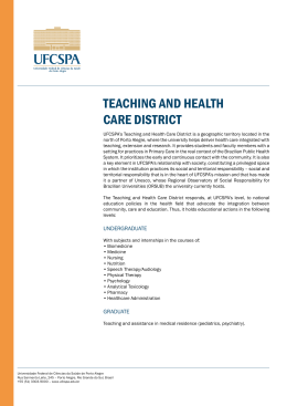
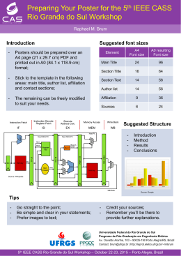
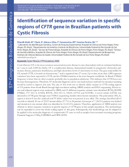
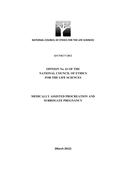
|CISTOADENOMA | RELATOS DE CASOS MUCINOSO DE PÂNCREAS... Coral et al. RELATOS DE CASOS Cistoadenoma mucinoso de pâncreas durante a gravidez Mucinous cystadenoma of the pancreas developing during pregnancy Roberto Pelegrini Coral1, Roberto Viña Coral2, Felipe W. De Bacco3, Natalino Rinaldi4, Cláudio G. Zettler5 RESUMO Cistoadenoma mucinoso de pâncreas durante a gravidez é extremamente raro e relatado somente cinco vezes na literatura. Nós descrevemos caso de uma mulher de 26 anos, na sua segunda gestação, com um acentuado aumento de volume intra-abdominal por massa cística, cuja gestação foi interrompida durante a trigésima primeira semana por severa pré-eclâmpsia. A cirurgia de retirada do cisto ocorreu 26 dias após o parto, com enucleação total da lesão. O exame anatomopatológico evidenciou o cisto como cistoadenoma mucinoso, que mediu 23x20x15 cm e pesou 9070 g. Até onde é do nosso conhecimento, é um dos maiores cistoadenomas mucinosos pancreáticos relatados durante a gestação. UNITERMOS: Pâncreas, Cistoadenoma Mucinoso, Gravidez, Neoplasia Cística Mucinosa. ABSTRACT Mucinous cystadenoma of the pancreas during pregnancy is an extremely rare condition and was reported only five times in the literature. We report the case of a 26-year-old female who, in her second pregnancy, had a sharp intra-abdominal volume increase due to cystic mass, whose pregnancy was interrupted at the thirty-first week of gestation because of severe pre-eclampsia. The surgery to remove the cyst was performed 26 days after delivery, with total enucleation of the lesion. The anatomico-pathological examination showed a mucinous cystadenoma that measured 23x20x15 cm and weighed 9,070 g. To the best of our knowledge this is one of the largest pancreatic mucinous cystadenomas reported during pregnancy. KEYWORDS: Pancreas, Mucinous Cystadenoma, Pregnancy, Mucinous Cystic Neoplasm. INTRODUCTION CASE REPORT The majority of the cystic lesions of the pancreas are inflammatory pseudocysts, while cystic neoplasm occurs in less then 10% of the cases. Pancreatic mucinous cystic neoplasm (MCN) represents up to 10% to 45% of these tumors, occur predominantly in middle-age women and normally are located in pancreatic body or tail. Histopathological analyses demonstrate mucing-producing columnar epithelium overlying an ovarian-type stroma, characteristically, but not always, lacking communication with pancreatic ductal system (1). According to the WHO classification, MCNs are differed as adenomas, borderline tumors and carcinomas. Although rare during pregnancy, the rapid growth of these kind of lesions during this period indicates the female hormone dependence, as already suggested (2-3). We here report the case of one of the hugest pancreatic cystadenomas during pregnancy, with special regard to the therapeutic management. A 26-year-old woman on her second pregnancy, one child borned, was referred to our institution because of an assymptomatic cystic mass measuring 12 cm of diameter in the upper abdomen detected in a routine ultrasound. Although the pregnancy was not noticed, backdating reveals the patient was approximately 3 weeks pregnant at that time. When the patient was admitted to our service, she was in the 30th week of gestation, complaining of progressive abdominal bulge and mild dyspnea. In her medical history she referred obesity (body mass index: 47 Kg/m2), systemic arterial hypertension, worsened in this gestation, and an uncomplicated previous pregnancy. On physical examination a palpable elastic mass occupied the entire left upper abdominal quadrant. Pregnancy’s 27th week ultrasound showed a 29-cm diameter cystic lesion in the left upper quadrant of the abdomen dislocating the left kidney to the pelvis and a healthy fetus. Beyond proteinuria, laboratory 1 Doutorado. Cirurgião Geral. Professor de Cirurgia Geral da ULBRA. Chefe do Serviço de Cirurgia Geral da Santa Casa, Porto Alegre. Médico Residente de Cirurgia Geral. Complexo Hospitalar Santa Casa de Porto Alegre. 3 Cirurgião Geral. Médico residente de cirurgia plástica do Hospital Ernesto Dorneles de Porto Alegre. 4 Cirurgião Geral do corpo clínico da Santa Casa de Porto Alegre. 5 Médico Patologista. Professor da UFCSPA e Membro do corpo clínico da Santa Casa de Porto Alegre. 2 328 015-491_cistoadenoma.pmd Revista da AMRIGS, Porto Alegre, 54 (3): 328-330, jul.-set. 2010 328 21/12/2010, 13:37 CISTOADENOMA MUCINOSO DE PÂNCREAS... Coral et al. RELATOS DE CASOS data were within normal limits. Pregnancy was interrupted because of severe preeclampsia in the 31th week. Cesarean section occurred with no complication, no ascites and no other operative findings were noticed. Post-partum computed tomography showed a 32x24x30 cm multiseptated cystic mass with smooth, thin walls, and intimate relation with retroperitoneum and mesentery root (Figura 1). Connection with the pancreas was not clearly detected. Laparotomy was performed 26 days after delivery, once child left neonatal intensive care unit and the patient was recovered. No ascites and no other pathological alteration were found, excepting the huge cystic mass in the left abdominal quadrant, dislocating cranial and to the right the spleen, the stomach and the pancreas and caudally the left colon. The lesion appeared to be growing on the exterior surface of the pancreatic body and tail with coarse adherence restricted to a limited region of the pancreatic tail, close to the splenic hilum. Aspiration of 3000 ml without spilling was required due to cyst’s volume to access the posterior surface, and dissection from the pancreas was easily performed, except to pancreatic distal tail and splenic hilum, when, after cystic enucleation, spleen ischemia succeeded, and splenectomy completed the procedure. Intraoperative frozen section examination and pathological cytology of the fluid showed no malignancy. Macroscopically, the cyst measured 23.0x18.0x15.0 cm and weighed 9070 g. Sections of the surgical specimen revealed a large cystic cavity filled with brown watery-mucoid fluid with few additional smaller cystic spaces projected into the lumen (Figura 2). Pathological analysis showed a well-differentiated columnar mucin-secreting epithelium supported by an ovarian-like stroma, no atypia neither evidence of invasion were noticed, and the final diagnosis was mucinous cystadenoma (Figura 3). Immunohistochemical studies showed positive staining for progesterone receptor and negative staining for estrogen receptor. The postoperative course was uneventful and six months after surgery patient and child are in good health. FIGURA 1 – Imagem TC, corte axial – Volumoso cistoadenoma mucinoso de pâncreas abaulando parede abdominal e comprimindo estruturas adjacentes. FIGURA 3 – TC – Corte coronal – Nota-se o cisto ocupando por totalidade o hemiabdômen esquerdo, deixando alças intestinais comprimidas à direita. FIGURA 2 – TC – corte sagital – Cisto ocupando praticamente todo abdômen superior. Revista da AMRIGS, Porto Alegre, 54 (3): 328-330, jul.-set. 2010 015-491_cistoadenoma.pmd 329 329 21/12/2010, 13:37 CISTOADENOMA MUCINOSO DE PÂNCREAS... Coral et al. RELATOS DE CASOS FIGURA 5 – Peça cirúrgica – Peça cistoadenoma mucinoso de pâncreas medindo 23x20x15cm, pesando 9070g. FINAL COMMENTS FIGURA 4 – Histologia – Corte histológico da peça, corado com hematoxilina-eosina, 100x. DISCUSSION As far as we know, there are five cases of mucinous cystadenoma of the pancreas during pregnancy reported in the English literature (2, 3, 4). A frequent characteristic observed is the rapid growth of the lesion, which is assumed to be consequence of the hormonal environment of pregnancy since receptors for progesterone and estrogen are found in tumor cells (2, 3). In our case, estrogen receptors were not showed. Despite few cases reported and consequently the absence of an established management, complete removal in the second trimester appears to be the most accepted approach to MCN during pregnancy. In our case, it was not possible due to an obstetric event – preeclampsia – which provoked the pregnancy interruption. Considering the surgical strategies, left pancreatectomy is pointed as the classical operation. Whereas there is no recurrence after complete resection and enucleation allows preserve pancreatic parenchyma and function, this technique has been considered safe and effective in the treatment of benign mucinous cystadenomas (5). Local invasion, inflammation and the degree of fibrous changes between tumor and adjacent pancreas should prevent enucleation and the MCN must be completely sampled for histologic examination to rule out foci of cancer. Lesion should be distant from the main pancreatic duct and not to deep in the parenchyma to safely perform enucleation. 330 015-491_cistoadenoma.pmd In conclusion, we reported the sixth – and one of the hugest – case of cystadenoma of the pancreas during pregnancy. The rapid growth observed during pregnancy indicates the influence of sex hormones over its tissue. Surgical excision is the treatment of choice, and enucleation should have place between surgical options of treatment for selected patients. REFERENCES 1. Brugge WR, Lauwers GY, Sahani D, et al. Cystic neoplasms of the pancreas. N Engl J Med. 2004;351(12):1218-1226. 2. Ganepola GA, Gritsman AY, Asimakopulos N, et al. Are pancreatic tumors hormone dependent?: A case report of unusual, rapidly growing pancreatic tumor during pregnancy, its possible relationship to female sex hormones, and review of the literature. Am Surg. 1999;65(2):105-11. 3. Kato M, Kubota K, Kita J, et al. Huge mucinous cystadenoma of the pancreas developing during pregnancy: a case report. Pancreas. 2005;30(2):186-188.. 4. Olsen M, Greer M, Feintuch T. Pancreatic mucinous cystadenoma during pregnancy. Am J Gynecol Health. 1993;4:27-30. 5. Kiely JM, Nakeeb A, Komorowski RA, et al. Cystic pancreatic neoplasms: enucleate or resect? J Gastrointest Surg. 2003;7(7): 890-897. Endereço para correspondência: Roberto Pelegrini Coral Av. Pereira Passos, 562 91900-240 – Porto Alegre, RS – Brasil (51) 9981-7823 [email protected] Recebido: 24/8/2009 – Aprovado: 6/9/2009 Revista da AMRIGS, Porto Alegre, 54 (3): 328-330, jul.-set. 2010 330 21/12/2010, 13:37
Baixar