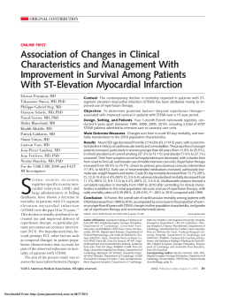
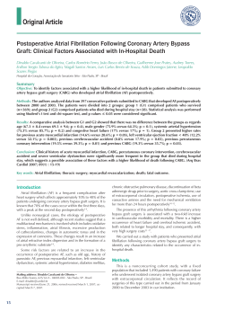
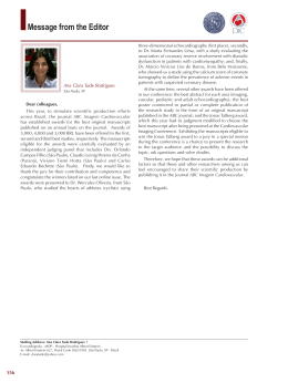
REVIEW ARTICLE
Rev Bras Cir Cardiovasc 2011;26(3):440-6
Absence of arteriosclerosis in intramyocardial
coronary arteries: a mystery to be solved?
Ausência de arteriosclerose na porção intramiocárdica das artérias coronárias: um mistério a ser
resolvido?
Edvaldo Luiz Ramalli Jr1, Leonardo Henrique Braga1, Patricia Martinez Evora2, Agnes Afrodite
Sumarelli Albuquerque3, Andrea Carla Celotto4, André Lupp Mota5, Paulo Roberto Barbosa Evora6
DOI: 10.5935/1678-9741.20110020
RBCCV 44205-1301
Abstract
Several studies show that portions of intramyocardial
coronary arteries are spared of arteriosclerosis, involving
morphological, embryological, biochemical and
pathophysiological aspects. Endothelial function is
significantly affected in the segment of transition, as
estimated by the vasoactive response to Ach. These findings
suggest that myocardial bridge can provide protection against
arteriosclerosis by counteracting the negative effects of
endothelial dysfunction. The intramyocardial portion’s
protection phenomenon deserves further scientific research
on all research fronts. Improved morphological,
biomechanical and especially physiological and
embryological knowledge may be the key to a future window
of opportunity for chronic arterial disease therapy and
prevention. In addition, this review discusses possible
therapeutic approaches for symptomatic coronary ischemia
caused by myocardial bridges.
Resumo
Diversos estudos demonstram que as porções
intramiocárdicas das artérias coronárias são poupadas da
arteriosclerose, envolvendo aspectos morfológicos,
embriológicos, biomecânicos e aspectos fisiopatológicos. A
função endotelial é significativamente afetada no segmento de
transição, tal como estimado pela resposta vasoativa para
acetilcolina (Ach). Esses achados sugerem que ponte miocárdica
pode fornecer proteção contra a arteriosclerose, por contrariar
os efeitos negativos da disfunção endotelial. O fenômeno dessa
proteção da porção intramiocárdica merece maior investigação
científica em todas as frentes de pesquisa. Maiores
conhecimentos sobre os aspectos morfológicos, biomecânicos
e, principalmente, fisiológicos e embriológicos podem ser a
chave para uma futura janela de oportunidades de terapia e
prevenção da doença arterial crônica. Nessa revisão, discutemse, também, possíveis abordagens terapêuticas para fenômenos
coronarianos isquêmicos causados por pontes miocárdicas.
Descriptors: Myocardial bridging. Coronary circulation.
Endothelium.
Descritores: Ponte miocárdica. Circulação coronária.
Endotélio.
1. Graduate student; Monitor, Surgery and Anatomy Department of
Faculdade de Medicina de Ribeirão Preto da Universidade de São
Paulo, Ribeirão Preto, SP, Brazil.
2. Veterinary Medicine; Graduate School of Veterinary Sciences of
Jaboticabal – UNESP, Jaboticabal, SP, Brazil.
3. Biologist; Biologist, Laboratory of Vascular Reactivity and
Endothelial Function, Ribeirão Preto, SP, Brazil.
4. PhD; Head of the Laboratory of Biomedical Vascular Reactivity
and Endothelial Function, Ribeirão Preto, SP, Brazil.
5. Physician; Resident of Surgery and Anatomy Department of
Faculdade de Medicina de Ribeirão Preto da Universidade de São
Paulo, Ribeirão Preto, SP, Brazil.
6. Full Professor; Department Chief of Faculdade de Medicina de Ribeirão
Preto da Universidade de São Paulo, Ribeirão Preto, SP, Brazil.
Work performed at Surgery and Anatomy Department of Faculdade
de Medicina de Ribeirão Preto da Universidade de São Paulo, Ribeirão
Preto, SP, Brazil.
440
Correspondence
Paulo Roberto B. Evora. Rua Rui Barbosa, 367, apto.15 – Ribeirão
Preto, SP, Brazil – CEP 14015-120
E-mail: [email protected]
Support: Fundação de Apoio ao Ensino, Pesquisa e Assistência do
HCFMRPUSP – FAEPA
Article received on February 17th, 2011
Article accepted on June 18th, 2011
Ramalli Jr EL, et al. - Absence of arteriosclerosis in intramyocardial
coronary arteries: a mystery to be solved?
INTRODUCTION
Several studies show that portions of intramyocardial
coronary arteries are spared of arteriosclerosis, this
phenomenon is easily demonstrated in the Anterior
Interventricular Artery, better known as anterior descending
artery (nomina to be used in the text) and the marginal
branches of the circumflex artery (Figure 1) [1,2]. The
“myocardial bridge” and the differences between the wall
thickness of intramyocardial coronary arteries and other
coronary arteries have been described approximately 60
years ago by Geiringer [3]. However, this “natural
prevention” that spares most intramyocardial portions from
arteriosclerosis, while the epicardial portions are severely
involved, has been poorly investigated and discussed [3,4].
Thus, the purpose of this review is to describe, briefly, the
possible pathophysiological mechanisms of this
phenomenon and its important implications for diagnosis
and treatment of coronary disease. In this way, the involved
morphological, embryological, biomechanical and
physiopharmacological aspects will be discussed. In
addition, possible therapeutic approaches to coronary
ischemic phenomena caused by myocardial bridges are also
discussed. Furthermore, this review followed, in general,
the textual formatting of the review published by Botta &
Elefteriades [1].
Rev Bras Cir Cardiovasc 2011;26(3):440-6
the 50s, Geiringer published that the intramyocardial
coronary artery wall is thinner than the wall from the
epicardial portion [3,5]. The vessel´s wall thickness itself is
an arteriosclerosis protective factor that is little elucidated
[1]. In 1951, Geiringer also reported that the intramyocardial
arteries lack vasa vasorum, as they have thin walls and
their supply of cell oxygen and nutrients occur by simple
diffusion [3,5]. Ever since the nineteenth century, there was
evidence that the vasa vasorum played a key role in the
arteriosclerosis development [6]. Laterin in the 80s, it was
proved that the vasa vasorum is involved in the coronary
disease progression and preventing its formation prevents
arteriosclerosis (Figure 2) [7].
Fig. 2 – Main anatomofunctional and embryological aspects
EMBRYOLOGICAL ASPECTS
Fig. 1 - Major anatomopathological aspects (adapted Bourassa
and colleagues [35])
MORPHOLOGICAL ASPECTS
The morphological features possibly related to the
intramyocardial portion´s “natural protection” in relation
to arteriosclerotic disease were the first to be described. In
The vascular smooth muscle cells of coronary arteries
are distinct from those of the proximal aorta through a series
of structural and functional criteria, mainly related to myosin
heavy chains isoforms in vascular smooth muscle
denominated SM1, SM2, and Smemb. This proves that the
coronary arteries are not derived from the aorta. These
morphofunctional differences may explain the increased
propensity for arteriosclerotic transformation. Currently, the
source of this variation among subpopulations of smooth
muscle is uncertain. Although the proximal aorta´s smooth
muscle is known to be derived from the neural crest, the
origin of coronary vascular smooth muscle remains
uncharacterized [8]. It is more likely that the adventitia and
media of coronary arteries are derived from pericardial
mesoderm, and if cell migration is incomplete, the
intramyocardial part of the coronary arteries could have
different structural composition from the remaining parts
of the coronary arteries. This may explain the lower
susceptibility of intramyocardial coronary to arteriosclerosis
[1,9] (Figure 2). The relationship between anatomic
441
Ramalli Jr EL, et al. - Absence of arteriosclerosis in intramyocardial
coronary arteries: a mystery to be solved?
Rev Bras Cir Cardiovasc 2011;26(3):440-6
variations of the myocardial bridges with a predisposition
to the risk of cardiac ischemia is still a detail to be clarified
[10,11].
oxide (NO), in protecting myocardial bridges are the main
hopes for intervention and possible coronary disease
solution.
BIOMECHANICAL ASPECTS
The two morphological factors mentioned (wall
thickness and absence of vasa vasorum) and embryological
factors, are unable to explain, by themselves, the protection
of the myocardial bridge in relation to arteriosclerosis. Thus,
the first assumptions regarding the biomechanical aspects
to elucidate this phenomenon emerged. It is known that
some mechanical forces such as blood flow are responsible
for shear stress and the pressure gradient is responsible
for mural stress. These phenomena play a key role in the
occurrence and location of arteriosclerosis, interfering with
the delicate functional balance between endothelium
protective factors and causes of arteriosclerosis. This is
easily evidenced on clinical observation of the
arteriosclerotic lesions predilection for local curvature,
bifurcations, branches and dilatation of the arteries. This
motivated the search for factors related to the pressure
gradient, the myocardial contraction and protection against
arteriosclerotic lesions. The pressure gradient through the
coronary arteries wall is given by the difference between
the intravascular blood pressure and tissue pressure,
according to modified Laplace’s law.
The myocardial bundles contraction, around
intramyocardial coronary arteries, produce a tissue pressure
close to, or even greater than, the intravascular blood
pressure, especially in systole. This significantly reduces
mural stress, and therefore plays a key role in protecting
the intramyocardial portion of the arteries from
arteriosclerotic lesions [4]. This reduction is directly
proportional to how deep the artery is in the myocardium.
Robicsek & Thubrikar [4] have published mural stress
reductions ranging from 63% to 80% depending on the
artery’s depth in the myocardium during systole and
concluded that the intramyocardial coronary always have
a significant positive pressure in their surrounding areas,
reducing the transmural gradient and, consequently, the
mural stress, thereby protecting the intramyocardial
portions from arteriosclerosis.
PHYSIOPHARMACOLOGICAL ASPECTS
The physiological aspects have arisen due to the
extension of arteriosclerosis pathogenesis research and the
research of biomechanical factors that protect the
intramyocardial portion, and currently have the greatest
perspective of evidence that could finally elucidate this
phenomenon. Paracrine signaling from adipose tissue, and
especially the role of vasoactive agents, including nitric
442
Paracrine signaling of epicardial adipose tissue
The physiological role of epicardial adipose tissue is
not yet fully elucidated. It has been proposed that epicardial
tissue acts as a buffer that protects the heart from its large
exposure to high levels of circulating fatty acids, or, as a
local energy resource for the myocardium during high
demand times. However, increasing evidence suggests that
epicardial adipose tissue may interact with the myocardium
and coronary arteries. This tissue could locally modulate
the heart and coronary circulation through paracrine
secretion of pro and anti-inflammatory mediators, leading
to the hypothesis of a dual role played by epicardial adipose
tissue [12]. Little is known regarding the relationship
between epicardial adipose tissue and arteriosclerosis. The
factors that influence on the balance between protective
effects and harmful effects of paracrine secretion are not
well elucidated. Inflammatory mediators such asadiponectin
and adrenomedullin seem to play a protective effect, while
the pro-inflammatory mediator such as interleukins1and 6
(IL-1, IL-6) and tumor necrosis factor alpha (TNF-á )
supposedly exert detrimental effect [12] (Figure 3).
Fig. 3 – Major pharmacological aspects
Expression of vasoactive agents
In 2001, Masuda et al. [13] investigated the relationship
between the expression of main vasoactive agents such as
endothelin-1 (ET-1), the angiotensin converting enzyme
(ACE) and endothelial nitric oxide synthase (eNOS), with
its location in relation to myocardial bridge and the degree
of involvement by arteriosclerosis. They found that in the
intramyocardial portion of the left anterior descending artery
there was no histopathological sign indicating the
beginning of arteriosclerosis in the intima and that the
Ramalli Jr EL, et al. - Absence of arteriosclerosis in intramyocardial
coronary arteries: a mystery to be solved?
Rev Bras Cir Cardiovasc 2011;26(3):440-6
arteriosclerosis rate is significantly smaller in the myocardial
bridge portion in relation to the proximal and distal segments
of the bridge.
They noted that the expression levels of eNOS, ET-1
and ACE are significantly higher in the portions proximal
and distal to myocardial bridge than in the portion
underneath the bridge. They established a significant
association between both the differential expression of
vasoactive agents along the anterior descending coronary
artery with less expression in the myocardial bridge as an
association between decreased expression of vasoactive
agents with lower arteriosclerosis rates. It was demonstrated
that the expression of vasoactive agents is related to an
altered hemodynamic stress on the vascular wall,
suggesting that the decrease of shear stress, beyond
interfering with the lipid transfer phase to the vascular wall
in arteriogenesis, also works to reduce the expression of
vasoactive agents (Figure 3) [13].
recognized as an important influence on endothelial
function. The high intravascular pressure has been
associated with a state of endothelial dysfunction with
impaired endothelium-dependent relaxation. As already
mentioned, the expression of vasoactive enzymes, such as
eNOS, ET-1 and ACE, is suppressed under a myocardial
bridge. It is known that endothelial dysfunction, signaled
by endothelium-dependent vasodilation, is an initiating
factor in the pathogenesis of atherosclerosis and coronary
artery intimal changes. Numerous reports and daily practice
of myocardial revascularization surgery showed that the
intima of internal arterial segments of the myocardial bridge
is spared from arteriosclerosis. This was confirmed by
histopathology, imaging assisted by computer analysis and
studies using intracoronary ultrassound (ICUS), however,
contrary evidence has been reported in some studies with
ICUS and anatomical pathology [16]. Thus, the relationship
between myocardial bridge and the pathogenesis of
arteriosclerosis remains unclear. Endothelial dysfunction
has been well proposed as an initial step in the
arteriosclerotic process, however the relationship between
endothelial dysfunction and arteriosclerosis in segments
of transition has been controversial. Even though the
relationship between endothelial dysfunction and
arteriosclerosis has been described in clinical conditions
such as diabetes and hypercholesterolemia, endothelial
dysfunction restricted to the site of mechanical compression
may reflect the proatherogenic effect, even if modest, in
this particular type of endothelial dysfunction. This
hypothesis needs to be validated [16-20].
MYOCARDIAL BRIDGE AND CORONARY
ENDOTHELIAL DYSFUNCTION
Myocardial bridge is a congenital defect, which may
present endothelial dysfunction of the coronary´s
intramyocardial segment. Some studies have reported that
endothelium-dependent vasorelaxation is impaired in this
segment of transition. This phenomenon is probably due
to a direct action of the myocardial bridge compressing
effect on endothelial function. Teragawa et al. [14] reported
that patients with myocardial bridge have coronary spasm
more frequently than patients without it at an estimated
rate of 73% vs. 40%. Herrmann et al. [15] also showed a
greater coronary vasoconstriction in response to Ach.
Moreover, the mean vasoconstriction in response to
maximum Ach was more prominent in inner segments of
myocardial bridge than in segments of control patients.
These pharmacological data can be related to endothelial
dysfunction and spasm in the segment of transition, and
maybe a reason for coronary events in patients with
myocardial bridge [15,16].
ENDOTHELIAL DYSFUNCTION AND ABSENCE OF
ARTERIOSCLEROSIS: AN APPARENT PARADOX
A myocardial bridge is characterized by focal systolic
compression of a coronary artery by an overlying
myocardial band. Chronic compression and relaxation of
the bridge can cause endothelial dysfunction by direct
stress. Despite the important relationship between
myocardial bridge and endothelial dysfunction, the bridge
segments are spared from arteriosclerotic plaque
formation [16].
Shear stress caused by related blood flow was
CLINICAL-SURGICAL IMPLICATIONS
The angiographic prevalence of myocardial bridge is
reported from 0.5% to 29.4% [25-27] and can result in
significant hemodynamic changes in coronary flow,
predisposing to cardiac events [28]. However, it is usually
considered a benign condition [25], though it has been
associated with myocardial ischemia [26], tachycardiainduced ischemia [27], conduction disturbances [28],
myocardial infarction [29,30] and sudden death (Table 1)
[31]. A recent Brazilian study analyzed the results of 3375
coronary angiographies performed in the period of 2003 to
2007, of which 123 showed the phenomenon of systolic
constriction of the left anterior descending artery with the
diagnosis of myocardial bridge. The diagnosis frequency
of myocardial bridge on cineangiocardiography performed
in this period was 3.6% [32]. These data confirm the clinicaltherapeutic importance of myocardial bridges.
In excellent “Letter to the Editor”, Barcin et al. [33]
summarize the implications of coronary artery
revascularization under myocardial bridges. This evaluation
was based in a study by Brodsky et al. [34] that suggests
443
Ramalli Jr EL, et al. - Absence of arteriosclerosis in intramyocardial
coronary arteries: a mystery to be solved?
Rev Bras Cir Cardiovasc 2011;26(3):440-6
that myocardial bridges may be an independent risk factor
for developing myocardial ischemia and interstitial fibrosis.
The authors came to this conclusion as a result of
pathological examination of six patients with intramural left
anterior descending artery compared with ten patients
without myocardial bridge. This finding is important
because data on the prognosis of myocardial bridge are
limited.
the proximal segment of the myocardial bridges in 12 (86%)
of 14 patients with systolic narrowing evidence. These
arteriosclerotic regions may also be the source of thrombus
and microemboli formation. In addition, the management of
patients with myocardial bridge showing a “milking effect”
is still controversial.
Medical therapy, especially beta-blockers is the first line
of treatment in this patient group. In patients that do not
respond to medical therapy, surgical revascularization or
stent implantation are the options. Even though coronary
stent is capable of suppressing ischemic symptoms, there
is a high rate of in-stent restenosis with stents implanted in
segments with myocardial bridge [40]. This finding was
corroborated by other studies, including pharmacological
stents [41,42]. It is speculated that the in-stent restenosis,
which was diffuse in most patients, may be due to
vasoactive substances released from the vessel wall,
squeezed between the stent and the myocardium, at each
contraction [40]. It seems that the stent implantation, at
least with metallic stents, should be avoided in myocardial
bridges. Studies comparing surgery with stent implantation
are still lacking. In a retrospective study, Wan and Wu [43]
have shown excellent short and long term results with the
surgery and concluded that surgery is better than stent.
In conclusion, the best approach for patients with
myocardial bridges is still controversial, even if they have
“milking effect” on angiography. Metal stents have higher
rates of restenosis and results with pharmacological stents
do not exist (Table 2).
Table 1. Main clinical aspects.
Clinical Aspects
•
Myocardial ischemia
•
Ischemia-induced tachycardia
•
Conduction disorders
•
Myocardial infarction
•
Sudden death
Accumulating case reports in medical literature, as has
been said more than once in this text, it is known that
myocardial bridges may cause angina pectoris, myocardial
infarction, fatal arrhythmias and even sudden cardiac death.
On the other hand, considering the high prevalence of these
bridges, especially in autopsy series (up to 16% in
angiographic series and 8.5% in autopsy series), it might
be speculated that most of the myocardial bridges should
be “harmless”. Although they seem to narrow the coronary
artery in systole, Bourassa et al. [35] demonstrated that the
coronary artery obstruction caused by myocardial bridge
(“milking effect”) extends to diastole. Doppler flow profile
revealed persistent reduction in initial diastolic diameter
followed by an abrupt acceleration of diastolic flow and
reduction of coronary flow reserve in the intramural
coronary arteries. It is not surprising, therefore, that
myocardial bridges may lead to coronary ischemia,
especially in conditions with increased heart rate where
the diastolic phase relatively shortens. The photon emission
tomography is the only test that demonstrated that perfusion
defects are reversible after exercise stress test in patients
with myocardial bridge [36]. To be considered “clinically
significant”, it is expected that the bridges cause
compression in coronary angiography, in addition to
ischemia symptoms [37].
An important point with the study of Brodsky et al. [34]
is that there is no information if there was an angiographic
narrowing in patients with myocardial bridge, all of whom
had interstitial fibrosis. This may raise the question of
whether myocardial bridges could lead to ischemia, even in
the absence of coronary narrowing in angiography. The
answer is probably “yes” because the bridges are
susceptible to be dynamic lesions. The “bridged” segment
maybe prone to spasm or narrowing, and can occur in
conditions where myocardial contractility increases [38].
Moreover, Ge et al. [39] showed arteriosclerotic plaques in
444
Table 2. Major therapeutic aspects.
Clinical Treatment
•
Beta-blockers, calcium antagonists and antiplatelet
•
Percutaneous angioplasty (?)
•
Surgical revascularization
•
Myectomy (with or without bypass)
CONCLUDING REMARKS
Endothelial function is significantly affected in the
segment of transition, as estimated by the vasoactive
response to Ach. However, the bridge segments are spared
from arteriosclerosis. These findings suggest that
myocardial bridge can provide protection against
arteriosclerosis by counteracting the negative effects of
endothelial dysfunction. The intramyocardial portion´s
protection phenomenon deserves further scientific research
on all research fronts. Improved morphological,
biomechanical and especially physiological and
embryological knowledge may be the key to a future window
of opportunity for chronic arterial disease therapy and
prevention.
Ramalli Jr EL, et al. - Absence of arteriosclerosis in intramyocardial
coronary arteries: a mystery to be solved?
Rev Bras Cir Cardiovasc 2011;26(3):440-6
The most accepted treatment today is the use
medications, especially beta-blockers in symptomatic
patients or with a history related to myocardial bridge. In
situations of inadequate response to medical treatment,
the options of surgical treatment with myocardial
revascularization or myotomy exist, and also the use of
stents, even with high restenosis rate, in cases of surgical
contraindication [44,45]. The reference 45 is a review article
with a much broader approach and its reading is mandatory
for anyone interested in the study of coronary myocardial
bridging.
Another unconsidered fact in medical literature is the
apparent paradox of the fact that the transition coronary
segments, in its path limited by myocardial bridge, do not
have arterosclerosis, but have endothelial dysfunction with
greater chance of spasm. This fact itself suggests that
clinical treatment with the option of using beta-blockers,
calcium antagonists and antiplatelet agents, is wiser.
isoforms as molecular markers for vascular development and
atherosclerosis. Circ Res.1993;73(6):1000-12.
10. Ishikawa Y, Akasaka Y, Suzuki K, Fujiwara M, Ogawa T, Yamazaki
K, et al. Anatomic properties of myocardial bridge predisposing
to myocardial infarction. Circulation. 2009;120(5):376-83.
11. Erbel R, Ge J, Möhlenkamp S. Myocardial bridging: a
congenital variant as an anatomic risk factor for myocardial
infarction? Circulation. 2009;120(5):357-9.
12. Iacobellis G, Barbaro G. The double role of epicardial adipose
tissue as pro- and anti-inflammatory organ. Horm Metab Res.
2008;40(7):442-5.
13. Masuda T, Ishikawa Y, Akasaka Y, Itoh K, Kiguchi H, Ishii T.
The effect of myocardial bridging of the coronary artery on
vasoactive agents and atherosclerosis localization. J Pathol.
2001;193(3):408-14.
14. Teragawa H, Fukuda Y, Matsuda K, Hirao H, Higashi Y,
Yamagata T, et al. Myocardial bridging increases the risk of
coronary spasm. Clin Cardiol. 2003;26(8):377-83.
REFERENCES
1. Botta DMJ, Elefteriades JA. Why are the intramyocardial
portions of the coronary arteries spared from arteriosclerosis?
Clinical implications. Int J Angiol. 2009;18(2):59-61.
2. Scher AM. Absence of atherosclerosis in human
intramyocardial coronary arteries: a neglected phenomenon.
Atherosclerosis. 2000;149(1):1-3.
3. Geiringer E. The mural coronary. Am Heart J. 1951;41(3):359-68.
4. Robicsek F, Thubrikar MJ. The freedom from atherosclerosis
of intramyocardial coronary arteries: reduction of mural stress
- a key factor. Eur J Cardiothorac Surg. 1994;8(5):228-35.
5. Geiringer E. Intimal vascularization and atherosclerosis. J
Pathol Bacteriol. 1951;63(2):201-11.
15. Herrmann J, Higano ST, Lenon RJ, Rihal CS, Lerman A.
Myocardial bridging is associated with alteration in coronary
vasoreactivity. Eur Heart J. 2004;25(23):2134-42.
16. Kim JW, Seo HS, Na JO, Suh SY, Choi CU, Kim EJ, et al.
Myocardial bridging is related to endothelial dysfunction but
not to plaque as assessed by intracoronary ultrasound. Heart.
2008;94(6):765-9.
17. Ishii T, Hosoda Y, Osaka T, Imai T, Shimada H, Takami A, et
al. The significance of myocardial bridge upon atherosclerosis
in the left anterior descending coronary artery. J Pathol.
1986;148(4):279-91.
18. de Winter RJ, Kok WE, Piek JJ. Coronary atherosclerosis
within a myocardial bridge, not a benign condition. Heart.
1998;80(1):91-3.
6. Rokitansky C, Swaine WE. A manual of pathological anatomy.
vol. 2. London: Blanchard & Lea;1855.
19. Burnsides C, Edwards JC, Lansing AI, Swarm RL.
Arteriosclerosis in the intramural and extramural portions of
coronary arteries in the human heart. Circulation.
1956;13(2):235-41.
7. Barger AC, Beeuwkes R 3rd, Lainey LL, Silverman KJ.
Hypothesis: vasa vasorum and neovascularization of human
coronary arteries. A possible role in the pathophysiology of
atherosclerosis. N Engl J Med. 1984;310(3):175-7.
20. Ishii T, Asuwa N, Masuda S, Ishikawa Y, Kiguchi H, Shimada
K. therosclerosis suppression in the left anterior descending
coronary artery by the presence of a myocardial bridge: an
ultrastructural study. Mod Pathol. 1991;4(4):424-31.
8. Mikawa T, Gourdie RG. Pericardial mesoderm generates a
population of coronary smooth muscle cells migrating into the
heart along with ingrowth of the epicardial organ. Dev Biol.
1996;174(2):221-32.
21. Irvin RG. The angiographic prevalence of myocardial bridging
in man. Chest. 1982;81(2):198-202.
9. Aikawa M, Sivam PN, Kuro-o M, Kimura K, Nakahara K,
Takewaki S, et al. Human smooth muscle myosin heavy chain
22. Noble J, Bourassa MG, Petitclerc R, Dyrda I. Myocardial
bridging and milking effect of the left anterior descending
coronary artery: normal variant or obstruction? Am J Cardiol.
1976;37(7):993-9.
445
Ramalli Jr EL, et al. - Absence of arteriosclerosis in intramyocardial
coronary arteries: a mystery to be solved?
Rev Bras Cir Cardiovasc 2011;26(3):440-6
23. Juilliére Y, Berder V, Suty-Selton C, Buffet P, Danchin N,
Cherrier F . Isolated myocardial bridges with angiographic
milking of the left anterior descending coronary artery: a longterm follow-up study. Am Heart J. 1995;129(4):663-5.
35. Bourassa MG, Butnaru A, Lespérance J, Tardif JC.
Symptomatic myocardial bridges: overview of ischemic
mechanisms and current diagnostic and treatment strategies. J
Am Coll Cardiol. 2003;41(3):351-9.
24. Li JJ. Is myocardial bridging a bridge connecting to cardiovascular
events? Chin Med J (Engl). 2010;123(7):964-8.
36. Huang WS, Chang HD, Yang SP, Tsao TP, Cheng CY, Cherng
SC. Abnormal 201TI myocardial single photon emission
computed tomography in energetic male patients with
myocardial bridge. Nucl Med Commun. 2002;23(11):1123-8.
25. Marshall ME, Headley RN. Intramural coronary artery as a
cause of unstable angina pectoris. South Med J.
1978;71(10):1304-6.
26. Feld H, Guadanino V, Hollander G, Greengart A, Lichstein E,
Shani J. Exercise-induced ventricular tachycardia in association
with a myocardial bridge. Chest. 1991;99(5):1295-6.
27. den Dulk K, Brugada P, Braat S, Heddle B, Wellens HJ .
Myocardial bridging as a cause of paroxysmal atrioventricular
block. J Am Coll Cardiol. 1983;1(3):965-9.
28. Bashour TT, Espinosa E, Blumenthal J, Wong T, Mason DT.
Myocardial infarction caused by coronary artery myocardial
bridge. Am Heart J. 1997;133(4):473-7.
29. Vasan RS, Bahl VK, Rajani M. Myocardial infarction associated
with a myocardial bridge. Int J Cardiol. 1989;25(2):240-1.
30. Morales AR, Romanelli R, Boucek RJ. The mural left anterior
descending coronary artery, strenuous exercise and sudden death.
Circulation. 1980;62(2):230-7.
31. Bestetti RB, Costa RS, Zucolotto S, Oliveira JS. Fatal outcome
associated with autopsy proven myocardial bridging of the
left anterior descending coronary artery. Eur Heart J.
1989;10(6):573-6.
32. Pereira AB, Castro DS, Menegotto ET, Amaral WM, Castro
GS. Myocardial bridging: therapeutic and clinical development.
Arq Bras Cardiol. 2010;94(2):175-81.
33. Barcin C, Kursaklioglu H, Kose S, Amasyali B. Coronary
myocardial bridge constitutes a risk: But how to manage it? Int
J Cardiol. 2010;138(2):215-6.
34. Brodsky SV, Roh L, Ashar K, Braun A, Ramaswamy G.
Myocardial bridging of coronary arteries: A risk factor for
myocardial fibrosis? Int J Cardiol. 2008;124(3):391-2.
446
37. Qian JY, Zhang F, Dong M, Ma JY, Ge L, Liu XB, et al.
Prevalence and characteristics of myocardial bridging in coronary
angiogram: data from consecutive 5525 patients. Chin Med J
(Engl). 2009;122(6):632-5
38. Sakuma M, Kamishirado H, Inoue T, Ichihara M, Takayanagi
K, Hayashi T, et al. Acute myocardial infarction associated
with myocardial bridge and coronary artery vasospasm. Int J
Clin Pract. 2002;56(9):721-2.
39. Ge J, Erbel R, Rupprecht HJ, Koch L, Kearney P, Görge G, et
al. Comparison of intravascular ultrasound and angiography in
the assessment of myocardial bridging. Circulation.
1994;89(4):1725-32.
40. Kursaklioglu H, Barcin C, Iyisoy A, Kose S, Amasyali B, Isik
E. Angiographic restenosis after myocardial bridge stenting.
Jpn Heart J. 2004;45(4):581-9.
41. Rondan J, Lozano I, Avanzas P, Lopez-Palop R, Vegas JM,
Moris C. Drug eluting stents may not be the answer for
myocardial bridges. Int J Cardiol. 2007;117(2):e76-8.
42. Kunamneni PB, Rajdev S, Krishnan P, Moreno PR, Kim MC,
Sharma SK, et al. Outcome of intracoronary stenting after failed
maximal medical therapy in patients with symptomatic myocardial
bridge. Catheter Cardiovasc Interv. 2008;71(2):185-90.
43. Wan L, Wu Q. Myocardial bridge, surgery or stenting? Interact
Cardiovasc Thorac Surg. 2005;4(6):517-20.
44. Crespo A, Aramendi JI, Hamzeh G, Voces R. Off-pump supraarterial myotomy for myocardial bridging. Eur J Cardiothorac
Surg. 2008;34(3):682-4.
45. Möhlenkamp S, Hort W, Ge J, Erbel R. Update on myocardial
bridging. Circulation. 2002;106(20):2616-22.
Baixar