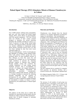
®
Therapy
Effects of Pulsed Signal
on
Gene Expression in Three-Dimensional Chondrocyte Cultures
I. Krüger
*•
#
M. Faensen
Introduction:
Results:
Pulsed electromagnetic fields (PEMF) have been used widely to
treat non-healing fractures and related problems in bone healing
since approval by the Food and Drug Administration (FDA) in
1979, with a success rate averaging 70-80% in a wide variety of
centers in several countries. A special pulsed magnetic field
configuration is used for Pulsed Signal Therapy . Many patients
treated with this Pulsed Signal Therapy (PST) lost their pains and
showed less osteoarthritic symptoms.
To determine the biological effects of PST on cartilage physiology
we used a three-dimensional chondrocyte culture as an in vitro
model for articular cartilage. Isolated chondrocytes of arthritic
cartilage proliferate in monolayer culture. In three-dimensional
culture cells redifferentiate again shown by the deposition of
cartilage-specific matrix components like collagen type II. Using
this cartilage model chondrocytes from different patients were
pooled to minimize variability between individual patients.
The aim of our study was to analyze the effects of the Pulsed
Signal Therapy (PST)1 on gene expression in chondrocyte cultures
derived from osteoarthritic (OA) articular cartilage.
Compared to the housekeeping gene GAPDH (glyceraldehyde-3-phosphate
dehydrogenase), the matrix proteins aggrecan, COMP, type I collagen and type III
collagen are very strongly expressed, while the expression profiles of type II and IX
collagen are at a very low level (collagen type IX: 0.001 to 0.0001% of GAPDH).
Gene expression of tested matrix proteins is oscillating during the study period. All
collagen genes are expressed at a higher level in the ostoarthritic groups than in the
normal groups. Concerning articular cartilage chondrocytes, periodic progression of
expression in PST-pellets has changed from the progression found in pellets of
osteoarthritic chondrocytes to the gene expression profile of normal donor pellets.
Type II and IX collagen genes showed a markedly stronger expression in the
untreated control pellets than in those treated with PST at the last evaluation dates.
aggrecan expression
COMP expression
ND
OA
% GAPDH
12
% GAPDH
ND
PST
10
8
6
4
OA
PST
150
100
50
2
Material and Methods:
Analyse:
rt PCR
gene
T1
Embl database
o
l
i
g
o
n
u
c
l
e
o
t
i
d
e
(
5
‘3
‘
)
product size (base pairs)
GAPDH
M33197
GGC GAT GCT GGC GCT GAG TAC
TGG TTC ACA CCC ATG ACG A
149
type II 1
collagen
X06268
CCG GGC AGA GGG CAA TAG CAG GTT
CAA TGA TGG GGA GGC GTG AG
128
type IX 3
collagen
L41162
AAT CAG GCT CTC GAA GCT CAT AAA A
CCT GCC ACA CCC CCG CTC CTT CAT
100
COMP
L32137
GGG TGG CCG CCT GGG GGT CTT
CTT GCC GCA GCT GAT GGG TCT C
116
aggrecan
X17406
CCA GTG CAC AGA GGG GTT TG
TCC GAG GGT GCC GTG AG
146
References:
1) Trock DH, et al. The effect of Pulsed Electromagnetic Fields in the Treatment of teoarthritis
of the Knee and Cervical Spine. Report of Randomized, Double Blind, Placebo Controlled
Trials. J Rheumatol 1994 ;21: 1903-1911
2) Schulze-Tanzil G, et al. Redifferentiation of dedifferentiated human chondrocytes in hightdensity cultures. Cell Tissue Res 2002;308: 371-397
3) Aigner T, et al. Anabolic and Catabolic Gene Expression Pattern Analysis in Normal Versus
Osteoarthritic Cartilage Using Complementary DNA-Array Technology. Arthritis Rheum
2001;44: 2777-2789
T2
T3
T4
T5
T6
T7
T8
ND
T1 T2 T3 T4 T5 T6 T7 T8 T9 T10
time
T9 T10
time
collagen IXa3 expression
collagen IIa1 expression
OA
ND
PST
1,4
1,2
1,0
0,8
0,6
0,4
0,2
0,0
% GAPDH
% GAPDH
Cartilage specimens were obtained from
different patients (mean age 71 years)
suffering from coxarthrosis and gonarthrosis,
respectively. Chondrocytes also were obtained
from healthy cartilage (ND) served as
controls.
Chondrocytes were isolated, expanded in
monolayer culture and pooled. Cells were
cultured three-dimensionally in high-density2
for 6 weeks. PST treatment cultures were
exposed to one hour of PST daily for 9
consecutive days.
The PST treatment device consisted of a
magnetic field generator, an electronic
interface, and a system of toroid coils. This
produces
unidirectional
DC
elliptical
magnetic fields of 10 –
15 Gauss with varying
frequencies between 10 and 30 Hz.
Treated as well as untreated OA and ND
chondrocytes were cultivated for up to
6 months and were used for subsequent semiquantitative rtPCR analysis (SYBR Green
PCR Core Kit and i-Cycler real-time PCR
System).
Expression of the marker genes collagen II
and IX, cartilage oligomeric matrix protein
(COMP) and aggrecan was measured prior to
and directly after PST treatment as well as 3
and 6 weeks, and 6 months after treatment.
PST
0
0
OA
PST
0,06
0,05
0,04
0,03
0,02
0,01
0,00
T1 T2 T3 T4 T5 T6 T7 T8 T9 T10
time
T1 T2
T3 T4 T5 T6 T7
T8 T9 T10
time
Diagrams: Marker gene expression (expression as percentage of GAPDH expression) in the PST-treated pellets (PST,
green) in relation to marker gene expression in the untreated control articular cartilage chondrocyte pellets (OA, red) and
in the normal donor chondrocyte pellets (ND, blue). Data values are given as the average ± standard deviation.
T1 - before PST treatment (PST), T2 - 5th day of PST, T3 - 9th day of PST, T4 - 2 days after PST, T5 - 4 days after PST,
T6 - 6 days after PST, T7 - 9 days after PST, T8 - 3 weeks after PST, T9 - 6 weeks after PST, T10 - 6 month after PST.
Conclusion:
The objective of our study was to investigate the role of PST on chondrocyte matrix
formation of adult human articular cartilage. As a hypothesis, PST treatment results in
an electromagnetic pulsed field, which may stimulate chondrocytes physiologically to
enhance their metabolic activity and the formation of cartilage extracellular matrix.
PST apparently had an effect on the expression of chondrocyte marker genes in OA
chondrocytes and reverted the expression of selected marker genes towards the
expression found in normal chondrocytes.
Strong expression of type II collagen is to be expected in chondrocytes from
osteoarthritic cartilage, because in such cartilage, the matrix shows an imbalance
between anabolism and catabolism, and the chondrocytes produce increased amounts
of matrix proteins.3 However, these are not retained in the extracellular matrix
because of enzymatic degradation, and there is a detectable loss of proteoglycans and
collagens.
Interestingly, however, in this study the gene expression of matrix proteins in
osteoarthritic cartilage seems to be down-regulated by PST. This surprising result
may be a first important indication for the possible mode of action of PST on the
cytobiological level. If the metabolic balance of the matrix was adequately restored as
to bind a sufficient amount of pericellular type II collagen in the cartilage matrix, the
gene expression could be reregulated. Since COMP seems to be involved in the crosslinkage of collagens, the slight increase of expression at these later dates may be an
indication of improved matrix synthesis. The same applies to type-II-associated type
IX collagen. A detailed determination of the expression of molecules linked to the
matrix and those involved in its catabolism may provide valuable data to verify this
theory.
The surprising results obtained here show that PST has an effect on the level of
matrix molecule gene expression. Their interpretation raises new questions that call
for more thorough investigations.
U
N
I
V
E
R
S
I
T
Y
M
E
D
I
C
A
L
C
E
N
T
E
R
•
C
H
A
R
I
T
É
•
F
A
C
U
L
T
Y
O
F
T
H
E
H
U
M
B
O
L
D
U
N
I
V
E
R
S
I
T
Y
•
B
E
R
L
I
N
*
University medical centre Charité of Humboldt University, Department of Rheumatology
#
DRK-Klinik Westend
Ina Krüger
Tissue Engineering Group
Tucholskystr. 2, 10117 Berlin, Germany
Email: [email protected]
Baixar