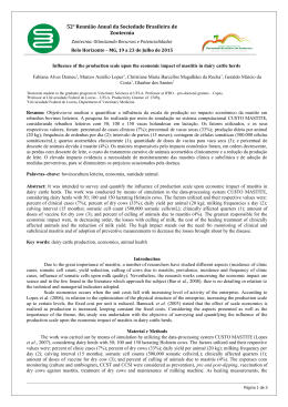
Corynebacterium pseudotuberculosis experimental infection of goats mamary gland.
CORYNEBACTERIUM PSEUDOTUBERCULOSIS EXPERIMENTAL
INFECTION OF GOATS MAMARY GLAND
J.W. Pinheiro Junior1, A.A.F. Oliveira 1, F.S.F. Alves2, L.B.G. Silva 1, S.S.A. Rabelo1, R.A. Mota 1
Universidade Federal Rural de Pernambuco, Departamento de Medicina Veterinária, Rua Dom Manuel, s/no,
CEP 52171-900, Recife, PE, Recife, Brazil. E-mail: [email protected]
1
ABSTRACT
The objective of this study is to describe some aspects of the pathogeny of Corynebacterium
pseudotuberculosis, isolated from a natural case of caseous lymphadenitis. The study was comprised
with 2 groups of 3 animals each; inoculated with a C. pseudotuberculosis strain, group 1 (GI) via intra
dermal and group 2 (GII) via the mammary gland. The animals were clinically examined on a daily
basis and hemograms carried out over a period of time at different intervals. Only 2 animals from
GII showed changes in the clinical examination characterized by hyperthermia. All the animals
in both groups developed papule and edema at the inoculation site. There were no significant
alterations in the erythrogram but in the leucogram there was leucocytosis indicated by the
presence of neutrophilia in 2 animals from GII. Results of the cellular analysis of the gland showed
that 2 animals from GII developed acute mastitis. This experiment has proven that C.
pseudotuberculosis, isolated from a natural infection of caseous lymphadenitis, could cause clinical
mastitis, losses in goat keeping and that the expression cytology test could be used as an alternative
diagnosis for mastitis.
KEY WORDS: Mastitis, caseous lymphadenitis, expression cytology.
RESUMO
INFECÇÃO EXPERIMENTAL DA GLÂNDULA MAMÁRIA EM CABRAS POR
CORYNEBACTERIUM PSEUDOTUBERCULOSIS. Objetivou-se com este trabalho descrever alguns aspectos da patogenia da infecção da glândula mamária por Corynebacterium pseudotuberculosis
isolado de um caso natural de linfadenite caseosa. Os animais em número de seis, foram separados
em dois grupos, no primeiro (GI) foi realizada inoculação da cepa de C. pseudotuberculosis pela via
intradérmica e no segundo grupo (GII) pela via intramamária. Esses animais foram acompanhados
diariamente por exame clínico e por hemogramas em diferentes tempos. Apenas dois animais do
GII apresentaram alteração ao exame clínico caracterizada por hipertermia. Todos os animais (GI
e GII) apresentaram pápula e edema no local da inoculação. No eritrograma, não foram observadas
alterações significativas, contudo no leucograma, observou-se leucocitose por neutrofilia em dois
animais do GII. A análise da celularidade das glândulas demonstrou que apenas dois animais do
GII apresentaram mastite aguda. Comprovou-se com este experimento que o C. pseudotuberculosis
isolado de infecção natural de linfadenite caseosa, pode ocasionar mastite clínica, causando
prejuízos para caprinocultura, assim como a citologia de expressão pode ser utilizada como
método de diagnóstico alternativo nos casos de mastite.
PALAVRAS-CHAVE: Mastite, linfadenite caseosa, citologia de expressão.
INTRODUCTION
Goat rearing is a developing activity throughout
the country, although there are a greater number of
goat farmers in the semi arid regions of the North East
Brazil (RIBEIRO et al., 2001). Some diseases, such as
mastitis and caseous lymphadenitis, reduce
2
production and productivity thus resulting in losses
to the producers and risks to Public Health.
Caseous lymphadenitis is a chronic disease with
a suppurative characteristic and necrotic
inflammation characterized by internal and external
abscesses and is caused by Corynebacterium
pseudotuberculosis (NFI; N DI, 1994). It causes economic
Embrapa, Centro Nacional de Pesquisa de Caprinos, Sobral, CE, Brazil.
Arq. Inst. Biol., São Paulo, v.73, n.4, p.395-400, out./dez., 2006
395
396
J.W. Pinheiro Junior et al.
losses by lowering milk and meat production, and the
value of the hide, condemnation of carcasses and
viscera are condemned, slaughtering of severely
affected animals, veterinary expenses and labor costs
in treating the abscesses and, in severe cases, death of
the animal (ALVES ; PINHEIRO, 1999).
ALMEIDA et al. (2005), in their study in Pernambuco
State, isolated C. pseudotuberculosis in 60% (3/5) of
lactating goats. These had mastitis and concomitant
caseous lesions in the supra mammary lymph node
which is characteristic of the disease.
These diseases, isolated or in conjunction with
other sanitary problems of small ruminants, contribute
considerably to lowering the quality of the milk, meat
and hide, thus leading to financial losses what could
even turn the breeding venture unviable.
Considering the importance of goat breeding in
Pernambuco State and the lack of regional research
that contributes in clarifying the epidemiology and
the disease control, the objective of this study is to
evaluate some important aspects of the C .
pseudotuberculosis pathogeny in the mammary gland
of milking goats.
MATERIAL AND METHODS
A strain of C. pseudotuberculosis obtained from a
clinical case of caseous lymphadenitis of animals
from Centro Nacional de Pesquisa em Caprinos was
used in the experimental infection. The strain was
identified according to the method recommended by
CARTER (1988). The infective dose was 3,6 x 10⁷ units
of formed colonies (BURRELL, 1978).
Six cross breed goats in different stages of lactation,
in good health condition, without any clinical sign of
mastitis and with milk microbiological examination
negative for C. pseudotuberculosis and other agents,
were used in the experiment. The animals were kept
in pens at the CNPC and were fed on elephant grass,
corn flour ration and cotton pie, and had water ad
libitum. The goats were separated into 2 groups of 3
animals each.
Using syringes and needles disposable, GI was
infected via intra dermal and GII was inoculated with
0.2 mL of infected material directly into the
parenchyma of the udder and approximately 5 cm to
the right of the supra mammary lymph node. The
infective dose was 3,6 x 10⁷ units of formed colonies
(BURRELL, 1978).
The clinical examination was carried out according
to ROSEMBERG (1993) taking into account physiological
parameters (body temperature, heart beat and
respiratory rate), palpations of the superficial lymph
nodes and mammary gland once a day, for one week
of adaptation and the three weeks post infection (PI).
Samples of milk were collected before the inoculation
to verify the state of health of the udder and after
inoculation to recover the microorganism inoculated.
These samples were seeded on agar base enriched
with 8% sheep blood and incubated for 48 hours at 37º
C. After this incubation period the morphological
characteristics of the colonies were recorded and
colonies smears stained by Gram technique according
to QUIN et al (1994).
Hemograms carried out according to JAIN (1993)
method were done for blood samples collected 72 and
8 hours before the inoculation and 48, 168, 326 and 672
hours post inoculation, comprising a total of 36 samples.
The procedure used in the expression cytology (EC)
was adapted from AZÍRA method (1976) in which, after
the teat cleaning with alcohol 70%, slight pressure is
applied with the thumb and index finger to strip the
entire canal of the teat. The material obtained was
placed on an opaque histology slide, fixed with absolute
methyl alcohol for ten minutes then stained with
Giemsa. These slides were examined for evaluating the
characteristics of stripping, composition and cell size.
Cell counts were carried out on ten fields under the
microscope and the mean cell count taken for the type
of inflammation classification as well as cell component.
The classification of the type of inflammation was
carried out according to COWELL; TYLER (2002).
The animals were slaughtered on the 28th day
post infection and samples of the supra mammary
lymph nodes were collected for bacteriological and
histopathological examination.
Statistical analysis was carried out on data of
animals within the same group using the Minitab
computer program. The data were tested for normality
of residues and the degree of freedom in opting for the
development the analysis by Nparl way analysis of
non parametric variance and utilizing the KruskalWalis test (SAMPAIO, 1998).
For results description the respective
measurements of the central trend were used: mean
and standard deviation for the variation in the
erythrogram and mean, lower and superior limits for
the leucogram. The level of significance used for
rejecting HO (null hypothesis) was 5%.
The present study was approved by EMBRAPA
Ethical Committee on March 2005.
RESULTS
Only 2 animals of GII had fever during the first 96
hours post infection. From the 5th day on, the animals
had normal rectal temperature. However, one animal
developed vital changes with tachycardia and
tachypnea and showed physical alteration of lacteous
secretion 48 hours PI. Some lumps were observed and
Arq. Inst. Biol., São Paulo, v.73, n.4, p.395-400, out./dez., 2006
397
Corynebacterium pseudotuberculosis experimental infection of goats mamary gland.
a reduction in milk production and turning into a
purulent viscose content after 72 hours. Apathy, loss
of appetite and a reduction in food consumption 72
hours post infection, remaining 48 without eating,
was also observed. The other 4 animals had no fever
or any other changes in their general health status.
No significant differences were observed in the
variables analyzed: haematocrit (Ht), erythrocytes (He)
and hemoglobin (Hb) for GI and GII animals on the
different sampling times (Table 1). However, there was
a Hemoglobin decrease in the GI 48 hours PI. In the
leucogram there was a significant difference in the
Group II animals in the rods and monocytes at different
sampling times (Table 2). A significant increase of the
total number of leucocytes was observed in both groups,
48 hours PI, characterizing a leucocytosis, with a
decrease in number 168 hours PI. Analysis of the
number of lymphocytes indicated an increase in GI
forty-eight (48) hours PI with a decrease 168 hours PI.
Examination of the site of inoculation revealed
papule formation 48 hours PI in all GI animals and
transformation of this into pustule on day 5 PI and
developing into an ulcer fairly soon after. All the GII
animals developed edema at the site of inoculation.
On day 20, GI animals showed ulcer cicatrisation
preceded by a dry crust formation.
An increase in the supra mammary lymph nodes
(lymphadenitis) was observed 48 hours post infection in
all the inoculated animals except for goats number 013
and 066 (GI), which showed it 168 hours post infection.
Analysis of the gland cells of the infected animals
revealed that for 2 GII animals more than 70% of the
inflammatory cells were neutrophils, considered
acute mastitis according to COWELL ; TYLER (2002)
criteria (Fig. 1). On the other hand, the neutrophils
counts increased progressively over the post
infection period for animals 68 and 119 and there
was also an increase, predominantly in neutrophils
in animal 180. The results for the EC are given in
Table 3. It was not possible to carry out EC on
animals 013 and 066 (GI) as these went dry during
the period of adaptation.
Table 1 – Average values of the erythrogram of goats inoculated intradermically (GI) and intra-mammary (GII) with
Corynebacterium pseudotuberculosis, CNPC, 2005.
Variables
Group1 (hours)
72
Hematócrito (%)
HCM (pg)
VCM (fl)
CHCM (g/dl)
He (x 106 )
Hb (g/dl)
8
48
336
23,00 24,00 22,33 21,67
9,53 8,97 8,57 12,90
28,37 28,37 24,70 26,57
33,77 31,67 34,83 33,00
8,13 8,52 9,28 9,13
7,77 7,60 7,80 7,19
Group2 (hours)
672
72
21,33
12,47
26,80
30,63
9,06
6,50
8
48
336
672
23,67 25,33 25,67 25,33 22,67
8,83
9,83 9,10 7,60 8,70
27,57 30,93 26,70 26,20 24,67
31,87 31,27 34,03 29,30 35,27
8,94 8,21 9,58 9,80 9,16
7,57 7,93 8,73 7,58 7,97
Table 2 – Leucogram median values of goats experimentally inoculated, intradermically (GI) and intramammary (GII)
with Corynebacterium pseudotuberculosis, CNPC, 2005.
Hours
Leucocytes
Variables
Segmented
Rods
Linphocytes
Basofile
Eosinofile
Monocytes
GI
72
8
48
168
336
672
9050,00
10750,00
24400,00
24200,00
11850,00
9100,00
3547,50
4212,00
6561,00
14762,00
5332,50
4185,00
0,00
0,00
121,50
0,00
169,00
0,00
4163,00
4730,00
7279,50
9196,00
6162,00
3875,00
0,00
0,00
121,50
0,00
169,00
0,00
271,50
108,00
316,50
242,00
118,50
186,00
82,50
107,50
0,00
0,00
0,00
0,00
GII
72
8
48
168
336
672
7550,00
6550,00
17200,00
13350,00
10800,00
16950,00
3895,50*
2947,50*
13167,00*
9078,00*
6696,00*
10000,00*
51,50
118,00
171,00
0,00
108,00
0,00
2719,50
3537,00
3784,00
4138,50
3564,00
6441,00
0,00
0,00
0,00
0,00
0,00
0,00
206,00
236,00
342,00
168,00
280,00
320,00
0,00*
0,00*
0,00*
0,00*
108,00*
169,50*
*Significant difference, with a confidence level of 5%.
Arq. Inst. Biol., São Paulo, v.73, n.4, p.395-400, out./dez., 2006
398
J.W. Pinheiro Junior et al.
Table 3 – Cytology Results from Expression Cytology (EC) of GI and GII animals, before and post experimental infection
with Corynebacterium pseudotuberculosis, CNPC, 2005.
Animal
013 RG (GI)
013 LG (GI)
066 RG (G)
066 LG (GI)
068 RG (GII)
068 LG (GII)
117 RG (GII)
117 LG (GII)
119 RG (GII)
119 LG (GII)
180 RG (GI)
180 LG (GI)
Hours
72 Pre-infection
48 Post-infection
432 Post-infection
600 Post-infection
NP
NP
NP
NP
40% neutrophils
10% neutrophils
10% neutrophils
NP
10% neutrophils
NP
NEN
NP
NP
NP
NP
NP
40% neutrophils
< 10% neutrophils
40% neutrophils
NP
20% neutrophils
NP
NEN
NP
NP
NP
NP
NP
50% neutrophils
NP
80% neutrophils
NP
40% neutrophils
NP
30% neutrophils
NP
NP
NP
NP
NP
NP
NP
NP
NP
70% neutrophils
NP
NEN
NP
RG – right gland; LG – left gland; NP – not performed; NEN – not enough to notice.
Fig. 1 – Intense inflammatory infiltered
polimorphnuclear, indicating acute infectious process.
Giemsa. (x 250). Animal 117 D.
It was possible to recover and identify C .
pseudotuberculosis from the pus of animal 66 seven (7)
days after the inoculation, from the milk secretion of
animal 117 forty eight (48) hours PI and from the supra
mammary lymph node and mammary gland of animals
119 and 117 respectively at the time of slaughtering.
All the animals showed the supra mammary lymph
nodes enlarged, but without abscesses. Visible
macroscopic alterations were not seen in the organs
examined, except a gelatinous edema at the sites of
inoculation. Animal 117 had a large quantity of
caseous secretion in the udder above the cut.
DISCUSSION
ETREBY & ABDEL-H AMID (1970) performed an experimental infection via galactofor canal in healthy
sheep with C. ovis and C. pyogenes and observed the
development of chronic purulent catarrhall acute
mastitis and chronic apostematous mastitis,
respectively.
The microorganism in the mammary parenchyma
demands chemotherapeutic action for neutrophil and
macrophags that perform phagocitosys and constitute
the initial focus of abscess formation that expand
through the secretor tissue (ETREBY; ABDEL -H AMID,
1970).
Even though the via of inoculation in this study
was different, the formation of pustules at the site of
inoculation was observed (GI all animals) and 1
animal from GII had a unilateral abscess of
approximately 5cm in diameter and with a purulent
content in the parenchyma of the gland. Also in this
animal there was a progressive increase of mammary
gland cells at different times of the EC examination.
There was a predominance of whole and some
degenerated neutrophils, characteristic of acute
inflammatory process (COWELL; TYLLER, 2002). Other
GI animals had also significant increase of cells, with
neutrophils predominance. Similar findings were
observed by BURREL (1978) in his study with experimental inoculation of caseous lymphadenitis in sheep.
The dose of C. pseudotuberculosis did not cause
suppurative and/or systemic caseous lesions by the
used inoculation via for GI and GII animals, even
though there was some alteration in the general health
status of the GII animals.
According to AUGUSTINE (1982) the concentration
of viable organisms in the purulent material is
estimated to be from 1 x 10⁶ to 5 x 10 7 per gram of pus
to cause the disease in animals. Other aspect that must
be considered is the period of observation that usually
Arq. Inst. Biol., São Paulo, v.73, n.4, p.395-400, out./dez., 2006
Corynebacterium pseudotuberculosis experimental infection of goats mamary gland.
is not long enough for the lesions formation in the
internal lymph nodes and other organs as was
described by LANGENEGGER; LANGENEGGER (1988) that
kept goats under observation for 126 days. JOHNSON et
al. (1993) intra dermically inoculated 4 x 10⁶ washed
bacteria and recorded a chronic disease with the
formation of internal and external abscesses without
any signs of the acute phase such as fever, loss of
appetite and hemolysis, the period between
inoculation and abscesses rupture was 3 to 6 weeks,
lesser than the one observed by ASHFAQ; CAMPBELL
(1979), 90 days. PEPIN et al. (1988) carried out a
subcutaneous experimental infection of C .
pseudotuberculosis on sheep at different dose rates and
observed local reaction and multiple abscesses
formation in the lymph nodes 21 days after
inoculation.
NAGY (1976) also carried out experimental infection
using different routes of infection and showed that
there exists a link between the point of inoculation
and the region lymph node affected. In the present
study the occurrence of lesions at the site of
inoculation, the formation of edema, erythema,
papules, ulcers and crust in GI animals lead to the
bacteria recovery from some of these lesions. However,
related to the recovery of C. pseudotuberculosis from the
lymph nodes and milk secretion, it was possible to
isolate the bacteria from 50% of the animals inoculated
which is in accordance with the findings of GARG;
CHANDIRAMANI (1985) who also did not recover the
bacteria from all the animals.
The formation of edema at the inoculation site
probably occurred due to the action of the toxin
produced by C. pseudotuberculosis, which affected the
blood vessels in vivo. JONHSON et al. (1993) in their study
discussed the role of this toxin in the pathogenesis of
naturally acquired disease.
GAMELL ; TARTOUR (1974) analysed the hemograms
and plasma protein of sheep experimentally infected
with C. pseudotuberculosis, and reported a severe
hemolytic anemia, described as transitory macrocitic
hypochromic, as well as hypochromic normocitic
anemia. This was not observed in our study.
In relation to the leucocytosis observed in the
present study, JAIN (1993) reported that, among other
causes, bacterial infections are responsible for
leucocytosis, generally related to neutrophilia. The
neutrophilia is the result of increased transference of
cells from the marginal pool to the circulation, reducing
the migration of cells to the tissues and increasing the
production and liberation of neutrophils by the bone
marrow. Also according to MEYER et al. (1995), intense
acute inflammation and those similarly found in GI
and GII animals, neutrophilia occurs from a stimulus
and continual tissue demand, elevated production
and liberation of neutrophils to suppress the tissue
demand. Even with the significant difference in the
evaluation of the monocytes obtained in GII during
the experimental phase, no monocytosis was
observed. According to KERR (2004) monocytosis
occurs just prior to chronic infections, more frequently
related to parasitic or bacterial infections.
CONCLUSION
From the results of this experiment it was shown
that C. pseudotuberculosis, isolated from a natural case
of caseous lymphadenitis, could cause acute clinical
mastitis. The results from the expression cytology
tests were satisfactory as an auxiliary diagnostic
method and could be considered for use in future
studies on goats.
REFERENCES
ALVES, F.S.F.; P INHEIRO, R.R. Linfadenite caseosa: recomendações e medidas profiláticas. Lavoura, n.631, p. 42-43,
1999.
ALMEIDA, M.C.S., S ILVA, L.B.G., M OTA, R.A., N UNES, J.R., S ILVA,
D.R. Estudo da relação entre a linfadenite caseosa e
mastite em cabras criadas no Estado de Pernambuco.
In: JORNADA DE INICIAÇÃO CIENTÍFICA/PIBIC/
FACEPE/CNPq, 2005, Recife. Anais. Recife, 2005.
p.81-82, 2005.
ASHFAQ, M.K.; CAMPBELL , S.G. A survey of caseous
lymphadenitis and its etiology in goats in the United
States. Veterinary Medicine of Small Animal Clinician,
v.74, p.1161-1165, 1979.
AUGUSTINE , J.L.; RICHARDS, A.B.; RENSHAW , H.W. Concentration
of Corynebacterium pseudotuberculosis obtained from
lesions of sheep and goats with caseous lymphadenitis.
In: INTERNATIONAL CONFERENCE ON GOAT
PRODUCTION AND DISEASE, 3., 1982, Tucson.
Proceedings. Tucson, 1982. p.525.
AZEVEDO, C.F. Criação de caprinos e ovinos no Nordeste. Natal,
Brasil, 1982. 54p.
AZEVEDO, E.O. Aspectos clínico epidemiológicos e diagnóstico
laboratorial da Agalaxia Contagiosa dos Ovinos e Caprinos
(ACOC) no Brasil. 2005. 136p. Tese (Doutorado). Universidade Federal Rural de Pernambuco, Recife, 2005.
AZÍRA, J. Diagnóstico citológico en patologia mamaria. Barcelona: Espaxs, 1976. p.15-25.
B URREL, D.H. Experimental induction of caseus
lymphadenitis in sheep by intralymphatic
inoculation of Corynebacterium ovis. Research in
Veterinary Science, v.24, p.269-276, 1978.
C ARTER, G.R. Fundamentos de bacteriologia e micologia veterinária. São Paulo: Roca, 1988. 249p.
C OWELL, R.L.; T YLER, R.D. Diagnostic cytology and hematology
of the horse. 2nd ed. Missouri: Mosby, 2002. 260p.
ETREBY , M.F.; ABDEL-HAMID, Y.M. Experimental Ovine
Mastitis – A pathologic study. Veterinary Pathology,
v.7, p.246-264, 1970.
Arq. Inst. Biol., São Paulo, v.73, n.4, p.395-400, out./dez., 2006
399
400
J.W. Pinheiro Junior et al.
GAMEL, A.A.; TARTOUR, G. Haematological and plasma
protein changes in sheeep experimentally infected
with Corynebacterium pseudotuberculosis. Journal
Comparative Pathology, v.84, p.477-483, 1974.
G ARG , D.N.; C H A N D I R A M A N I, N.K. Occurrence of
Corynebacterium ovis infection in sheep and goats.
Indian Journal Animal Science. v.55, n.6, p.398-402, 1985.
JAIN , N.C. Essentials of veterinary hematology. 5.ed.
Philadelphia: Lea & Febiger, 1993. 417p.
JOHNSON , E.H.; VIDAL, C.E.S.; SANTA ROSA, J.; KASS, P.H.
Observations on goats experimentally infected with
Corynebacterium pseudotuberculosis. Small Ruminant
Research, v.12, p.357-369, 1993.
KERR, M.G. Exames laboratoriais em medicina veterinária: bioquímica clínica e hematologia, 2. ed. São Paulo: Roca,
2004. 436p.
LANGENEGGER , J.; LANGENEGGER , C.H. Reprodução da
linfadenite caseosa em caprinos com pequeno número de Corynebacterium pseudotuberculosis. Pesquisa
Veterinária Brasileira, v.8, n.1/2, p.23-26, 1988.
MEYER, D.J.; COLES, E.H.; RICH, L.J. Medicina de laboratório
veterinário: interpretação e diagnóstico. São Paulo: Roca,
1995. 308p.
NAGY, G. Caseous lymphadenitis in sheep – methods of
infection. Journal of the South African Veterinary
Association, v.47, n.3, p.197-199, 1976.
NFI , A.N.; ND I, C.N. Caseous lymphadenitis in sheep
and goats in Mankon Cameron. Bulletin of Animal
and Health Production in África, v.42, p.163-166,
1994.
PÉPIN , M.; P ARDON P.; M ARLY , J.; L ANTIER, F. Corynebacterium
pseudotuberculosis infection in adult ewes by
inoculation in the external ear. American Journal of
Veterinary Research, v.49, n.4, p.459-463, 1988.
QU I N, P.J. C ARTER, M.E.; MARKEY, B.Clinical veterinary
microbiology London: Wolfe, 1994. 648p.
RIBEIRO, M.G.; DIAS JÚNIOR, J.G.; PAES, A.C.; BARBOSA, P.G.;
NARDI JÚNIOR, G.; LISONI, F.J.P. Punção aspirativa com
agulha fina no diagnóstico do Corynebacterium
pseudotuberculosis na linfadenite caseosa caprina.
Arquivos do Instituto Biológico, São Paulo, v.68, n.1,
p.23-28, 2001.
ROSEMBERG, G. Exame clínico dos bovinos. 3. ed. Rio de Janeiro:
Guanabara-Koogan, 1993. 419p.
SAMPAIO, I.B.M. Estatística aplicada à experimentação animal.
Belo Horizonte: Fundação de Ensino e Pesquisa em
Medicina Veterinária e Zootecnia, 1998. 185p.
Received on 4/7/06
Accepted on 15/10/06
Arq. Inst. Biol., São Paulo, v.73, n.4, p.395-400, out./dez., 2006
Baixar