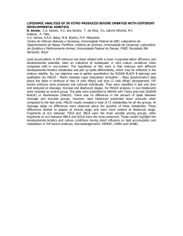
Anim. Reprod., v.11, n.2, p.96-103, Apr./Jun. 2014 Recombinant expression and purification of the bovine acidic Seminal Fluid Protein I.C. Bustamante-Filho1,2,3,5, G.D. Salton2, F.M. Munari2, M.R. Schneider3, R.C. Mattos1, J.P. Laurino4, E.O. Cirne-Lima1,2, M.I.M. Jobim1 1 Faculdade de Veterinária, Universidade Federal do Rio Grande do Sul, Porto Alegre, RS, Brazil. 2 Centro de Pesquisa Experimental, Hospital de Clínicas de Porto Alegre, RS, Brazil. 3 Gene Center, Ludwig-Maximilians Universität München, Munich, Bayern, Germany. 4 Instituto de Biotecnologia, Universidade de Caxias do Sul, Caxias do Sul, RS, Brazil. 5 Centro de Ciências Biológicas e da Saúde, Centro Universitário UNIVATES, Lajeado, RS, Brazil. Abstract The acidic Seminal Fluid Protein (aSFP), a 12.9 kDa protein is a maker for bovine semen freezability possibly due to its antioxidant activity and effect on sperm mitochondrial function. However, its precise function on sperm preservation during freezingthaw is poorly understood. The use of recombinant DNA technology allows new approaches on the study of function and structure of proteins, and its production in procaryote systems offers several advantages. The present work describes the recombinant expression of the bovine aSFP and its binding properties. A cDNA library from the bovine seminal vesicle was used as template for amplification of the aSFP coding region. The amplicon was cloned into a pET23a(+) vector and transformed into E.coli BL21 pLysS strain. The recombinant expression was obtained in E coli. One step ion immobilized affinity chromatography was performed, resulting in high yield of purified protein. To determine the bioactivity of the raSFP, the protein was incubated in different concentrations with 107 spermtozoa at 37°C for 5 h. Western blotting and fluorescence microscopy analyses showed the ability of the recombinant aSFP to attach to the spermatozoa. Based on our results, the described method can be used to obtain mg levels of recombinant aSFP. Keywords: bovine, recombinant protein, seminal plasma, spermatozoa. Introduction Seminal plasma (SP), a physiological secretion from multiple glands of the male reproductive tract, is a very complex fluid and is the natural medium for final maturation of spermatozoa through hormonal, enzymatic and surface-modifying events (Mann and Lutwak-Mann, 1981). The proteomic approach of seminal plasma is a valuable tool to identify proteins playing key roles on sperm maturation, viability and fertilization. Although the identification of SP proteins (Assumpção et al., 2005; Kelly et al., 2006; Moura et al., 2006, 2007, 2010) provided many insights about their correlation with biological phenomena, most of ________________________________ 8 Corresponding author: [email protected] Received: November 5, 2013 Accepted: May 6, 2014 their precise functions are still unknown. Fertilityassociated proteins were described in this fluid, as well as glycoproteins which participate in sperm-egg recognition, modulation of spermatozoa capacitation and the acrosome reaction (Killian et al., 1993; Therien et al., 1995; Calvete et al., 1996; Iborra et al., 1996). The contribution of SP proteins to freezability is also a matter of study in several species, including bull (Roncoletta et al., 2000; Jobim et al., 2004; Bergeron et al., 2007; Manjunath et al., 2007) ram (Barrios et al., 2000; Rebolledo et al., 2007), buffalo (Harshan et al., 2006; Asadpour et al., 2007) and boar (Casas et al., 2009). Comparing the SP protein profile from bulls with high and low semen freezabilty, Jobim et al., (2004) suggested the acidic Seminal Fluid Protein (aSFP) as a marker of good semen freezability. The 12.9 kDa aSFP is an acidic nonglycosylated protein (pI 4.8; Einspanier et al., 1991), mainly synthesized by the ampulla and seminal vesicle epithelium of the bull in high concentrations (2-7 mg/ml in SP; Einspanier et al., 1993; Dostàlovà et al., 1994) and has recently been described in epididymal fluid (Moura et al., 2010). This protein has not been detected in any other bovine tissues nor in other mammalian species, such as goat, sheep, pig, rat, dog, or human (Wempe et al., 1992; Einspanier et al., 1993). It presents approximately 50% amino acid sequence identity with polypeptides of the spermadhesin family (Einspanier et al., 1994), but with different biological characteristics, aSFP binds only loosely to the acrosomal cap of ejaculated bovine sperm and is quantitatively released during in vitro capacitation (Dostàlovà et al., 1994). In addition, aSFP possesses neither carbohydrate nor zona pellucida binding abilities, strongly indicating that aSFP may not be involved in gamete interaction (Dostàlovà et al., 1994; Ekhlasi-Hundrieser et al., 2008). Recently, the production of recombinant forms of seminal plasma proteins has been adopted as a new tool for understanding the biological activities of spermadhesins (Ekhlasi-Hundrieser et al., 2008; Cajazeiras et al., 2009). Considering its possible role on semen freezability, the aim of this work was to purify a bioactive recombinant form of aSFP and to study its binding properties on bovine semen. Bustamante-Filho et al. Recombinant expression of aSFP. Materials and Methods Materials Expression vector pET23a(+), as well as Escherichia coli strains BL21 (DE3) pLysS, BL21 (DE3) were from Novagen (EMD Biosciences, La Jolla, CA, USA); JM109 strains were from Promega (Madison, WI, USA). TOPO TA kit, all modification enzymes, antibody anti His-tag, secondary anti mouse and E. coli strains BL21 (DE3) Star and BL21 SI were from Invitrogen (Carlsbad, CA, USA). RNA extraction kit, Miniprep DNA isolation kit and Miniprep Plasmid DNA purification kit were from Macherey-Nagel (Düren, Germany). Reagents to detect chemioluminescence (ECL) were purchased from Amersham Pharmacia Biotech (Piscataway, NJ, USA). Hybond-C nitrocellulose membranes were from Hybond ECL (Hybond ECL nitrocellulose membrane, Amersham Biosciences, Freiburg, Germany). X-ray films were purchased from Kodak (Kodak X-Omat, Rochester, NY, USA). Clonning of cDNA sequence into expression vector For expression of aSFP with N-terminal Histag, bovine seminal vesicle cDNA was used as a template for polymerase chain reaction (PCR) amplification of aSFP. Synthetic oligonucleotide primers (forward: 5’GTCCATATGAAGCTGTCCAGCGTCAT-3’; reverse: were 5’-AGTCTCGAGAGCTTGTGGATCCT-3’) designed based on the aSFP sequence available on the GeneBank (accession number NM_174616; Wempe et al., 1992) to amplify the codon region. The 5’ NdeI and 3’ XhoI restriction sites are underlined, and the start codon is shown in bold. PCR amplification was performed in a thermocycler TC-4000 (Techne UK, Staffordshire, UK) using Taq DNA polymerase, under the following conditions: 94°C, 2 min; 36 cycles of 94°C, 30 s; 56°C 30 s; 72°C, 1 min, and the final elongation of 72°C for 5 min. The resulted aSFP amplicon (404 bp) was cloned into pCR2.1 vector, excised from this vector using the restriction enzyme pair mentioned above, and ligated into the expression vector pET23(a)+ linearized with the same enzyme pair. Digested insert and vector were run on a 1.5% agarose gel, gel purified using a commercial kit and then incubated overnight at 16°C using T4 DNA ligase. Ligation reactions were transformed into electrocompetent E. coli JM109 cells, and plasmid DNA was isolated. The resulting construct pET-aSFP was sequenced using the Big Dye Terminator® v 3.1 Cycle Sequencing Kit (Applied Biosystems, Foster City, CA, USA) and the automated sequencer ABI PRISM® 3100 Genetic analyzer (Applied Biosystems). Dying reactions were performed using a GeneAmp PCR System 9700 (Applied Biosystems) and data analyzed using Data Anim. Reprod., v.11, n.2, p.96-103, Apr./Jun. 2014 Collection v1.0.1 (Applied Biosystems). The following primers were used for sequence confirmation: forward 5'-TAATACGACTCACTATAGGG-3'; reverse 5'GCTAGTTATTGCTCAGCGG-3'; forward 5'CGCCACCTATTACGGACCGAAAAC-3', reverse 5'TCCCTAAGACAGGAGATCCTGGCA-3'. Protein expression in E. coli For protein expression assays, plasmids were transformed into E. coli BL21 pLysS competent cells using methods described elsewhere (Sambrook and Russell, 2001). Transformed bacteria were grown overnight on LB-agar plates containing 100 µg/ml ampicillin and/or 50 μg/ml chloramphenicol, after which single colonies were used to inoculate 2x YT medium containing the same antibiotic. For protein expression, 250 ml 2x YT medium was inoculated with 1/100 volume of overnight culture; bacteria were grown at 37°C with shaking at 200 rpm until the OD600 reached 0.6 - 0.8 and then the lactose analogue, isopropyl-β-Dthiogalactopyranoside (IPTG) was added to a final concentration of 0.5 mM to induce expression. Three hours after induction, cell cultures were harvested by centrifugation at 5,000 x g for 10 min at 4°C. Protein purification using immobilized metal affinity chromatography Cell pellet from 250 ml induced culture was suspended in 10 ml of denaturing lysis buffer (50 mM NaH2PO4, 8 M urea, pH 8.0), incubated for 30 min in the presence of lysozyme (1 mg/ml), disrupted by sonication (6 cycles of 10 s on ice), and centrifuged at 20,000 x g for 30 min at 4°C, as described in the Invitrogen protocol handbook. Supernatant was loaded onto an Econopack column (Bio-Rad) filled with 1 mL of Ni-NTA agarose resin (Invitrogen), prepared and equilibrated according to manufacturer’s instructions. Washing steps were performed using 5 to 8 bed volumes of denaturing washing buffer (50 mM NaH2PO4, 8 M urea) with decreasing pH (pH 6.0 and pH 5.3). Recombinant His-tagged aSFP (raSFP) was eluted in five elution steps using 500 μl of denaturing elution buffer (50 mM NaH2PO4, 8 M urea, pH 4.5). All samples were analyzed by SDS-PAGE and western blotting. Elution aliquots were polled and centrifuged in an Amicon tube (MWCO 3 kDa) for concentration and urea removal. Determination of protein concentration, electrophoresis and Western blotting Protein concentration was determined by the method of Lowry et al. (1951) and SDS-PAGE 14% were carried out according to Laemmli (1970) using the Mini Protean 3 system (Bio-Rad, USA). Samples were always loaded in duplicates in two gels, one for 97 Bustamante-Filho et al. Recombinant expression of aSFP. Coomassie Brilliant Blue staining, and the other for electrotransferring to nitrocellulose membranes using a Mini Trans Blot apparatus (Bio-Rad). Membranes were incubated for 60 min at 4°C in blocking solution - Trisbuffered saline containing 5% non-fat milk (w/v) and 0.1% Tween-20 (v/v) - and further incubated with the primary antibody diluted in blocking solution overnight at 4°C. The primary antibody used was His-tag monoclonal antibody (Invitrogen), used at a concentration of 1:10,000. The membranes were washed and incubated with horseradish peroxidase-conjugated anti-mouse antibody (1:5,000) for 2 h. The chemiluminescence (ECL) was detected using X-ray films. Binding of raSFP to bovine spermatozoa The ability of spermatozoa to bind raSFP was accessed by incubating 1 x 10⁷ spermatozoa with 700; 2,000; 5,000 and 7,000 ng of purified protein in a final volume of 1 ml of phosphate saline buffer (PBS) at 37°C for 5 h. As negative controls, one sperm sample was incubated without raSFP, and one sample was incubated with another recombinant His-tag fusioned protein (7,000 ng of tick calreticulin). Semen samples from two bulls were obtained by artificial vagina, and the ejaculate was washed three times in PBS to remove seminal plasma. Following incubation, samples were washed with PBS by centrifugation at 1,000 x g for 5 min at 20°C (Dostàlovà et al., 1994). To confirm the binding of raSFP to spermatozoa, 5 x 10⁶ cells were lysed and protein extracts were analyzed by Western blotting as described above. The topography of raSFP binding pattern was studied by indirect immunofluorescence (Dostàlovà et al., 1994). Briefly, 10 µl of samples (approximately 5 x 10⁶ cells) were spread on slides, air dried and fixed in methanol. Cells were incubated for 2 h at 37°C with a 1:500 (v/v) dilution of mouse antiHis-tag antibody in PBS containing 5% BSA. The slides were washed and were incubated for another 2 h at 37°C with a 1:200 (v/v) dilution of an R-Phycoerithrin labeled goat anti-mouse IgG antibody (Sigma, USA). Slides were observed under a fluorescence microscope (Olympus, Japan). One hundred raSFP marked cells per slide were counted and group (four slides per group) values were statistically compared through ANOVA, followed by Tukey test assuming P < 0.05 as significant (Prism 6 sofware, GraphPad, USA). Results PCR amplification of aSFP cDNA from a bovine seminal vesicle library resulted in one band of approximately 400 bp by agarose gel electrophoresis analysis. The amplicon was successfully cloned into the prokaryotic expression plasmid pET-23a(+; Fig. 1). Figure 1. Construction of pET-aSFP expression vector. (A) Restriction analysis of recombinant plasmid pET-aSFP in Agarose Gel 1%. Lane MW= DNA marker; Lane 1= plasmid pET-aSFP digested with NdeI and XhoI. Arrow indicates the target gene. (B) Schematic diagram of the construct pET-asFP, showing the coding region of aSFP gene under control of T7 promoter. Induction of raSFP expression was optimized after several expression conditions assays and best results were obtained with addition of 0.5 mM IPTG to the culture at 37°C for 3 h. Following sonication and centrifugation of the bacterial culture, the supernatants and inclusion bodies 98 were run on 12% SDS-PAGE. We obtained an expression level of approximately 30% of the total bacteria proteins (Fig. 2A). Western blotting analysis revealed that the majority of raSFP was in the insoluble fraction as a consequence of inclusion bodies formation (Fig. 2B and C). Anim. Reprod., v.11, n.2, p.96-103, Apr./Jun. 2014 Bustamante-Filho et al. Recombinant expression of aSFP. Figure 2. Analysis of raSFP expression. (A) Coomassie-stained SDS-PAGE of bacterial extracts. Lane MW= Molecular weight standard; Lane 1= Non induced control, 1 h; Lane 2= 1.0 mM IPTG, 1 h; Lane 3= 0.5 mM IPTG, 1 h; Lane 4= Non induced control, 3 h; Lane 5= 1.0 mM IPTG, 3 h; Lane 6= 0.5 mM IPTG, 3 h. (B) Anti-His-tag Western blot of BL21 pLysS bacterial extracts induced with 0.5 mM IPTG for 3 h. Lane 1= BL21 pLysS harboring pET-aSFP; Lane 2= BL21 pLysS harboring pET23a(+); Lane 3= BL21 pLysS without plasmid. (C) Anti-His-tag Western blot of BL21 pLysS lysates. Lane 1= supernatant; Lane 2= pellet. Arrows indicate the recombinant protein. pET23(a)+ expression vector permits the production of N-terminal 6xHis-tagged recombinant proteins, which can be purified by one step metal chelating affinity chromatography (IMAC). Recombinant aSFP was purified using denaturing conditions, after inclusion bodies solubilization with 8 M urea (Fig. 3A). Following protein concentration and removal of urea from solution, the SDS-PAGE profile of the pool of elution fractions showed a purity of 70% (Fig. 3B). Figure 3. Purification of raSFP under denaturing conditions. (A) Coomassie-stained SDS-PAGE of column fractions following purification of raSFP on a His-Select column. (B) Coomassie-stained SDS-PAGE gel of column fractions before and after urea removal and concentration of the elution fractions. Lane 1= before concentration; Lane 2= after concentration. Western blotting demonstrated that raSFP bind to ejaculated spermatozoa. Protein binding were detected in samples incubated with 700; 2,000; 5,000 and 7,000 ng of purified raSFP (Fig. 4). The fluorescence microscopy Anim. Reprod., v.11, n.2, p.96-103, Apr./Jun. 2014 confirmed the binding of raSFP to spermatozoa incubated with of recombinant protein (Fig. 5A), with a higher number of raSFP-bound spermatozoa in samples incubated with 7,000 ng of protein (P < 0.001, Fig. 5D). No 99 Bustamante-Filho et al. Recombinant expression of aSFP. fluorescence signal was detected in samples incubated with 700; 2,000 ng of recombinant protein (data not shown). Nonspecific cross reactivity was discarded by the absence of labeling on negative controls (Fig. 4, 5B and 5C). Figure 4. Binding of raSFP to bovine spermatozoa. Cell protein extracts of 5 x 10⁶ spermatozoa were run in 14% SDS-PAGE, transferred to nitrocellulose membrane and incubated with different concentrations of raSFP from 700 to 7000 ng. Peroxidase conjugated with anti-His-tag antibody was used as a detection system. PBS= Negative control without raSFP; His= Negative control (incubation with 7,000 ng of his-tagged recombinant tick calreticulin); run raSFP= Positive control. Figure 5. Representative images of immunofluorescence microscopy of bovine spermatozoa incubated with raSFP. Columns from left to right, microphotography under visible light, UV and merged image. A= Semen sample incubated with 7,000 ng of raSFP; B= Semen sample incubated in PBS; C= Semen sample incubated with Histagged tick calreticulin. Magnification of 400X. D= Comparison of the percentage of raSFP-marked sperm between groups. Letters mean statistical difference (P < 0.001). Discussion Seminal plasma proteins hold important functions on sperm viability in both male and female reproductive tracts. Proteomic studies provided a broader view of the bovine seminal plasma protein content (Desnoyers et al., 1994; Frazer et al., 1996; Manjunath and Thérien, 2002; Jobim et al., 2004; Moura et al., 2007) and provided valuable information for a better understanding of their role on sperm physiology and fertilization. Here we describe the cloning and purification of the bovine aSFP. The protein was expressed as inclusion bodies, which has many advantages including easy separation by centrifugation, protection from proteolytic degradation and lower contamination with 100 other E. coli proteins (Addona et al., 2009; Meehan et al., 2010). Results from Western blotting confirmed the ability of raSFP to attach to sperm cells after incubation with 700 ng of purified protein. The acidic Seminal Fluid Protein structure is mainly formed by a CUB domain (Romero et al., 1997). Therefore, since the CUB domain may be responsible for the protein attachment to cellular membrane, we assume that raSFP was correctly folded after urea removal. The CUB domain is a widely occurring structural motif found almost exclusively in extracellular and plasma membrane-associated proteins (Bork and Beckmann, 1993). These proteins participate in a wide range of biological functions, including complement activation, tissue repair, cell signaling, inflammation and receptor-mediated endocytosis (Blanc et al., 2007). Although the roles of the CUB domains Anim. Reprod., v.11, n.2, p.96-103, Apr./Jun. 2014 Bustamante-Filho et al. Recombinant expression of aSFP. are yet to be completely elucidated, a number of them have been shown to be involved in oligomerization and/or recognition of substrates and binding partners (Bork and Beckmann, 1993; Blanc et al., 2007). Recombinant aSFP attached clearly to spermatozoa’s mid-piece, a binding pattern similar to other spermadhesins like porcine AQN1 and AWN, which bind to the acrosome and participate in the fertilization process (Sanz et al., 1992; Haase, 2005). aSFP is not related to the acrosomal function and oocyte fusion since it is believed that the protein detaches from sperm head in utero (Dostàlovà et al., 1994). Thus, the topographical binding pattern of raSFP supports its known influence on mitochondrial activity of sperm cells. It is noteworthy to mention that spermadhesins are multifunctional proteins in other species (TopferPetersen et al., 1998; Haase et al., 2005) , so different binding behaviors could be possible. To elucidate the precise function of aSFP on motility preservation of frozen semen, it is important to investigate the binding mechanism to sperm membrane and which signaling pathways aSFP might be interacting with. Differently from other spermadhesins, native and recombinant aSFP do not posses mannose binding properties (EkhlasiHundrieser et al., 2008), so a protein-protein interaction is more likely. However, no specific protein receptor for aSFP in sperm membrane has been described so far. Proteomic studies suggested aSFP as a marker for high semen freezability (Jobim et al., 2004), however its potential role on sperm preservation is yet to be determined. The influence of aSFP in bovine semen was demonstrated by (Schöneck et al., 1996). The addition of a high concentration of bovine SP purified aSFP in semen, decreased sperm motility and mitochondrial activity. Since aSFP is secreted at the epididymis, a natural sperm reservoir, these effects lead to a lower production of reactive oxygen species (ROS) by stored sperm cells. In addition, aSFP showed a remarkable antioxidant behavior due to its redox equilibrium exhibited by the cysteine residues Cys54 and Cys75 (Calvete and Sanz, 2007). These Cys residues are responsible for aSFP alternation between oxidized and reduced states, acting as ROS scavenger. As a result, bulls with higher semen freezability may ejaculate spermatozoa with lower oxidative damage on membrane lipids and proteins. These effects are an important strategy for semen storage in the epidydimis, resulting in a better preservation of sperm motility and viability (Schöneck et al., 1996). Thus, the role of aSFP on semen freezability might be associated with the quality of the stored sperm and with a minor effect after ejaculation. Egg yolk-based semen extenders present a marked antioxidant activity (Bustamante-Filho et al., 2009), so aSFP antioxidant protection may only be beneficial before semen dilution. In this article we report, for the first time, the recombinant expression and purification of aSFP. The Anim. Reprod., v.11, n.2, p.96-103, Apr./Jun. 2014 plasmid (pET23a) used in the expression study was successfully constructed by the ligation of the mRNA aSFP cDNA, resulting in N-terminal tagging with 6xHis. The expression of the raSFP in bacterial system (E. coli BL21 pLysS) and further purification was successfully accomplished and may help us understand the mechanism by which this protein modulates sperm metabolism and cryopreservation success in further studies. Acknowledgments Financial support for this work was provided by CNPq, CAPES, DAAD, FIPE-HCPA and FAPERGS. The authors would like to thank Dr. Luciana Meirelles Richer and Prof. Dr. Itabajara da Silva Vaz Junior for protein purification troubleshooting; Steffen Schiller, Joseph Millauer and Jefferson Beck for technical assistance. References Addona TA, Abbatiello SE, Schilling B, Skates SJ, Mani DR, Bunk DM, Spiegelman CH, Zimmerman LJ, Ham AJ, Keshishian H, Hall SC, Allen S, Blackman RK, Borchers CH, Buck C, Cardasis HL, Cusack MP, Dodder NG, Gibson BW, Held JM, Hiltke T, Jackson A, Johansen EB, Kinsinger CR, Li J, Mesri M, Neubert TA, Niles RK, Pulsipher TC, Ransohoff D, Rodriguez H, Rudnick PA, Smith D, Tabb DL, Tegeler TJ, Variyath AM, Vega-Montoto LJ, Wahlander A, Waldemarson S, Wang M, Whiteaker JR, Zhao L, Anderson NL, Fisher SJ, Liebler DC, Paulovich AG, Regnier FE, Tempst P, Carr SA. 2009. Multi-site assessment of the precision and reproducibility of multiple reaction monitoringbased measurements of proteins in plasma. Nat Biotechnol, 27:633-641. Asadpour R, Alavi-Shoushtari SM, Rezaii SA, Ansari MH. 2007. SDS-polyacrylamide gel electrophoresis of buffalo bulls seminal plasma proteins and their relation with semen freezability. Anim Reprod Sci, 102:308-313. Assumpção TI, Fontes W, Sousa MV, Ricart CAO. 2005. Proteome analysis of Nelore bull (Bos taurus indicus). seminal plasma. Protein Pept Lett, 12:813-817. Barrios B, Pérez-Pé R, Gallego M, Tato A, Osada J, Muiño-Blanco T, Cebrián-Pérez JA. 2000. Seminal plasma proteins revert the cold-shock damage on ram sperm membrane. Biol Reprod, 63:1531-1537. Bergeron A, Brindle Y, Blondin P, Manjunath P. 2007. Milk caseins decrease the binding of the major bovine seminal plasma proteins to sperm and prevent lipid loss from the sperm membrane during sperm storage. Biol Reprod, 77:120-126. Blanc G, Font B, Eichenberger D, Moreau C, Ricard-Blum S, Hulmes DJS, Moali C. 2007. Insights into how CUB domains can exert specific functions 101 Bustamante-Filho et al. Recombinant expression of aSFP. while sharing a common fold. J Biol Chem, 282:1692416933. Bork P, Beckmann G. 1993. The CUB domain. A widespread module in developmentally regulated proteins. J Mol Biol, 231:539-545. Bustamante-Filho IC, Pederzolli CD, Sgaravatti ÂM, Gregory RM, Dutra-Filho CS, Jobim MIM, Mattos RC. 2009. Skim milk-egg yolk based semen extender compensates for non-enzymatic antioxidant activity loss during equine semen cryopreservation. Anim Reprod, 6:392-399. Cajazeiras JB, Melo LM, Albuquerque ES, RadisBaptista G, Cavada BS, Freitas VJ. 2009. Analysis of protein expression and a new prokaryotic expression system for goat (Capra hircus) spermadhesin Bdh-2 cDNA. Genet Mol Res, 8:1147-1157. Calvete JJ, Varela PF, Sanz L, Romero A, Mann K, Töfper-Petersen E. 1996. A procedure for the largescale isolation of major bovine seminal plasma proteins. Protein Expr Purif, 8:48-56. Calvete JJ, Sanz L. 2007. Insights into structurefunction correlations of ungulate seminal plasma proteins. Soc Reprod Fertil Suppl, 65:201-215. Casas I, Sancho S, Briz M, Pinart E, Bussalleu E, Yeste M, Bonet S. 2009. Freezability prediction of boar ejaculates assessed by functional sperm parameters and sperm proteins. Theriogenology, 72:930-948. Desnoyers L, Therien I, Manjunath P. 1994. Characterization of the major proteins of bovine seminal fluid by two-dimensional polyacrylamide gel electrophoresis. Mol Reprod Dev, 37:425-435. Dostàlovà Z, Calvete JJ, Sanz L, Hettel C, Riedel D, Schoneck C, Einspanier R, Topfer-Petersen E. 1994. Immunolocalization and quantitation of acidic seminal fluid protein (aSFP) in ejaculated, swim-up, and capacitated bull spermatozoa. Biol Chem Hoppe Seyler, 375:457-461. Einspanier R, Einspanier A, Wempe F, Scheit KH. 1991. Characterization of a new bioactive protein from bovine seminal fluid. Biochem Biophys Res Commun, 179:1006-1010. Einspanier R, Amselgruber W, Sinowatz F, Henle T, Ropke R, Schams D. 1993. Localization and concentration of a new bioactive acetic seminal fluid protein (aSFP) in bulls (Bos taurus). J Reprod Fertil, 98:241-244. Einspanier R, Krause I, Calvete JJ, Töpfer-Petersen E, Klostermeyer H, Karg H. 1994. Bovine seminal plasma aSFP: localization of disulfide bridges and detection of three different isoelectric forms. FEBS Lett, 344:61-64. Ekhlasi-Hundrieser M, Calvete JJ, Von Rad B, Hettel C, Nimtz M, Töpfer-Petersen E. 2008. Point mutations abolishing the mannose-binding capability of boar spermadhesin AQN-1. Biochim Biophys Acta, 1784:856-862. Frazer GS, Bucci DM, Brooks CL. 1996. Twodimensional polyacrylamide gel electrophoresis of 102 bovine semen after cryopreservation in half-milliliter straws. Theriogenology, 46:1103-1115. Haase B, Schlötterer C, Hundrieser ME, Kuiper H, Distl O, Töpfer-Petersen E, Leeb T. 2005. Evolution of the spermadhesin gene family. Gene, 352:20-29. Harshan HM, Singh LP, Arangasamy A, Ansari MR, Kumar S. 2006. Effect of buffalo seminal plasma heparin binding protein (HBP) on freezability and in vitro fertility of buffalo cauda spermatozoa. Anim Reprod Sci, 93:124-133. Iborra A, Morte C, Fuentes P, Garcia-Framis V, Andolz P, Martinez P. 1996. Human sperm coating antigen from seminal plasma origin. Am J Reprod Immunol, 36:118-125. Jobim MIM, Oberst ER, Salbego CG, Souza DO, Wald VB, Tramontina F, Mattos RC. 2004. Twodimensional polyacrylamide gel electrophoresis of bovine seminal plasma proteins and their relation with semen freezability. Theriogenology, 61:255-266. Kelly VC, Kuy S, Palmer DJ, Xu Z, Davis SR, Cooper GJ. 2006. Characterization of bovine seminal plasma by proteomics. Proteomics, 6:5826 - 5833. Killian GJ, Chapman DA, Rogowski LA. 1993. Fertility-associated proteins in Holstein bull seminal plasma. Biol Reprod, 49:1202-1207. Laemmli UK. 1970. Cleavage of structural proteins during the assembly of the head of bacteriophage T4. Nature, 227:680-685. Lowry OH, Rosenbrough NJ, Farr AL, Randall RJ. 1951. Protein measurement with the Folin phenol reagent. J Biol Chem, 193:265-275. Manjunath P, Thérien I. 2002. Role of seminal plasma phospholipid-binding proteins in sperm membrane lipid modification that occurs during capacitation. J Reprod Immunol, 53:109-119 Manjunath P, Bergeron A, Lefebvre J, Fan J. 2007. Seminal plasma proteins: functions and interaction with protective agents during semen preservation. Soc Reprod Fertil Suppl, 65:217-228. Mann T, Lutwak-Mann C. 1981. Male Reproductive Function and Semen. Berlin: Springer-Verlag. Meehan KL, Rainczuk A, Salamonsen LA, Stephens AN. 2010. Proteomics and the search for biomarkers of female reproductive diseases. Reproduction, 140:505519. Moura AA, Koc H, Chapman DA, Killian GJ. 2006. Identification of proteins in the accessory sex gland fluid associated with fertility indexes of dairy bulls: a proteomic approach. J Androl, 27:201-211. Moura AA, Chapman DA, Koc H, Killian GJ. 2007. A comprehensive proteomic analysis of the accessory sex gland fluid from mature Holstein bulls. Anim Reprod Sci, 98:169-188. Moura AA, Souza CE, Stanley BA, Chapman DA, Killian GJ. 2010. Proteomics of cauda epididymal fluid from mature Holstein bulls. J Proteomics, 73:20062020. Rebolledo AD, Sierra LN, Tamayo AC, Loria AA, Anim. Reprod., v.11, n.2, p.96-103, Apr./Jun. 2014 Bustamante-Filho et al. Recombinant expression of aSFP. Denis SE, Oses RB, Parra EG, Monsreal LP, Ugalde JR. 2007. Fertility in hair sheep inseminated with freeze spermatozoa rediluited with seminal plasma. Rev Cient Fac Cienc Vet, 17:73-76. Romero A, Romao MJ, Varela PF, Kolln I, Dias JM, Carvalho AL, Sanz L, Topfer-Petersen E, Calvete JJ. 1997. The crystal structures of two spermadhesins reveal the CUB domain fold. Nat Struct Biol, 4:783788. Roncoletta M, Morani EdSC, Franceschini PH, Ramos PRR. 2000. Caracterização da proteína 26 kDa do plasma seminal e sua relação com a congelabilidade do sêmen de touros. Arq Fac Vet UFRGS, 28:323-332. Sambrook J, Russell DW. 2001. Molecular Cloning: a laboratory manual. 3rd.ed. New York, Cold Spring Harbor. Sanz L, Calvete JJ, Mann K, Schäfer W, Schmid ER, Amselgruber W, Sinowatz F, Ehrhard M, TöpferPetersen E. 1992. The complete primary structure of the spermadhesin AWN, a zona pellucida-binding Anim. Reprod., v.11, n.2, p.96-103, Apr./Jun. 2014 protein isolated from boar spermatozoa. FEBS Lett, 300:213-218. Schöneck C, Braun J, Einspanier R. 1996. Sperm viability is influenced in vitro by the bovine seminal plasma protein aSFP : Effects on motility, mithocondrial activity and lipid peroxidation. Theriogenology, 45:633642. Therien I, Bleau G, Manjunath P. 1995. Phosphatidylcholine-binding proteins of bovine seminal plasma modulate capacitation of spermatozoa by heparin. Biol Reprod, 52:1372-1379. Topfer-Petersen E, Romero A, Varela PF, EkhlasiHundrieser M, Dostàlovà Z, Sanz L, Calvete JJ. 1998. Spermadhesins: a new protein family. Facts, hypotheses and perspectives. Andrologia, 30:217-224. Wempe F, Einspanier R, Scheit KH. 1992. Characterization by cDNA cloning of the mRNA of a new growth factor from bovine seminal plasma: acidic seminal fluid protein. Biochem Biophys Res Commun, 183:232-237. 103
Baixar