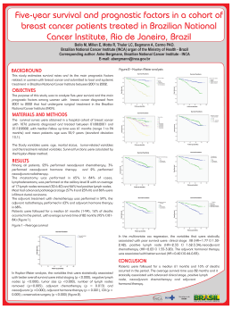
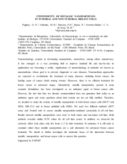
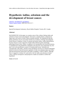
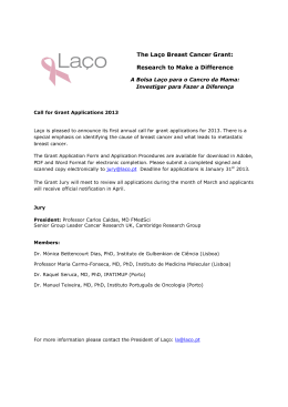
CLINICAL SCIENCE
Expression of aldehyde dehydrogenase after neoadjuvant chemotherapy is associated with expression
of hypoxia-inducible factors 1 and 2 alpha and
predicts prognosis in locally advanced breast cancer
Daniel Guimarães Tiezzi,I Willian Simões Clagnan,I Larissa Raquel Mouro Mandarano,I Christiani Bisinoto
de Sousa,I Heitor Ricardo Cosiski Marana,I Marcelo Guimarães Tiezzi,II Jurandyr Moreira de AndradeI
I
Universidade de São Paulo, Hospital das Clı́nicas of the Ribeirão Preto School of Medicine, Breast Disease Division Department of Gynecology and
Obstetrics, Ribeirão Preto/SP, Brazil. II Laboratório de Anatomia Patológica e Citopatologia de Presidente Prudente, São Paulo/SP, Brazil.
OBJECTIVE: To analyze the expression of hypoxia-inducible factors (hypoxia-inducible factor 1A and hypoxiainducible factor 2A) and aldehyde dehydrogenase proteins in patients with locally advanced breast carcinoma
who were subjected to neoadjuvant chemotherapy.
METHODS: We included 90 patients with histologically confirmed stage II and III breast carcinoma who were
treated with neoadjuvant chemotherapy between 2000 and 2005. Immunohistochemistry for aldehyde
dehydrogenase, hypoxia-inducible factor 1A, and hypoxia-inducible factor 2A was performed before and after
neoadjuvant chemotherapy. We analyzed the influence of clinical and pathological features on clinical and
pathological response, disease-free survival, and overall survival.
RESULTS: An objective clinical response to neoadjuvant chemotherapy was observed in 80% of patients, with
12% showing a complete pathological response. Among all clinical and pathological parameters, only the
expression of hypoxia-inducible factor 1A was associated with a pathological response. A positive association
was found between expression of aldehyde dehydrogenase and that of hypoxia-inducible factor 1A before and
after chemotherapy. Aldehyde dehydrogenase expression was associated with expression of hypoxia induciblefactor 2A in tumors after neoadjuvant treatment. In a univariate analysis, prognosis was influenced by age,
pathological response, metastasis to axillary lymph nodes after neoadjuvant chemotherapy, overexpression of
hypoxia-inducible factor 2, and the presence of aldehyde dehydrogenase-positive cells within the primary
tumor after neoadjuvant chemotherapy. In a multivariate analysis, only age and the presence of aldehyde
dehydrogenase-positive cells after chemotherapy were associated with reduced overall survival.
CONCLUSION: The presence of aldehyde dehydrogenase-positive cells within the residual tumor after
neoadjuvant chemotherapy is associated with an increase in the expression of hypoxia-inducible factor 2A
and with poor prognosis in patients with locally advanced breast cancer.
KEYWORDS: Breast Cancer; Neoadjuvant Chemotherapy; Prognostic Factors.
Tiezzi DG, Clagnan WS, Mandarano LR, Sousa CB, Marana HR, Tiezzi MG, et al. Expression of aldehyde dehydrogenase after neoadjuvant
chemotherapy is associated with expression of hypoxia-inducible factors 1 and 2 alpha and predicts prognosis in locally advanced breast
cancer. Clinics. 2013;68(5):592-598.
Received for publication on December 2, 2012; First review completed on December 2, 2012; Accepted for publication on January 3, 2013
E-mail: [email protected]
Tel.: 55 16 3602-2488
relevant factor in cancer resistance to therapy and in
recurrence (1). The presence of a subpopulation of cells with
stem cell properties within the tumor has been widely
demonstrated to be responsible for tumor heterogeneity
(2–4). In breast carcinomas, the expression or activity of
aldehyde dehydrogenase (ALDH) has been reported to be a
marker for stem-like cancer cells, based on the observation that
cells with ALDH expression have self-renewal and lineage
differentiation capacity, as well as the ability to produce
tumors in xenograft models. Moreover, the presence of
ALDH-positive cells within inflammatory breast carcinomas
has been significantly associated with poor survival (5,6).
& INTRODUCTION
Malignant solid tumors are composed of a heterogeneous
cell population. Tumor heterogeneity is considered to be a
Copyright ß 2013 CLINICS – This is an Open Access article distributed under
the terms of the Creative Commons Attribution Non-Commercial License (http://
creativecommons.org/licenses/by-nc/3.0/) which permits unrestricted noncommercial use, distribution, and reproduction in any medium, provided the
original work is properly cited.
No potential conflict of interest was reported.
DOI: 10.6061/clinics/2013(05)03
592
CLINICS 2013;68(5):592-598
ALDH and HIFs expression in breast cancer
Tiezzi DG et al.
the objective response group, which consisted of all patients
with a complete or partial response; and the no-response
group, which consisted of all patients with stable or
progressive disease. All surgical specimens from the definitive breast procedures were submitted for pathological
evaluation and were classified as having complete response
(pCR) when no residual invasive carcinoma was observed.
All the patients received adjuvant chemotherapy to
complete a total of nine cycles: CMF (cyclophosphamide,
600 mg/m2; methotrexate, 50 mg/m2; fluorouracil, 600 mg/
m2 iv on D1 every 21 days) for patients with negative or less
than four positive axillary lymph nodes (ALNs), or FEC
(fluorouracil, 600 mg/m2; epirubicin, 50 mg/m2; cyclophosphamide, 600 mg/m2 iv on D1 every 21 days) if there were
more than three positive lymph nodes. Tamoxifen (20 mg/
day) for 60 months was offered to all patients with
hormone-positive tumors (ER or PgR). No patients received
HER2 target therapy. Radiation therapy was performed in
the residual breast parenchyma of all patients who underwent breast-conserving surgery (50 Gy). Patients who
underwent mastectomies received adjuvant chest wall
radiotherapy if the residual tumors had a diameter of at
least 5 cm (at the highest pathological diameter), if ALN
metastasis was present, or if skin, muscle, or chest wall
invasion was present (50 Gy). The supraclavicular fossa was
irradiated if four or more positive ALNs were present.
Although the presence of cancer stem cells could explain
tumor heterogeneity and cancer relapse, evidence that tumor
cell phenotype is influenced by microenvironmental changes
supports the hypothesis that cell plasticity is involved in tumor
heterogeneity (7,8). The conversion to a stem-like state can be
driven by the epithelial-to-mesenchymal transition (EMT)
(9,10). Microenvironmental changes and signaling, such as
tissue hypoxia and hypoxia-inducible factor-mediated signaling, can trigger EMT, thereby promoting metastasis and
stemness (11,12). It was recently demonstrated that differentiated cells could convert into stem-like cells in normal and
neoplastic tissues (7). Additionally, the use of antiangiogenic
agents resulted in an increase in cancer-initiating cells in breast
cancer xenografts through the generation of tumor hypoxia
(13). Hypoxia-inducible factors (HIFs) mediate the processes
involved in oxygen homeostasis. HIFs transcriptionally
regulate the expression of target genes and represent a link
between oxygen sensors and effectors in the cellular adaptation to hypoxia (14). Such observations support the idea that
tumor hypoxia and hypoxia signaling are important for cancer
heterogeneity, aggressiveness, and dissemination.
Neoadjuvant chemotherapy (NACT) is currently being
used as a clinical approach to reduce tumor volume in
locally advanced breast cancer and as an alternative method
for facilitating the use of breast-conserving therapies (15,16).
Although primary chemotherapy has no influence on
overall survival, it has been reported that patients who
achieve complete pathological response (pCR) have better
prognoses than those who do not (17). The results of studies
on pathological response and ALDH expression have been
controversial (18,19). However, the presence of ALDHpositive cells within a tumor was demonstrated to be a
prognostic factor in locally advanced breast cancer patients
subjected to primary cytotoxic therapy (18,19). These studies
suggest that stem cells could be responsible for tumor
resistance and relapse. However, the relationship between
the presence of ALDH-positive cells within the primary
tumor and the expression of hypoxia-inducible factors
(HIFs) has not yet been studied. We analyzed the expression
of HIF1A, HIF2A, and ALDH in patients with locally
advanced breast carcinoma who were subjected to NACT.
Immunohistochemistry
All tissue samples were routinely fixed in 4% neutral
formalin and embedded in paraffin. Briefly, 3-mm-thick
sections were cut from paraffin blocks containing representative tumor samples. The paraffin sections were de-waxed in
xylene, rehydrated through a series of graded alcohols,
placed in 10 mM citrate buffer, and subjected to heat retrieval
using a vapor lock for 40 min. After heating, the slides were
allowed to cool to room temperature and were briefly
washed with Tris-buffered saline. Endogenous peroxidase
activity was blocked with 3% hydrogen peroxide in methanol
for 5 min. Normal serum (Novostain Super ABC kit;
Novocastra, Newcastle upon Tyne, UK) was used for
30 min to block nonspecific signals. The following primary
antibodies were incubated overnight at room temperature:
mouse anti-human HIF1A (1:300, clone H1alpha67; Abcam,
Cambridge, MA), mouse anti-human HIF2A (1:800, clone
ep190b, Abcam, Cambridge, MA), and mouse anti-human
ALDH (1:500, clone 44/ALDH; BD, Franklin Lakes, NJ).
Immunohistochemical staining was performed using the
Biocare Medical Mach 4 Universal Polymer Detection kit
(Biocare, Concord, CA) according to the manufacturer’s
protocol. The following tissues were used as positive
controls: melanoma and tonsil for HIF1A, placenta for
HIF2A, and liver for ALDH. Negative controls for immunostaining were prepared by omitting the primary antibody.
& MATERIALS AND METHODS
We prospectively included 90 patients with histologically
proven invasive breast carcinoma who were subjected to
anthracycline/taxane NACT at our institution between 2000
and 2005. In 2011, we obtained the clinical, pathological, and
immunohistochemical (estrogen receptor [ER], progesterone
receptor [PgR], and HER2) information from the patients’
files and retrieved all paraffin blocks from the primary
tumors before and after chemotherapy. A total of 75 blocks
before treatment and 67 blocks after treatment provided
adequate tumor sampling for immunohistochemical analysis of the expression of ALDH, HIF1A, and HIF2A.
All of the patients were treated with intravenous (iv)
epirubicin (50 mg/m2) plus docetaxel (75 mg/m2) on day 1
and then every 21 days in a neoadjuvant setting. The clinical
response was evaluated at every visit. The number of NACTs
varied from two to seven (median of three cycles) according
to the clinical response, on the basis of the primary tumor
diameter and the axillary status. The clinical response was
defined according to the International Union Against Cancer
(UICC) criteria. The patients were classified into two groups:
Scoring methods
HIF1A was present in the cytoplasm and/or nuclei in a
homogeneous pattern in tumor cells. The results were
interpreted according to staining intensity and were scored
as 0, 1+, 2+, or 3+. Any intensity of staining was considered
positive. HIF2A staining occurred mainly in the nucleus,
and we quantified HIF2A expression in terms of the
percentage (P) and intensity (I) of positive cells. The
quick score (QS = P6I) was obtained in samples before and
after neoadjuvant therapy. Figures 1 and 2 show the
593
ALDH and HIFs expression in breast cancer
Tiezzi DG et al.
CLINICS 2013;68(5):592-598
Figure 1 - HIF1A expression in breast carcinoma. Note that
homogeneous staining can be observed in all samples. We
classified the samples according to staining intensity from 0 to
3+. Tumors classified as 1+ to 3+ were considered to have positive
HIF1A expression. (a) Negative expression (2006); (b) weak
positive (1+) expression (2006); moderate positive (2+) expression; (d) strong positive (3+) expression (2006).
Figure 3 - ALDH expression in breast carcinoma. (a) Negative
expression. Note the positive expression in mesenchymal cells
(internal positive control, 2006). (b) Focal positive expression.
Note that some clusters show positive expression in malignant
cells (4006). (c) Focal positive cells. Note the variable (weak to
strong) brown staining in clusters of malignant cells (2006). (d)
Invasive ductal carcinoma with more than 90% of cells showing
positive expression of ALDH (4006).
immunohistochemistry for HIF1A and HIF2A in invasive
ductal carcinomas. ALDH was present in the cytoplasm. A
positive control (mesenchymal cells) was considered for all
of the samples analyzed. We considered the cases with
evident cytoplasmic staining on at least five tumor cells
arranged in a cluster to be positive. Figure 3 shows one
negative and some positive patterns for ALDH staining in
invasive ductal carcinomas.
combination of docetaxel and epirubicin. The second
objective was to evaluate the impact of the presence of
ALDH-positive cells on disease-free and overall survival.
We also studied the association between ALDH and HIF
expression.
Statistical analysis
The expression of ALDH and HIF protein and other
categorical variables were evaluated using a standard x2
test, median test, or Fisher’s exact test. Changes in ALDH
and HIF1A expression after neoadjuvant treatment were
analyzed using McNemar’s test. The disease-free survival
and overall survival intervals were calculated from the date
of diagnosis, and survival curves were derived from
Kaplan-Meier estimates and compared using log-rank tests.
The influence of prognostic factors on survival was assessed
by multivariate analysis (Cox proportional hazard models).
JMP, version 7.0.2, SAS, and MedCalc, version 12.1.4, were
used for the statistical analyses. The level of significance
was established as p,0.05.
Study endpoints
The primary goal was to test the associations between the
expression of ALDH, HIF1A, and HIF2A protein and the
clinical and pathological response to NACT with the
Ethics
The ethics committee of the Hospital das Clı́nicas
approved this study (protocol #5764/2000) in accordance
with the ethical guidelines of the 1975 Declaration of
Helsinki, revised in 1983, and all of the patients signed a
consent form.
& RESULTS
The patients’ mean age was 49¡11.5 years; most of them
were postmenopausal (55%). According to clinical stage
(CS), 11 patients were stage IIa, 33 patients were stage IIb, 15
patients were stage IIIa, and 31 patients were stage IIIb.
Invasive ductal carcinoma was diagnosed in 80 (89%)
patients, and grade 2 was the most frequent (57.8%)
histological grade. The positive rates of ER, PgR, and
HER2 were 63.3%, 43.3%, and 27.7%, respectively. An
Figure 2 - HIF2A in breast carcinoma. Note the progressive (0, 1+,
2+, and 3+) expression in invasive carcinoma. The quick score (QS)
was calculated as the product of the staining intensity and the
percentage of positive cells (QS = I6P). (a) Negative expression
(2006); (b) weak positive (1+) expression (2006); (c) moderate
positive (2+) expression (2006); (d) strong positive expression
(2006).
594
CLINICS 2013;68(5):592-598
ALDH and HIFs expression in breast cancer
Tiezzi DG et al.
analyzed the accuracy of ALDH, HIF1A, and HIF2A
expression as predictive of response on the basis of pCR.
ALDH and HIF2A expression did not predict the response.
However, the pCR rate in HIF1A-positive patients was 21%,
whereas it was 5% in HIF1A-negative patients (p = 0.03).
Table 2 lists the results for ALDH and HIF expression as
well as the clinical and pathological parameters.
Relationship between ALDH expression and HIF expression. Before NACT, we found positive expression of ALDH
in 25 tumors. In this subset of samples, 17 tumors (68%)
were positive for HIF1A. HIF1A positivity was observed in
42.8% of ALDH-negative samples. A positive association
was observed between ALDH and HIF1A expression (x2
test, p = 0.03). The same expression pattern was observed
after chemotherapy. HIF1A positivity was observed in 50%
of ALDH-positive tumors and in 21.7% of ALDH-negative
tumors (p = 0.03).
The median QS for HIF2A expression in ALDH-positive
and -negative tumors was 80 before NACT (range 0–270 and
0–240, respectively; median test, p = 0.9). However, after
treatment, the median QS was 55 (range 0–270) in ALDHpositive tumors and 2.5 (range 0–225) in ALDH-negative
tumors (median test, p = 0.01).
The presence of ALDH-positive cells after NACT predicted disease-free and overall survival. We further analyzed the influence of clinical and histopathological patterns
on disease-free and overall survival. In a univariate analysis,
age, axillary status, HER2 protein expression, pathological
response, and ALDH protein expression after NACT were
identified as significant prognostic factors. Additionally, the
presence of ALN metastasis after NACT was more frequent
in patients with residual tumors containing ALDH-positive
cells (59% versus 41% of ALN metastasis cases, p = 0.04). The
worst prognosis was observed among patients with ALDHpositive cells within the residual tumor after NACT and
with positive ALNs (overall survival, estimated using a
Kaplan-Meier curve, of 24% at 100 months of follow-up).
Figure 4 shows the disease-free and overall survival after
treatment of patients treated with NACT, according to
ALDH protein expression. Multivariate analysis showed
that only age (HR = 0.94; CI 95%: 0.91–0.98 for overall
survival) and protein expression of ALDH (HR = 2.54; CI
95%: 1.04–6.23 for overall survival) after NACT were
significant prognostic factors.
objective clinical response to therapy was observed in 72
patients (80%), and 11 patients (12.2%) achieved pCR.
Breast-conserving surgery was possible in 47 patients
(52.8%), and 38 patients (42%) were free of ALN metastasis
after NACT. The patients’ characteristics are summarized in
Table 1.
ALDH, HIF1A, and HIF2A protein expression in locally
advanced breast carcinomas. ALDH expression was analyzed in 75 and 67 samples before and after treatment,
respectively. Positive expression was found in 25 tumors
(33%) before treatment and in 32 (47%) tumors after
chemotherapy. Paired analysis showed an increase in
ALDH protein expression after NACT (McNemar’s test,
x2 = 3.8; p = 0.04).
Positive expression of HIF1A was observed in 50% and
38% of tumors before and after chemotherapy, respectively
(McNemar’s test, x2 = 0.4; p = 0.5). The median HIF2A QS
was 80 (range 0–270) before NACT and 10 (range 0–270)
after NACT. There was a significant reduction in HIF2A
expression after NACT (Wilcoxon test, p = 0.0001).
Expression of ALDH, HIF1A, and HIF2A before NACT
according to the patients’ characteristics. We observed an
association between age at diagnosis and the presence of
ALDH. In patients with positive ALDH expression, the
mean age was 45¡9.9 years, compared with 50.8¡11.4
years among ALDH-negative patients (p = 0.02). Positive
HIF1A expression was found in 76% of grade 3 tumors.
Furthermore, positive HIF1A expression was found in 45%
and 35% of grade 2 and grade 1 tumors, respectively
(p = 0.03).
Expression of ALDH, HIF1A, and HIF2A, and response to
NACT. An objective clinical response was observed in 80%
of patients. The complete clinical response rate was 23%,
and 11 patients (12%) achieved pCR after NACT. The pCR
rate was higher in ER-negative than in ER-positive tumors
(24% versus 5%, respectively; p = 0.01). No association was
observed between PgR or HER2 expression and pCR. We
Table 1 - Characteristics of 90 patients with locally
advanced breast cancer subjected to neoadjuvant
chemotherapy with a combination of epirubicin and
docetaxel.
Age [mean (SD)]
Menopausal status [n (%)]
Pre
Post
Clinical stage [n (%)]
II
III
Histology [n (%)]
Ductal
Others
Grade [n (%)]
1
2
3
ER positive [n (%)]
PgR positive [n (%)]
HER2 positive [n (%)]
Objective response [n (%)]
Breast-conserving surgery rate [n (%)]
pCR [n (%)]
Negative ALN [n (%)]
49 (11.5)
& DISCUSSION
40 (44.4%)
50 (55.6%)
Breast cancer is a disease with an adverse prognosis. The
presence of stem-like cells or tumor-initiating cells within
tumors, defined as ALDH-positive or CD44+/CD242 cells,
has been proposed as a relevant factor for treatment
resistance as well as disease recurrence and dissemination
(4,5,20). In our series, we analyzed the presence of ALDHpositive cells within locally advanced breast cancer, and we
observed no relationship between ALDH expression and
response to therapy. However, we demonstrated that the
presence of ALDH-positive cells within tumors after NACT
is an independent prognostic factor. Moreover, the presence
of ALDH-positive cells is associated with increased expression of HIF1A and HIF2A, which suggests that hypoxia and
HIFs help to maintain stem-like cancer cells within primary
tumors.
The existence of cancer stem cells was first demonstrated
in acute myeloid leukemia, in which a subset of cells was
44 (48.9%)
46 (51.1%)
80 (88.9%)
10 (11.1%)
16 (17.8%)
52 (57.8%)
22 (24.4%)
57 (63.3%)
39 (43.3%)
25 (27.7%)
72 (80%)
47 (52.8%)
11 (12.2%)
38 (42%)
ER, estrogen receptor; PgR, progesterone receptor; pCR, complete
pathological response; ALN, axillary lymph node.
595
ALDH and HIFs expression in breast cancer
Tiezzi DG et al.
CLINICS 2013;68(5):592-598
Table 2 - Associations among ALDH, HIF1A, and HIF2A before neoadjuvant chemotherapy, with clinical and
pathological parameters, in 75 patients with locally advanced breast cancer subjected to neoadjuvant epirubicin and
docetaxel combination.
ALDH
Parameter
Age (mean ¡ SD)
Menopausal status (n)
Pre
Post
Clinical stage (n)
II
III
Grade (n)
1/2
3
ER positive (n)
PgR positive (n)
HER2 positive (n)
Clinical response (n)
OR
NR
pCR (n)
ALN (n)
Positive
Negative
HIF1A
HIF2A
Positive(n = 25)
Negative(n = 50)
p*
Positive(n = 38)
Negative(n = 37)
p*
QS (median)
p**
45¡10
51¡11
0.02
47¡11
51¡11
0.1
r = 20.11
0.3
18
7
26
24
0.09
24
14
19
18
0.3
100
55
0.1
14
11
23
27
0.4
17
21
20
17
0.5
80
80
0.9
17
8
16
11
7
40
10
32
23
13
0.2
1.0
1.0
1.0
25
13
21
15
12
33
4
28
20
8
0.02
0.08
0.2
0.4
65
100
80
75
60
0.2
0.8
0.9
0.4
20
5
4
41
9
6
1.0
0.7
33
5
8
28
9
2
0.2
0.03
80
70
90
0.8
0.6
12
12
31
19
0.4
18
19
25
12
0.09
65
100
0.08
ER, estrogen receptor; OR, objective response; NR, no response; pCR, complete pathological response; ALN, axillary lymph node; * Fisher’s exact test;
**median test.
found that shared some characteristics with normal stem/
progenitor cells, such as the expression of stem cell markers,
self-renewal ability, and lineage differentiation (2). A similar
population of cells was prospectively identified in breast
carcinoma (4). According to recent reports, ALDH expression in breast carcinomas can identify a subpopulation of
cells enriched with cancer stem cells. Immunohistochemical
analyses have shown that ALDH-positive cells are present
in 12% to 34% of breast carcinomas (6,18,19). In our study,
we observed positive expression in 33% of the tumor
samples. Patients with different stages of the disease and
different scoring methods may explain the differences
among studies.
Although the prospective study of Li et al. demonstrated
an increase in the proportion of CD44+/CD242 tumor cells
after NACT (21), recent studies have reported controversial
data concerning the presence of ALDH-positive cells and
response to NACT. Tanei et al. observed significantly lower
pCR in ALDH-positive breast tumors (9.5% versus 32% in
ALDH-negative tumors) in patients treated with paclitaxel
followed by FEC. In contrast, Lee et al. observed a pCR rate
of 10% and 41.6% in ALDH-negative and ALDH-positive
tumors, respectively, in patients treated with two different
regimens of chemotherapy. The pCR rate was only different
in patients treated with anthracycline plus taxane (18,19).
We did not find any association between ALDH expression
and response to therapy. However, we observed that
ALDH-positive cells within the residual tumor were
associated with an increased risk of developing ALN
metastasis after NACT.
The presence of ALN metastasis after NACT is a wellknown prognostic factor (17), and a compatible result was
obtained in our study. However, we observed that the
presence of ALDH-positive cells within the residual
primary tumor was a limiting factor for prognosis in
ALN-positive patients. We analyzed the overall survival
among ALN-positive patients according to the presence of
ALDH-positive cells after NACT, and we observed a 62%
Figure 4 - Disease-free and overall survival of 67 patients with locally advanced breast cancer subjected to neoadjuvant chemotherapy
according to ALDH protein expression after treatment (log-rank test).
596
CLINICS 2013;68(5):592-598
ALDH and HIFs expression in breast cancer
Tiezzi DG et al.
overall survival rate in ALDH-negative patients, compared
with an overall survival rate of 24% in ALDH-positive
patients (p = 0.02).
The role of low oxygen tension in the resistance of cancer
to therapy has been described for decades (23,24). Tissue
oxygen tension less than 20–30 mm Hg has been directly
associated with increased resistance to radiation and
chemotherapy (25–27). Several biological mechanisms have
been described as being relevant to treatment resistance.
The direct effect of low oxygen levels and consequent
reduced reactive oxygen species (ROS) and free radical
production in tumor cells, as well as the expression of genes
modulated by the hypoxia-signaling pathway, have been
reported to be potential mechanisms (28–30).
Under normoxia, the alpha fractions of HIFs are hydroxylated at two proline-specific residues (Pro-402 and Pro564), which confers high affinity for von Hippel Lindau
(VHL) protein. The HIF-VHL complex is rapidly degraded
by the ubiquitin-proteasome machinery. Under low oxygen
tension, prolyl-hydroxylase activity is reduced, which
consequently decreases HIF-VHL affinity (31,32). HIF alpha
protein accumulates, and its dimerization with HIF beta in
the nucleus confers a transcriptional function that activates
some biological processes, including angiogenesis (through
VEGF), inhibition of apoptosis, a glucose metabolism shift
favoring glycolysis, and invasion/metastasis (33).
Docetaxel is a cytotoxic drug from the taxane class that is
a semisynthetic analog of paclitaxel, which is extracted from
the bark of the rare Pacific yew tree Taxus brevifolia (24).
Docetaxel has a potent antimitotic action, as it binds to and
stabilizes microtubules during metaphase and anaphase
(34,35), and in combination with anthracycline, it comprises
one of the most potent chemotherapy regimens for breast
cancer. Combined docetaxel and anthracycline therapy can
reduce blood flow and may account for HIF modulation
(23,25). Suppression of HIF2A has been demonstrated to
lead to differentiation of neuroblastoma and glioma stem/
initiating cells (12,36,37). In breast cancer, HIFs are key
mediators of angiogenesis and metastasis (33). Tumor
hypoxia can facilitate intravasation, and normalization of
the vasculature can reduce metastasis (38). Anti-VEGF
therapy increases tumor invasiveness and metastasis, and
a recent report demonstrated that anti-VEGF therapy
increased the cancer stem cell population through hypoxia
pathway signaling in xenograft models (13,39,40).
Additionally, clinical studies have demonstrated only
limited benefits of antiangiogenic therapy for breast cancer
(41).
We observed an overall reduction in HIF1A and HIF2A
expression after NACT using a combination of a taxane and
anthracycline. However, ALDH protein expression was
increased after treatment, and the presence of ALDHpositive cells within the residual tumor was the most
important prognostic factor in patients subjected to NACT.
Interestingly, HIF2A expression did not decrease in tumors
with residual ALDH+ cells after NACT. The role of HIFs in
controlling the stem-like cancer cell population has become
evident in recent publications. Louie et al. demonstrated
that the stem-like breast cancer cell subpopulation could be
expanded through repetitive hypoxia/reoxygenation cycles
without genetic manipulation (42). These data support
previous experimental findings that HIF signaling can
control the ‘‘stemness’’ phenotype (12,37). Therefore, the
use of HIF-targeted therapy may be effective in reducing or
eradicating stem-like cells, thereby improving overall
survival.
The presence of ALDH-positive cells within residual
tumors after NACT is associated with increased HIF2A
expression and poor prognoses in patients with locally
advanced breast cancer. An association was found between
ALDH expression in tumor cells and HIF1A expression in
locally advanced breast carcinomas.
& ACKNOWLEDGMENTS
The authors thank Ms. Ana Maria Anselmi Dorigan for her excellent
technical assistance. This research was supported by a grant from
Fundação de Amparo à Pesquisa do Estado de São Paulo (FAPESP)
(process 2008/09294-9) and by Fundação de Amparo ao Ensino, Pesquisa
e Assistência (FAEPA) do HCFMRP – USP, Brazil.
& AUTHOR CONTRIBUTIONS
Tiezzi DG participated in the design of the study and the recruitment and
treatment of patients, drafted the manuscript and performed the statistical
analysis. Andrade JM participated in the design and coordination of the
study and helped to draft the manuscript. Tiezzi MG performed the
immunohistochemical analysis. Marana HR participated in the design of
the study and in the recruitment and treatment of patients. Mandarano
LR, Sousa CB and Clagnan WS helped with the data collection and
paraffin block selection.
& REFERENCES
1. Sorlie T, Perou CM, Tibshirani R, Aas T, Geisler S, Johnsen H, et al. Gene
expression patterns of breast carcinomas distinguish tumor subclasses
with clinical implications. Proc Natl Acad Sci U S A. 2001;98(19):1086974, http://dx.doi.org/10.1073/pnas.191367098.
2. Lapidot T, Sirard C, Vormoor J, Murdoch B, Hoang T, Caceres-Cortes J,
et al. A cell initiating human acute myeloid leukaemia after transplantation into SCID mice. Nature. 1994;367(6464):645-8, http://dx.doi.org/10.
1038/367645a0.
3. Singh S, Hawkins C, Clarke I, Squire J, Bayani J, Hide T, et al.
Identification of human brain tumour initiating cells. Nature.
2004;432(7015):396-401, http://dx.doi.org/10.1038/nature03128.
4. Al-Hajj M, Wicha M, Benito-Hernandez A, Morrison S, Clarke M.
Prospective identification of tumorigenic breast cancer cells. Proc Natl
Acad Sci USA. 2003;100(7):3983-8, http://dx.doi.org/10.1073/pnas.
0530291100.
5. Charafe-Jauffret E, Ginestier C, Iovino F, Tarpin C, Diebel M, Esterni B,
et al. Aldehyde dehydrogenase 1-positive cancer stem cells mediate
metastasis and poor clinical outcome in inflammatory breast cancer. Clin
Cancer Res. 2010 Jan 1;16(1):45-55, http://dx.doi.org/10.1158/1078-0432.
CCR-09-1630.
6. Ginestier C, Hur MH, Charafe-Jauffret E, Monville F, Dutcher J, Brown
M, et al. ALDH1 Is a Marker of Normal and Malignant Human
Mammary Stem Cells and a Predictor of Poor Clinical Outcome. Cell
Stem Cell. 2007;1(5):555-67, http://dx.doi.org/10.1016/j.stem.2007.08.
014.
7. Chaffer CL, Brueckmann I, Scheel C, Kaestli AJ, Wiggins PA, Rodrigues
LO, et al. Normal and neoplastic nonstem cells can spontaneously
convert to a stem-like state. Proc Natl Acad Sci U S A. 2011;108(19):79505, http://dx.doi.org/10.1073/pnas.1102454108.
8. Polyak K, Weinberg RA. Transitions between epithelial and mesenchymal states: acquisition of malignant and stem cell traits. Nat Rev
Cancer. 2009;9(4):265-73, http://dx.doi.org/10.1038/nrc2620.
9. Thiery JP, Sleeman JP. Complex networks orchestrate epithelialmesenchymal transitions. Nat Rev Mol Cell Biol. 2006;7(2):131-42,
http://dx.doi.org/10.1038/nrm1835.
10. Brabletz T, Jung A, Reu S, Porzner M, Hlubek F, Kunz-Schughart LA,
et al. Variable beta-catenin expression in colorectal cancers indicates
tumor progression driven by the tumor environment. Proc Natl Acad
Sci U S A. 2001;98(18):10356-61, http://dx.doi.org/10.1073/pnas.
171610498.
11. Yang MH, Wu MZ, Chiou SH, Chen PM, Chang SY, Liu CJ, et al. Direct
regulation of TWIST by HIF-1alpha promotes metastasis. Nat Cell Biol.
2008;10(3):295-305, http://dx.doi.org/10.1038/ncb1691.
12. Li Z, Bao S, Wu Q, Wang H, Eyler C, Sathornsumetee S, et al. Hypoxiainducible factors regulate tumorigenic capacity of glioma stem cells.
Cancer Cell. 2009;15(6):501-13, http://dx.doi.org/10.1016/j.ccr.2009.03.
018.
597
ALDH and HIFs expression in breast cancer
Tiezzi DG et al.
CLINICS 2013;68(5):592-598
13. Conley SJ, Gheordunescu E, Kakarala P, Newman B, Korkaya H, Heath
AN, et al. Antiangiogenic agents increase breast cancer stem cells via the
generation of tumor hypoxia. Proc Natl Acad Sci U S A. 2012.
14. Semenza GL. Hypoxia-inducible factor 1: control of oxygen homeostasis
in health and disease. Pediatr Res. 2001;49(5):614-7, http://dx.doi.org/
10.1203/00006450-200105000-00002.
15. Fisher B, Brown A, Mamounas E, Wieand S, Robidoux A, Margolese RG,
et al. Effect of preoperative chemotherapy on local-regional disease in
women with operable breast cancer: findings from National Surgical
Adjuvant Breast and Bowel Project B-18. J Clin Oncol. 1997;15(7):2483-93.
16. Tiezzi DG, Andrade JM, Marana HR, Zola FE, Peria FM. Breast
conserving surgery after neoadjuvant therapy for large primary breast
cancer. Eur J Surg Oncol. 2008;34(8):863-7.
17. Fisher B, Bryant J, Wolmark N, Mamounas E, Brown A, Fisher ER, et al.
Effect of preoperative chemotherapy on the outcome of women with
operable breast cancer. J Clin Oncol. 1998;16(8):2672-85.
18. Tanei T, Morimoto K, Shimazu K, Kim SJ, Tanji Y, Taguchi T, et al.
Association of breast cancer stem cells identified by aldehyde dehydrogenase 1 expression with resistance to sequential Paclitaxel and
epirubicin-based chemotherapy for breast cancers. Clin Cancer Res.
2009;15(12):4234-41, http://dx.doi.org/10.1158/1078-0432.CCR-08-1479.
19. Lee HE, Kim JH, Kim YJ, Choi SY, Kim SW, Kang E, et al. An increase in
cancer stem cell population after primary systemic therapy is a poor
prognostic factor in breast cancer. Br J Cancer. 2011;104(11):1730-8,
http://dx.doi.org/10.1038/bjc.2011.159.
20. Seo YL, Choi CS, Yoon DY, Yun EJ, Lee YJ, Park SJ, et al. Benign breast
diseases associated with cyclosporine therapy in renal transplant
recipients. Transplantation proceedings. 2005;37(10):4315-9, http://dx.
doi.org/10.1016/j.transproceed.2005.10.111.
21. Li X, Lewis MT, Huang J, Gutierrez C, Osborne CK, Wu MF, et al.
Intrinsic resistance of tumorigenic breast cancer cells to chemotherapy.
J Natl Cancer Inst. 2008;100(9):672-9.
22. Clarke SJ, Beale PJ, Rivory LP. Clinical and preclinical pharmacokinetics
of raltitrexed. Clinical pharmacokinetics. 2000;39(6):429-43, http://dx.
doi.org/10.2165/00003088-200039060-00004.
23. Mollica F, Jain RK, Netti PA. A model for temporal heterogeneities of
tumor blood flow. Microvascular research. 2003;65(1):56-60, http://dx.
doi.org/10.1016/S0026-2862(02)00012-2.
24. Clarke SJ, Rivory LP. Clinical pharmacokinetics of docetaxel. Clinical
pharmacokinetics. 1999;36(2):99-114, http://dx.doi.org/10.2165/00003088199936020-00002.
25. Generali D, Berruti A, Brizzi MP, Campo L, Bonardi S, Wigfield S, et al.
Hypoxia-inducible factor-1alpha expression predicts a poor response to
primary chemoendocrine therapy and disease-free survival in primary
human breast cancer. Clin Cancer Res. 2006;12(15):4562-8, http://dx.doi.
org/10.1158/1078-0432.CCR-05-2690.
26. Lord SJ, Lei W, Craft P, Cawson JN, Morris I, Walleser S, et al. A
systematic review of the effectiveness of magnetic resonance imaging
(MRI) as an addition to mammography and ultrasound in screening
young women at high risk of breast cancer. Eur J Cancer.
2007;43(13):1905-17.
27. Cosse JP, Michiels C. Tumour hypoxia affects the responsiveness of
cancer cells to chemotherapy and promotes cancer progression. Anticancer agents in medicinal chemistry. 2008;8(7):790-7, http://dx.doi.org/
10.2174/187152008785914798.
28. Grizzle WE, Srivastava S, Manne U. The biology of incipient, preinvasive or intraepithelial neoplasia. Cancer Biomark. 2011;9(1-6):21-39.
29. Brekelmans CT, Seynaeve C, Bartels CC, Tilanus-Linthorst MM, MeijersHeijboer EJ, Crepin CM, et al. Effectiveness of breast cancer surveillance
in BRCA1/2 gene mutation carriers and women with high familial risk.
J Clin Oncol. 2001;19(4):924-30.
30. Scheuer L, Kauff N, Robson M, Kelly B, Barakat R, Satagopan J, et al.
Outcome of preventive surgery and screening for breast and ovarian
cancer in BRCA mutation carriers. J Clin Oncol. 2002;20(5):1260-8,
http://dx.doi.org/10.1200/JCO.20.5.1260.
31. DeSantis C, Siegel R, Bandi P, Jemal A. Breast cancer statistics, 2011. CA
Cancer J Clin. 2011;61(6):409-18.
32. Rossouw JE, Anderson GL, Prentice RL, LaCroix AZ, Kooperberg C,
Stefanick ML, et al. Risks and benefits of estrogen plus progestin in
healthy postmenopausal women: principal results From the Women’s
Health Initiative randomized controlled trial. Jama. 2002;288(3):321-33.
33. Huang LE, Gu J, Schau M, Bunn HF. Regulation of hypoxia-inducible
factor 1alpha is mediated by an O2-dependent degradation domain via
the ubiquitin-proteasome pathway. Proc Natl Acad Sci U S A.
1998;95(14):7987-92, http://dx.doi.org/10.1073/pnas.95.14.7987.
34. Poli G, Leonarduzzi G, Biasi F, Chiarpotto E. Oxidative stress and cell
signalling. Curr Med Chem. 2004;11(9):1163-82, http://dx.doi.org/10.
2174/0929867043365323.
35. Achuthan S, Santhoshkumar TR, Prabhakar J, Nair SA, Pillai MR. Druginduced senescence generates chemoresistant stemlike cells with low
reactive oxygen species. J Biol Chem. 2011;286(43):37813-29, http://dx.
doi.org/10.1074/jbc.M110.200675.
36. Pietras A, Gisselsson D, Ora I, Noguera R, Beckman S, Navarro S, et al.
High levels of HIF-2alpha highlight an immature neural crest-like
neuroblastoma cell cohort located in a perivascular niche. J Pathol.
2008;214(4):482-8.
37. Heddleston JM, Li Z, McLendon RE, Hjelmeland AB, Rich JN. The
hypoxic microenvironment maintains glioblastoma stem cells and
promotes reprogramming towards a cancer stem cell phenotype. Cell
Cycle. 2009;8(20).
38. Ferlay J, International Agency for Research on Cancer., International
Association of Cancer Registries. CI5VII electronic database of Cancer
incidence in five continents, vol. VII. Lyon, France: International Agency
for Research on Cancer, 1997.
39. Degnim AC, Visscher DW, Berman HK, Frost MH, Sellers TA, Vierkant
RA, et al. Stratification of breast cancer risk in women with atypia: a
Mayo cohort study. J Clin Oncol. 2007;25(19):2671-7, http://dx.doi.org/
10.1200/JCO.2006.09.0217.
40. Courtillot C, Plu-Bureau G, Binart N, Balleyguier C, Sigal-Zafrani B,
Goffin V, et al. Benign breast diseases. J Mammary Gland Biol Neoplasia.
2005;10(4):325-35, http://dx.doi.org/10.1007/s10911-006-9006-4.
41. Tamimi RM, Byrne C, Baer HJ, Rosner B, Schnitt SJ, Connolly JL, et al.
Benign breast disease, recent alcohol consumption, and risk of breast
cancer: a nested case-control study. Breast Cancer Res. 2005;7(4):R555-62,
http://dx.doi.org/10.1186/bcr1039.
42. Louie E, Nik S, Chen JS, Schmidt M, Song B, Pacson C, et al.
Identification of a stem-like cell population by exposing metastatic
breast cancer cell lines to repetitive cycles of hypoxia and reoxygenation.
Breast Cancer Res. 2010;12(6):R94, http://dx.doi.org/10.1186/bcr2773.
598
Baixar