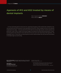
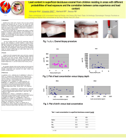
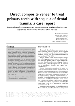
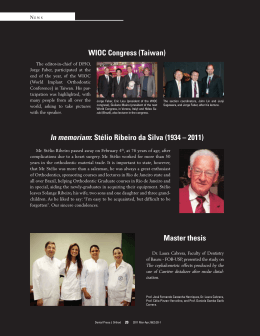
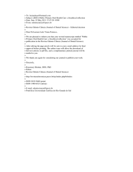
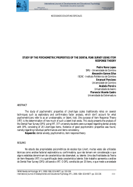
ISSN 1983-5183
Rev. Odontol. Univ. Cid. São Paulo
2014; 26(1): 96-102, jan-abr
AESTHETIC SOLUTION TO FLUOROSIS IN A CHILD
SOLUÇÃO ESTÉTICA PARA A FLUOROSE EM CRIANÇA
Ana Carolina Valinoti
Juliana Cardoso Neves do Amaral
Erika Calvano Küchler
Lívia Azeredo Alves Antunes
Leonardo dos Santos Antunes
Marcelo de Castro Costa
*
**
***
****
*****
******
Abstract
Dental fluorosis is a defect of enamel formation caused by chronic ingestion of fluoride from different sources
during tooth development, which results in interference in proper mineralization. Clinically the tooth enamel
may present as white striae along the lines of enamel, opaque white spot and in more severe cases as brown
spots. Diagnosis is based on clinical characteristics associated with a history of exposure to fluoride. The aim
of this report is to present a treatment in a child with severe aesthetic compromising in anterior maxillary permanent teeth caused by fluorosis. The treatment was based on a combination of three techniques in order to be
minimally invasive, enhance aesthetics and preserving the dental structure.
Descriptors: Enamel, Fluorosis, Microabrasion, Vital bleaching, Composite resin, Child, Odontopediatrics.
Resumo
Fluorose dental é um defeito na formação do esmalte causado pela ingestão crônica de flúor de diferentes
origens durante o desenvolvimento dentário, o que resulta em uma interferência na mineralização adequada.
Clinicamente, o esmalte dentário pode assumir uma tonalidade esbranquiçada ou exibir pequenas manchas
ou linhas brancas e, nos casos mais severos, manchas amarronzadas. O diagnóstico é baseado em características clínicas associadas com a história de exposição ao flúor. O foco deste estudo é mostrar o tratamento em
uma criança com grande comprometimento estético em dentes permanentes anteriores superiores acometidos
por fluorose. O tratamento baseou-se numa combinação de três técnicas, a fim de ser minimamente invasiva,
melhorar a estética e preservar a estrutura dentária.
Descritores: Esmalte, fluorose, microabrasão, clareamento vital, resina composta, criança, Odontopediatria.
****** Mestre em Odontopediatria pela Universidade Federal do Rio de Janeiro, Rio de Janeiro. [email protected]
****** Graduada pela Universidade Gama Filho, Rio de Janeiro. [email protected]
****** D
outora em Ciências Médicas pela Universidade Federal Fluminense, Niterói, RJ. [email protected]
****** P
rofessora Adjunta, Departamento de Formação Específica, Faculdade de Odontologia, Universidade Federal Fluminense, Nova Friburgo, RJ. [email protected]
****** P
hD, Professor Adjunto, Departamento de Formação Específica, Faculdade de Odontologia, Universidade Federal Fluminense, Nova Friburgo, RJ. [email protected]
****** P
hD, Professor Associado, Departamento de Odontopediatria e Ortodontia, Universidade Federal do Rio de Janeiro, Rio de Janeiro. pttpo2009@yahoo.
com.br
96
ISSN 1983-5183
INTRODUCTION
Fluoride has been used worldwide to
prevent dental caries, despite its link with
dental fluorosis1. The literature has revealed increasing prevalence of dental fluorosis, ranging between 7.7 and 80.9% in the
areas with fluoridated water and between
2.9 and 42% in the areas without water
fluoridation2,3. Enamel fluorosis is a defect
of enamel formation, in which fluoride
ingestion during critical periods of mineralization results in interference in proper
mineralization4. It is characterized by hypomineralization caused by the retention
of amelogenin proteins by fluoride5 and
it is believed to be caused by chronic ingestion of fluoride during tooth development1.
Clinically the tooth enamel may present fluorosis as white striae along the lines of enamel perikymata, opaque white
spots and in more severe cases it results
in brown spots due to extrinsic pigmenta-
tion . The severity depends on the concentration of fluoride exposure, the stage
of ameloblastic activity, and individual
variations in susceptibility8,9. The diagnosis is confirmed on the basis of the clinical
characteristics associated with the complete histories of exposure to fluoride10.
Since these defects in dental enamel
may have significant aesthetic, functional11 and psychological impact7, it is necessary for the dentist to intervene12. In the
past, restoring teeth with fluorosis could
not be done conservatively13. Nowadays
minimally invasive techniques are the preferred treatment options for children and
adolescents14. Among these techniques,
we have the dental bleaching and microabrasion associated with resin restoration.
The aim of this report is to present a
conservative treatment performed in a
child with severely compromised aesthetic appearance of the maxillary permanent
teeth caused by fluorosis, associating three
6,7
Valinoti AC
Amaral JCN
Kuchler EC
Antunes LAA
Antunes LS
Costa MC
Aesthetic
solution to
fluorosis in a
child
•• 97 ••
Figure 1. Intra-oral view of the maxillary central incisors with structural defects and generalized white spots in all teeth.
Rev. Odontol.
Univ. Cid. São
Paulo
2014; 26(1): 96102, jan-abr
ISSN 1983-5183
Valinoti AC
Amaral JCN
Kuchler EC
Antunes LAA
Antunes LS
Costa MC
techniques in order to be minimally invasive, to obtain a more favourable aesthetic
result and to preserve dental structure.
Aesthetic
solution to
fluorosis in a
child
A 9-year-old girl was attended at the
Continuous Education Pediatric Dentistry
Clinics of the Federal University of Rio de
Janeiro, Brazil, with the chief complaint
being aesthetic problems in the anterior
maxillary teeth. The patient’s and her parents’ desire was that she should have an
aesthetically pleasing smile.
Her medical history was non-contributory and there was no family history
of dental abnormalities. Intra-oral examination revealed generalized hypomineralized permanent teeth with opaque white spots. Both maxillary central incisors
presented brown spots in the incisal and
CASE REPORT
•• 98 ••
middle thirds. The maxillary canines also
presented this same pattern in the incisal
region. She presented normal psychosocial behaviour, initially being introspective and in some situations, being ashamed
of her smile and concerned about the brownish stains on her teeth.
A detailed history was obtained, with
an emphasis on the possibility of having
swallowed fluoride as child up to the age
of 2 to 3 years of age. According to her
mother, she usually hid away to eat the
children’s toothpaste (1100 ppm fluoride)
because of its delicious taste, in addition
to ingesting it every day during the tooth
brushing until 4 years of age. Moreover,
she lived in Rio de Janeiro, a region with
water fluoridation (0.7 ppm fluoride). Based on the symmetrical and bilateral pattern of enamel hypomineralization and
the presence of a contributory fluoride
history, we confirmed the diagnosis of severe dental fluorosis according to Dean’s
classification 15. All teeth were affected
presenting white spots while maxillary
central incisors presented structural defects and brown stains and pits in the incisal cuspides of maxillary canines and pre
molars (Figure 1).
The option was to perform a more conservative treatment, comprising 3 steps:
vital tooth bleaching and enamel microabrasion associated with composite resin
restoration of the maxillary central incisors that were affected by structural defects and brown stains.
Although the mandibular teeth also
presented dental fluorosis, they were not
treated, since they did not affect the aesthetics of the smile and caused no pain.
Treatment
Rev. Odontol.
Univ. Cid. São
Paulo
2014; 26(1): 96102, jan-abr
Figure 2. A
- Intra-oral view of the maxillary
central incisors after 3 weeks of
take-home tooth whitening treatment. B - Technique of microabrasion with phosphoric acid 35% and
pumice into a slurry with rubber
cup prophylaxis. C - Post-treatment
view of patient’s anterior teeth
after 3 sessions of microabrasion.
D – Remineralization with topical
fluoride.
Initially, the brown stains on the maxillary central incisors and opaque white
spots on the other teeth were treated with
a home tooth whitening system (16% carbamide peroxide) for 3 weeks. During this
period, the child was monitored as regards
tooth sensitivity and pulp vitality, and was
advised to avoid foods and drinks containing dyes. Vitality testing of the maxillary
central incisors undertaken at each review
appointment indicated no change in pulp
ISSN 1983-5183
Valinoti AC
Amaral JCN
Kuchler EC
Antunes LAA
Antunes LS
Costa MC
Aesthetic
solution to
fluorosis in a
child
Figure 3. Post-treatment intra-oral view showing the improved aesthetics following tooth whitening, microabrasion and resin restoration.
status. The aim of dental whitening was to
uniformize the colour and resulted in slightly whitening the cervical region of the
teeth (Figure 2A).
In the next step, teeth were microabraded, following the technique of the UK
National Clinical Guidelines in Paediatric
Dentistry11. The maxillary central and lateral incisors and canines were isolated
with a rubber dam and Vaseline was applied to the gingiva. The teeth were cleaned with pumice and water, washed and
dried. A mixture of phosphoric acid 35%
and pumice was put into a slurry, and a
small amount was applied to the labial
surface of affected teeth with rubber cup
prophylaxis, using a low speed rotary handpiece, for five seconds (Figure 2B). The
tooth surfaces were then washed for five
seconds directly into the aspirator. Ten
applications of the microabrasion paste
were made, lasting 5 seconds each. After
treatment, topical fluoride was applied for
3 minutes, to enhance remineralization
(Figure 2D). This was repeated every week
for another 3 sessions. Between the sessions, any degree of tooth sensitivity was
reported.
In the review one week after the last
session, the stained area was reduced in
size but small brown areas persisted and
the patient requested a further treatment
session. Microabrasion significantly redu-
ced the brown stains and no tooth sensitivity was reported (Figure 2C).
The third step of the treatment was to
restore the maxillary incisors and canines
with composite resin, to improve the uniformity of tooth colour. It was not necessary to perform bevel or any other mechanical preparation (Figure 3).
The microabrasion and resin restoration techniques were performed under
anaesthesia by papillary infiltration only.
The patient was delighted with the aesthetic improvement and reported that she
was smiling more often and feeling that
she was integrating better with her friends.
It was recommended to return for quarterly reviews.
•• 99 ••
DISCUSSION
With regard to the suggested hypotheses about how fluoride might alter enamel
matrix formation, one possibility is that
fluoride might specifically interact with
ameloblasts to affect the synthesis and
secretion of enamel matrix proteins and
proteinases4,16. Severe fluorosis is not a
frequent occurrence, but dental fluorosis
in general seems to be on the rise as a result of the increase in exposure to fluoride
from all sources17. Dental fluorosis is a defect of enamel formation, in which fluoride ingestion during critical periods of enamel mineralization results in interference
Rev. Odontol.
Univ. Cid. São
Paulo
2014; 26(1): 96102, jan-abr
ISSN 1983-5183
Valinoti AC
Amaral JCN
Kuchler EC
Antunes LAA
Antunes LS
Costa MC
Aesthetic
solution to
fluorosis in a
child
•• 100 ••
Rev. Odontol.
Univ. Cid. São
Paulo
2014; 26(1): 96102, jan-abr
in the proper organization of the enamel
proteins, their removal or their proper mineralization4.
In this clinical case, the final diagnosis
of fluorosis was based on the clinical findings in addition to the detailed anamnesis. The symmetrical white spots scattered
throughout the permanent maxillary incisors along the lines of perikymata were
thus initially diagnosed and classified as
fluorosis. These clinical features were associated with the history of chronic fluoride ingestion during the period of 2 to 3 years of age, which is the time of formation
of these dental elements. It corroborates
the findings of Hong et al18,19 , who suggested that the most critical period for susceptibility to fluorosis in human maxillary
central incisors corresponds to the first 24
months of age, and Evans et al.19,20 who
reported the age range to be between 15
to 24 months for girls and 21 to 30 months
for boys. These findings are also in agreement with those of Bardsen20 (1999), who
reported that children who ingest fluoride
during the first two years of life have more
chance of developing fluorosis in maxillary permanent incisors compared with
those who were exposed after two years
of age. Furthermore, fluorosis can not be
attributed only to the form of fluoride use,
because, although it is possible to quantify
the fluoride present in toothpastes and the
water supply, there are other sources that
can increase total ingestion, such as teas6.
The aim of clinical management of
tooth discolouration is to produce an acceptable cosmetic result as conservatively
as possible21. According Periasamy et al.22
(2001), depending on the type and severity of the enamel defects, there are several options of treatment that range from
simple selective polishing, bleaching and
microabrasion to manufacturing porcelain
crowns. It is important to consider that
porcelain veneers were contraindicated
at this time, due to the patient’s immature gingival contour and pulpal size11. In
the clinical case presented, in spite of the
serious compromise caused by the fluorosis, the choice was to associate bleaching,
microabrasion and resin restoration. This
association of treatments was successfully
performed in order to preserve the dental
structure, correct surface enamel hypomineralization and colouring defects by
removing superficial enamel21,23. Microabrasion could be safely performed, since
the maxillary central incisors presented
complete root formation. Another advantage was performing these techniques under anaesthesia by papillary infiltration
only.
Bleaching is often used after mechanical abrasion in an attempt to whiten the
teeth when the white spot lesions remain
problematic24, 25 and to provide a more
uniform colour. Microabrasion reduced
the area and intensity of the black and
brown stains. Nevertheless, when small
brown areas persisted, it was decided to
stop microabrasion in order to avoid exposing more subsurface hypomineralized
enamel26. Thus, the ability to remove the
fluorosis-induced chalky spots in a conservative manner allowed the teeth to be
restored with the use of a direct composite
resin in an aesthetic and functional manner.
Lambert14 (2006) reported a conservative aesthetic solution for dental fluorosis
in a 15-year-old girl, by means of microabrasion and resin restoration. However,
a diamond bur was used to eradicate fluorosis and bevelling the surface to restore
the tooth with composite resin afterwards.
In this report, it was preferred to not perform this mechanical preparation because
the young age of the child and to be minimally invasive, conserving as much of the
dental structure as possible.
CONCLUSION
A successful conservative approach to
dental fluorosis in a child was obtained
with a combination of home tooth whitening and microabrasion techniques. Further improvement in aesthetics was achieved with composite resin.
ISSN 1983-5183
REFERÊNCIAS
1.Den
Besten PK. Dental fluorosis: its
use as a biomarker. Adv Dent Res
1994 Jun;8(1):105-10.
2.Clark DC. Trends in prevalence of den-
tal fluorosis in North America. Community Dent Oral Epidemiol 1994
Jun;22(3):148-52.
3.Pendrys
DG. Risk of enamel fluorosis
in nonfluoridated and optimally fluoridated populations: considerations for
the dental professional. J Am Dent Assoc 2000 Jun;131(6):746-55.
4.Aoba
T, Fejerskov O. Dental fluorosis:
chemistry and biology. Crit Rev Oral
Biol Med 2002 13(2):155-70.
5.Neville
B. Oral & maxillofacial pathology. Philadelphia: Saunders; 2002.
6.Fejerskov
O, Larsen MJ, Richards A,
Baelum V. Dental tissue effects of fluoride. Adv Dent Res 1994 Jun;8(1):1531.
7.Rahmatulla
AH. Clinical evaluation
of two different techniques for the removal of fluorosis stains. Egypt Dent J
1995 Jul;41(3):1287-94.
8.Cutress
TW, Suckling GW. Differential
diagnosis of dental fluorosis. J Dent
Res 1990 Feb;69 Spec No(714-20; discussion 21.
9.Moller
IJ. Fluorides and dental fluorosis. Int Dent J 1982 Jun;32(2):135-47.
10.
Limeback
H, Vieira AP, Lawrence H.
Improving esthetically objectionable
human enamel fluorosis with a simple
microabrasion technique. Eur J Oral
Sci 2006 May;114 Suppl 1(123-6; discussion 7-9, 380.
11.
Wray
A, Welbury R. UK National Clinical Guidelines in Paediatric Dentistry: Treatment of intrinsic discoloration
in permanent anterior teeth in children
and adolescents. Int J Paediatr Dent
2001 Jul;11(4):309-15.
12.
Martinez-Mier
EA, Maupome G, Soto-Rojas AE, Urena-Cirett JL, Katz BP,
Stookey GK. Development of a questionnaire to measure perceptions of,
and concerns derived from, dental fluorosis. Community Dent Health 2004
Dec;21(4):299-305.
Valinoti AC
Amaral JCN
Kuchler EC
Antunes LAA
Antunes LS
Costa MC
Aesthetic
solution to
fluorosis in a
child
13.
Allen
K, Agosta C, Estafan D. Using
microabrasive material to remove fluorosis stains. J Am Dent Assoc 2004
Mar;135(3):319-23.
14.
Lambert
DL. Conservative aesthetic
solutions for the adolescent and young
adult utilizing composite resins. Dent
Clin North Am 2006 Jan;50(1):87-118,
vi-vii.
15.
Dean
H. Classification of mottled
enamel diagnosis. J Amer Dent Assoc
1934 21(8):1421-26.
16.
DenBesten
PK, Thariani H. Biological
mechanisms of fluorosis and level and
timing of systemic exposure to fluoride with respect to fluorosis. J Dent Res
1992 May;71(5):1238-43.
17.
Rozier
RG. The prevalence and severity of enamel fluorosis in North American children. J Public Health Dent
1999 Fall;59(4):239-46.
•• 101 ••
18.
Hong
L, Levy SM, Broffitt B, Warren JJ,
Kanellis MJ, Wefel JS, et al. Timing of
fluoride intake in relation to development of fluorosis on maxillary central
incisors. Community Dent Oral Epidemiol 2006 Aug;34(4):299-309.
19.
Evans
RW, Darvell BW. Refining the
estimate of the critical period for susceptibility to enamel fluorosis in human maxillary central incisors. J Public Health Dent 1995 55(4):238-49.
20.
Bardsen
A. “Risk periods” associated
with the development of dental fluorosis in maxillary permanent central incisors: a meta-analysis. Acta Odontol
Scand 1999 Oct;57(5):247-56.
21.
Rodd
HD, Davidson LE. The aesthetic
management of severe dental fluorosis in the young patient. Dent Update
1997 Dec;24(10):408-11.
Rev. Odontol.
Univ. Cid. São
Paulo
2014; 26(1): 96102, jan-abr
ISSN 1983-5183
Valinoti AC
Amaral JCN
Kuchler EC
Antunes LAA
Antunes LS
Costa MC
Aesthetic
solution to
fluorosis in a
child
22.
Peariasamy
K, Anderson P, Brook AH.
A quantitative study of the effect of pumicing and etching on the remineralisation of enamel opacities. Int J Paediatr Dent 2001 May;11(3):193-200.
23.
Croll
TP. Esthetic correction for teeth
with fluorosis and fluorosis-like enamel dysmineralization. J Esthet Dent
1998 10(1):21-9.
H, Randel H. Rotary reduction, enamel microabrasion, and
dental bleaching for tooth color improvement. Compend Contin Educ
Dent 1998 Jan;19(1):62-7.
25.
Bodden
MK, Haywood VB. Treatment
of endemic fluorosis and tetracycline
staining with macroabrasion and nightguard vital bleaching: a case report.
Quintessence Int 2003 Feb;34(2):8791.
26.
Ng
F, Manton DJ. Aesthetic management of severely fluorosed incisors
in an adolescent female. Aust Dent J
2007 Sep;52(3):243-8.
24.
Rosenthaler
•• 102 ••
Rev. Odontol.
Univ. Cid. São
Paulo
2014; 26(1): 96102, jan-abr
Recebido:11/03/2014
Aceito :24/04/2014
Baixar